HAL Id: tel-02953333
https://tel.archives-ouvertes.fr/tel-02953333
Submitted on 30 Sep 2020HAL is a multi-disciplinary open access archive for the deposit and dissemination of sci-entific research documents, whether they are pub-lished or not. The documents may come from teaching and research institutions in France or abroad, or from public or private research centers.
L’archive ouverte pluridisciplinaire HAL, est destinée au dépôt et à la diffusion de documents scientifiques de niveau recherche, publiés ou non, émanant des établissements d’enseignement et de recherche français ou étrangers, des laboratoires publics ou privés.
Unravelling the protective role of APRIL in the central
nervous system
Mashal Ahmed
To cite this version:
Mashal Ahmed. Unravelling the protective role of APRIL in the central nervous system. Virology. Université Grenoble Alpes, 2019. English. �NNT : 2019GREAV055�. �tel-02953333�
THÈSE
Pour obtenir le grade de
DOCTEUR DE LA COMMUNAUTE UNIVERSITE
GRENOBLE ALPES
Spécialité : Virologie - Microbiologie - Immunologie Arrêté ministériel : 25 mai 2016
Présentée par
Mashal Claude Ahmed
Thèse dirigée par Bertrand HUARD, Professeur des universités, UGA
préparée au sein du Laboratoire CRI IAB - Centre de Recherche Oncologie/Développement - Institute for Advanced
Biosciences
dans l'École Doctorale Chimie et Sciences du Vivant
Rôle neuroprotecteur de APRIL dans le
système nerveux central
Unravelling the protective role of APRIL in
the central nervous system
Thèse soutenue publiquement le 9 décembre 2019, devant le jury composé de :
Madame, Jessica KWOK
Associate Professor, University of Leeds, Rapporteur Madame, Myriam CAYRE
Directeur de Recherche, Institut de Biologie du Développement de Marseille (IBDM), Rapporteur
Monsieur, Alain BUISSON
Professeur, Grenoble Institut des Neurosciences (GIN), Examinateur, Président du jury
Monsieur, Romain VIVES
Directeur de Recherche, Institut de Biologie Structurale (IBS), Examinateur
3
Acknowledgements
First and foremost, I wish to thank the members of the jury for having accepted to evaluate this thesis. To Dr Jessica Kwok and Dr Myriam Cayre, who have accepted to travel to Grenoble from the UK and Marseille respectively to be reviewers of this manuscript, I am honoured to have the chance to meet you and discuss the current work with you. To Dr Romain Vives and Dr Alain Buisson who have accepted to be the examiners, I thank you for your continued interest in the current project, your long-term moral support and scientific counsel.
The scientific work detailed in this manuscript was a collaborative work. Bright, curious minds and generous personalities were pillars that raised this project up from a hypothesis to real results.
I first want to thank Pascal Schneider of the University of Lausanne, Switzerland, for sharing his protocol for DNA mutagenesis, as well as for his gifts in the form of purified recombinant proteins. His counsel has always been swift and invaluable.
The in vitro functional assays described in the current work were made possible by the kind collaboration of members of Annie Andrieux’s team, from the Grenoble Institute of Neuroscience (GIN). I want to give a very warm thanks to Dr Leticia Peris who provided the training as well as the resources for these experiments, and I also wish to extend my gratitude to Dr Pierre Heemeryck, and Dr Jean-Christophe Deloulme for their aid and generosity.
The ex vivo experiments to study remyelination were made possible by the collaboration of members of Dr Charbel Massad’s team at the University of Paris Descartes. I wish to especially thank Dr Delphine Meffre for her very generous contributions, and extend my gratitude to Aida Padilla Ferrer who provided invaluable effort, training and experience. I also want to thank Dr Mehrnaz Jafarian-Tehrani and Dr Alex Carrete for their key roles in setting up this collaborative work.
I am also grateful to the members of the SAGAG team of the Institute of Structural Biology (IBS) who provided resources and expertise for experiments concerning molecular interactions and biotinylated materials. I heartedly thank Dr Romain Vives, and Evelyne Gout, as well as Dr Hugues Lortat-Jacob for their contributions, their time and effort, and their generous counsel.
4 I wish to give great thanks to Professor Bertrand Huard and Vice director Patrice Marche for taking me on this project and letting me be a part of the IAPC team. As my supervisor, Bertrand gave me much trust and free rein on this project, while providing the bedrock upon which it was built. I extend my gratitude to Patrice for always sharing advice, and for his constant encouragement.
On a more personal note, my time at the Institute for Advanced Biosciences (IAB) was shared with many fine colleagues who I would go on to call my friends. First, thanks to the wonderful friends who I shared my office with. Marion Ressejac, Arindam Dey, Alexis Leroy, and Christine Charrat all made life at the IAB so enjoyable. They added warmth, spice, seriousness, and laughter to every day. I also want to thank Keerthi, Lydie, Arnaud, and Alexouille la Fripouille (Dr Alexis Gonon), as well as Alexandre, Michael, Norbert, all examples of the good people I got to work with at the institute.
Heartfelt thanks to Dr Laurie Baert a certain Dr Mathieu Dangin, who were both excellent teachers and continue to be excellent friends. I wish them the best of luck in their endeavours, as I follow behind them.
Dr Ben Manfroid de Montmiraille, also known as Benoit Manfroi(d), was the push on my back as the mountain grew steep, and the winds grew harsh. I reserve my deepest gratitude for the colleague whose friendship and scientific counsel is without measure.
Finally, a message to my family. Thank you, Papa, and Maman, my loving parents who always made me proud of where I am and who I am. I have come this far thanks to both of you, and for both you. Manu, you got your wish; your name is on my thesis. Adeline, you more than anyone else were by my side for these gruelling years. You more than anyone else, I thank for every morning I could wake up for a day of work, and you I thank for every evening I returned to a loving fiancée.
6
Table of Contents
Summary ...8 Résumé en français ...10 Index of Figures ...21 Index of Tables ...21 Chapter 1: Introduction ...231 Chondroitin Sulfate Proteoglycans ...23
1.1 CSPGs and the ECM of the CNS ...26
1.2 CS-GAGs: Biodiversity in structure and function ...28
1.3 Pathologies of the Central nervous system: the role of CSPGs ...37
1.4 Known Partners of CSPGs ...42
1.5 Potential Partners of CSPGs ...52
2 APRIL: A proliferation inducing ligand ...56
2.1 Structure and physio-biology ...56
2.2 The ATAMS trial ...57
2.3 Previous work by the laboratory ...59
3 Objective of study ...60
Chapter 2: Material and Methods ...61
1 Production and purification of recombinant Fc-fused candidate proteins ...61
1.1 Generation of mouse brain cDNA library ...61
1.2 Polymerase chain reaction (PCR) ...62
1.3 Molecular Cloning ...65
1.4 pTOPO subcloning (TA cloning) ...66
1.5 Mutation or de-mutation by double PCR ...68
1.6 Transient Transfection of HEK293T cells ...71
1.7 Purification and validation of recombinant proteins ...72
7
2.1 Biotinylation of CS-GAGs ...74
2.2 ELISA...74
3 Neurite Outgrowth Assay ...76
4 OPC Outgrowth Assay ...78
5 Ex vivo Organotypic Culture and Myelination Assay ...80
5.1 Organotypic culture ...80
5.2 Immunofluorescence and analysis ...81
6 Notes ...84
Chapter 3: Results ...89
1 APRIL-Fc prevents binding of CS types to inhibitory receptors ...89
2 APRIL-Fc prevents CS-mediated inhibition of neurite outgrowth ...94
3 Preliminary Results: OPC outgrowth assays ...99
4 APRIL-Fc promotes myelination in an ex vivo organotypic model ... 102
Chapter 4: Conclusions, discussion and perspectives ... 106
Bibliography ... 111
Résumé / Abstract ... 148
Abstract in English... 148
Abstract en français ... 148
This thesis manuscript will begin with an overview in English and a more detailed summary in French, which will be followed by four main chapters covering the introduction, the material and methods used, the results, and a discussion. Abstracts in French and English are presented on the last page of the document.
8
Summary
Chondroitin sulfate proteoglycans (CSPGs) are diverse, major constituents of the extracellular matrix (ECM) with key roles in the central nervous system (CNS) such as tissue structure, neural development, plasticity, and regeneration [1, 2]. They are especially studied for their role in inhibiting repair in the CNS [2–8]. Their upregulation following trauma in the CNS contributes to the formation of the glial scar, a cellular and biochemical barrier to regenerating neurons as well as myelinating cells [3, 4, 9, 10]. Additionally, CSPGs may play pathophysiological roles in modulating repair in neurodegenerative diseases [11]. For example, they have been shown to be upregulated in multiple sclerosis (MS) lesions [12–14]. The inhibitory activities of CSPGs are known to be mediated by their chondroitin sulfate (CS) chains [17–22]. The digestion of CS chains by chondroitinase ABC promotes neural regeneration and remyelination in vitro, as well as in the mouse spinal cord injury (SCI) model [4, 6, 10, 18, 23–26]. CS are sulfated glycosaminoglycans (GAGs) consisting of repeating disaccharide units of glucuronic acid and N-acetylgalactosamine, and come in a variety of types depending on their sulfation pattern and epimerization [27]. Chondroitin sulfate A (CS-A) and B (CS-B, also known as dermatan sulfate) consist of mono-sulfated disaccharide units that differ by an epimerization, yielding either glucuronic acid or iduronic acid respectively. Chondroitin sulfate D (CS-D) and E (CS-E) on the other hand, are rich in disulfated disaccharide units, but differ from each other by the positions of their sulfations.
The inhibition mediated by CS GAGs depends on their ability to bind to a variety of receptors and ligands with roles in modulation of repair and regeneration. Among them, the best characterized are receptor-type tyrosine-protein phosphatase sigma (PTPRS), a member of the protein tyrosine phosphatase (PTP) family, Nogo receptor 1 (NgR1, also known as reticulon 4 receptor), a member of the Nogo receptor family, and semaphorin 3A (Sema3A), a member of the semaphorins family. CSPGs have been shown to mediate their inhibition of neural and myelin repair through PTPRS [8, 28–33] and NgR1 [34]. Other families of proteins with known affinity for sulfated proteoglycans and that regulate neural plasticity include the slits, and the ephrins [35–37]. Ephrin-B2 (EFNB2), a member of the ephrins subfamily of receptor protein-tyrosine kinases, has a role in the formation of the glial scar [38, 39], and Slit
9 guidance ligand 2 (Slit2, also known as slit homologue 2), a member of the Slit family has roles in axonal guidance and migration of oligodendrocyte precursors [37, 40, 41].
This study aims to show the therapeutic potential of blocking CS interactions with repair-inhibiting proteins of the CNS that mediate their activity. We show that differently sulfated CS GAGs have different binding preferences for various inhibitory proteins, eliciting the need for an agent capable of indiscriminate and stable binding to CS types. We have previously shown that a proliferation inducing ligand (APRIL, also known as TNFSF13) binds to CSPGs [42]. A recombinant, hexameric form of APRIL (APRIL-Fc) exhibits broad and strong affinities for CS GAGs. We show that APRIL-Fc is able to effectively block CS binding to repair-inhibiting partners and is able to neutralize the inhibitory effects of CS types on neurons in in
vitro functional assays, as well as on oligodendrocyte progenitor cells in pilot experiments. In
an ex vivo organotypic model, APRIL-Fc treatment boosted remyelination.
Key words: a proliferation inducing ligand (APRIL), chondroitin sulfate proteoglycan (CSPG),
10
Résumé en français
Chapitre 1 : Introduction
Les chondroïtines sulfates protéoglycanes (CSPG)
Les chondroïtines sulfates protéoglycanes (CSPG) sont des composants majeurs de la matrice extracellulaire (MEC) et sont omniprésents dans le règne animal. Ils sont constitués d’un cœur protéique décoré par des chaînes glycosaminoglycanes chondroïtines sulfates (CS-GAG) [11, 43–45]. La MEC du système nerveux central (SNC) se distingue de celle d’autres types de tissus par des quantités plus élevées de GAGs, parmi lesquels les CS-GAGs sont les plus abondants [46–48]. Ces CS-GAGs ont des rôles variés comme par exemple dans le développement, la plasticité mais aussi la régénération. Ce dernier sera le sujet de cette étude [17, 49, 50].
La plupart des fonctions des CSPG sont médiées par leur CS-GAGs et ses motifs sulfatés ; ces derniers sont cruciaux pour la fixation de protéines partenaires [2, 17–22]. Les CS-GAGs sont composés de répétitions de disaccharides (N-acétylgalactosamine, GalNAc et acide glucuronique, GlcA). Ils forment différents types de CS-GAGs selon leur sulfation et épimérisation [27, 51, 52]. Les CS-A et CS-C sont mono-sulfatés à la position 4 ou 6 du GalNAc respectivement. Le CS-D est di-sulfaté aux positions 2 et 6 du GlcA et GalNAc respectivement, tandis que le CS-E est di-sulfaté aux positions 4 et 6 du GalNAc. Le CS-B, aussi appelé dermatane sulfate (DS), est une variante où le GlcA est épimérisé à un acide iduronique (IdoA) [27, 53–56]. Le CS-B est sulfaté a la position 4 du GalNAc comme CS-A, mais il peut aussi être sulfaté à la position 2 du IdoA [55].
Ces variations mènent à une diversité d’interactions avec des partenaires protéiques. Les chaînes de CS-GAGs peuvent alors avoir des préférences de liaison distinctes ou qui se chevauchent selon la présentation et la distribution des diffèrent CS-types, formant un ‘code sulfaté’ [20, 22, 37, 57]. En conséquence, il a été montré que les différent CS-GAGs n’ont pas les mêmes effets sur les cellules du SNC, étant soit inhibants, soit bénéfiques, ou même neutres. Cependant, différentes études se contredisent dans leur caractérisation de ces différences (voir tableau 3).
11
Les pathologies du système nerveux central : le rôle des CSPG
De nombreuses études ont démontré que les CSPG sont surexprimés dans les tissus nerveux endommagés ; c’est le cas pour des blessures traumatiques et neurodégénératives. Leur expression est considérée comme étant responsable de l’échec de la régénération. Nous allons considérer dans cette étude les exemples de la lésion de la moelle épinière et de la sclérose en plaques et discuter des rôles des CSPG dans ces pathologies.
Lésion de la moelle épinière : Les lésions de la moelle épinière sont majoritairement
causées par le traumatisme physique, comme des chutes, accidents de véhicules, ou des blessures par balle [58]. Le tissu nerveux endommagé devient un site de transformation épigénétique et d’activité gliale, comprenant la mobilisation des astrocytes réactifs, microglies, et des cellules du système immune infiltrants. Ces effets contribuent à la formation d’un environnement inhibiteur, appelé « cicatrice gliale », qui constituera finalement une barrière à la régénération neuronale [59–61]. Parmi ces cellules, les astrocytes réactifs en particulier produisent des molécules inhibitrices comme les CSPG, des ephrines, et des semaphorines qui rendent l’environnement défavorable à la repousse des axones et à leur remyélinisation [2, 4, 7, 61–63].
Plusieurs études ont identifié les CSPG comme étant clés dans l’inhibition de la régénération suivant une blessure traumatique. Les neurones cultivés sur des substrats recouverts de CSPG présentent des défauts dans l’élongation axonale, des cônes de croissance dystrophiques, une croissance des neurites réduite, et une adhésion cellulaire perturbée [3, 15, 64–68]. Ces effets sont abolis par la dégradation des CS-GAGs par l’enzyme bactérienne, la chondroitinase ABC (ChABC). Les études in vivo ont aussi démontré l’efficacité de ce traitement pour augmenter la régénération dans des modèles des blessures traumatiques du SNC [18, 23, 69–71].
En plus de l’endommagement neuronal, les blessures traumatiques impliquent aussi la démyélinisation [72, 73], un trait dégénératif qu’on retrouve aussi dans la sclérose en plaques, le sujet du prochain paragraphe.
12
La sclérose en plaques : La sclérose en plaques est la maladie immunitaire la plus
commune du SNC, et se caractérise par une attaque auto-immune sur les gaines de myéline qui entourent les neurones [74, 75], et en conséquence, par l’apparition des lésions (‘sclerae’) démyélinisées dans la matière blanche et grise du SNC [76–79]. La démyélinisation et l’inflammation du tissu nerveux conduisent finalement à une dégénération progressive des axones, ce qui se manifeste cliniquement par une variété de symptômes, comme la cécité, l'engourdissement, la perte de contrôle moteur et/ou des fonctions cognitives [74, 75, 80]. Typique des maladies neurodégénératives, la sclérose en plaques a une pathogenèse insaisissable, et n’a pas de remède.
Les CSPG sont surexprimés dans les lésions des patients, et s’associent avec l’astrocytose. C’est également le cas dans les moelles épinières des souris atteintes de l’encéphalomyélite auto-immune expérimental (EAE, modèle murin de la sclérose en plaques) [12, 14, 81]. Plusieurs études in vitro ont démontré que les CSPG inhibent l’adhésion et la croissance des cellules progénitrices d’oligodendrocytes (OPC), ainsi que leur différentiation, et leur capacité a myéliniser des axones [5, 24, 26, 82]. Comme dans le cas de l’inhibition des neurones, la dégradation des CS par ChABC abolit leurs effets néfastes.
Alors, comme pour les blessures traumatiques, les CSPG empêchent le recrutement des OPC aux tissus démyélinisés ainsi que leur différentiation, compromettant leur potentiel réparateur. Ces observations font des CSPG une cible intéressante dans la recherche de thérapies régénératives pour des pathologies impliquant la démyélinisation chronique, et jusque-là des études in vivo testant des inhibiteurs des CSPG dans les modèles de remyelination ont démontré une efficacité prometteuse (par exemple la xyloside [5], et la fluorosamine [82]).
Des récepteurs des CSPG connus
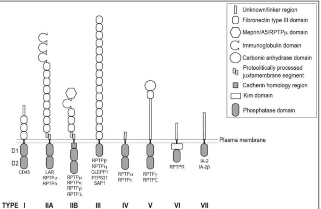
PTPRS : Le type récepteur tyrosine phosphatase protéine sigma (PTPRS) fait partie du
sous-famille de type récepteur (RPTP) de la famille des phosphatases protéine-tyrosine (PTP) [83–86]. Cette sous-famille est importante dans la développement du SNC et des processus neuronaux variés [86–88]. Dans ce groupe se trouvent PTPRS et deux autres protéines,
13 leucocyte antigen-related (LAR), et RPTP-delta (PTPRD), qui forment la famille des LAR, et qui sont connues pour leurs interactions fonctionnelles avec les GAGs sulfates [89]. PTPRS est surexprimée dans des pathologies du SNC. Cette protéine s’accumule dans les axones dystrophiques qui ne peuvent pénétrer les lésions de la moelle épinière dans la souris [33], et elle est surexprimée par les OPC dans les modèles murins de la sclérose en plaques [8]. Le niveau d’expression de PTPRS est élevé dans les lésions de souris modèle EAE, ou lors d’un traitement lysolécithine (LPC) qui induit une démyélinisation focalisée. En interagissant avec les CSPG, qui sont eux aussi surexprimés dans ces modèles, il a été démontré que PTPRS joue un rôle dans l’inhibition de la réparation [31, 44, 89]. Plusieurs types des neurones exhibent une résistance à l’inhibition pas des CSPG in vitro s’ils n’ont pas une PTPRS fonctionnel [28, 65, 90–93]. PTPRS a aussi été impliquée dans la modulation des cellules déclinantes du SNC. Dans une étude, l’inhibition de PTPRS a inversé l’inhibition de croissance des OPC médiée par les CSPG in vitro [30]. Plusieurs études in vivo confirment cette observation, par exemple, les souris avec une PTPRS non fonctionnelle présentent une meilleure régénération après une lésion du nerf optique [94], ou de la moelle épinière [90, 92]. Les traitements bloquants les interactions entre PTPRS et les CSPG ont démontré un potentiel pour l’amélioration de la régénération neuronale, et aussi de la remyélinisation [30, 32, 33, 95].
NgR1 : La famille des récepteurs Nogo (NgRs) est constituée de trois membres, NgR1,
NgR2, NgR3 [96, 97]. Ces protéines sont exprimées à la surface d’une variété de neurones [97– 99], et interagissent avec des inhibiteurs associés à la myéline (MAIs), inhibant la régénération neuronale et la remyélinisation [100–104]. Une étude percutante par l’équipe de Dickendesher à l’université de Michigan a démontré que NgR1 et NgR3 sont aussi des récepteurs des CSPG [34]. Les neurones aux NgRs non fonctionnels ont montré une résistance à l’inhibition par les CSPG, et la double délétion de ces NgRs et de PTPRS a encore amélioré la régénération après une blessure par écrasement du nerf optique. Au sujet des maladies neurodégénératives, des études ont montré une surexpression des NgRs et de leurs partenaires dans les lésions des patients atteints de sclérose en plaques, et des souris modèles EAE [99, 101, 105–107]. Ciblant ces protéines et leur voies de signalisation dans l’EAE a promu la régénération axonale, et la remyélinisation [106, 108, 109], et les souris sans NgR1 fonctionnelle présentent un phénotype EAE atténué [106, 107].
14
Sema3A : Les semaphorines sont une grande famille de protéines sécrétées,
transmembranaires, ou associées aux surfaces cellulaires, avec des rôles variés dans plusieurs tissus des invertébrés et des vertébrés [110, 111]. Semaphorin 3A (Sema3A) est un des membre avec des rôles clés dans le guide et la migration des neurones et OPC pendent le développement [112–115]. Il s’associe avec la matrice extracellulaire, en particulier avec les filets péri-neuronaux (PNNs) [116, 117]. Les CSPG sont des composants majeurs des PNNs, et ont été décrits comme les sites d’amarrage pour Sema3A [116, 118]. L’accumulation et la présentation de Sema3A sur les CS-GAGs peuvent médier les interactions avec des récepteurs comme neuropilin-1 (Nrp1) et plexin A1, exprimés par des neurones et des OPC. Cette coopération entre les CSPG et Sema3A augmente la répulsion des neurones et induit la dystrophie axonale in vitro [119, 120]. Plusieurs études dans les modèles de rat ont montré que Sema3A est surexprimée dans le SNC blessé [121–123]. L’accumulation de Sema3A dans les lésions corrèle avec l'incapacité des neurones à pénétrer ces zones, indicatif du rôle inhibiteur de Sema3A dans la régénération. Les semaphorines ont aussi été impliquées dans les maladies neurodégénératives [124]. La forte surexpression de Sema3A dans les lésions des patients atteints de sclérose en plaques [125, 126], et aussi des modèles rongeurs de démyélinisation [125, 127, 128], corrèle avec l’échec de remyélinisation. Validant ce lien, l’introduction de Sema3A dans les lésions démyélinisantes chez la souris [129] ou le rat [130] a eu comme effet d’inhiber le recrutement des OPC au site endommagé, et d’empêcher la remyélinisation.
Tout ceci montre que les CSPG travaillent main dans la main avec une variété de protéines de différentes familles avec des rôles inhibiteurs dans la régénération.
Des partenaires potentiels des CSPG
EFNB2 : Les ephrins (EFNs) sont des ligands transmembranaires qui exhibent une
signalisation bidirectionnelle en interagissant en trans avec les récepteurs Eph. Dans le SNC les ephrines et leur récepteurs sont surexprimés sur les neurones, les astrocytes, et les OPC suite à une blessure [38, 39, 131–133]. Ces protéines sont surexprimées aussi dans et autour des lésions dans la sclérose en plaques [132, 134]. EFNB2, la protéine considérée dans l’étude actuelle, est impliquée dans l’inhibition de la réparation [39]. Cette protéine est exprimée par des astrocytes réactifs, et a un rôle clé dans la formation de la cicatrice gliale suite à la lésion
15 de la moelle épinière chez le rat [38]. Un récepteur de EFNB2 pertinent dans cette étude, EphA4, interagit avec les CSPG pour inhiber la croissance neuronale [135]. Il est possible que les CSPG puissent être impliqués dans la modulation d’activité des Ephs et leurs ligands. Dans cette étude nous allons creuser cette hypothèse en testant les interactions entre EFNB2 et différents types de CS. A notre connaissance, celle-ci sera la première investigation sur cette question.
Slit2 : Les slits sont une famille de protéines secrétées bien connues pour leurs rôles
comme molécules-guides dans le développement du system nerveux. Ils sont exprimés par les cellules gliales ainsi que par les neurones, se lient aux récepteurs ‘roundabout’ (Robo) et influencent la croissance et la migration des neurones et des OPC [41, 136–138]. Les slits ont été étudiées pour leurs effets sur la plasticité neuronale [138, 139], et ont besoin d’interagir avec les protéoglycanes sulfatés comme corécepteurs pour effectuer leurs fonctions [36, 37, 140, 141]. C’est le cas pour Slit2, qui a besoin de syndecan-1 (un protéoglycane heparan sulfate (HSPG) qui contient aussi des motifs CS) pour ces effets inhibiteurs sur les neurones et OPC. Les protéines qui interagissent avec les HSPGs présentent souvent une affinité pour les CSPG (par exemple, la famille LAR des PTP, APRIL, et certaines semaphorines)., L’ajout de CS ‘decoys’ a aboli l’inhibition de la croissance axonale par Slit2, suggérant que les CSPG peuvent moduler les activités des slits, renforçant cette idée. Dans cette étude nous allons tester, pour la première fois à notre connaissance, des interactions entre Slit2 et différents types de CS.
APRIL: ‘A proliferation inducing ligand’
‘A proliferation inducing ligand’ (APRIL, TNFS13), le treizième membre de la famille des facteurs de nécrose tumorale (TNSF), est une protéine sécrétée par les cellules myéloïdes, et a un rôle dans la différentiation et la survie des cellules productrices d’anticorps [142–144]. Deux récepteurs TNF ont été décrits pour APRIL : ‘B‐cell maturation antigen’ (BCMA) et ‘transmembrane activator, calcium modulator and cyclophilin ligand interactor’ (TACI). APRIL a une affinité pour les glycosaminoglycanes (GAGs) sulfatés, ce qui est important pour sa fonction [145]. Par exemple, son interaction avec les HSPGs est critique pour la bonne costimulation des cellules B [142–144].
L’auto-immunité est le résultat d’une réponse immune adaptive vers un antigène de « soi », réponse souvent médiée par la production des auto-anticorps [146]. Ces anticorps
16 sont produits par des cellules B, et cibler cette lignée s'est révélé être une thérapie effective dans certain maladies, par exemple la sclérose en plaques (Ocrelizumab) [147–150].
L’atacicept, une forme de TACI soluble fusionnée à une immunoglobuline, a été conçue pour cibler les cellules B matures et les plasmocytes producteurs d’anticorps en agissant comme une barrière pour APRIL [151]. L’essai clinique en phase II désignée ‘ATAMS’ (atacicept in multiple sclerosis, IMP28063, ClinicalTrials.gov identifier: NCT00642902) a été mis en place pour évaluer les effets d’atacicept dans la sclérose en plaques [151, 152]. Dans une tournure inattendue, le traitement a abouti à une augmentation de l’inflammation et une exacerbation des symptômes, conduisant à l'arrêt de l’essai clinique.
Cette observation a indiqué un rôle neuroprotecteur pour APRIL dans le CNS. Pour tester cette possibilité, la surexpression et la délétion d’APRIL dans les souris EAE a été investiguée. Notre équipe a trouvé que l’absence d’APRIL aboutit à une exacerbation de la maladie, tandis qu’une surexpression a un effet protecteur [42]. Dans les biopsies des patients atteints de sclérose en plaques, APRIL s’accumule au niveau des lésions, et colocalise avec des CSPG, et en particulier le CS de type E. Nous pouvons constater que APRIL, en se liant au CSPG au niveau des lésions, peut avoir comme effet d’empêcher les CSPG d’interagir avec ces protéines partenaires inhibitrices.
Objective de l’Etude
L’étude actuelle a pour objectif de prendre avantage de l’affinité d’APRIL pour les CSPG. Un APRIL recombinant, fusionné à une immunoglobuline (APRIL-Fc) démontre une capacité à se fixer à différents types des CS. Nous allons évaluer le potentiel de APRIL-Fc comme inhibiteur des interactions entres les CSPG leurs partenaires protéiques connus (comme PTPRS et NgR1) ou potentiels (EFNB2, Slit2). De plus, nous allons tester APRIL-Fc comme traitement in vitro sur les neurones et les OPC inhibés par les CS. Enfin, nous allons utiliser APRIL-Fc dans un modèle ex vivo de démyélinisation, pour tester sa capacité à promouvoir la remyélinisation.
L’objectif de cette étude est de démontrer un potentiel thérapeutique pour APRIL-Fc dans le contexte de pathologies du SNC.
17 Chapitre 2 : Matériel et Méthodes
Ce chapitre décrira en détail les protocoles mis en place pour cette étude. Brièvement, on a produit une librairie ADNc dérivé de cerveaux de souris et de la lignée de glioblastome humain, U251. Les amorces conçues pour l’amplification des gènes candidats choisis pour l’étude (PTPRS, NgR1, EFNB2, Slit2) sont décrits dans tableau 5. Les amplicons ont été insérés par clonage moléculaire dans les vecteurs pCRIII contenant des séquences pour le fusionnement de tag-Fc. Ces plasmides d’expression ont été utilisés pour transfecter les cellules HEK293T pour la production des protéines recombinantes solubles, fusionnées à une immunoglobuline. Celles-ci ont été utilisées dans des tests ELISA pour des liaisons avec différents types de CS, et dans des tests de compétition impliquant APRIL-Fc.
Des tests in vitro fonctionnels ont requis l’extraction et culture des neurones et OPC des cortex de souris embryonnaires. Ces cellules ont été cultivées sur des lamelles recouvertes de CS, et traitées avec APRIL-Fc ou un contrôle. Les neurones ont été fixés 3 jours plus tard, tandis que les OPC ont été fixés 1 jour plus tard. Leur croissance a été mesurée et analysée sous imageJ.
Concernant les tests ex vivo utilisant les coupes de cervelets organotypiques des souris P10, les coupes de cervelets ont été soumises à un traitement LPC démyélinisant, puis cultivées en présence de APRIL-Fc ou un contrôle. Après fixation, l'imagerie confocale et l'analyse de la myélinisation ont été réalisées.
Chapitre 3 : Résultats
Nous avons cloné des versions solubles fusionnées à Fc de récepteurs connus pour leurs interactions avec les CSPG (PTPRS et NgR1), et aussi de protéines partenaires potentiels (EFNB2 et Slit2). Leur capacité à se lier aux différents types de CS (A, B, D et E) a été évaluée par ELISA. Ces protéines recombinantes ont toutes démontré une affinité pour des CS, en exhibant des préférences différentielles pour les différents types. Dans ces tests ELISA, APRIL-Fc a démontré une affinité pour tous les CS testés, et cette liaison a eu comme effet d’abolir efficacement la capacité des CS à fixer ces partenaires protéiques recombinants.
Pour voir si cette observation peut se traduire en un blocage fonctionnel d’activité inhibitrice des CS in vitro, nous avons cultivé des neurones dérivés des cortex embryonnaires
18 de souris sur les substrats recouverts de CS, et traités avec APRIL-Fc ou un contrôle. La croissance neuronale a été inhibée par les CS de types A, B et E, mais pas par le type D. Le traitement des CS par APRIL-Fc a efficacement neutralisé leurs effets inhibiteurs dans une manière dose-dépendante. Dans des expériences préliminaires, nous avons observé une inhibition de croissance des cellules progénitrices d'oligodendrocytes (OPC) dérivées des cortex de souris par les CS de types B, D et E. Ici aussi, APRIL-Fc démontre une capacité à abolir les effets des CS.
L’inhibition par les CSPG de la mobilisation des OPC et de leur maturation morphologique est un obstacle à la régénération de myéline dans le SNC. Les cultures organotypiques des coupes de cervelets démyélinisées par le traitement LPC est un modèle
ex vivo de remyélinisation. Nous avons trouvé que l’expression de CSPG est augmentée dans
la matière grise démyélinisée. Le traitement APRIL-Fc suivant la démyélinisation par LPC a eu comme effet d’accélérer la remyélinisation.
En conclusion, nos résultats démontrent l’efficacité d’APRIL-Fc comme un agent bloquant des CSPG, et un potentiel thérapeutique pour cette molécule dans les pathologies du SNC où la régénération est empêchée.
Chapitre 4 : Conclusions et discussion
En cohérence avec les études précédentes, nous montrons que les CS peuvent inhiber la croissance des neurones et les OPC in vitro. Dans le cas des neurones, l’étude actuelle est un complément à plusieurs autres qui ont testé les effets des différents types de CS (tableau 3). Nous démontrons les effets inhibiteurs des types A, B et E sur les neurones corticaux de souris embryonnaires, ainsi que l’inactivité du type D sur ces cellules. Il faut noter pour la variabilité dans la caractérisation des types de CS dans la littérature, qu’il y a des différences dans les procédures expérimentales utilisées. De nombreuses preuves suggèrent que les neurones des différents types ou espèces d’origines différentes peuvent répondre différemment à un CS. De plus, l’origine et la méthode de préparation du CS peuvent aussi influencer son activité [54].
Concernant les expériences préliminaires sur les OPC, notre étude est à notre connaissance la première à tester les différents types de CS sur la croissance de ces cellules.
19 Une observation intéressante est l’inhibition de la croissance des OPC dans la présence de CS-D, un type qui a régulièrement été rapporté comme neutre (comme dans notre étude) ou bénéfique pour les neurones (tableau 3). Inversement, les OPC n’ont pas été inhibés par le CS-A, une observation qui diffère de nos expériences avec les neurones. Parce qu’il a régulièrement été montré que les CSPG sont inhibiteurs à la fois de la régénération neuronale, et aussi de la mobilisation des cellules remyélinisâtes du SNC, nos observations éclairent les mécanismes à la base de la caractéristique à double tranchant des CSPG
Il est important de noter que les CS sont des molécules chargées négativement, et peuvent donc influencer l’adhésion cellulaire in vitro. Nous avons observé une perte d’attachement cellulaire et une augmentation de l’agrégation des neurones et des OPC quand ils sont cultivés sur des concentrations élevées de CS, en accord avec des études précédentes [82, 153]. Cependant, les effets inhibiteurs des CSPG ne peuvent pas être attribués uniquement à leurs charges anioniques. Il a été démontré que la neutralisation des charges anioniques, bien que bénéfique à l’adhésion cellulaire, n’inverse pas l’inhibition de croissance [82, 153].
L’objectif clé de cette étude a été d’évaluer APRIL-Fc comme agent bloquant des CSPG. Nous démontrons l’efficacité de cette molécule pour empêcher les effets néfastes de tous les types de CS testés de survenir sur les neurones et les OPC. Nous proposons un mécanisme moléculaire pour cette activité neutralisante. Par les expériences ELISA nous avons démontré que APRIL-Fc peut se lier aux différents types de CS, et peut interférer efficacement avec leur interaction avec des partenaires protéiques recombinants. Parmi ces protéines, nous avons découvert qu’un membre des ephrines, EFNB2, et un membre des slits, Slit2, ont la capacité de se lier à certains types de CS. Ceci peut être révélateur des interactions fonctionnelles entre ces protéines inhibitrices de régénération et les CSPG.
Dans un modèle ex vivo utilisant les cultures des coupes des cervelets organotypiques, nous voyons une augmentation d’expression des CSPG pendant 3 jours à la suite d’une démyélinisation par LPC. Nous rapportons que le traitement APRIL-Fc pendant cette période améliore la régénération de la myéline. La SNC a une capacité formidable de se remyéliniser suite a des blessures, et ceci se voit même dans la sclérose en plaques. Cette réparation intrinsèque se produit grâce à la mobilisation, au recrutement, et à la différentiation des OPC. Cependant, la remyélinisation finit par échouer dans les stades ultérieurs, non pour manque d’OPC disponible. Il semble que les OPC perdent leur potentiel régénérateur à cause des
20 altérations biochimiques dans l’environnement endommagé, particulièrement dans les lésions actives chroniques. Les preuves actuelles suggèrent que la surexpression des CSPG est principalement responsable, et cibler les CSPG dans des modèles de remyélinisation ont régulièrement démontré des effets bénéfiques. Puisque APRIL-Fc montre une capacité de bloquer des CSPG et leurs effets inhibiteurs in vitro, nous n'avons pas été surpris de trouver que le traitement des coupes de cervelets démyélinisés pendant une période de surexpression de CSPG ont exhibés une meilleure régénération. Le blocage des CS par APRIL-Fc peut prévenir leur interaction avec des protéines qui ont un rôle de médiation de leurs fonctions. Cela peut alors faciliter le recrutement des cellules réparatrices comme les OPC, et une remyélinisation sans entrave. L’étude de ces cellules et leur dynamique pendant le traitement présente une perspective intéressante.
Le mécanisme d’action d’APRIL-Fc le distingue des autres traitements ciblant les CSPG. Par exemple, la xyloside et la fluorosamine sont des inhibiteurs de la synthèse des CSPG, et la ChABC est une enzyme qui dégrade les CS. Des anticorps anti-CS-E ont démontré l’efficacité de bloquer les effets inhibiteurs d’un type de CS sur des neurones [65, 118], cependant, APRIL-Fc a le potentiel de bloquer plusieurs types de CS simultanément, et d’empêcher une plus grande variété d’interactions CS-protéines. Cela peut surmonter les problèmes de compensation et de redondance qui peuvent se produire en bloquant une seule voie de signalisation. L’équipe de Dickendesher a mis en évidence l’ effet cumulatif de bloquer plusieurs partenaires des CSPG (NgR1 et PTPRS) [34].
Tout cela pris en compte, nous décrivons le potentiel d’un APRIL recombinant d’être un agent bloquant des CSPG, et proposons que cela présente une stratégie thérapeutique viable pour favoriser la régénération dans des pathologies du SNC.
Mots-clefs : a proliferation inducing ligand (APRIL), chondroïtines sulfates protéoglycanes
(CSPG), system nerveuse central (SNC), remyélinisation, lésion de la moelle épinière, sclérose en plaques
21
Index of Figures
Figure 1: Composition of glycosaminoglycans. ...24 Figure 2: Schematic representation of the members of the lectican family. ...26 Figure 3: Chondroitin sulfate disaccharide types and their enzymes. ...29 Figure 4: Structural schematisation and classification of human RPTP family members. ...42 Figure 5: Schematic representation of molecular partners of NgR1. ...46 Figure 6: Schematic representation of the perineuronal net. ...49 Figure 7: Eph receptor and ephrin ligand binding preferences. ...53 Figure 8: A comparison of an N-terminal region of mature APRIL and BAFF. ...57 Figure 9: Principle of mutation/de-mutation by double PCR. ...70 Figure 10: Quantification of myelination in organotypic cerebellar slices. ...83 Figure 11: Production of recombinant proteins by HEK293T Cells. ...90 Figure 12: APRIL-Fc prevents binding of CS types to inhibitory receptors ...93 Figure 13: APRIL-mediated interference is not due to binding to recombinant proteins. ...93 Figure 14: CS-GAGs affect cell adhesion and aggregation...95 Figure 15: APRIL-Fc prevents CS-mediated inhibition of neurite outgrowth. ...97 Figure 16: The differential effects of CS-A and CS-E on OPCs. ... 100 Figure 17: APRIL-Fc prevents CS-mediated inhibition of OPC outgrowth. ... 101 Figure 18: CSPG expression is upregulated following cerebellar demyelination. ... 103 Figure 19: APRIL improves myelination in LPC-treated organotypic slice cultures. ... 105
Index of Tables
Table 1: Structural characteristics of human proteoglycans. ...25 Table 2: CSPGs of the central nervous system. ...27 Table 3: a summary of in vitro evidences for CS-type-specific effects on neurons. ...31 Table 4: A list of in vivo evidences for CSPG expression and sulfation in the injured CNS ...36 Table 5: Primer designs for amplification of sequences for soluble protein production. ...63 Table 6: Specific PCR conditions and reagents for the amplification of candidate genes. ...64
23
Chapter 1: Introduction
1 Chondroitin Sulfate Proteoglycans
Virtually all extracellular matrices of bilateral organisms, including multicellular animals, contain proteoglycans [154]. These are molecules comprised of a protein core covalently decorated with one or more glycosaminoglycan (GAG) chains that are often sulfated. Both the protein core and the composition of GAG chains attached to it come with some structural diversity, creating families of proteoglycans that can have differential distributions in tissues and organs, as well as varying functions [155].
GAG chains are constituted by repeating units of disaccharides and these can come in five types, hyaluronan (HA), heparan sulphate (HS) [156], chondroitin sulphate (CS)[45], dermatan sulphate (DS)[55], and keratin sulfate (KS) (figure 1) [157]. Of all the GAGs in the human body, HS are the most complex, diverse, and ubiquitous. CS-GAGs are the most abundant overall, and are especially important in the composition of the brain ECM [20]. DS are the predominant GAG in the skin, but are also found in various other tissues such as the brain and blood vessel walls [53]. KS are widespread in tissues such as the cornea, cartilage and brain, however compared to CS and HS, the functions of KS have so far not been as well characterized [158, 159]. Broadly speaking, a proteoglycan (PG) is classified as a HSPG, CSPG, DSPG, or KSPG depending on the type of GAG chains associated with it, though it is common to find PGs with more than one type of GAG. For example, the CSPG aggrecan contains KS, and the HSPG syndecan-1 can contain CS and DS (table 1) [154, 160].
24
Figure 1: Composition of glycosaminoglycans.
Source: Couchman & Pataki, 2012 [154]. Heparan sulfate (HS), dermatan sulfate (DS), chondroitin sulfate (CS) share a common ‘stub’ consisting of a xylose unit, two galactose units, and a glucuronic acid. Their polymeric chains are constituted by different disaccharide units as shown, and are variably sulfated. The N-acetylglucosamine of HS can be sulfated at the 6 position (6S), but can also be deacetylated and N-sulfated (NS). Iduronic acid, which occurs in both DS and HS, can be sulfated at the 2 position. N-acetylgalactosamine residues are found in both DS and CS, and can be monosulphated or disulfated. Keratan sulfate (KS) is made of galactose and N-acetylglucosamine residues which can both be sulfated at the 6 position. Hyaluronan is comprised of repeating N-acetylglucosamine and glucuronic acid residues, but is not sulfated as its synthesis occurs at the cell surface.
25
Table 1: Structural characteristics of human proteoglycans.
Adapted from: Couchman & Pataki, 2012 [154]. Proteoglycans come in a wide variety of sizes, types, and modifications. Glypicans are HSPGs anchored to cell surfaces by glycosylphosphatidylinositol (GPI) residues. Syndecans are transmembrane HSPGs that can also harbour CS and DS-GAGs in their polymeric chains. Lecticans represent the most important family of CSPGs of the central nervous system, and are usually associated with the extracellular matrix or cell surfaces. Decorin and biglycan are examples of DS-containing small leucine-rich repeat proteoglycans (SLRPs).
26
1.1 CSPGs and the ECM of the CNS
Chondroitin sulphate proteoglycans (CSPGs) are a major and ubiquitous component of the ECM in the animal kingdom, and are constituted by a core protein decorated by chains of CS-GAGs [11, 43–45]. The CSPGs of the CNS are majoritarily comprised of the lecticans family (aggrecan, brevican, neurocan, and versican) (figure 2), as well as neuron-glial antigen 2 (NG2), and phosphacan. Some of these, namely brevican, versican isoform V2, neurocan, and phosphacan are exclusively expressed in the nervous system [161–163].
Lecticans are secreted proteins with a shared affinity for hyaluronan, to which they bind and contribute to the formation of complex ECM structures called perineuronal nets (PNNs) (see figure 6) [164]. They differ from each other by their protein cores and number of attached GAG chains, which can be as few as one to as many as a hundred depending on lectican [2, 51, 165].
Figure 2: Schematic representation of the members of the lectican family. Source: Grimpe & Silver, 2002 [166].
27 Other CSPGs can be membrane bound, such as NG2, which is a marker for neural progenitor cells, especially oligodendrocyte progenitor cells (OPCs) [166–168], and phosphacan, which is an extracellular variant of receptor-type tyrosine-protein phosphatase zeta, (PTPRζ/PTPRZ) [169].
Most cells of the human body bathe in complex information provided to them by proteoglycans like CSPGs written in a language consisting of a vast variety of proteins bound to their GAG chains. These include growth factors, cytokines, hormones, enzymes, repulsive and attractive guidance molecules, and viral proteins [20, 170]. The capture, sequestration, and presentation of bound factors serve as signals, modulating cell activities and processes. As such, CSPGs mediate a vast array of biological processes, such as cellular metabolism, cell migration, development, cancer progression, and the maintenance of the structural integrity of tissues to name but a few [2, 102].
The ECM of the CNS is distinct from that of other tissues, containing relatively high amounts of GAGs of which CS-GAG are the most abundant (table 2) [46–48]. These have various important roles such as in development and plasticity, as well as regeneration and repair which will be the focus of this study [17, 49, 50]. Most CSPG-mediated processes depend on the CS-GAGs and their sulphation motifs, which are crucial for binding to a wide variety of proteins [2, 17–22], though there have been reports of functions mediated by the protein cores of NG2 and versican V2 [67, 171], as well as neurocan and phosphacan [172].
Table 2: CSPGs of the central nervous system.
28
1.2 CS-GAGs: Biodiversity in structure and function
CS-GAGs are composed of repeating units of N-acetylgalactosamine (GalNAc) and glucuronic acid (GlcA) disaccharides, and come in a variety of types depending on their sulfation pattern and epimerization (figure 3) [27, 51, 52]. Various enzymes, such as glycosyltransferases and sulfotransferases, are responsible for the assembly and sulfation of these structures [173], and their genetic ablation can result in severe developmental defects and neurological disorders in both humans and mice [174–176]. There are five types of CS disaccharides in mammals. CS-A and CS-C are monosulphated at the 4or 6 position of the GalNAc unit respectively. D and E are commonly referred to as the oversulfated CS-types; CS-D is disulphated at the 2 and 6 position of GlcA and GAlNAc, respectively, and CS-E is disulphated at the 4 and 6 position of GalNAc. CS-B, also known as dermatan sulphate (DS), is a variant where the GlcA is epimerized to iduronic acid (IdoA), creating a similarity to HS (differing from the latter by the lack of a glucosamine unit) [27, 53–56]. CS-B is sulphated at the 4 position of GalNAc like CS-A, but can also be sulphated at the 2 position of IdoA [55]. Different CS-types, including DS, often coexist in the same polymeric structures, for example in versican [27, 177].
29
Figure 3: Chondroitin sulfate disaccharide types and their enzymes.
Adapted from Dyck & Karimi-Abdolrezaee, 2015 [102]. CS-A and CS-C are monosulphated by the indicated sulfotransferases at the 4 or 6 position of the GalNAc unit respectively. The additional sulfation of CS-A and CS-C gives rise to CS-E and CS-D respectively. CS-B, also known as dermatan sulphate (DS), is a variant where the GlcA is epimerized to IdoA; it is sulphated at the 4 position of GalNAc by dermatan 4-sulfotransferase.
30 These variances confer a diversity in interactions with protein partners. For example CS-E interacts with midkine (MK) and pleiotrophin (PTN) [178], while CS-B is a co-receptor for FGF-7 and hepatocyte growth factor (HGF) with roles in wound repair [53, 179]. Thus CS-GAG chains interact with various partners, and can exhibit different or overlapping binding preferences depending on the presentation and distribution of different CS-types, a ‘sulfation code’ [20, 22, 37, 57].
Likely in consequence of this, different CS-GAGs have been shown to have different effects on cells of the CNS, being either inhibitory, growth-promoting, or neutral in nature [2, 180]. However, studies have been conflicting in their conclusions when characterizing these differences (table 3). These contradictions may be explained by the differential effects of a CS-type depending on its source and preparation [54], as well as evidence that the effects of a CS-type may vary depending on the neuron subjected to it. For example, CS-E was reported to be beneficial, inhibitory, and neutral in rat hippocampal neurones, rat CGNs, and mouse CGNs, respectively (in separate studies) [64, 65, 181]. In addition, the effects of a CS can be modulated by the expression of certain receptors on the cell surface. As an example, J.M. Brown’s team reported that CS-E-mediated inhibition of outgrowth is reduced in PTPRS-deficient neurons [182].
To add to the complexity granted by the variations in sulphation, the spatial and temporal expression of different CSPGs and their sulfation codes evolve during development, and in the context of injury or disease (table 4) [17, 52, 62, 161, 183–185].
31
Table 3: a summary of in vitro evidences for CS-type-specific effects on neurons.
CS Reference Source of CS Assay type Cells tested Observations Remarks
CS-A V.P. Swarup 2013 [180] Bovine trachea, Sigma Neuron guidance on immobilised stripes of CS Rat E18 hippocampal neurons Neurons preferred to grow on CS-A over PLL only CS were modified by thiolation J.M. Brown 2012 [65] Whale cartilage, Seikagaku Neurite outgrowth on CS-coated substrate Chick E7 dorsal root ganglion cells (DRGs) No effect Concentrations up to 100 µg/ml used Growth cone collapse
Chick E7 DRGs No effect Concentrations
up to 100 µg/ml used Axon crossing (spot assay) Rat P5-9 cerebellar granule neurons (CGNs) No effect 1 mg/ml of CS spotted L. Karumbaiah 2007 [186] Bovine trachea, Sigma
Spot assay Rat E18
cortical neurons No effect on axonal crossing, fasciculation and cell attachment CS-GAGs were biotinylated using biotin hydrazide concentrations of up to 2.8 mg/ml were used Wang 2008 [64]
Seikagaku Spot assay Mouse P5-8
CGNs
Repellent activity
Concentration used not cited
A.M. Clement 1998 [187] Bovine trachea, Sigma Neurite outgrowth Rat E18 hippocampal neurons No effect 16 µg/ml of CS spotted A.M. Clement 1999 [181]
Not cited Neurite
outgrowth Rat E18 hippocampal neurons No effect CS used at 5 µg/ml C. Ueoka 2000 [68] Whale cartilage, and bovine tracheal cartilage, Seikagaku Inhibition Assay of MK-mediated adhesion and outgrowth Rat E17-18 cerebral cortical neurons No effect Concentrations up to 50 mg/ml used
32 CS-B (DS) V.P. Swarup 2013 [180] Porcine intestinal mucosa, Sigma Neuron guidance on immobilised stripes of CS Rat E18 hippocampal neurons Neurons preferred to grow on CS-B over PLL only CS were modified by thiolation RJ Gilbert 2005 [68] Sigma Neurite extension in a 3D culture system with CS-GAGs immobilized on agarose hydrogels Chick E9 DRG explants Moderate but significant inhibitory effect 0.5 mg/ml of CS used Yu 2001 [66] Sigma Neurite Crossing of 3D agarose hydrogel Chick E9 DRG explants Significant inhibition Dose dependent inhibition A.M. Clement 1998 [187] Bovine mucosa, Sigma Neurite outgrowth Rat E18 hippocampal neurons No effect 16 µg/ml of CS spotted C. Ueoka 2000 [188] Porcine skin, Seikagaku Inhibition Assay of MK-mediated adhesion and outgrowth Rat E17-18 cerebral cortical neurons No effect Concentrations up to 50 mg/ml used M. Hikino 2003 [54] Porcine skin, Seikagaku, Various DS preparations from lower marine organisms Neurite outgrowth Mouse E16 hippocampal neurons Except for porcine skin CS-B, all other DS variants promoted neurite outgrowth CS-B/DS from different organisms exhibited different effects on morphology 2 µg of CS coated F. Lafont 1992 [189] Bovine mucosa, Sigma Neurite outgrowth Rat E14 mesencephalo n neurons Stimulated dendritic outgrowth CS used at 10 µg/ml
33 CS-C V.P. Swarup 2013 [180] Bovine, Pfaltz and Bauer Neuron guidance on immobilised stripes of CS Rat E18 hippocampal neurons Repellent activity CS were modified by thiolation J.M. Brown 2012 [65] Shark cartilage, Seikagaku Neurite outgrowth on CS-coated substrate Chick E7 dorsal root ganglion cells (DRGs) No effect Concentrations up to 100 µg/ml used Growth cone collapse
Chick E7 DRGs No effect Concentrations
up to 100 µg/ml used Axon crossing (spot assay) Rat P5-9 cerebellar granule neurons (CGNs) No effect 1 mg/ml of CS spotted Wang 2008 [64]
Seikagaku Spot assay Mouse P5-8
CGNs
No effect Concentration
used not cited
F. Lafont 1992 [189] Shark cartilage, Sigma Neurite outgrowth Rat E14 mesencephalo n neurons Enhanced neurite outgrowth CS used at 10 µg/ml RJ Gilbert 2005 [68] Seikagaku Neurite extension in a 3D culture system with CS-GAGs immobilized on agarose hydrogels Chick E9 DRG explants No effect 0.5 mg/ml of CS used A.M. Clement 1998 [187] Shark cartilage, Sigma Neurite outgrowth Rat E18 hippocampal neurons No effect 16 µg/ml of CS spotted A.M. Clement 1999 [181]
Not cited Neurite
outgrowth Rat E18 hippocampal neurons No effect CS used at 5 µg/ml C. Ueoka 2000 [188] Shark cartilage, Seikagaku Inhibition Assay of MK-mediated adhesion and outgrowth Rat E17-18 cerebral cortical neurons No effect Concentrations up to 50 mg/ml used
34 CS-D V.P. Swarup 2013 [180] Shark cartilage, Seikagaku Neuron guidance on immobilised stripes of CS Rat E18 hippocampal neurons No effect CS were modified by thiolation Wang 2008 [64] Seikagaku Axonal guidance assay Mouse P5-8 CGNs
No effect Result is only
cited in discussion section A.M. Clement 1998 [187] Shark cartilage, Seikagaku Neurite outgrowth Rat E18 hippocampal neurons Modest (15 %) promotion of axonal elongation 16 µg/ml of CS spotted A.M. Clement 1999 [181]
Not cited Neurite
outgrowth Rat E18 hippocampal neurons Modest promotion of neurite length CS used at 5 µg/ml Chikako Ueoka 2000 [188] Shark cartilage, Seikagaku Inhibition Assay of MK-mediated adhesion and outgrowth Rat E17-18 cerebral cortical neurons No effect Concentrations up to 50 mg/ml used M. Hikino 2003 [54] Shark cartilage, Seikagaku Neurite outgrowth Mouse E16 hippocampal neurons Promotion of axonal elongation, no effect on number of neurites 2 µg of CS coated
35 CS-E V.P. Swarup 2013 [180] Squid cartilage, Seikagaku Neuron guidance on immobilised stripes of CS Rat E18 hippocampal neurons Neurons preferred to grow on CS-E over PLL only CS were modified by thiolation J.M. Brown 2012 [65] Squid cartilage, Seikagaku Neurite outgrowth on CS-coated substrate Chick E7 dorsal root ganglion cells (DRGs) Inhibition of outgrowth Concentrations less than 10 µg/ml were effective Growth cone collapse Chick E7 DRGs Increased growth cone collapse Concentrations less than 10 µg/ml were effective Axon crossing (spot assay) Rat P5-9 cerebellar granule neurons (CGNs) Repellent activity 1 mg/ml of CS spotted L. Karumbaiah 2017 [186] Squid cartilage, Seikagaku
Spot assay Rat E18
cortical neurons Inhibited attachment and exhibited repellent activity CS-GAGs were biotinylated Concentrations of 0.7 mg/ml and above were effective Wang 2008 [64] Seikagaku Axonal guidance assay Mouse P5-8 CGNs
No effect Result is only
cited in discussion section RJ Gilbert 2005 [68] Seikagaku Neurite extension in a 3D culture system Chick E9 DRG explants Potent inhibitory activity 0.5 mg/ml of CS used A.M. Clement 1999 [181]
Not cited Neurite
outgrowth Rat E18 hippocampal neurons Substantial promotion of neurite length CS used at 5 µg/ml C. Ueoka 2000 [188] Squid cartilage, Seikagaku Inhibition Assay of MK-mediated adhesion and outgrowth Rat E17-18 cerebral cortical neurons Potent inhibitory activity 2.5 mg/ml of CS was effective M. Hikino 2003 [54] Squid cartilage, Seikagaku Neurite outgrowth Mouse E16 hippocampal neurons Promotion of axonal elongation, reduced number of neurites 2 µg of CS coated G. Dick 2013 [118] PNN-GAGs extracted from adult rat brain Neurite outgrowth assay using a CS-E blocking antibody Adult rat DRG explants Blocking CS-E improved neurite outgrowth An indirect demonstration of inhibitory activity for CS-E
36
Table 4: A list of in vivo evidences for CSPG expression and sulfation in the injured CNS
Study Injury type Observations
L.L. Jones 2003 [161]
Dorsal column spinal cord transection in the rat
Neurocan, brevican, and versican expression increased around the lesion site within days following injury, peaking at 2 weeks. Neurocan and versican upregulation persisted for 4 weeks, while brevican expression lasted for at least 2 months. L.L. Jones 2002
[190]
Dorsal column spinal cord transection in the rat
NG2 was highly expressed by macrophages and
oligodendrocyte progenitors within 24 hrs of injury, peaking at 1 week, and persisting for 8 weeks.
Versican, neurocan and brevican were moderately upregulated after 7 days, while phosphacan was downregulated.
J.M. Levine, 1994 [191]
Puncture lesion of the adult rat cerebellum
Upregulation of NG2 expression adjacent to the lesion site that peaked at 7 days.
N. Harris, 2009 [62] Cerebral contusion
injury in the rat
Upregulation of neurocan and versican in the glial scar. High expression of neurocan, versican, aggrecan, and phosphacan in regions delimiting the contusion site 7 days after injury, mostly by astrocytes.
C.C. Stichel, 1995 [185]
Transection of post-commissural fornix of the adult rat
Rapid upregulation of decorin by astrocytes in a wide area around the lesion, followed by an accumulation of biglycan that was confined to the lesion site. Both proteoglycans persisted in the lesion site for 6 months after transection (decorin and biglycan are DS/CS containing small leucine-rich repeat proteoglycans, or SLRPs).
A. Buss, 2009 [63] Biopsies of human
patients with traumatic, complete SCI
In SCI, NG2 and phosphacan were found in the glial scar. Neurocan and versican were found only in the lesion epicentre.
Jae-Hyuk Yi, 2012 [184]
A controlled cortical impact injury over the left sensory motor cortex in mice, resulting in a traumatic
brain injury (TBI)
Neurocan and NG2 expression was increased (and overlapped with an upregulation of 4-sulfated CS-GAGs) in a tight band surrounding the injury core within 7 days after injury, and this persisted for 4 weeks. Aggrecan and phosphacan expression was decreased in the regions surrounding the injury site. 2, 6-disulfated CS-GAGs (CS-D) were also reported to be upregulated in the injured cortex.
H. Wang, 2008 [64]
Dorsal hemisection of the mouse spinal cord
High astrocytic expression of 4-sulfated CS-GAGs delimiting the lesion within 24 hours following injury. Low levels of 6-sulfated CS-GAGs observed.
R.J Gilbert, 2005 [68]
Nitrocellulose filters implanted into adult rat cortices to stimulate glial scar formation
Chondroitin 4-sulfate (CS-A) was dominant in the uninjured brain, while chondroitin 6-sulfate C) and 4, 6-disulfate (CS-E) were overexpressed in the glial scar.
F. Properzi, 2005 [183]
Stab wound to adult rat cerebral cortex
CS-C synthesis and expression is increased around the injury site, but not other CS types. 6-sulphated CS-GAGs were highly expressed on the surface of OPCs.
H. Li, 2013 [18] Transection of
nigrostriatal pathway in the adult mice midbrain
DS and CS accumulated in the glial scar and the perilesional area of the injured brain.
37
1.3 Pathologies of the Central nervous system: the role of CSPGs
CNS pathologies can broadly be divided into traumatic, neurovascular, infectious and neurodegenerative. Traumatic injuries are majoritarily contusive in nature, but can also involve lacerations. Sharp physical forces that cause sudden compression, displacement, and shearing of nervous tissue will compromise neural networks and can cause a wide variety of debilitating symptoms depending on the extent and site of the injury [192]. Neurodegenerative disorders such as Alzheimer’s disease (AD), multiple sclerosis (MS) and Parkinson’s disease share many features, including an elusive cause for pathogenesis, the progressive degeneration of neurons, the lack of a cure, and a limited capacity for regeneration by the nervous tissue [193].
Innumerous studies have found that CSPGs are upregulated in and around damaged nervous tissues in both traumatic and neurodegenerative injuries, and they are believed to be at the heart of regenerative failure [17, 52, 62, 161, 183–185]. In the following section we will look at the examples of spinal cord injury and multiple sclerosis, and review the evidence for the role of CSPGs in these pathologies.
Spinal cord Injury
Spinal cord injuries (SCIs) are majoritarily caused by physical trauma, such as from falling, vehicle accidents, or gunshot wounds. The severity of the injury depends on location, and on whether it is classified as complete (causing a total loss of affected motor or sensorial functions) or incomplete injury (partial loss of function as not all nervous communication is severed by the traumatic event) [58].
The damaged neural tissue becomes the site of much epigenetic transformation and glial cell activity, including the mobilisation of reactive astrocytes, microglia, fibroblasts and infiltrating cells of the innate immune system which contribute to the formation of an inhibitory environment, a ‘quarantine zone’ delimited by cellular and chemical barriers, called the glial scar [59–61].
38 The latter is a protective response, isolating the damaged tissue and separating it from healthy tissue so as to prevent the spread of a potential microbial invasion and confine the immune response [194, 195]. Interfering with reactive astrocytes or their ablation disrupts scar formation, which resulted in an increased lesion size, dysregulation of local inflammatory immune responses, disordered repair of the blood brain barrier, increased neuronal loss, and worse clinical outcomes [194–197].
However, the glial scar ultimately becomes a barrier to neuronal regeneration. The upregulated production of a variety of inhibitory molecules like CSPGs, PTPRS, KSPGs, semaphorins, ephrins and myelin-associated inhibitors, especially by reactive astrocytes, create an environment unfavourable to regrowing axons and remyelinating cells [2, 4, 7, 61– 63].
For this reason, such injuries are commonly associated with permanent debilitation, a grim perception brought about by the inability of CNS neurons to reextend across the damaged tissue and reform lost connections. This regenerative failure was first observed by Ramon y Cajal, who described severed axons apparently aborting repair at the glial scar, destined to degenerate [198].
Many more recent studies, however, have revealed that neurons of the CNS are intrinsically inclined to regenerate [61, 199, 200]. Their capacity to do so has been shown to depend on several factors, including the permissibility of the local environment, especially in and around the glial scar [7, 201, 202].
Many studies have identified CSPGs as key players in the inhibition of neuronal regeneration following injury. In vivo studies reporting that CSPGs are upregulated following injury [52, 64, 68, 161, 183, 184] give importance to in vitro findings demonstrating their inhibitory effects on cells of the CNS. Neurons cultured on CSPG-coated substrates exhibit many defects, including stunted axonal elongation, dystrophic growth cones, reduced neurite outgrowth, and disrupted adhesion [3, 15, 64–68]. Consistently, these effects have been shown to be abolished by the digestion of GAG chains with the bacterial enzyme chondroitinase ABC (ChABC). These observations have been translated into many in vivo studies demonstrating the effectiveness of ChABC treatment in traumatic CNS injuries of both the brain and spinal cord [18, 23, 69, 70]. For example, the digestion of CS-GAGs was shown
39 to improve functional recovery, including locomotion, following contusion [203], dorsal column crush [70], and hemisection SCI in rats [204, 205].
The main disadvantages of ChABC treatment are the invasiveness of applying the enzyme locally, the incomplete digestion of CSPGs, and the unwanted digestion of other proteoglycans that may be beneficial for healing. Nonetheless, the treatment demonstrates the potential of targeting CS-GAG chains for regenerative therapy.
In addition to neuronal damage, traumatic injuries also involve progressive demyelination [72, 73], a degenerative feature shared with MS, which will be the subject of the next section.
Multiple sclerosis
MS is the most common immune-mediated pathology of the central nervous system, driven by an autoimmune attack on the myelin sheaths that are essential for the normal saltatory conduction of action potentials along axons, and the latter’s metabolic maintenance [74, 75]. The name comes from the multiple lesions (‘sclerae’ as in scars, also referred to as plaques) that appear in the white matter and grey matter of the spinal cord and brain [76– 79].
The immune-mediated demyelination and inflammation of nervous tissue eventually lead to a progressive degeneration of axons, causing a wide variety of symptoms depending on the areas affected, such as blindness, numbness, loss of motor control and other cognitive functions [74, 75, 80]. Though the average life span is only reduced by 5-10 years [206, 207], the chronic disease is severely debilitating, and compromises the patient’s independence and quality of life. Depression is also a sever secondary effect, and suicide rates are increased in MS patients [208]. The disease most commonly is diagnosed in young people between twenty to thirty years of age, and is two-to-three times more common in women than in men [207]. The majority of patients initially present a relapsing-remitting MS (RRMS), but up to half of these evolve into a progressive type if untreated (secondary progressive MS, SPMS) that is characterized by a continuous deterioration of symptoms and brain atrophy [209]. About 15% of patients exhibit a progressive type of disease at onset (primary progressive MS,