RESEARCH OUTPUTS / RÉSULTATS DE RECHERCHE
Author(s) - Auteur(s) :
Publication date - Date de publication :
Permanent link - Permalien :
Rights / License - Licence de droit d’auteur :
Bibliothèque Universitaire Moretus Plantin
Dépôt Institutionnel - Portail de la Recherche
researchportal.unamur.be
University of Namur
Structural variety of clofaziminium salts
Bodart, Laurie; Tumanov, Nikolay; Wouters, Johan
Published in:Acta Crystallographica Section B: Structural Science, Crystal Engineering and Materials
DOI:
10.1107/s2052520619007649
Publication date:
2019
Document Version
Peer reviewed version Link to publication
Citation for pulished version (HARVARD):
Bodart, L, Tumanov, N & Wouters, J 2019, 'Structural variety of clofaziminium salts: Effect of the counter-ion on clofaziminium conformation and crystal packing', Acta Crystallographica Section B: Structural Science, Crystal
Engineering and Materials, vol. 75, pp. 674-686. https://doi.org/10.1107/s2052520619007649
General rights
Copyright and moral rights for the publications made accessible in the public portal are retained by the authors and/or other copyright owners and it is a condition of accessing publications that users recognise and abide by the legal requirements associated with these rights. • Users may download and print one copy of any publication from the public portal for the purpose of private study or research. • You may not further distribute the material or use it for any profit-making activity or commercial gain
• You may freely distribute the URL identifying the publication in the public portal ? Take down policy
If you believe that this document breaches copyright please contact us providing details, and we will remove access to the work immediately and investigate your claim.
IMPORTANT: this document contains embedded data - to preserve data integrity, please ensure where possible that the IUCr
Word tools (available from http://journals.iucr.org/services/docxtemplate/) are installed when editing this document. 1
Structural variety of clofaziminium salts: effect of the counter-ion
on clofaziminium conformation and on crystal packing
Authors
Laurie Bodarta*, Nikolay Tumanova and Johan Woutersa aChimie, UNamur, Rue de Bruxelles 61, Namur, 5000, Belgium
Correspondence email: [email protected]
Funding information Fonds De La Recherche Scientifique - FNRS, research fellow grant.
Crystal packing and conformational comparison of known and new clofaziminium salts.
Abstract Clofazimine (CFZ), is a water-insoluble antimycobacterial agent gaining attention as a multi-drug and extensively-drug resistant tuberculosis treatment. Novel salts of clofazimine are reported with fumaric, succinic, 2,4-dihydroxybenzoic and terephthalic acids as well as with saccharin. The salt structures were obtained by single-crystal X-ray diffraction. Salt with 2,4-dihydroxybenzoic acid and with saccharin are solvated (methanol and acetonitrile respectively). Clofazimine reaction with terephthalic acid led to two cocrystals of salt, one solvated and a non-solvated one. New clofaziminium salts were compared to the currently known ones in terms of crystal packing and clofazimine/ium conformations. Clofaziminium hydrogen succinate presents isostructurality with clofaziminium hydrogen malonate, an already described salt. In structures of clofaziminium terephthalate terephthalic acid cocrystal of salt, solvent evaporation leads to packing and H-bonding modifications. In all structures, clofaziminium conformation is quite well conserved and steric hindrance is observed around the protonated site. Conformational optimization of clofaziminium reveals that this steric hindrance energy penalty is compensated by H-bond interactions with clofaziminium counter-ion.
Keywords: Clofazimine; salt; conformational comparison; crystal packing comparison, counter-ion effect on clofaziminium conformation.
2
1. Introduction
Clofazimine (CFZ, Figure 1) is an antimycobacterial and anti-inflammatory agent belonging to the family of riminophenazine. First synthesized by Barry et al. in 1957, clofazimine has been used worldwide in combination with dapsone and rifampicin as a treatment against leprosy thanks to its bactericidal effect against M. leprae (Barry et al., 1957). Besides its anti-leprotic properties, clofazimine is also a drug used to treat multi-drug resistant (MDR) tuberculosis (World Health Organization, 2015). Recently, the apparition of resistant strains of M. tuberculosis resulted in a renewal of interest for this drug, since clofazimine exhibit good in vitro activity against MDR Mycobacterium tuberculosis (Redd et al., 1999; Cholo et al., 2012). However, clofazimine is a very lipophilic molecule resulting in poor solubility and very long half-time (around 70 days in human (Levy, 1974)). This property is associated with undesirable effects such as recrystallization of the compound in the liver and in macrophages (Horstman et al., 2017; Keswani et al., 2015) as well as bioaccumulation resulting in strong side effects, skin discoloration (Job et al., 1990) and poor compliance. Currently, clofazimine has a limited clinical use probably because of its lack of bioavailability and so its limited efficiency. Several methods were investigated to increase clofazimine solubility with various success. Among those were described, for example, clofazimine complexation in cyclodextrins (Salem et al., 2003), preparation of amorphous solid dispersion (Narang & Srivastava, 2002) or clofazimine co-administration with lipid vehicles (O’Reilly et al., 1994).
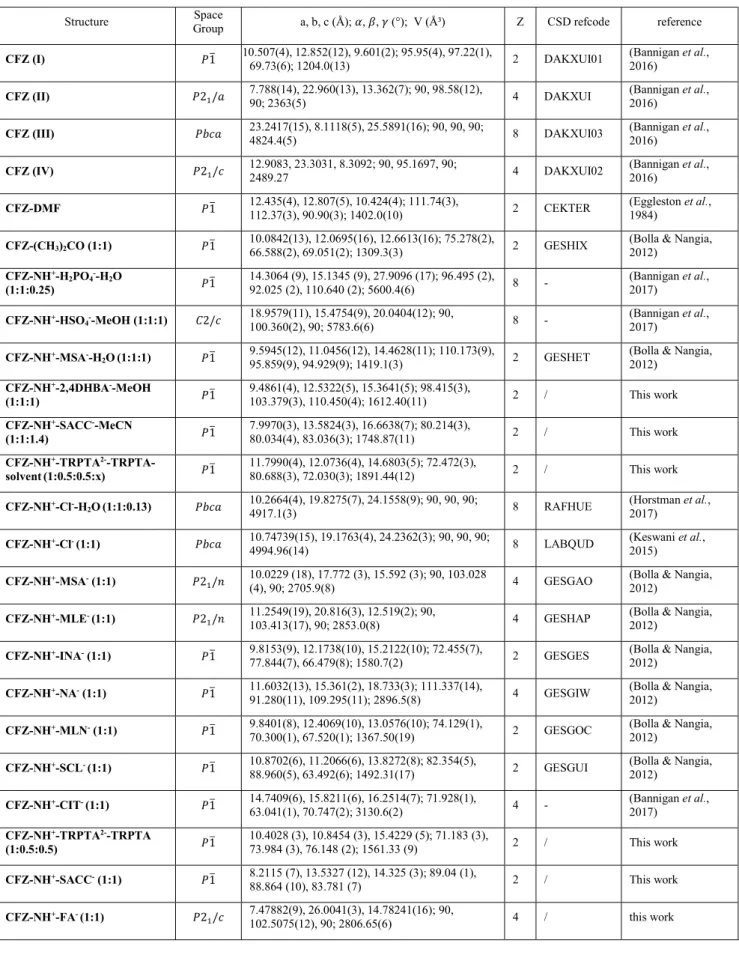
Because clofazimine is gaining attention as a MDR and extensively drug-resistant M. tuberculosis treatment, groups of researchers recently tried new approaches to improve its pharmacological properties. As clofazimine is a weakly basic compound (pKa 9.29 (Keswani et al., 2015)), a possible strategy to increase its solubility is salt formation. In 2012, G. Bolla et al. published eight structures of salts, one of these, clofazimine mesylate was 99 time more soluble in 60% EtOH-water media than pure clofazimine (Bolla & Nangia, 2012). In 2016, Bannigan et al. investigated clofazimine polymorphism (Bannigan et al., 2016) and more recently they studied the solubility of several clofaziminium salts (Bannigan et al., 2017). Here, we report seven new structures: three salts (clofaziminium hydrogen fumarate, CFZ-NH+-FA- (1:1), clofaziminium hydrogen succinate, CFZ-NH+-SA- (1:1)) and clofaziminium saccharinate, CFZ-NH+-SACC- (1:1), two solvated salts (clofaziminium 2,4-dihydroxybenzoate methanol solvate, CFZ-NH+-2,4DHBA--MeOH (1:1:1) and clofaziminium saccharinate acetonitrile solvate, CFZ-NH+-SACC--MeCN (1:1:1.4)), and two cocrystals of salt (structure composed of one cation, one anion and a non-ionized molecule). One is nonstoichiometrically solvated and the other is non-solvated (clofaziminium terephthalate terephthalic acid solvate CFZ-NH+-TRPTA2--TRPTA-solvent (1:0.5:0.5:x) and clofaziminium terephthalate terephthalic acid, CFZ-NH+-TRPTA2--TRPTA (1:0.5:0.5)). Those new salts are compared to the currently known ones in terms of crystal packing and clofaziminium conformation.
3 Figure 1 (a) Numbering scheme of clofazimine/ium(CFZ/CFZ-NH+) (torsions angles: T1, C8-N3-C19-C20; T2, C9-N4-C25-C27; T3, C12-N1-C13-C18 T4, H-N4-C9-C8 and T5, H-N3-C8-C9) and selected acids in the present work : fumaric acid, succinic acid, DL-malic acid, 2,4-dihydroxybenzoic acid, saccharin, L-aspartic acid, terephthalic acid and citric acid. (b) Acids crystallized with clofazimine by G. Bolla et al. (Bolla & Nangia, 2012) and (c) acids crystallized with clofazimine by P. Bannigan et al. (Bannigan et al., 2017). CFZ-NH+-CIT- (1:1) salt was already described by P. Bannigan et al. (Bannigan et al., 2017), in the present study salt formation of CFZ/CIT in 2/1 and 3/1 molar ratios was attempted.
2. Experimental section 2.1. Materials
Clofazimine was purchased from TCI Europe N.V. (Zwindrecht, Belgium), fumaric, 2,4-dihydroxybenzoic and terephthalic acids as well as saccharin were from Sigma-Aldrich (Steinheim, Germany), while succinic acid was purchased from J.T. Baker Chemicals (Deventer, Holland). Crystallization solvents (acetonitrile, methanol, ethyl acetate, and diethyl ether) are commercially available (Acros Organics, Geel, Belgium) and were used without further purification.
2.2. General routes for clofazimine salification.
All samples were prepared by liquid-assisted grinding (LAG) (Frišcic et al., 2009; Shan et al., 2002; Trask et al., 2004; James et al., 2012) with a Retsch MM 400 Mixer Mill apparatus with two grinding jars in which five 2mL Eppendorf tubes can be installed. Each sample grinding was performed with around 100 mg of powder (75mg of clofazimine and corresponding mass of acid to respect 1/1, 2/1 or 3/1 CFZ/acid molar ratios) in presence of 6 to 8 stainless steel grinding balls (2 mm diameter).
4 Following solvents were used for liquid-assisted ball milling: MeCN for CFZ/FA, CFZ/SA, CFZ/SACC, CFZ/L-ASP, and CFZ/DL-MAL equimolar mixtures as well as for CFZ/CA 2/1 and 3/1 mixtures; MeOH for CFZ/2,4-DHBA 1/1 and EtOAc for CFZ/TRPTA. Powders leading to new diffraction patterns were then involved in crystallization assays by slow evaporation at room temperature in a mixture of MeCN and MeOH, except for CFZ/TRPTA 1/1 powder for which EtOAc was used. For CFZ/DL-MAL crystallization in Et2O at 4°C as well as in MeCN at room temperature was also attempted.
2.3. Single-crystal X-ray diffraction (SCXRD).
Selected crystals of suitable size were mounted on an Oxford Diffraction Gemini Ultra R system (4-circle kappa platform, Ruby CCD detector). By default Mo radiation was used for data collection, but when the crystal diffracted poorly, Cu radiation was used as it allows enhanced intensities with this Gemini system. In consequence, data were collected using Mo 𝐾𝛼 (𝜆 = 0.71073 Å) (for CFZ-NH+ -SA- (1:1), CFZ-NH+-TRPTA2--TRPTA-solvent (1:0.5:0.5:x) and CFZ-NH+-TRPTA2--TRPTA (1:0.5:0.5)) or Cu 𝐾𝛼 (𝜆 = 1.54184 Å) for the other salts (CFZ-NH+-FA- (1:1), CFZ-NH+-2,4DHBA --MeOH (1:1:1
),
CFZ-NH+-SACC- (1:1) and CFZ-NH+-SACC--MeCN (1:1:1.4)). Full data sets were collected either at room temperature (CFZ-NH+-FA- (1:1), CFZ-NH+-SA- (1:1), CFZ-NH+ -SACC- (1:1), CFZ-NH+-2,4DHBA--MeOH (1:1:1) and CFZ-NH+-TRPTA2--TRPTA (1:0.5:0.5)) or at 100K (CFZ-NH+-SACC--MeCN (1:1:1.4) and CFZ-NH+-TRPTA2--TRPTA-solvent (1:0.5:0.5:x)). Analytical absorption correction was performed on SCXRD data for structures CFZ-NH+-FA- (1:1), CFZ-NH+-SA- (1:1), CFZ-NH+-TRPTA2--TRPTA-solvent (1:0.5:0.5:x) and CFZ-NH+-TRPTA2--TRPTA (1:0.5:0.5) using CrysAlis PRO 1.171.38.46 ((Rigaku Oxford Diffraction, 2015)) and 1.171.39.46 ((Rigaku Oxford Diffraction, 2018)). Analytical numeric absorption correction using a multifaceted crystal model based on expressions derived by R.C. Clark & J.S. Reid. (Clark, R. C. & Reid, J. S. (1995). Acta Cryst. A51, 887-897). Empirical absorption correction using spherical harmonics, implemented in SCALE3 ABSPACK scaling algorithm. Gaussian absorption correction was performed on SCXRD data for structures CFZ-NH+-SACC--MeCN (1:1:1.4) and CFZ-NH+-2,4DHBA--MeOH (1:1:1) using CrysAlis PRO 1.171.39.46 ((Rigaku Oxford Diffraction, 2018)). Numerical absorption correction based on gaussian integration over a multifaceted crystal model. Empirical absorption correction using spherical harmonics, implemented in SCALE3 ABSPACK scaling algorithm. Multi-scan absorption correction was performed on SCXRD data for structure CFZ-NH+-SACC- (1:1) (CrysAlis PRO 1.171.39.46 (Rigaku Oxford Diffraction, 2018) Empirical absorption correction using spherical harmonics, implemented in SCALE3 ABSPACK scaling algorithm.). Structures were solved by dual-space method using SHELXT (Sheldrick, 2015b) and then refined by least square method using SHELXL-2018/1 (Sheldrick, 2015a) within Olex2 (Dolomanov et al., 2009) and SHELXLE (Hübschle et al., 2011). Non-hydrogen atoms were anisotropically refined. Hydrogen atoms implied in strong hydrogen bonds were localized by Fourier5 difference maps while those not implied in H-bonds were refined as riding body by fixing thermal ellipsoids to 1.2 times the one of preceding atom (or 1.5 for methyl group). Several structures are disordered, those structures are: CFZ-NH+-SA- (1:1) (C29 and C30 of hydrogen succinate), CFZ-NH+-2,4DHBA--MeOH (1:1:1) (2,4DHBA- and MeOH show minor disorder (hydrogen atom of the OH group of disordered MeOH could not be localised by Fourier difference map)), CFZ-NH+-SACC -(1:1) (SACC- is disordered) and CFZ-NH+-SACC--MeCN (1:1:1.4)(chlorophenyl, 1, on Figure 1 and isopropyl groups of clofazimine as well as acetonitrile, which is disordered over three positions). The structure of cocrystal of salt CFZ-NH+-TRPTA2--TRPTA-solvent (1:0.5:0.5:x) presents solvent accessible voids forming channels but solvent of crystallization could not be unambiguously assigned. Therefore, the original reflection file of CFZ-NH+-TRPTA2--TRPTA (1:0.5:0.5) was submitted to the PLATON SQUEEZE procedure (Spek, 2015). Total potential accessible void volume was determined as 489 ų with an electron count per cell of 141, which corresponds to 2.9 EtOAc molecules in the unit cell. Only low resolution data collection (till 0.9Å) could be performed for the structure CFZ-NH+-SACC- (1:1) because crystals were obtained by desolvating crystals of CFZ-NH+-SACC--MeCN (1:1:1.4) (other methods did not lead to unsolvated crystals).
2.4. Powder X-ray diffraction (PXRD).
Powder diffraction data were collected on an X’PERT PRO PANalytical Bragg-Brentano diffractometer with Cu 𝐾𝛼 radiation (𝜆 = 1.54184 Å) at 45 kV and 30 mA with a X’Celerator linear detector. Data were collected from 4 to 40° 2 𝜃 angles with a step size of 0.0167°. Variable temperature measurement using AntonPaar system were realized at 25°C and then between 30 and 200°C with data collection each 10°C. Calculated powder patterns from SCXRD data were generated with the program Mercury CSD 3.10.2 (Macrae et al., 2008).
2.5. Search in the Cambridge structural database (CSD).
Known structures of clofazimine and clofaziminium salts were retrieved from CSD using ConQuest. In total 15 structures implying clofazimine were retrieved (clofazimine, clofaziminium salts and/or solvates). Three other clofaziminium salts (CFZ-NH+-CIT- (1:1), CFZ-NH+-H
2PO4--H2O (1:1:0.25) and CFZ-NH+-HSO
4--MeOH (1:1:1)) were available as supplementary data from the publication of Bannigan et al.) (Bannigan et al., 2017). All these 18 structures were further analyzed.
2.6. Structure visualization, voids calculation and full interaction maps (FIMs) generation.
Structures were visualized with the CCDC Mercury CSD 3.10.2 software and images were generated using the same program (Macrae et al., 2008). Figures illustrating voids were generated with the display voids option in CCDC Mercury CSD 3.10.2 with a probe radius of 1.2 Å and a grid spacing of 0.1 Å. For the structures of CFZ-NH+-TRPTA2--TRPTA-solvent (1:0.5:0.5:x) and CFZ-NH+ -TRPTA2--TRPTA (1:0.5:0.5), least square planes passing through atoms N1, C5 and C6 of
6 clofaziminium were calculated with CCDC Mercury CSD 3.10.2 in order to compare crystal packing of the two structures. Full interaction maps were generated within CCDC Mercury CSD 3.10.2. Probes used for FIMs generation were uncharged NH nitrogen, alcohol oxygen, and carbonyl oxygen with a contour level of 6.0. FIMs were generated using coordinates of a clofazimine molecule (from polymorph DAKXUI03) (Figure 2a), a clofaziminium ion (GESHET, CFZ-NH+-MSA--H
2O(1:1:1)) (Figures 2b and 6a) and of a clofaziminium ion from CFZ-NH+-CIT- (1:1) (Figure 6b) (for those salts, only clofaziminium was considered for FIMs generation).
2.7. Crystal packing comparison.
Crystal packing comparison and figure of crystal lattice overlay were performed with the crystal packing similarity tool in CCDC Mercury (Macrae et al., 2008) with a packing shell size of 15 molecules and a distance and angle tolerance of 30% and 30° respectively. Molecular differences and structure inversion were allowed while bond types, hydrogen positions, atom’s hydrogen count and atom’s bond count were ignored. As multi-components systems with different counter-ions were analyzed, the smallest molecular component was ignored for packing overlay. Only CFZ-NH+-SA -(1:1)showed a molecular overlay of 15 molecules out of 15 with CFZ-NH+-MLN- (1:1) (GESGOC).
2.8. Melting point measurement.
Melting points of non-solvated salts were determined visually by using a Büchi Melting point B545 apparatus.
2.9. Clofazimine/clofaziminium overlay.
For each structure of clofazimine/clofaziminium salt (structures of this paper, and these found in the literature) coordinates of one molecule/ion of clofazimine/clofaziminium were selected from asymmetric unit. All selected molecules/ions were overlaid using the Small Molecule overlay tool in Discovery Studio v18 (Dassault Systèmes BIOVIA, 2016). Neither rotatable bonds nor flexible torsions were allowed and the alignment by consensus was performed by a field 50% steric and 50% electrostatic.
2.10. Conformational analysis by quantum mechanics calculations.
Optimization calculations of clofazimine and clofaziminium were performed using density functional theory (DFT) with the m06 functional (Zhao & Truhlar, 2008) and the 6-311G(d) basis set (Frisch et al., 2016). To this aim coordinates of the neutral form of clofazimine was extracted from DAKXUI03 structure while coordinates of the protonated form (clofaziminium) were extracted from CFZ-NH+ -Cl- (1:1) (LABQUD) crystal structure.
7
3. Results and discussion
Acidic compounds to react with clofazimine were selected after full interaction map analysis of clofazimine and clofaziminium. Salification assays were performed by liquid-assisted grinding and powders leading to new diffraction patterns were used in crystallization experiments. In this section, results obtained after salification assays and PXRD measurement are first summarized. Then, new structures are described and compared to the known ones in terms of interaction and crystal packing similarity. Finally, clofazimine and clofaziminium conformations are compared with each other and with the optimized conformations of clofazimine and clofaziminium (in presence or absence of its counter-ion).
3.1. Full interaction maps analysis of CFZ and CFZ-NH+ and choice of acids to react with CFZ.
Full interaction maps (FIMs) were generated to investigate the propensity of clofazimine/clofaziminium to interact with uncharged NH nitrogen, alcohol oxygen, and carbonyl oxygen. FIMs calculations at the 6.0 contour level indicate two main sites of interaction, namely around nitrogen atoms N3 and N4 for the first site and around N2 for the second site (Figure 2). Both sites are common for clofazimine and clofaziminium. However, the interaction between the first site and carbonyl oxygen or OH probes is stronger in the case of clofaziminium (Figure 2 (a) and (b)). From those FIMs analyses, it can be assumed that clofazimine should be able to form salts with several carboxylic acids. Clofaziminium salts with organic acids (methanesulfonic acid, maleic acid, isonicotinic acid, nicotinic acid, salicylic acid and malonic acid) were reported by G. Bolla et al. (Bolla & Nangia, 2012) while Bannigan et al. obtained clofaziminium salts by reacting clofazimine with organic and inorganic acids (hydrochloric acid, sulphuric acid, nitric acid, oxalic acid, phosphoric acid, citric, formic and acetic acids) (Bannigan et al., 2017). Here, other carboxylic acids (which should protonate N4 of CFZ) were selected to form new clofaziminium salts. 2,4DHBA and DL-MAL were selected because of their alcohol functions which could potentially interact with the N2 site of CFZ. L-ASP was chosen to assess the ability of clofazimine to react with amino-acids (reaction is expected with the carboxylic acid of the side chain). CIT was selected to be reacted in 2/1 and 3/1 CFZ/CIT molar ratios with CFZ to investigate CIT propensity to be unprotonated at multiple sites. Moreover, the potential new structures of CFZ/CIT could be compared to the one obtained by Bannigan et al. (Bannigan et al., 2017). FA and SA were chosen for their similarity and so for their potential ability to form isostructural structures. TRPTA was selected to compare CFZ salts obtained with aliphatic vs aromatic dicarboxylic acids. Finally, saccharin was chosen to evaluate its propensity to react with clofazimine. Indeed, saccharin has acidic properties despite its lack of carboxylic acid function. Moreover, except terephthalic and 2,4-dihydroxybenzoic acids, the selected compounds are classified either as food additives (Ash & Ash, 2008) or as generally regarded as safe (GRAS) (FDA, 2018).
8 Figure 2 FIMs calculated around clofazimine (a) and clofaziminium (b). Carbonyl oxygen,
uncharged NH and alcohol OH probes in red, blue and orange respectively.
3.2. CFZ salification assays and corresponding results.
All liquid-assisted ball milling led to solids with new powder patterns except for CFZ/L-ASP 1/1 (Figure S2). Crystallization assays of all powders except this one were attempted and only, CFZ/DL-MAL did not crystallize in our conditions. Single-crystal X-ray diffraction data were collected on crystals of suitable size. Seven new clofaziminium salt structures were refined. Among those, three are salts (clofaziminium hydrogen fumarate, CFZ-NH+-FA- (1:1), clofaziminium hydrogen succinate, CFZ-NH+-SA-(1:1) and clofaziminium saccharinate, CFZ-NH+-SACC- (1:1)), two are solvated salts (clofaziminium 2,4-dihydroxybenzoate methanol solvate, CFZ-NH+-2,4DHBA--MeOH (1:1:1) and clofaziminium saccharinate acetonitrile solvate, CFZ-NH+-SACC--MeCN(1:1:1.4)), and two are cocrystals of salt (structure composed of one cation, one anion, and a non-ionized molecule). One is nonstoichiometrically solvated and the other is non-solvated (clofaziminium terephthalate terephthalic acid solvate CFZ-NH+-TRPTA2--TRPTA-solvent (1:0.5:0.5:x) and clofaziminium terephthalate terephthalic acid, CFZ-NH+-TRPTA2--TRPTA(1:0.5:0.5)). The latter was obtained by slowly drying corresponding solvated crystals at room temperature. Crystals of CFZ/CIT 2/1 were obtained. However, the structure is intrinsically disordered and could not be refined even using data collected at 100 K. Its cell parameters and were determined (a: 19.2962 (5), b: 12.3140 (3), c: 23.9105 (6); 𝛼: 90, 𝛽: 110.667 (3), 𝛾: 90) and Powder pattern of the as synthesized product matches the one calculated from single-crystal data (Figure S2). Finally, CFZ/CIT 3/1 has a powder pattern highly similar to the one obtained after grinding CFZ/CIT 2/1. However, some new peaks are observed (Figure S2). Crystallization assays of CFZ/CIT 3/1 led to crystals of CFZ/CIT 2/1. Results of CFZ salification assays are summarized in Table 1.
9 Table 1 Clofazimine salification assays and corresponding results.
Acid pKas (Haynes) CFZ/Acid ratio New PXRD? Crystals? Structure refined? FA 3.02; 4.38 1/1 yes yes yes SA 4.21; 5.64 1/1 yes yes yes 2,4-DHBA 3.11 1/1 yes yes yes DL-MAL 3.40; 5.11 1/1 yes no no
SACC 1.94 (Rowe et
al., 2006) 1/1 yes yes yes L-ASP 1.99; 3.90 1/1 no no no TRPTA 3.54; 4.34 1/1 yes yes yes
CIT 3.13; 4.76; 6.40 2/1 3/1 yes yes yes yes** no* no * Structure of CFZ-NH+- CIT- (2:1) is intrinsically disordered. ** Crystallized as CFZ-NH+- CIT- (2:1).
3.3. Crystal packing description of new CFZ salts. 3.3.1. CFZ-NH+-FA- (1:1) salt.
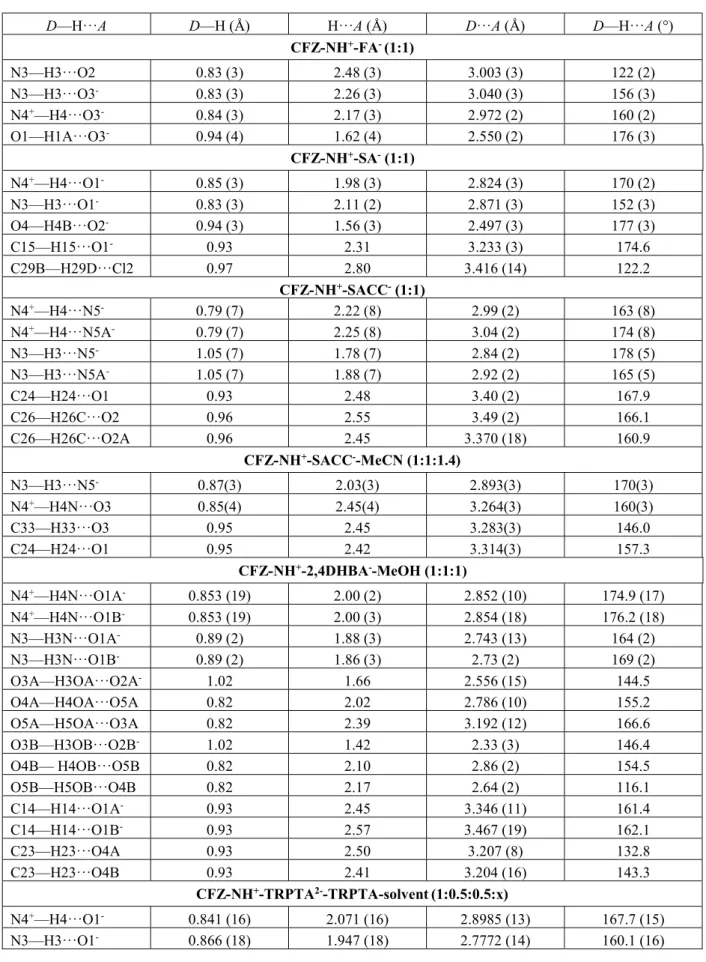
Clofaziminium hydrogen fumarate salt (Figure 3 (a)) crystallizes in 𝑃2 /𝑐 space group (Table 3). Proton transfer between fumaric acid (O3) and the isopropyl imine of CFZ (N4) occurs. As already observed by P. Bannigan et al. (Bannigan et al., 2017), this proton transfer is accompanied by an increase of the iminium angle (angle C9-N4-C25 = 126.02(18)) in comparison with the one of the uncharged clofazimine (C9-N4-C25 around 120° (Table S3)). A 𝑅 (7) H-bond motif is observed between FA- and CFZ-NH+ (N4+-H⋯O3- and N3-H⋯O3-) (Table S2). Hydrogen fumarate forms chains along the a-axis through H-bonds (𝐶 (7) motif, O1-H⋯O3-) (Table S2). A 𝐷(2) motif is also present (N3-H⋯O2) (Table S2). CFZ-NH+ are stacked along a-axis in a head-to-tail fashion. PXRD pattern of the as synthesized powder matches the one calculated from single-crystal data (Figure S1). Melting point of CFZ-NH+-FA- (1:1) is 246°C, which is much higher than the starting API (Table 2).
10 Table 2 Melting points of non-solvated clofaziminium salts.
Compound analyzed Melting point (°C) CFZ (DAKXUI01) 219
Fumaric acid 210 * (Temesvári et al., 1971)
Succinic acid 186.9
Saccharin 229.5 (Basavoju et al., 2008)
Terephthalic acid 276 * (Kimyonok & Ulutürk, 2016) CFZ-NH+-FA- (1:1) 246 CFZ-NH+-SA-(1:1)- 214 CFZ-NH+-SACC- (1:1) 252 CFZ-NH+-TRPTA2--TRPTA (1:0.5:0.5) 240 * sublimation 3.3.2. CFZ-NH+-SA- (1:1) salt.
Clofaziminium hydrogen succinate salt (Figure 3 (b)) crystallizes in 𝑃1 space group (Table 3). Proton transfer between succinic acid (O1) and the isopropyl imine of CFZ (N4) occurs, which is confirmed by bond distances reflecting resonance in the carboxylate and by an increase of the iminium angle (Table S3). A 𝑅 (7) H-bond motif is observed between SA- and CFZ-NH+ (N4+-H⋯O1- and N3-H⋯O1-) and an intramolecular H-bond is also present in hydrogen succinate ion (𝑆 (7) motif, O4-H⋯O2-) (Table S2). 2D sheets are formed through weak C-H⋯O and C-H⋯Cl H-bonds (Table S2). PXRD pattern of the as synthesized product matches the one calculated from single-crystal data (Figure S1). This salt melts at 214°C, this melting point is 5°C less than the one of the starting API CFZ (Table 2).
11 Figure 3 H-bond interactions in the structures of CFZ-NH+-FA- (1:1) (a), CFZ-NH+-SA- (1:1) (b), CFZ-NH+-SACC--MeCN (1:1:1.4) (c), CFZ-NH+-SACC- (1:1) (d)CFZ-NH+-2,4DHBA--MeOH (1:1:1) (e), CFZ-NH+-TRPTA2--TRPTA-solvent(1:0.5:0.5:x) (f) and CFZ-NH+-TRPTA2--TRPTA (1:0.5:0.5) (g). Ellipsoids are drawn at the 50% probability level.
12 Table 3 Experimental details.
Experiments were carried out using an Xcalibur, Ruby, Gemini Ultra diffractometer. H atoms were treated by a mixture of independent and constrained refinement.
CFZ-NH+-FA- (1:1) CFZ-NH+-SA- (1:1) CFZ-NH+-SACC--MeCN(1:1:1.4)
Crystal data
Chemical formula C27H23Cl2N4+·C4H3O4- C27H23Cl2N4+·C4H5O4- C27H23Cl2N4+·C7H4NO3S-·
1.393(C2H3N)
Mr 589.46 591.47 713.78
Crystal system, space group Monoclinic, P21/c Triclinic, P1 Triclinic, P1
Temperature (K) 295 295 100 a, b, c (Å) 7.47882 (9), 26.0041 (3), 14.78241 (16) 10.6376 (6), 12.2781 (5), 12.7975 (8) 7.9970 (3), 13.5824 (3), 16.6638 (7) , , (°) 90, 102.5075 (12), 90 90.079 (4), 113.211 (6), 108.284 (4) 80.214 (3), 80.034 (4), 83.036 (3) V (Å3) 2806.65 (6) 1443.57 (15) 1748.87 (11) Z 4 2 2 Radiation type Cu K Mo K Cu K (mm-1) 2.45 0.27 2.61 Crystal size (mm) 0.57 × 0.13 × 0.04 0.57 × 0.42 × 0.03 0.70 × 0.26 × 0.05 Data collection
Absorption correction Analytical Analytical Gaussian Tmin, Tmax 0.502, 0.915 0.897, 0.990 0.295, 1.000
No. of measured,
independent and observed [I > 2(I)] reflections 15078, 4944, 4394 10778, 5090, 3520 17257, 6202, 5378 Rint 0.021 0.022 0.033 (sin /)max (Å-1) 0.597 0.595 0.598 Refinement R[F2 > 2(F2)], wR(F2), S 0.049, 0.136, 1.01 0.045, 0.122, 1.03 0.053, 0.142, 1.05 No. of reflections 4944 5090 6202 No. of parameters 383 412 594 No. of restraints 0 4 137 𝜌max, Δ𝜌min (e Å-3) 0.46, -0.51 0.24, -0.24 0.61, -0.67
13 CFZ-NH+-SACC- (1:1) CFZ-NH+ -2,4DHBA--MeOH (1:1:1) CFZ-NH+-TRPTA 2--TRPTA-solvent (1:0.5:0.5:x) CFZ-NH+-TRPTA 2--TRPTA (1:0.5:0.5) Crystal data Chemical formula C27H23Cl2N4+· C7H4NO3S -C27H23Cl2N4+· C7H5O4-·CH4O C27H23Cl2N4+· 0.5(C8H4O42-)· 0.5(C8H6O4) C27H23Cl2N4+· 0.5(C8H4O42-) ·0.5(C8H6O4) Mr 656.56 659.54 639.51 639.51
Crystal system, space group Triclinic, P1 Triclinic, P1 Triclinic, P1 Triclinic, P1 Temperature (K) 295 295 100 295 a, b, c (Å) 8.2115 (7), 13.5327 (12), 14.325 (3) 9.4861 (4), 12.5322 (5), 15.3641 (5) 11.7990 (4), 12.0736 (4), 14.6803 (5) 10.4028 (3), 10.8454 (3), 15.4229 (5) , , (°) 89.04 (1), 88.864 (10), 83.781 (7) 98.415 (3), 103.379 (3), 110.450 (4) 72.472 (3), 80.688 (3), 72.030 (3) 71.183 (3), 73.984 (3), 76.148 (2) V (Å3) 1582.0 (3) 1612.41 (12) 1891.44 (12) 1561.33 (9) Z 2 2 2 2 Radiation type Cu K Cu K Mo K Mo K (mm-1) 2.82 2.22 0.21 0.25 Crystal size (mm) 0.54 × 0.29 × 0.05 0.49 × 0.12 × 0.06 0.57 × 0.39 × 0.26 0.72 × 0.48 × 0.41 Data collection
Absorption correction Multi-scan Gaussian Analytical Analytical Tmin, Tmax 0.549, 1.000 0.539, 0.991 0.917, 0.960 0.886, 0.921
No. of measured,
independent and observed [I > 2(I)] reflections 7488, 3783, 2265 15458, 5692, 4965 20823, 11543, 9100 13059, 5704, 4554 Rint 0.038 0.021 0.022 0.018 (sin /)max (Å-1) 0.526 0.597 0.714 0.602 Refinement R[F2 > 2(F2)], wR(F2), S 0.064, 0.190, 1.08 0.037, 0.111, 1.04 0.044, 0.118, 1.03 0.040, 0.109, 1.02 No. of reflections 3783 5692 11543 5704 No. of parameters 525 555 420 420 No. of restraints 483 66 0 0 𝜌max, 𝜌min (e Å-3) 0.23, -0.22 0.24, -0.32 0.41, -0.30 0.23, -0.27
Computer programs: CrysAlis PRO 1.171.38.46 (Rigaku OD, 2015), CrysAlis PRO 1.171.39.46 (Rigaku OD, 2018), SHELXT (Sheldrick, 2015), SHELXT 2014/5 (Sheldrick, 2014), SHELXL2016/6 (Sheldrick, 2016).
IMPORTANT: this document contains embedded data - to preserve data integrity, please ensure where possible that the IUCr
Word tools (available from http://journals.iucr.org/services/docxtemplate/) are installed when editing this document. 14
3.3.3. (CFZ-NH+)
1(SACC-)1(MeCN)1.4 solvated salt.
Clofaziminium saccharinate acetonitrile solvated salt (Figure 3 (c)) crystallizes in 𝑃1 space group (Table 3). Proton transfer between saccharin (N5) and the isopropyl imine of CFZ (N4) occurs and is confirmed by an increase of the iminium angle (Table S3). Acetonitrile solvate is disordered over three positions (first, second and third position occupancies were refined to the following values 0.4640, 0.4890 and 0.440). A 𝑅 (9) H-bond motif is observed between SACC- and CFZ-NH+ (N4+ -H⋯O3 and N3-H⋯N5- (Figure 3 (c) and Table S2)). Weak C-H⋯O H-bonds stabilize head-to-tail stacking of CFZ-NH+ (Table S2).
3.3.4. CFZ-NH+-SACC- (1:1) salt
Clofaziminium saccharinate (Figure 3 (d)) was obtained by drying crystals of CFZ-NH+-SACC- -MeCN (1:1:1.4) at room temperature. The unsolvated salt (CFZ-NH+-SACC- (1:1)) crystallizes in 𝑃1 space group (Table 3). Proton transfer between saccharin (N5) and the isopropyl imine of CFZ (N4) occurs and is confirmed by an increase of the iminium angle (Table S3). A 𝑅 (7) H-bond motif is observed between SACC- and CFZ-NH+ (N4+-H⋯N5- and N3-H⋯N5- (Figure 3 (c) and Table S2)). Weak C-H⋯O H-bonds stabilize head-to-tail stacking of CFZ-NH+ (Table S2). It is interesting to notice that liquid-assisted grinding experiment leads to the unsolvated salt. Indeed, the powder pattern of the as synthesized product (CFZ/SACC 1/1 LAG MeCN) matches the one calculated from SCXRD data of CFZ-NH+-SACC- (1:1) and not the one calculated from SCXRD data of CFZ-NH+-SACC- -MeCN (1:1:1.4).
3.3.5. CFZ-NH+-2,4DHBA--MeOH (1:1:1) solvated salt.
Clofaziminium 2,4-dihydroxybenzoate methanol solvated salt (Figure 3 (e)) crystallizes in 𝑃1 space group (Table 3). Proton transfer between 2,4-dihydroxybenzoic acid (O1) and the isopropyl imine of CFZ (N4) is confirmed by bond distances reflecting resonance in the carboxylate and by an increase of the iminium angle in comparison with the unprotonated form of clofazimine (Table S3). MeOH and 2,4-DHBA- are slightly disordered. A 𝑅 (7) H-bond is observed between 2,4-DHBA- and CFZ-NH+ (N4+-H⋯O1A- and N3-H⋯O1A- as well as N4+-H⋯O1B- and N3-H⋯O1B-) (Table S2). An intramolecular H-bond is also present in 2,4-dihydroxybenzoate (𝑆 (6) motif, O3A-H⋯O2A- as well as O3B-H⋯O2B-) (Table S2). Methanol molecules serve as a bridge between two 2,4-DHBA-. Indeed, a 𝐶 (8) motif (O4A-H⋯O5A and O5A-H⋯O3A) is observed (Table S2) (Figure 4). In the second position of 2,4DHBA- and MeOH (disordered), the H- bond interactions form between O4B-H ⋯ O5B and O5B-O4B-H ⋯ O4B (Table S2). CFZ-NO4B-H+ are stacked in a head-to-tail fashion. This arrangement is further stabilized by weak C-H⋯O H-bonds (Table S2). Powder pattern of the as synthesized product matches the one calculated from single-crystal data (Figure S1).
15 Figure 4 MeOH molecules serve as bridges between 2,4-DHBA- in the structure of CFZ-NH+ -2,4DHBA--MeOH (1:1:1).
3.3.6. CFZ-NH+-TRPTA2--TRPTA-solvent (1:0.5:0.5:x) nonstoichiometrically solvated cocrystal
of salt.
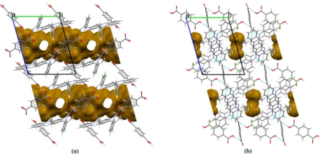
Clofaziminium terephthalate terephthalic acid nonstoichiometrically solvated cocrystal of salt crystallizes in 𝑃1 space group (Table 3). Its asymmetric unit is composed of one ion of clofaziminium with a half terephthalate ion and a half molecule of terephthalic acid (Figure 3 (f)). Indeed, terephtalic acid and terephthalate are both positioned on an inversion center. Proton transfer between terephthalate (O1) and the isopropyl imine of CFZ (N4) occurs and is confirmed by an increase of the iminium angle (Table S3). Actually, terephthalate anion serves as a linker between two clofaziminium cations, generating three-component CFZ-NH+-TRPTA2--CFZ-NH+ assemblies (Figure 5 (a)). In those, a 𝑅 (7) H-bond motif is observed between TRPTA2- and CFZ-NH+ (N4+-H⋯O1- and N3-H⋯O1-) (Table S2). Those three-component assemblies are interconnected through H-bonds between terephthalate anions and terephthalic acid molecules (𝐷 (2) H-bond motif, O1A-H⋯O1-) (Figure 5 (a) and Table S2). Weak C-H⋯O bonds are also present in the structure (Table S2). This arrangement results in solvent accessible voids forming channels (Figure 5 (b) and S4(a)). Although one molecule of ethyl acetate could be localized, high residual electron densities remained in the structure. As all crystallization solvent molecules could not be assigned unambiguously despite low temperature measurement (100K), Platon Squeeze (Spek, 2015) procedure was applied to the data.
16 Figure 5 Three-component assemblies (capped stick) linked by terephthalic acid (ball and stick) of CFZ-NH+-TRPTA2--TRPTA-solvent (1:0.5:0.5:x) (a) and of CFZ-NH+-TRPTA2--TRPTA
(1:0.5:0.5) (c). Voids along b-axis (top) and c-axis (bottom) of CFZ-NH+-TRPTA2--TRPTA-solvent (1:0.5:0.5:x) (b) and of CFZ-NH+-TRPTA2--TRPTA (1:0.5:0.5) (d).
3.3.7. CFZ-NH+-TRPTA2--TRPTA (1:0.5:0.5) cocrystal of salt.
Clofaziminium terephthalate terephthalic acid cocrystal of salt (Figure 3 (g)) was obtained by drying CFZ-NH+-TRPTA2--TRPTA-solvent (1:0.5:0.5:x) at room temperature. CFZ-NH+-TRPTA2- -TRPTA (1:0.5:0.5) cocrystal of salt crystallizes in 𝑃1 space group (Table 3). Its asymmetric unit is composed of one ion of clofaziminium with a half terephthalate ion and a half molecule of terephthalic acid (Figure 3 (f)). Indeed, terephtalic acid and terephthalate are both positioned on an inversion center. Proton transfer between terephthalate (O1) and the isopropyl imine of CFZ (N4) occurs and is confirmed by an increase of the iminium angle (Table S3). Like in the solvated structure, terephthalate anion serves as a linker between two clofaziminium cations, leading to three-component assemblies. Bifurcated H-bonds between TRPTA2- and CFZ-NH+ are observed (𝑅 (9) motif, N4-H+⋯O1- and N3-H⋯O2-) in addition to the 𝑅 (7), N4+-H⋯O1- and N3-H⋯O1- (Table S2). Three-component assemblies are interconnected through H-bonds with terephthalic acid molecules (𝐷 (2) H-bond motif, O1B-H⋯O2-) (Figure 5 (c)) (Table S2). Voids are present in the structure of CFZ-NH+-TRPTA2--TRPTA (1:0.5:0.5)
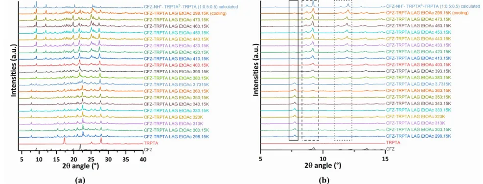
, however, they are not interconnected and no channel is observed (Figure 5 (d) and S4(b)). Interestingly, as shown by variable temperature powder X-ray diffraction, this non-solvated structure can be obtained by heating the batch powder synthesized by LAG with EtOAc as solvent (Figure S3).
17
3.4. CFZ-counter ion interaction comparison.
Full interaction map analyses revealed two possible sites of interaction around clofazimine, namely, near nitrogen atoms N3 and N4 for the first site and around N2 for the second one. Analysis of the 18 known structures (15 on CSD and 3 supplied as supplementary data from the article of Bannigan et al. (Bannigan et al., 2017)) and the seven new ones described in this paper reveals that first site is implied in all salt structures while the second one is only implied in two structures. One structure is the hydrated salt CFZ-NH+-MSA--H
2O (1:1:1). Indeed, N2 interacts through H-bonds with a water molecule (𝐷 (2), 2.899(4) Å and 173(2)°) (Figure 6 (a)). In this case, the water molecule is placed at an optimal position (even for a contour level as high as 30). The second structure is CFZ-NH+-CIT -(1:1) in which N2 interacts through H-bond with one carboxylic group of dihydrogen citrate ion (𝐷 (2), 2.898(4) Å and 161.6°, Figure 6 (b)). In this structure, the OH from the carboxylic acid interacting with N2 is, however, less optimally placed. While H-bonds between N2 from CFZ and OH from 2,4-DHBA or MeOH could have been expected, such interaction is not observed in the structure of CFZ-NH+-2,4DHBA--MeOH (1:1:1).
Figure 6 FIMs calculated around clofaziminium from CFZ-NH+-MSA--H
2O (1:1:1) (a) and CFZ-NH+ of CFZ-NH+-CIT-(1:1) (b) carbonyl oxygen, uncharged NH and alcohol OH probes in red, blue and orange respectively.
Concerning clofaziminium salts obtained with aliphatic dicarboxylic acids, in currently known structures, proton transfer occurred from one carboxylic acid function, while the other remained protonated. However, in the structures involving the aromatic dicarboxylic acid CFZ-NH+-TRPTA2- -TRPTA (1:0.5:0.5) and CFZ-NH+-TRPTA2--TRPTA-solvent (1:0.5:0.5:x)), terephthalate anion is fully unprotonated, leading to three component assemblies (CFZ-NH+-TRPTA2--CFZ-NH+) bridged together by the remaining terephthalic acid molecule (CFZ and TRPTA were ball milled in 1/1 molar ratio). Double deprotonation of terephthalic acid could eventually be explained by the two low pKa values for this compound (3.54 and 4.34). But interestingly, fumaric acid, which has second pKa value similar to the one of terephthalic acid (pKa of 3.02 and 4.38 for FA) is unprotonated at only one site, forming thus hydrogen fumarate salt of clofazimine. This observation could be explained by the longer distance between carboxylates in TRPTA in comparison with the COOH-COOH distance in
18 FA (5.803(3) in the structure of CFZ-NH+-TRPTA2--TRPTA (1:0.5:0.5) vs. 3.883(3) in the structure of CFZ-NH+-FA- (1:1)).
3.5. Crystal packing comparison
All known clofaziminium salt structures, as well as those described in this paper, were compared by submitting them to the crystal packing similarity tool in CCDC Mercury (Macrae et al., 2008) with a packing shell size of 15 molecules and a distance and angle tolerance of 30% and 30° respectively. Only CFZ-NH+-SA- (1:1) showed a molecular overlay of 15 molecules out of 15 with CFZ-NH+ -MLN- (1:1) (GESGOC). This result was quite interesting. Indeed, isostructurality could have been expected between salts implying hydrogen succinate and hydrogen fumarate as counter-ion (as were previously described by J.Galcera et al.) (Galcera & Molins, 2009). However, crystal packing comparison of clofaziminium salts CFZ-NH+-FA- (1:1) and CFZ-NH+-SA- (1:1) indicates a lack of isostructurality. The negative charge on the carboxylate part of hemisuccinate favors the formation of a strong intramolecular H-bond at the expense of intermolecular H-bonds (in contrast to the one observed in CFZ-NH+-FA- (1:1)) preventing isostructurality with CFZ-NH+-FA- (1:1). This intramolecular H-bond could be similar to the one observed in hydrogen maleate in the structure of CFZ-NH+-MLE-(1:1), described by G. Bolla and A. Nangia (Bolla & Nangia, 2012). However, no isostructurality is observed with CFZ-NH+-MLE- (1:1).
Figure 7 Structure of salt CFZ-NH+-SA- (1:1)
1 (a),of overlay between CFZ-NH+-SA- (1:1) and CFZ-NH+-MLN- (1:1) (b), and of CFZ-NH+-MLN- (1:1) (c). Crystal packing comparison of CFZ-NH+-SA- (1:1)(green) with CFZ-NH+-MLN- (1:1) salt (grey) (d).
19 More surprisingly, CFZ-NH+-SA- (1:1) presents isostructurality with CFZ-NH+-MLN- (1:1), a clofaziminium salt previously described by G. Bolla and A. Nangia (Bolla & Nangia, 2012). The overlay between the crystal lattice of CFZ-NH+-SA- (1:1) and the one of CFZ-NH+-MLN- (1:1) is illustrated in Figure 7. Other crystal packings that are interesting to compare are those of CFZ-NH+ -SACC—MeCN (1:1:1.4) and of CFZ-NH+-TRPTA2--TRPTA-solvent (1:0.5:0.5:x) with their non-solvated analogues (CFZ-NH+-SACC- (1:1) and CFZ-NH+-TRPTA2--TRPTA (1:0.5:0.5)). In terms of composition, those salts differ only by the presence or absence of solvent in the structure. However, this solvent may have a strong impact on the crystal packing.Indeed in the non-solvated structure of CFZ-NH+-SACC-, a 𝑅 (7) H-bond motif is observed between SACC- and CFZ-NH+ (N4+-H⋯N5 -and N3-H⋯N5-) while in the solvated salt a 𝑅 (9) motif (N4+-H⋯O3 and N3-H⋯N5-) is observed. Such modification of H-bond results in a slight shift in the position of SACC- (Figure 8). However both structures are quite similar as 14 clofaziminium ions over 15 overlay when solvated and non-solvated structures are analysed by the crystal packing similarity tool in CCDC mercury (Macrae et al., 2008). More structural changes are observed between CFZ-NH+-TRPTA2--TRPTA-solvent (1:0.5:0.5:x) and CFZ-NH+-TRPTA2--TRPTA- (1:0.5:0.5) (Figure 9 (c)). In the non-solvated structure, H-bonds observed between TRPTA2- and CFZ-NH+ are bifurcated, which is not the case in its solvated analog. Those bifurcated H-bonds lead to major modifications in terms of distance between the planes passing through the main core of CFZ-NH+ (those planes were calculated by taking N1, C5 and C6 atoms of clofaziminium as reference). Indeed, while the inter-plane distance is 6.049Å in the structure of CFZ-NH+-TRPTA2--TRPTA-solvent (1:0.5:0.5:x) (d, Figure 9 (a)), it is only 2.217 Å in the one of CFZ- NH+-TRPTA2--TRPTA (1:0.5:0.5) (d, Figure 9(b)). The differences in crystal packing observed between those two structures result in major changes in terms of (solvent accessible) voids. Indeed in the structure of CFZ-NH+-TRPTA2--TRPTA-solvent (1:0.5:0.5:x), solvent accessible voids form channels as illustrated at Figure 5 (b) while in that of CFZ-NH+ -TRPTA2--TRPTA (1:0.5:0.5) voids are not interconnected and no channel is observed (Figure 5 (d)).
Figure 8 Crystal packing overlay of solvated (green) and non-solvated structuresof clofaziminium saccharinate.
20 Figure 9 Distance, d, between the planes passing through the main core of CFZ-NH+ in the
structures of CFZ-NH+-TRPTA2--TRPTA-solvent (1:0.5:0.5:x) (a) and CFZ-NH+-TRPTA2- -TRPTA (1:0.5:0.5) (b). Crystal packing overlay of solvated (green) and non-solvated structures(c).
3.6. Conformational analysis of clofazimine/clofaziminium.
Clofazimine/clofaziminium conformation is quite restrained, however, conformational changes could be expected around three main torsion angles (T1, T2 and T5 Figure 1) for clofazimine and around four torsion angles for clofaziminium (T1, T2, T4 and T5 Figure 1). T3 torsion angle (Figure 1) is however expected to have values around 90° (because of steric hindrance that would result from T3 torsion angles values different from 90°). To assess the conformational versatility of clofazimine(ium), coordinates were extracted from known structures (from CSD data, and supplementary data from the article of Bannigan et al. (Bannigan et al., 2017)) and overlaid (Figure 10 (a) and (b)). Results indicate similar conformations of all clofazimine molecules (Figure 10 (a)) which could be explained by the formation of an intramolecular H-bond between N3-H and N4. Main differences are observed around T1 and T2 torsion angles. T1 and T2 have values around 150 or -150° with two possible combinations: for negative T2 values, T1 can be either positive (CFZ I and II), or negative (Table S3). Clofaziminium ions also adopt similar conformations (Figure 10 (b)). T1 torsion angle is quite well conserved in salts structures with main values being comprised between 130 and 160°.
21 Figure 10 Front and side views after superimposition of clofazimine (a) and clofaziminium (b) conformations, optimized geometries of clofazimine (c), of clofaziminium without any counter-ion (d) and of clofaziminium with Cl- counter-ion (e).
However, flexibility is observed around T2. Indeed, T2 torsion angle can adopt different values around 150-160°, 90-100°, and 70-80°. More surprising are the values of T4 and T5 torsion angles. Indeed T4 torsion angles are comprised between 0 and 15° while T5 values are mainly comprised between 0 and 20° in absolute value (there are few exceptions: 38° in CFZ-NH+-SACC--MeCN (1:1:1.4), 31° in CFZ-NH+-SACC-(1:1) -28° and 34° in CFZ-NH+-TRPTA2--TRPTA-solvent (1:0.5:0.5:x) and CFZ-NH+-FA- (1:1)). Such values of T4 and T5 result in steric hindrance between the hydrogen atoms positioned on N3 and N4 (distance around those hydrogen atoms is around 1.9 Å (Table S3)). Because of this steric effect, other values of T4 and T5 could be expected, indeed N3-H could be tilted outside the plane of clofazimine. To assess the effect of the counter-ion on CFZ-NH+ conformation, optimization calculations were performed in absence and in presence of the counter-ion (Cl-), starting from coordinates of clofaziminium chloride (LABQUD) (Figure 9 (d) and (e) and table S5 and S6). Optimization of clofazimine was also performed (starting from coordinates of DAKXUI03) (Figure 10 (c) and Table S4) for the sake of comparison. Optimized CFZ-NH+ conformation confirms the hypothesis of possible higher T5 torsion angle values (T4 = 15.40° and T5= 51.35°). Such a high value of T5 is however never observed in crystal structures. This suggests a quite strong effect of the counter-ion on clofaziminium conformation. Indeed, in clofaziminium salt
22 crystal structures, the hydrogen atom positioned on N3 remains almost in the plane of clofazimine. This could be explained by the presence of H-bonds between CFZ-NH+ and its counter-ion, which forces the N3-H to stay close to the clofaziminium plane and compensates for the energy penalty due to steric hindrance. This assumption is supported by the fact that the optimized geometry of CFZ-NH+ in presence of Cl- counter-ion results in T5 torsion values much closer, in absolute value, to the ones observed in crystal structures (T4=-3.41° and T5=-11.34° in optimized CFZ-NH+-Cl- (1:1)) (Figure 10 (e) compared to Figure 10 (b) and Table S3).
4. Conclusions
Six new (solvated/cocrystal of) salts of clofazimine drug with fumaric, succinic, 2,4-dihydroxybenzoic and terephthalic acids as well as with saccharin are reported and compared to the currently known clofaziminium salt structures. While full interaction map analyses reveal two main sites of interaction around clofaziminium, CFZ-NH+ interaction with its counter-ion mainly occurs at the site located around N3 and N4. Interaction at the second site of clofaziminium is only observed in two structures (CFZ-NH+-CIT- (1:1) and CFZ-NH+-MSA--H
2O (1:1:1)) that were already reported. Acid/base reaction between clofazimine and dicarboxylic acids, more often results in single proton transfer (one carboxylic acid function is unprotonated while the other remains protonated) except in the structures implying terephthalic acid (in which COOH-COOH distance is longer than in aliphatic dicarboxylic acids evaluated in the present work). In structures implying terephthalic acid, one terephthalate ion is fully unprotonated while the remaining terephthalic acid molecules serve as bridges between terephthalate anions. Crystal packing comparison of all known structures revealed that CFZ-NH+-SA- (1:1) and CFZ-NH+-MLE- (1:1) are isostructural. CFZ-NH+-SACC--MeCN (1:1:1.4) and CFZ-NH+-TRPTA2--TRPTA-solvent (1:0.5:0.5:x) can be converted to CFZ-NH+ -SACC- (1:1) and CFZ-NH+-TRPTA2--TRPTA (1:0.5:0.5) by solvent evaporation. In both case solvent evaporation leads to structural changes and in H-bond interactions. Crystal packing comparison of those two structures reveals that in clofaziminium saccharinate, anion position is slightly changed while in clofaziminium terephthalate, solvent has a strong impact on H-bond interactions and packing. Indeed, H-bond interactions between CFZ-NH+ and TRPTA2- change upon drying, leading to the disappearance of solvent accessible channels in favor of isolated voids. In all known structures, clofazimine and clofaziminium adopt similar conformations. The one observed in salt structures may be surprising at first glance because it results in steric hindrance around the protonated site. However conformational optimization of clofaziminium showed the strong impact of the counter-ion on CFZ-NH+ conformation. Indeed, H-bonds occurring between clofaziminium and the counter-ion compensate the energy penalty due to the steric hindrance. Altogether, these results indicate that despite its quite restrained conformation, clofazimine can crystallize as salts with a high variety of packing, which could lead to different physicochemical and pharmacokinetic properties.
23
Acknowledgements This work was performed on XRD equipment from the PC2 platform at UNamur. The authors thank CECI platform and Jean Quertinmont for their help with conformational calculations.
Conflicts of interest
The authors declare that the research was conducted in the absence of any commercial or financial relationships that could be construed as a potential conflict of interest.
References
Ash, M. & Ash, I. (2008). Handbook of Food Additives New-York: Synapse Information Resources. Bannigan, P., Durack, E., Madden, C., Lusi, M. & Hudson, S. P. (2017). ACS Omega. 2, 8969–8981. Bannigan, P., Zeglinski, J., Lusi, M., O’Brien, J. & Hudson, S. P. (2016). Cryst. Growth Des. 16,
7240–7250.
Barry, V. C., Belton, J. G., Conalty, M. L., Denneny, J. M., Edward, D. W., O’Sullivan, J. F., Twomey, D. & Winder, F. (1957). Nature. 179, 1013–1015.
Basavoju, S., Bostro, D. & Velaga, S. (2008). Pharm. Res. 25, 530–541. Bolla, G. & Nangia, A. (2012). Cryst. Growth Des. 12, 6250–6259.
Cholo, M. C., Steel, H. C., Fourie, P. B., Germishuizen, W. A. & Anderson, R. (2012). J. Antimicrob. Chemother. 67, 290–298.
Dassault Systèmes BIOVIA (2016).
Dolomanov, O. V., Bourhis, L. J., Gildea, R. J., Howard, J. A. K. & Puschmann, H. (2009). J. Appl. Crystallogr. 42, 339–341.
FDA (2018). SCOGS (Select Committee on GRAS Substances).
Frisch, M. J., Trucks, G. W., Schlegel, H. B., Scuseria, G. E., Robb, M. A., Cheesemen, J. R., Scalmani, G., Barone, V., Petersson, G. A., Nakatsuji, H., Li, X., Caricato, M., Marenich, A. V, Bloino, J., Janesco, B. G., Gomperts, R., Mennucci, B., Hratchian, H. P., Ortiz, J. V, Izmaylov, A. F., Sonnenberg, J. L., Williams-Young, D., Ding, F., Lipparini, F., Egidi, F., Goings, J., Peng, B., Petrone, A., Henderson, T., Ranasinghe, D., Zakrzewski, V. G., Gao, J., Rega, N., Zheng, G., Liang, W., Hada, M., Ehara, M., Toyota, K., Fukuda, R., Hasegawa, J., Ishida, M., Nakajima, T., Honda, Y., Kitao, O., Nakai, H., Vreven, T., Throssell, K., Montgomery, J. A., Peralta, J. J. E., Ogliaro, F., Bearpark, M. J., Heyd, J. J., Brothers, E. N., Kudin, K. N., Staroverov, V. N., Keith, T. A., Kobayashi, R., Normand, J., Raghavachari, K., Rendell, A. P., Burant, J. C., Iyengar, S. S., Tomasi, J., Cossi, M., Millam, J. M., Klene, M., Adamo, C., Cammi, R., Ochterski, J. W., Martin, R. L., Morokuma, K., Farkas, O., Foresman, J. B. & Fox, D. J. (2016). Gaussian 16, Revision A.03 Wallingford CT: Gaussian, Inc.
Frišcic, T., Childs, S. L., Rizvi, S. A. A. & Jones, W. (2009). CrystEngComm. 11, 418–426. Galcera, J. & Molins, E. (2009). Cryst. Growth Des. 9, 327–334.
24 Haynes, W. M. Handbook of Chemistry and Physics Boca Raton.
Horstman, E. M., Keswani, R. K., Frey, B. A., Rzeczycki, P. M., LaLone, V., Bertke, J. A., Kenis, P. J. A. & Rosania, G. R. (2017). Angew. Chemie - Int. Ed. 56, 1815–1819.
Hübschle, C. B., Sheldrick, G. M. & Dittrich, B. (2011). J. Appl. Crystallogr. 44, 1281–1284.
James, S. L., Adams, C. J., Bolm, C., Braga, D., Collier, P., Frišcic, T., Grepioni, F., Harris, K. D. M., Hyett, G., Jones, W., Krebs, A., Mack, J., Maini, L., Orpen, A. G., Parkin, I. P., Shearouse, W. C., Steed, J. W. & Waddell, D. C. (2012). Chem. Soc. Rev. 41, 413–447.
Job, C. K., Yoder, L., Jacobson, R. R. & Hastings, R. C. (1990). J. Am. Acad. Dermatol. 23, 236–241. Keswani, R. K., Baik, J., Yeomans, L., Hitzman, C., Johnson, A. M., Pawate, A. S., Kenis, P. J. A.,
Rodriguez-Hornedo, N., Stringer, K. A. & Rosania, G. R. (2015). Mol. Pharm. 12, 2528–2536. Kimyonok, A. B. E. & Ulutürk, M. (2016). J. Energ. Mater. 34, 113–122.
Levy, L. (1974). Am. J. Trop. Med. Hyg. 23, 1097–1109.
Macrae, C. F., Bruno, I. J., Chisholm, J. A., Edgington, P. R., McCabe, P., Pidcock, E., Rodriguez-Monge, L., Taylor, R., Van De Streek, J. & Wood, P. A. (2008). J. Appl. Crystallogr. 41, 466– 470.
Narang, A. S. & Srivastava, A. K. (2002). Drug Dev. Ind. Pharm. 28, 1001–1013. O’Reilly, J. R., Corrigan, O. I. & O’Driscoll, C. M. (1994). Int. J. Pharm. 105, 137–146.
Redd, V. M., O’Sullivan, J. F. & Gangadharam, P. R. J. (1999). J. Antimicrob. Chemother. 43, 615– 623.
Rigaku Oxford Diffraction (2015). CrysAlis PRO. Rigaku Oxford Diffraction Ltd, Yarnton, England. Rigaku Oxford Diffraction (2018). CrysAlis PRO. Rigaku Oxford Diffraction Ltd, Yarnton, England. Rowe, R. C., Sheskey, P. J. & Owen, S. C. (2006). Handbook of Pharmaceutical Excipients
Pharmaceutical Press and American Pharmacists Association.
Salem, I. I., Steffan, G. & Düzgünes, N. (2003). Int. J. Pharm. 260, 105–114. Shan, N., Toda, F. & Jones, W. (2002). Chem. Commun. 2, 2372–2373. Sheldrick, G. M. (2015a). Acta Crystallogr. Sect. C Struct. Chem. 71, 3–8. Sheldrick, G. M. (2015b). Acta Crystallogr. Sect. A Found. Crystallogr. 71, 3–8. Spek, A. L. (2015). Acta Crystallogr. Sect. C Struct. Chem. 71, 9–18.
Temesvári, I., Liptay, G. & Pungor, E. (1971). E. J. Therm. Anal. 3, 293.
Trask, A. V, Motherwell, W. D. S. & Jones, W. (2004). Chem. Commun. 890–891. World Health Organization (2015). Treatment of Tuberculosis.
Zhao, Y. & Truhlar, D. G. (2008). Theor. Chem. Acc. 120, 215–241.
List of Figure and Table captions:
Figure 1: (a) Numbering scheme of clofazimine/ium(CFZ/CFZ-NH+) (torsions angles: T1, C8-N3-C19-C20; T2, C9-N4-C25-C27; T3, C12-N1-C13-C18 T4, H-N4-C9-C8 and T5, H-N3-C8-C9) and
25 selected acids in the present work : fumaric acid, succinic acid, DL-malic acid, 2,4-dihydroxybenzoic acid, saccharin, L-aspartic acid, terephthalic acid and citric acid. (b) Acids crystallized with clofazimine by G. Bolla et al. (Bolla & Nangia, 2012) and (c) acids crystallized with clofazimine by P. Bannigan et al. (Bannigan et al., 2017). CFZ-NH+-CIT- (1:1) salt was already described by P. Bannigan et al. (Bannigan et al., 2017), in the present study salt formation of CFZ/CIT in 2/1 and 3/1 molar ratios was attempted.
Figure 2: FIMs calculated around clofazimine (a) and clofaziminium (b). Carbonyl oxygen, uncharged NH and alcohol OH probes in red, blue and orange respectively.
Figure 3: H-bond interactions in the structures of CFZ-NH+-FA- (1:1) (a), CFZ-NH+-SA- (1:1) (b), CFZ-NH+-SACC--MeCN (1:1:1.4) (c), CFZ-NH+-SACC- (1:1)- (d)CFZ-NH+-2,4DHBA--MeOH (1:1:1) (e), CFZ-NH+-TRPTA2--TRPTA-solvent(1:0.5:0.5:x) (f) and CFZ-NH+-TRPTA2--TRPTA (1:0.5:0.5) (g). Ellipsoids are drawn at the 50% probability level.
Figure 4: MeOH molecules serve as bridges between 2,4-DHBA- in the structure of CFZ-NH+ -2,4DHBA--MeOH (1:1:1).
Figure 5: Three-component assemblies (capped stick) linked by terephthalic acid (ball and stick) of CFZ-NH+-TRPTA2--TRPTA-solvent (1:0.5:0.5:x) (a) and of CFZ-NH+-TRPTA2--TRPTA
(1:0.5:0.5) (c). Voids along b-axis (top) and c-axis (bottom) of CFZ-NH+-TRPTA2--TRPTA-solvent (1:0.5:0.5:x) (b) and of CFZ-NH+-TRPTA2--TRPTA (1:0.5:0.5) (d).
Figure 6: FIMs calculated around clofaziminium from CFZ-NH+-MSA--H
2O (1:1:1) (a) and CFZ-NH+ of CFZ-NH+-CIT- (1:1) (b) carbonyl oxygen, uncharged NH and alcohol OH probes in red, blue and orange respectively.
Figure 7: Structure of salt CFZ-NH+-SA- (1:1)(a),of overlay between CFZ-NH+-SA- (1:1) and CFZ-NH+-MLN- (1:1) (b), and of CFZ-NH+-MLN- (1:1) (c). Crystal packing comparison of CFZ-NH+-SA- (1:1)(green) with CFZ-NH+-MLN- (1:1) salt (grey) (d).
Figure 8: Crystal packing overlay of solvated (green) and non-solvated structuresof clofaziminium saccharinate.
Figure 9: Distance, d, between the planes passing through the main core of CFZ-NH+ in the structures of CFZ-NH+-TRPTA2--TRPTA-solvent (1:0.5:0.5:x) (a) and CFZ-NH+-TRPTA2- -TRPTA (1:0.5:0.5) (b). Crystal packing overlay of solvated (green) and non-solvated structures(c).
26 Figure 10: Front and side views after superimposition of clofazimine (a) and clofaziminium (b) conformations, optimized geometries of clofazimine (c), of clofaziminium without any counter-ion (d) and of clofaziminium with Cl- counter-ion (e).
Table 1: Clofazimine salification assays and corresponding results. Table 2: Melting points of non-solvated clofaziminium salts. Table 3: Experimental details.
Table S1: Published and new structures implying clofazimine or clofaziminium. Table S2: Selected hydrogen-bond parameters.
Table S3: T1, T2, T3, T4, and T5 torsions angles, C9-N4-C25 angle, and H-H distance between N3-H and N4-N3-H in clofazimine/clofaziminium in published and new structures as well as in optimized structures. Data from structures presented in this work are written in italic. A, B, C and D annotations indicate different molecules in the asymmetric unit while 1 and 2 superscripts indicate the two positions observed in case of disorder. NA means that hydrogen is not present in cif file. Table S4: Atomic coordinates of optimized clofazimine (DAKXUI03).
Table S5: Atomic coordinates of optimized geometry of clofaziminium without any counter-ion. Table S6: Atomic coordinates of optimized geometry of clofaziminium with Cl- counter-ion (LABQUD).
Figure S1: Powder patterns of (a), from bottom to top, CFZ, FA, CFZ/FA LAG MeCN batch powder and CFZ-NH+FA- (1:1) calculated pattern from SCXRD data, (b) CFZ, SA, CFZ/SA LAG MeCN batch powder and CFZ-NH+-SA- (1:1) calculated pattern from SCXRD data, (c) CFZ, SACC, CFZ/SACC LAG MeCN batch powder and CFZ-NH+-SACC- (1:1) and CFZ-NH+-SACC--MeCN (1:1:1.4) calculated patterns from SCXRD data, (d) CFZ, 2,4-DHBA, CFZ/2,4-DHBA LAG MeOH batch powder and CFZ-NH+-2,4DHBA--MeOH (1:1:1) calculated pattern from SCXRD data Figure S2: Powder patterns of: (a), CFZ, DL-MAL and CFZ/DL-MAL LAG MeCN batch powder, (b) CFZ, L-ASP, CFZ/L-ASP LAG MeCN batch powder, (c) CFZ, TRPTA, CFZ/TRPTA LAG EtOAc, CFZ-NH+-TRPTA2--TRPTA-solvent (1:0.5:0.5:x)calculated pattern from SCXRD data, CFZ/TRPTA LAG EtOAc batch powder at 25°C after heating to 200°C and CFZ-NH+-TRPTA2- -TRPTA (1:0.5:0.5)calculated from SCXRD data, (d) CFZ, CIT, CFZ-NH+-CIT- (1:1) calculated from SCXRD data (Bannigan et al., 2017), CFZ/CIT 2/1 LAG MeOH/MeCN 50/50, CFZ-CIT 2/1 calculated from SCXRD data obtained at 100K (structure intrinsically disordered) and CFZ/CIT 3/1 LAG MeOH/MeCN 50/50. * SCXRD data from cif file available in the publication of Bannigan et al. (Bannigan et al., 2017).
27 Figure S3: (a)Variable temperature powder X-ray diffraction of CFZ/TRPTA LAG EtOAc batch powder and comparison with calculated pattern of CFZ-NH+-TRPTA2--TRPTA(1:0.5:0.5). Desolvatation of a solvated crystalline powder occurred to give a non-solvated crystalline powder matching the calculated powder pattern of CFZ-NH+-TRPTA2--TRPTA (1:0.5:0.5). (b) Zoom on the 5-15° 2𝜃 region, disappearing peak upon heating at 2𝜃 value of 7.7° highlighted by plain line frame, appearing peaks upon heating at 2𝜃 values of 8.8 and 9.2° (dashed frame) as well as 11.5 and 12.0° (dotted frame).
Figure S4: Channel arrangement and solvent accessible voids in the structure of CFZ-NH+-TRPTA 2--TRPTA-solvent (1:0.5:0.5:x) (view along a-axis) (a) and in the one of CFZ-NH+-TRPTA2- -TRPTA (1:0.5:0.5) (view along a-axis) (b).
Figure S5: Effect of different solvents while grinding (a) CFZ with FA, (b) CFZ with SA, (c) CFZ with SACC.
Figure S6: Effect of different solvents while grinding (a) CFZ with TRPTA, (b) CFZ with CIT in 1/1 molar ratio, (c) CFZ with CIT in 2/1 molar ratio and (d) CFZ with CIT in 3/1 molar ratio.
Figure S7: Hirshfeld surface analysis (red regions highlighting close contacts), 2D-fingerprint plots (based on the surface generated on the molecules/ions present in the asymmetric unit) and percentage contribution to the Hirshfeld surface area for the different close contacts in the structures of (a) CFZ-NH+-FA- (1:1) (b) CFZ-NH+-SA- (1:1), (c) CFZ-NH+-SACC--MeCN (1:1:1.4), (d) CFZ-NH+ -SACC-(1:1), (e) CFZ-NH+-2,4DHBA--MeOH (1:1:1) and (f) CFZ-NH+-TRPTA2--TRPTA (1:0.5:0.5).
28
References
Ash, M. & Ash, I. (2008). Handbook of Food Additives New-York: Synapse Information Resources. Bannigan, P., Durack, E., Madden, C., Lusi, M. & Hudson, S. P. (2017). ACS Omega. 2, 8969–8981. Bannigan, P., Zeglinski, J., Lusi, M., O’Brien, J. & Hudson, S. P. (2016). Cryst. Growth Des. 16,
7240–7250.
Barry, V. C., Belton, J. G., Conalty, M. L., Denneny, J. M., Edward, D. W., O’Sullivan, J. F., Twomey, D. & Winder, F. (1957). Nature. 179, 1013–1015.
Basavoju, S., Bostro, D. & Velaga, S. (2008). Pharm. Res. 25, 530–541. Bolla, G. & Nangia, A. (2012). Cryst. Growth Des. 12, 6250–6259.
Cholo, M. C., Steel, H. C., Fourie, P. B., Germishuizen, W. A. & Anderson, R. (2012). J. Antimicrob. Chemother. 67, 290–298.
Dassault Systèmes BIOVIA (2016).
Dolomanov, O. V., Bourhis, L. J., Gildea, R. J., Howard, J. A. K. & Puschmann, H. (2009). J. Appl. Crystallogr. 42, 339–341.
FDA (2018). SCOGS (Select Committee on GRAS Substances).
Frisch, M. J., Trucks, G. W., Schlegel, H. B., Scuseria, G. E., Robb, M. A., Cheesemen, J. R., Scalmani, G., Barone, V., Petersson, G. A., Nakatsuji, H., Li, X., Caricato, M., Marenich, A. V, Bloino, J., Janesco, B. G., Gomperts, R., Mennucci, B., Hratchian, H. P., Ortiz, J. V, Izmaylov, A. F., Sonnenberg, J. L., Williams-Young, D., Ding, F., Lipparini, F., Egidi, F., Goings, J., Peng, B., Petrone, A., Henderson, T., Ranasinghe, D., Zakrzewski, V. G., Gao, J., Rega, N., Zheng, G., Liang, W., Hada, M., Ehara, M., Toyota, K., Fukuda, R., Hasegawa, J., Ishida, M., Nakajima, T., Honda, Y., Kitao, O., Nakai, H., Vreven, T., Throssell, K., Montgomery, J. A., Peralta, J. J. E., Ogliaro, F., Bearpark, M. J., Heyd, J. J., Brothers, E. N., Kudin, K. N., Staroverov, V. N., Keith, T. A., Kobayashi, R., Normand, J., Raghavachari, K., Rendell, A. P., Burant, J. C., Iyengar, S. S., Tomasi, J., Cossi, M., Millam, J. M., Klene, M., Adamo, C., Cammi, R., Ochterski, J. W., Martin, R. L., Morokuma, K., Farkas, O., Foresman, J. B. & Fox, D. J. (2016). Gaussian 16, Revision A.03 Wallingford CT: Gaussian, Inc.
Frišcic, T., Childs, S. L., Rizvi, S. A. A. & Jones, W. (2009). CrystEngComm. 11, 418–426. Galcera, J. & Molins, E. (2009). Cryst. Growth Des. 9, 327–334.
29
Horstman, E. M., Keswani, R. K., Frey, B. A., Rzeczycki, P. M., LaLone, V., Bertke, J. A., Kenis, P. J. A. & Rosania, G. R. (2017). Angew. Chemie - Int. Ed. 56, 1815–1819.
Hübschle, C. B., Sheldrick, G. M. & Dittrich, B. (2011). J. Appl. Crystallogr. 44, 1281–1284.
James, S. L., Adams, C. J., Bolm, C., Braga, D., Collier, P., Frišcic, T., Grepioni, F., Harris, K. D. M., Hyett, G., Jones, W., Krebs, A., Mack, J., Maini, L., Orpen, A. G., Parkin, I. P., Shearouse, W. C., Steed, J. W. & Waddell, D. C. (2012). Chem. Soc. Rev. 41, 413–447.
Job, C. K., Yoder, L., Jacobson, R. R. & Hastings, R. C. (1990). J. Am. Acad. Dermatol. 23, 236–241. Keswani, R. K., Baik, J., Yeomans, L., Hitzman, C., Johnson, A. M., Pawate, A. S., Kenis, P. J. A.,
Rodriguez-Hornedo, N., Stringer, K. A. & Rosania, G. R. (2015). Mol. Pharm. 12, 2528–2536. Kimyonok, A. B. E. & Ulutürk, M. (2016). J. Energ. Mater. 34, 113–122.
Levy, L. (1974). Am. J. Trop. Med. Hyg. 23, 1097–1109.
Macrae, C. F., Bruno, I. J., Chisholm, J. A., Edgington, P. R., McCabe, P., Pidcock, E., Rodriguez-Monge, L., Taylor, R., Van De Streek, J. & Wood, P. A. (2008). J. Appl. Crystallogr. 41, 466– 470.
Narang, A. S. & Srivastava, A. K. (2002). Drug Dev. Ind. Pharm. 28, 1001–1013. O’Reilly, J. R., Corrigan, O. I. & O’Driscoll, C. M. (1994). Int. J. Pharm. 105, 137–146.
Redd, V. M., O’Sullivan, J. F. & Gangadharam, P. R. J. (1999). J. Antimicrob. Chemother. 43, 615– 623.
Rigaku Oxford Diffraction (2015). CrysAlis PRO. Rigaku Oxford Diffraction Ltd, Yarnton, England. Rigaku Oxford Diffraction (2018). CrysAlis PRO. Rigaku Oxford Diffraction Ltd, Yarnton, England. Rowe, R. C., Sheskey, P. J. & Owen, S. C. (2006). Handbook of Pharmaceutical Excipients
Pharmaceutical Press and American Pharmacists Association.
Salem, I. I., Steffan, G. & Düzgünes, N. (2003). Int. J. Pharm. 260, 105–114. Shan, N., Toda, F. & Jones, W. (2002). Chem. Commun. 2, 2372–2373. Sheldrick, G. M. (2015a). Acta Crystallogr. Sect. C Struct. Chem. 71, 3–8. Sheldrick, G. M. (2015b). Acta Crystallogr. Sect. A Found. Crystallogr. 71, 3–8. Spek, A. L. (2015). Acta Crystallogr. Sect. C Struct. Chem. 71, 9–18.