HAL Id: tel-01674158
https://tel.archives-ouvertes.fr/tel-01674158v2
Submitted on 2 Jan 2018HAL is a multi-disciplinary open access archive for the deposit and dissemination of sci-entific research documents, whether they are pub-lished or not. The documents may come from teaching and research institutions in France or abroad, or from public or private research centers.
L’archive ouverte pluridisciplinaire HAL, est destinée au dépôt et à la diffusion de documents scientifiques de niveau recherche, publiés ou non, émanant des établissements d’enseignement et de recherche français ou étrangers, des laboratoires publics ou privés.
Unraveling development and ageing dynamics of the
rodent dentition
Pauline Marangoni
To cite this version:
Pauline Marangoni. Unraveling development and ageing dynamics of the rodent dentition. Populations and Evolution [q-bio.PE]. Ecole normale supérieure de lyon - ENS LYON, 2014. English. �NNT : 2014ENSL0965�. �tel-01674158v2�
THÈSE
en vue de l'obtention du grade de
Docteur de l’Université de Lyon, délivré par l’École Normale Supérieure de Lyon
Discipline : Sciences de la vie
Laboratoire : Institut de Génomique Fonctionnelle de Lyon École Doctorale : Biologie Moléculaire Intégrative et Cellulaire
présentée et soutenue publiquement le 05 décembre 2014 par Madame Pauline MARANGONI
_____________________________________________
Unraveling development and ageing dynamics
of the rodent dentition
______________________________________________
Directeur de thèse : M. Laurent VIRIOT
Devant la commission d'examen formée de : Mme. Ariane BERDAL Rapporteur
M. Cyril CHARLES Co-encadrant
M. Yann HERAULT Examinateur
M. Ophir D. KLEIN Examinateur M. Frédéric MICHON Rapporteur M. Laurent VIRIOT Directeur
« La science, mon garçon, est faite d’erreurs, mais d’erreurs qu’il est bon de commettre parce qu’elles mènent peu à peu à la vérité » Jules Verne (in Voyage au centre de la Terre, 1864)
As it is the tradition, I would like to start by thanking my two supervisors... Laurent, you made me enter the research world working on a biological question I did not even know I could be interested in. And here I am, about six years after you first talked to me about the importance of the fossil record in evolutionary biology. And then Cyril, we met in San Francisco where you let the chatty French intern crush on your coach. If I recall well, Laurent told me that it would be difficult to make you talk. I guess I am contagious. Thank you both for supervising me during this PhD, I really appreciated the independence you let me have, and I value the advices you gave me on the road.
I am also very grateful to the members of my jury, for having agreed to be part of this adventure. Ophir, thank you for coming such a long way just for my defense, it means a lot to me. Fred, thank you for having a nice word to say every single time, and for asking me questions that make me re-think entire parts of my projects in the blink of an eye. Yann, you monitored closely this PhD project, thank you for your advices during my PhD committee meetings, and thank you for being part of this jury to complete the circle! Ariane, I am very grateful that you agreed on coming, and I am looking forward to hearing your questions and remarks.
I am honored to have been part of the IGFL. During all the internships I performed at the old LR4, and in our shiny and new building, I have met and worked with amazing people. Starting with the team Evo-Devo of the Vertebrate Dentition. Béatrice, I am so sad that you will not be at my defense. It has been a pleasure sharing your office, talking about dance, cooking, theater, music, and occasionally about science! Johan, you are the PhD student in charge now, thanks for supporting me even in my poor sport choices Ana Rosa, this has been a real pleasure having you around, I will miss your accent for sure. Florence, you are officially my qPCR role-model. Thank you for being so kind with me. And of course, thanks to those who have already left the team, but are still present in our minds: Antoine, for the insightful discussions we have had, Helder, especially for the pep talk before my PhD funding examination, Manu, for the amazing ride to La Londe in that van, Maïtena, for your contagious laugh… I shall not forget Henry and Marie-Lise: your kindness is beyond words, and I really enjoyed working next to you. We all make a hell of a team, not quite conventional I have to admit, but who cares?
I value the time I spent with Guillaume, Mélisandre, Juli, and Julie. We all came here for various reasons, but at least we got to create friendships that will hopefully last longer
than our respective PhDs… Cyrielle & Domi (and of cours Kévin & Jeff’), please know that you’re missed every single day, and that I cannot wait to be on the same continent than you! One last thing: when I grow up, I want to be just like you Delphine, thank you for being an active member of my PhD follow up committee. You have been incredibly supportive, and you helped me more than you can imagine. Thank you Sandrine and Benjamin for letting me be part of the sequencing platform experience, I really enjoyed working with you. Of course, I shall not forget our local Charlie’s Angels: Martine, Joanne, Sarah, Sonia, Fabienne, you make our lives in the lab easier. Christian, thank you for being so kind with us. Roxane and Myriam, thanks for kindly answering all my PhD-related questions, we are lucky to have you. Kerstin, Adriane, Alice and Chunying, I am super impatient to be back, working with you the 1st time was great, so I have amazing expectations for the 2nd time to come! I also would like to thank Violaine Nicolas (MNHN) and Wim Wendelen (MRAC), for kindly letting me access collection specimens. Lastly, I am perfectly aware that I am here now because I have met amazing biology teachers who passed on their love for their subject to me: Mrs Gueth, Mr Vidal, thanks for supporting my crazy idea to become a researcher and helping me get there.
Of course, there are also people outside of the working sphere that I want to acknowledge, because despite a tight schedule, I have manage to meet, dance, sing, or just have fun with them. Lisou, Julie, Kévin, Guillaume, Nor’, Nathalie, Cyrielle, Morgane, Greg, Camille, Wiki, Valentin, Léo, Loïc, Agnès, Sarah, Jérém’, Ellen, Hélène, MJ, Flo, Paulinette, Lise, Bibine, Bérénice, Férouze, Muriel, Sandra, Aurél’, Poulpi, Diane, Sam, Anneke, Jana, Eloïse, Manue, Estelle, you are the most wonderful friends anyone could ever ask for. Alberto, thank you for reminding me that what I love the most is to surpass myself; I cannot wait to be able to dance with you again. And of course, my sincerest apologies to those of you I might have forgotten while writing this, it’s definitely harder than it seems…
Before closing this happy chapter of my thesis, let’s be emotional for a little bit longer. Martin, thank you for going through my mood swings with a smile on your face. We don’t know exactly what the future holds, but we will find a way. Mum and Dad, I am blessed to be your daughter. You taught me to work hard to get where I wanted to be, and I hope I did not disappoint. Thank you for having always being so comprehensive with me. Anne-Claire, going through our PhD together was, for sure, the craziest experience we have ever had! Congrats again on your defense earlier this year, and a million thanks for all your help.
UNRAVELING DEVELOPMENT AND AGEING DYNAMICS OF THE RODENT DENTITION
The evolution of the vertebrate dentition is among the most exciting topics in the evo-devo field, with particular attention being drawn to the mouse model. The mouse dentition includes four ever-growing incisors and twelve molars with a specific cusp pattern. Incisors and molars develop according to a tightly regulated molecular network.
The ERK-MAPK cascade is involved at various stages of tooth development. Molar tooth phenotype comparisons in mutant mice for genes acting at various levels of the cascade highlighted a dental phenotype signature, which consists in the presence of a supernumerary tooth and shared cusp pattern defects. Some of these recall characters present in fossil rodents, supporting the ERK-MAPK as a good candidate to explain some evolutionary trends of the rodent dentition. By working on a mouse line over-expressing one of this pathway inhibitor in the oral epithelium, I perfect our understanding of Fgf gene role in specifying signaling center formation at the right stage, and in achieving correct mineralization.
When considering evergrowing incisors, mouse dentition is also dynamic at the lifetime scale. I monitored the ageing process of the mouse upper incisors, and provided a chronology of occurrence of the variety of age-related defects display. These defects are set up from the six months on, the most frequent abnormality being the presence of an enamel groove along the surface of the incisor. Using Next Generation Sequencing technologies, I detected transcriptomic changes in the stem cell niches affecting cell proliferation and metabolism, as well as the stem cell niche functioning. The correlation found between the groove occurrence and a large immune response in dental tissues expands our concern for dental stem cell ageing.
KEY WORDS
Rodent dentition, ERK-MAPK pathway, comparative dental anatomy, incisor stem cell niches, ageing, gene expression profile
TABLE OF CONTENTS
List of the tables and figures
Introduction & objectives
Part A: Tooth evo-devo and the RTK/ ERK-MAPK pathway A.1 Rsk2 is a modulator of tooth development
A.1.1 Material and methods A.1.2 Results
A.1.3 Discussion A.1.4 Conclusions
A.2 Unraveling the evolutionary potential of the ERK-MAPK pathway . Article: Phenotypic and evolutionary implications of modulating the ERK-MAPK cascade using the dentition as a model
A.3 Modulations of tooth development in K14-Spry4 mice .
Article: Implications of FGF signaling pathway in tooth shape and mineralization: insights from the K14-Spry4 transgenic mice A.4 Conclusions
Part B: Rodent incisors: evolution, diversity & ageing
B.1 Inter- and intra-specific variation of the rodent incisors B.1.1 Morphometrics
B.1.2 Incisor ornamentation B.1.3 Color
B.2 Phenotypic disruptions of the upper incisors in ageing mice
Article: Disruption of the incisor stem cell niches in ageing mice B.3 Transcriptomic disruptions of the upper incisor in ageing mice
Article: Independent transcriptomic analyses reveal gene expression changes in ageing mouse incisor stem cell niches
B.4 Conclusions
General conclusion & Perspectives
References iii 1 7 12 13 13 16 18 19 2 20 48 2 50 73 75 79 80 81 85 87 9 89 110 111 154 157 161
Annexes
Annex 1: Continuous dental replacement in mammals . Annex 2: Rsk2 and the mouse craniofacial development Annex 3: Control of the incisor number by Sprouty genes
Annex 4: Collection numbers of the rodent specimens coming from natural history museums 179 179 191 213 231
List of the Tables and Figures
Introduction & objectives
FIG. 1: Simplified view of dental evolution in mammals FIG. 2: X-ray microtomographic view of a mouse skull Part A: Tooth evo-devo and the RTK/ ERK-MAPK pathway
FIG. A.1: Mouse first lower molar development
TAB. A.1: The expression of RTK-activated MAPK-ERK cascade members, modulators and effectors during mouse tooth development
FIG. A.2: Simplified view of the ERK-MAPK cascade .
A.1 Rsk2 is a modulator of tooth development
FIG. A.3: Variation of molar shape and number in Rsk2-/Y and Rsk1,2,3 null mice analyzed by X-ray microtomography
FIG. A.4: Comparison of molar length (mesio-distal distance) and width (vestibulo-lingual distance) in the Rsk2-/Y sample
FIG. A.5: Variation of M1 root slope in Rsk2-/Y mice
A.2 Unraveling the evolutionary potential of the ERK-MAPK pathway
Article: Phenotypic and evolutionary implications of modulating the ERK-MAPK cascade using the dentition as a model
Fig. 1: Dental character matrix.
Fig. 2: Molar tooth proportions in the Spry1-/-, Spry2-/-, Spry4-/- and Rsk2-/Y mutant mice
Fig. 3: Abnormal phenotype in the Spry1-/-, Spry2-/-, Spry4-/- and Rsk2-/Y mutant mice Fig. 4: ST phenotype and surface in the Spry2-/-, Spry4-/- and Rsk2-/Y mutant mice Suppl. Fig. 1: Details of some shared mutant features in the Rsk2-/Y mice
Fig. 5: Mutant dentition recapitulates the evolution of murine tooth characters
A.3 Modulations of tooth development in K14-Spry4 mice
Article: Implications of FGF signaling pathway in tooth shape and mineralization: insights from the K14-Spry4 transgenic mice
3 5 9 14 15 16 27 10 11 30 31 33 35 41
Fig. 1: Most prevalent phenotypes in the K14-Spry4 mice compared to the phenotype of their WT littermates
Fig. 2: Additional cusp defects in the transgenic M1 and M1
Fig. 3: Comparison of the molar surface in the transgenic to the molar surface of their WT littermates
Fig. 4: Comparison of transgenic and WT first molar histo-morphogenesis from the bud to prior to the bell stage
Fig. 5: Abnormal shape of the transgenic molar germ cervical loops
Tab. 1: Evolution of both the proportion of transgenic embryos in the collected litters, and the mean number of embryos per stage
Fig. 6: Shh gene expression variations in the lower tooth germ Fig. 7: Fusion of the upper and lower jaws in the transgenic mice
A.4 Conclusions
Part B: Rodent incisors: evolution, diversity & ageing FIG. B.1 Development of the mouse lower incisor FIG. B.2 Incisors are asymmetric teeth
B.1 Inter- and intra-specific variation of the rodent incisors
FIG. B.3: Differential insertion of the upper incisors in various rodent species FIG. B.4 Presence of enamel groove across the rodent phylogeny
FIG. B.5 Diversity of grooved incisor phenotype among the order Rodentia
FIG. B.6 Inter- and intra-specific variation in the color of the rodent incisor enamel
B.2 Phenotypic disruptions of the upper incisors in ageing mice
Article: Disruption of the incisor stem cell niches in ageing mice
Fig. 1: Mouse incisors are asymmetric teeth that continuously grow throughout the lifespan of the animal
Fig. 2: Mouse upper incisors display various age-related abnormalities from about 6 months of age onwards
Fig. 3: Enamel grooves are reversible age-related abnormalities Suppl. Fig. 1: Grooves are traceable to the growth region
Fig. 4: X-ray microtomography allows a better characterization of the possible age-related abnormalities found in mice
Fig. 5: Psammomys obesus also displays age-related abnormalities on its upper incisors
Fig. 6: Upper incisor elongation continuously progresses as the mouse ages Fig. 7: A fold in the ameloblast layer causes enamel groove
86 57 81 83 91 58 59 60 61 62 63 64 75 76 84 95 96 97 98 99 100 102
Fig. 8: Abnormalities of tissue organization can be detected in the growth region and the ameloblast layer even when the protruding phenotype appears normal
B.3 Transcriptomic disruptions of the upper incisor in ageing mice
Article: Independent transcriptomic analyses reveal gene expression changes in ageing mouse incisor stem cell niches
Fig. 1: Schematized organization of the upper incisor growth region, and age- related defects.
Fig. 2: Comparison of the two experimental strategies
Suppl. Tab. 1: List of the primers used for RT-qPCR validation of the candidate gene expression levels
Tab. 1: Number of differentially expressed genes in the growth region transcriptomic analysis
Fig. 3: Genetic variation and differentially expressed genes depending on the specimen age
Tab. 2: Number of differentially expressed genes in the entire incisor transcriptomic analysis
Fig. 4: Genetic variation and differentially expressed genes depending on the specimen age and phenotype
Tab. 3: Genes of interest to understand the molecular impact of ageing on the stem cell niches, and the setting of the aplastic groove
Suppl. Tab. 2: List of the differentially expressed genes between two age conditions from the growth region transcriptomic analysis
Suppl. Tab. 3: List of the genes differentially expressed between two age conditions from the entire incisor transcriptomic analysis
Suppl. Tab. 4: List of the genes differentially expressed in the presence or the absence of aplastic groove
B.4 Conclusions
General conclusion & Perspectives References Annexes 103 123 125 127 128 130 131 132 133 114 117 121
I
NTRODUCTION
&
ince the very first steps of vertebrate paleontology (Cuvier, 1812), the comparative anatomy of teeth and dentitions occupies a very important position in mammal paleobiology. This status stands for several reasons: firstly because teeth are made of the hardest mineralized tissues of the body and as a consequence often become selectively preserved in comparison with other parts of the skeleton during fossilization; secondly because teeth – and especially postcanine teeth (premolars and molars) – provide appropriate characters to study mammalian diet, taxonomy and evolution (Gregory, 1934; McKenna, 1975). Given the morphological diversity among extant species and the available fossil record, biologists and paleobiologists have started to address the question of the mechanisms underlying the setting up of the mammalian dental diversity.
To understand the basis of mammal dental evolution, it is necessary to consider mammals in the larger framework of the synapsid evolution, thus supplementing the living species with many extinct taxa with no current descendants. The earliest members of the lineage leading to mammals lived about 300 million years (Ma) ago (Pough et al., 2005). They displayed a dentition made up of numerous simple, conical and pointed teeth (haplodonty), which were almost identical along the tooth row (homodonty), and which were continuously replaced throughout their life (polyphyodonty) (Ungar, 2010). The earliest mammals sensu lato, known at about 200 Ma, had a dentition made of teeth with various shapes (heterodonty) including tricuspid postcanine teeth (plexodonty) that were replaced
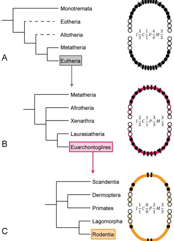
once during their lifetime (diphyodonty) (Luo et al., 2004). Further evolutionary trends of the dentition from these earliest mammals sensu lato to the placental mammals (eutherians) is characterized by a reduction of the dental formula coupled with a specialization of postcanine teeth leading to the acquisition of the mammalian mastication (Weijs, 1994). Basal eutherians had a fixed maximal number of teeth (FIG. 1A) and this number continuously decreased over the evolution of most modern eutherian groups such as euarchontoglires (FIG. 1B). Along with this decrease, a higher specialization of the postcanine teeth (especially the molars) was achieved.
Mammals thus progressively acquired a limited dentition replacement, a sectorization of the dentition into three dental types (incisor, canine and postcanine), as well as an increased complexity of their postcanine crown pattern. Because the various steps of these evolutionary processes are only documented by fossils, the evolution of the mammalian dentition has first mainly interested paleontologists, and for the past two decades this interest has expanded to the evo-devo community.
Evo-devo is a dual discipline that compares individual development with phenotypic changes during evolution (Müller, 2007). In order to understand how evolution acted on the biological properties of extinct and extant species, investigations aim at understanding how developmental processes are set up and modulated in extant species. Established correlations allow us to infer that similar processes were acting in the course of evolution (Carroll, 2008). Besides being a major tool to study mammalian evolution, teeth are also a well-studied example of ectodermal organs arising from the interaction of an epithelial layer with the underlying mesenchyme, similarly to many exocrine glands, feathers, scales, nails, and hair (Pispa and Thesleff, 2003).
FIG. 1: Simplified view of dental evolution in mammals. Phylogenies are simplified from Bininda-Edmonds et al. 2007. (A) Phylogeny and maximal dental formula in mammalian infraclasses. (B) Phylogeny and maximal dental formula of the various eutherian superorders. (C) Euarchontoglire order Phylogeny and maximal dental formula in the various euarchontoglire orders. I – rectangles: incisors, C – triangles: canines, P – black circles: premolars, M – white circles: molars.
Historically, the evo-devo community first asserted itself by comparing various body plans and analyzing the importance of toolkit genes in setting them (Duboule and Dollé, 1989). Dental evo-devo rapidly grew as a major focus of the discipline. As the field developed, a new interest arose on the genetic and epigenetic determination of odontogenesis (Maas and Bei, 1997; Peters and Balling, 1999; Jernvall and Thesleff, 2000; Thesleff, 2003; Tucker and Sharpe, 2004; Catón and Tucker, 2009; Jheon et al., 2011), and computational tools helped to better understand the evolution of dentition features (Salazar-Ciudad and Jernvall, 2010). Since there are about 5,500 species of mammals (Wilson and Reeder, 2005), researchers have to focus on a few species and use them as proxies to collect data (Minelli and Baedke, 2014). A classic extrapolation has been to work on rodents. Indeed, the order Rodentia is a rather large order encompassing about 40% of all mammal species diversity (Wilson and Reeder, 2005). This order – and especially murine rodents – shows a reduction of the maximal dental formula compared to the euarchontoglires (FIG. 1C). Rodents gained the status of well-established mammal representatives within the “tooth community” because of the very interesting dental diversity they display, but also because one of their members, the mouse (Mus musculus) has been used as a model for mammalian development for decades (Hedrich, 2004).
Mice are extremely derived in terms of dentition properties. They display only one highly specialized continuously growing incisor, as well as three complex molars that are not replaced per jaw quadrant (FIG. 2). If rare mammalian species display continuous dental replacement (see annex 1: Rodrigues et al., 2011), monophyodonty marks the final limitation in tooth renewal, and is observed in many taxa including muroid rodents, bats or shrews (Jernvall and Thesleff, 2012). Mouse dentition recapitulates the evolutionary trends that are characteristic of mammalian dental evolution, and as a consequence, the understanding of this evolutionary dynamics is of prime interest.
The present work aims at addressing two levels of mouse dentition dynamics: (1) the evolutionary dynamics of tooth number determination and shape setting in the postcanine area, and (2) the lifespan dynamics of the continuously growing incisors. Using a classic evo-devo approach on postcanine tooth number and shape, I first tackle the evolutionary potential of the well-documented FGF-activated ERK-MAPK pathway. This is an example of huge signaling pathway modulating most of the cellular processes, also involved at various stages of tooth development (Laugel-Haushalter et al., 2014). By scrutinizing the dentition of mice bearing mutation for various genes expressed in the same signaling pathway, I address the effects of genes acting at different steps of the pathway. From the mineralized tooth phenotype, I draw conclusions regarding the plausible evolutionary relevance of these genes. In a second part, I focus on the mouse continuously growing incisors. By looking at the diversity they display in terms of ornamentation, color and shape, I highlight their importance in the evo-devo field. Switching focus to the modification of their phenotype during the animal lifetime, I address the question of stem cell ageing in combining traditional in vivo monitoring and histological studies with cutting-edge transcriptomics to provide a further insight into the use of the mouse model.
FIG. 2: X-ray microtomographic view of a mouse skull. Incisors (yellow) and molars (pink) are separated by a toothless gap called diastema. Scale bar: 0.5 mm.
P
ART
A
T
OOTH
E
VO
-D
EVO
&
THE
RTK/ERK-MAPK
rganogenesis is one major step achieved during the development of a given organism. It is a dynamic and complex succession of molecular and cellular events leading to the organ identity, properties and functions. The mechanisms involved are well-conserved in all vertebrates. The organs undergoing budding and branching morphogenesis develop according to a series of morphogenetic changes similar to what has been well-documented in the mouse molars.
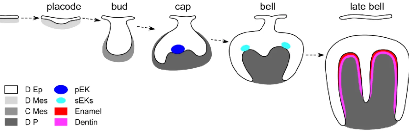
Tooth development (odontogenesis) is ruled by specific time-space interactions between the oral epithelium of the first branchial arch and the underlying mesenchyme deriving from the neural crests (Peters and Balling, 1999). Odontogenesis begins with a succession of histo-morphogenetical transformations named from the shape of the dental epithelium (FIG. A.1). Mouse gestation lasts around 20 days, and tooth development is initiated around Embryonic day 9.5 (Tucker and Sharpe, 2004). The secretion of growth factors combined with the activity of transcription factors in both upper and lower cheek teeth presumptive areas induces cell proliferation in both odontogenic epithelium and underlying mesenchyme (Thesleff, 2003). The first histo-morphogenetical sign of tooth development is the thickening of the dental epithelium until it reaches the placode stage. Then the placode grows toward the mesenchyme to form a bud (E13). At that stage, two distinct populations of epithelial cells can be observed: (1) a well-organized basal epithelium which remains at the interface with the odontogenic mesenchyme, and (2) a loosely arrange group of cells located towards the mouth cavity called stellate reticulum. Later on, the basal surface of the bud invaginates at its lingual and vestibular ends (cervical loops) as the underlying dental mesenchyme condenses. At the cap stage, the primary enamel knot (pEK, dark blue in FIG. A.1) forms in the basal dental
epithelium (Jernvall et al., 1994; Jernvall et al., 1998; Vaahtokari et al., 1996a; Vaahtokari et al., 1996b). The pEK is a dense non-proliferative group of cells secreting many molecules including Sonic Hedgehog (SHH), Fibroblast Growth Factors (FGFs), Bone Morphogenetic Proteins (BMPs) and WNTs (Thesleff et al., 2001). The combination of these molecules plays a major role in inducing the proliferation of surrounding cells, and thus in initiating the presumptive crown shape (Coin et al., 1999). From that stage on, odontogenesis is ruled by this epithelial signaling center.
Then, the cervical loops continue to expand into the mesenchyme, and the cap takes a bell shape. As the cytodifferentiation goes further, secondary enamel knots (sEKs, light blue in FIG. A.1) are formed at the top of the presumptive cusps (Tucker and Sharpe, 2004). At the late bell stage, the dimensions of the presumptive crown are set and the mineralization of the secreted dentin and enamel matrixes starts (Tucker and Sharpe, 1999). These matrixes are respectively secreted by odontoblasts and ameloblasts, two cell types which constitute the epithelio-mesenchymal interface (Arana-Chavez and Massa, 2004; Goldberg et al., 2002). Ameloblasts derive from the basal epithelial layer, while odontoblasts derive the mesenchymal dental papilla (Thesleff et al., 2001). By the end of this sequence, the crown formation is achieved. After the eruption (starting at about postnatal day 10 for the first molar) is achieved, the mouse possesses one incisor separated from three molars by a toothless gap called diastema in each dental quadrant (Nanci, 2007). Incisor development follows the same overall developmental series, the specificities of which will be discussed below.
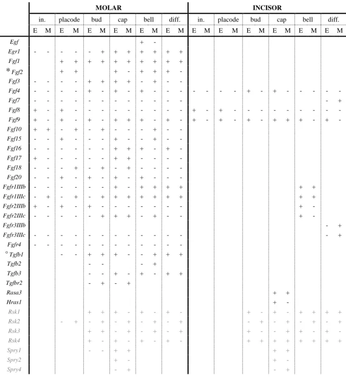
This sequence of morphological changes is driven by precise molecular networks acting together to tightly regulate its successive steps (Bei, 2009). One example of important network is the extracellular signal-regulated kinase/mitogen-activated protein kinase (ERK-MAPK) signaling pathway that can be triggered by receptor tyrosine kinase (RTK) activation. This cascade has the specificity of encompassing two signaling pathways necessary for proper tooth development: FGF signaling pathway (Li et al., 2014), and the ERK signaling pathway (Corson et al., 2003). Numerous RTK-activated ERK-MAPK members are expressed in dental tissues. They have been demonstrated to act during tooth development (TAB. A.1).
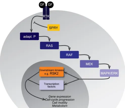
In Part A, I address the developmental and the evolutionary potential of this RTK-activated ERK-MAPK cascade during odontogenesis. The fixation of a growth factor (GF) on its RTK triggers the phosphorylation of successive kinases down to the final activation of effective kinases, as well as the expression of transcription factors (Sebolt-Leopold and Herrera, 2004, see FIG. A.2).
FIG. A.1: Mouse first lower molar development. The dental germ undergoes successive histo-morphogenetical modifications leading to formation of a mineralized matrixes-covered tooth ready to erupt. D Ep: dental epithelium; D Mes: dental mesenchyme; C Mes: condensed mesenchyme; D P: dental papilla; pEK: primary enamel knot; sEKs: secondary enamel knots.
MOLAR INCISOR
in. placode bud cap bell diff. in. placode bud cap bell diff.
E M E M E M E M E M E M E M E M E M E M E M E M Egf + - Egr1 - - - + + + + + + + Fgf1 + + + + + + + + + + * Fgf2 + + + - + + + - Fgf3 - - - - + + + + - + - - Fgf4 - - - - + - + - + - - - + - + - - - - - Fgf7 - - - + Fgf8 + - + - - - + - + - - - - Fgf9 + - + - + - + + + - + - + - + - + - + + + - + - Fgf10 + + - + - + - - - + - - Fgf15 - - + - - - + - - + - - Fgf16 - - - + + + - + - Fgf17 + - - - + + - - - - Fgf18 - - - + - + - + - - - - Fgf20 - - + - + - + - + - - - Fgfr1IIIb - - - + - + + + + + + Fgfr1IIIc - + - + - + + + + + + + + + Fgfr2IIIb + - + - + - - - + - Fgfr2IIIc - - - + + + - + - - + - Fgfr3IIIb - + Fgfr3IIIc - - - + Fgfr4 - - - - ° Tgfb1 - - + + + - - + + + Tgfb2 - - - + Tgfb3 - - + - + - + + Tgfbr2 - + - + Rasa3 + + Hras1 + - Rsk1 + + + - + - + - + - + - + + + + Rsk2 - + - + - + - + - + - + - + - + - + Rsk3 + + - + - + - + + - - + + - + - Rsk4 + - + - + - + - + + + + + + + + Spry1 - - + + + + Spry2 + - + - Spry4 - + - +
TAB. A.1: The expression of RTK-activated MAPK-ERK cascade members, modulators and effectors during mouse tooth development. The table summarize the detection (+) or absence (-) of gene expression during molar and incisor development. in.: initiation; diff.: differentiation; E: epithelium; M: mesenchyme; *: the fixation method used might impair the results reported; °: discrepancies can be found in the literature. Table compiled from Snead et al., 1989; Pelton et al., 1991; Vaahtokari et al., 1991; Cam et al., 1992; Karavanova et al,. 1992; Wang et al., 1995; Vaahtokari et al., 1996; Peters et al., 1992; Orr-Urtreger et al., 1993; Jernvall et al., 1994; Bei and Maas 1998; Jernvall et
al., 1998; Kettunen and Thesleff 1998; Kettunen et al., 1998; Xu et al., 1998; Kettunen et al., 2000;
Zeniou et al., 2002; Kohn et al., 2003; Åberg et al., 2004; Klein et al., 2006; Porntaveetus et al., 2011; Laugel-Haushalter et al., 2014; Li et al., 2014; and the Eurexpress project website www.eurexpress.org/ee/ (Diez-Roux et al., 2011). In black are members of the pathway, and in gray some of their modulators.
I take advantage of the availability of mutant mice for upstream regulator of RTKs (SPRY, yellow in FIG. A.2), and for a downstream effector kinase (RSK2, orange in FIG. A.2) to compare their adult dental phenotypes. By examining the presence of common phenotypes, I address plausible evolutionary role of the cascade in building the dentition of modern mice. I start with a conventional approach on knockout (KO) mice to characterize Rsk2 action on craniofacial development, and more specifically on tooth formation. Then, I compare the Spry1, Spry2, and Spry4 mutant molar phenotypes to the one of Rsk2-/Y mice. In order to perfect our understanding of the role of ERK-MAPK cascade members, I lastly investigate molar phenotype in transgenic mice over-expressing the Spry4 gene in the oral epithelium.
FIG. A.2: Simplified view of the ERK-MAPK cascade. Growth factors binding their receptor (RTK) trigger the activation of the phosphorylation cascade down to the final activation of the pathway effectors. This pathway regulates most of the cellular processes, including gene expression, cell cycle progression, cell motility and metabolism. Our interest is drawn to the Sprouty family (Spry) of RTK inhibitors (yellow), and to one of the effector kinase RSK2 (orange).
A.1 R
SK
2
IS A MODULATOR OF TOOTH DEVELOPMENT
This part of my work focuses on a study realized in collaboration with a team of the Developmental Biology and Stem Cells department from the IGBMC (Illkirch, France) published earlier this year in PLoS One (see annex 2: Laugel-Haushalter et al., 2014). The study aims at addressing the role of Rsk2 gene in mouse craniofacial development in order to question the relevance of Rsk2-/Y and Rsk1,2,3 null mutants as models for the X-linked craniofacial disorder called Coffin-Lowry syndrome (OMIM #303600). Mutations in RSK2 gene are responsible for the development of this syndrome in human (Trivier et al., 1996; Jacquot et al., 1998; Delaunoy et al., 2001). The disorder has an estimated prevalence of 1 in 40,000 to 50,000 people, with females displaying a higher feature variability than males (Hanauer and Young, 2002). Clinically, some defects resulting from this syndrome are detectable at birth, while others will progressively be established until the patient reaches the age of 2 (Hunter, 2002). Growth defects, skeletal and craniofacial dismorphia, as well as mental retardation make up the wide range of features associated with the syndrome (Hanauer and Young, 2002; Herrera-Soto et al., 2007).
Our collaborators analyzed the skull phenotype of the two Rsk mutant mice, and they designed a microarray experiment to address molecular changes in the regulatory network. I aimed at documenting adult molar phenotype in Rsk2-/Y as well as triple Rsk1,2,3 KO mutants in order to complete the description of Rsk2 gene role in craniofacial development.
A.1.1 Material and methods
A first cohort of 6 Rsk2-/Y male mice and 6 WT littermates has been studied. They have been bred at the IGBMC mouse breeding facility (Illkirch, France). A second cohort including 7 Rsk1,2,3-/- compound mutants was also provided to us. Molar rows were imaged using high-resolution X-ray microtomography with the Nanotom S (GE) equipment available at the UMS3444 (Lyon). A cubic voxel of 3µm was used. 3D-reconstructions from the acquired 2,000 projections were obtained using datos|x software and algorithm with a beam hardening correction. Pictures of the reconstructed molar row were taken in occlusal view to observe dental features of each specimen. Measures of the tooth length (mesio-distal distance) and width (vestibule-lingual distance) have been made on occlusal-oriented pictures of the molar rows. Statistical significance of the measures has been verified using the Student t-test for 2 group comparisons, with a p-value threshold of 0.05.
A.1.2 Results
Despite the absence of abnormal phenotype in incisors, defects are seen in the molar rows. The most striking abnormality observed in both Rsk2-/Y and Rsk1,2,3 null mutant mice is the presence of a ST in both upper and lower jaws (arrowheads, FIG. A.3). This extra tooth is erupted in alignment with the molar row, in a mesial position. The upper ST occurs in 65% of Rsk2-/Y mice and in 83% of the compound mutant mice. The lower ST occurs in 31% of the simple mutant mice and in 11% of the Rsk1,2,3 null mice. Overall, the shape of the ST ranges
from a monocuspid tooth to complex molar-like tooth. The inactivation of Rsk2 alone is sufficient to generate the abnormal ST-displaying phenotype. It is also sufficient to alter tooth proportions (FIG A.4). In compound mutants, the M3 is absent in 39% of the specimens (e.g. 1283 in FIG. A.3). The absence of any tooth socket calls for agenesis, which is a missing tooth due to a developmental failure.
FIG. A.3: Variation of molar shape and number in Rsk2-/Y and Rsk1,2,3 null mice analyzed by X-ray microtomography. All molar rows are similarly oriented (top corresponds to mesial, and left to lingual side). On the left are WT molars, other rows are mutant molars as indicated. Arrowheads point to ST; dottend ellipses show the reduction of the mesial-most affected cusp. Numbers refer to specimen ID. Scale bar: 0.7 mm.
Shape abnormalities mainly occur in the mesial-most chevron of both M1 and M1. Indeed, in both mutant backgrounds, the first chevron appears flattened when a ST occurs (dotted ellipses, FIG. A.3). In the upper jaw, a similar yet less pronounced flattening is also observed in some specimens that do not display any ST (e.g. 728 in FIG. A.3). I found this character to be correlated with the inclination of the M1 main root (FIG. A.5). Only Rsk2-/Y mutants display the 3 phenotypes depicted. When the root tends to be set vertically, the first
FIG. A.4: Comparison of molar length (mesio-distal distance) and width (vestibulo-lingual distance) in the Rsk2-/Y sample. A: measurements in the upper rows; B: measurements in the lower rows. WT tooth are depicted with filled circles, Rsk2-/Y without ST with blanked circles, and Rsk2-/Y with ST with blanked circles with a dot. Overall, the biggest changes in tooth proportions occur on both M1 and M1. Error bars represent the measurement standard deviation.
chevron appears straighter, reducing its size in occlusal view. In the lower molar rows, flattening is only seen when a complex-shaped ST occurs (e.g. 155 in FIG.A.3).
A.1.3 Discussion
Coffin-Lowry syndrome orodental features do not relate to Rsk-KO mutant phenotype
Coffin-Lowry syndrome is characterized by psychomotor and growth retardation, digit abnormalities, progressive skeletal malformations, as well as craniofacial dysmorphism (Temtamy et al., 1974; Temtamy et al., 1975; Hanauer and Young, 2002). Focusing on those craniofacial dysmorphism, cases report a high narrow palate, a midline lingual furrow, malocclusion, peg-shaped incisors, and hypodontia (Young, 1988). Due to rare availability of FIG. A.5: Variation of M1 root slope in Rsk2-/Y mice. Vestibular side of the molars is facing. On the top is a WT M1, then the gradient of slope is presented in the mutants. Dotted lines help visualizing the inclination from the tip of the first chevron to the basis of the main root. Scale bar: 0.5mm
the triple Rsk1,2,3 null mutants, the complete craniofacial and dental characterization has only been performed here on Rsk2-/Y mutants (see Annex 1 for details). The study shows an overall length reduction from the nasal to the occipital bone (the frontal bone being the most affected), as well as a strong nasal deviation for the most severe phenotypes seen. In the dental field, ST occurs in both Rsk2-/Y mutant and in the few compound mutants available, while tooth agenesis only occurs in the triple mutants. Comparing the human abnormalities to the ones highlighted in the murine model, it is clearly difficult to relate them, the only overlap remaining the occurrence of tooth agenesis.
Modulation of Rsk2 dosage in mouse mirrors evolution of mammalian dentition
Mice are the most common mammalian model used in tooth evo-devo field. The inactivation of Rsk2 gene only is sufficient to generate a dental phenotype of interest when looking at the evolution of dentition. The maximal dental formula found in Eutheria is 𝐼33𝐶11𝑃44𝑀33, meaning that modern mice lost 2 incisors, 1 canine and 4 premolars along their
evolutionary history. In our mutants, the ST occurs in a position corresponding to a 4th premolar, at the back of the diastema. A rudimentary tooth primordium was reported to develop in that same distal part of the diastema though it is aborted by the cap stage (E14) (Peterka et al., 2000; Viriot et al., 2000; Peterková et al., 2003). The ST found in Rsk2-/Y and Rsk1,2,3 null mutants is likely to be erupting from the completed development of this rudimentary primordium. This finding recalls another mutant studied in the light of human craniofacial syndrome that finally revealed its evolutionary potential: the Tabby mice. Heterozygous Tabby mice display ST in their upper molar rows, while K14-Eda mice display ST in their lower molar rows (Pispa et al., 1999; Kangas et al., 2004; Charles et al., 2009a). But these mice also display a lot of cusp defects, which do not seem to occur in Rsk-knockout mutants. However, the specimens in our sample were not collected at the same age. Due to
the different wear state of the molar rows, it is thus difficult to rule out the existence of such cusp defects.
A.1.4 Conclusions
Rsk2-/Y and Rsk1,2,3 null mice were screened to test whether their craniofacial phenotypes match the clinical picture of the Coffin-Lowry syndrome. Only two of the displayed phenotypes mimic what is observed in human patients. The occurrence of tooth agenesis has indeed been addressed, but the mice are also displaying variable skull phenotype ranging from WT-like to severe dysmorphy associated with nasal bone deviation. Thus, these mutant mice cannot be considered as the best models to address the Coffin-Lowry syndrome.
Interestingly, the occurrence of ST mesially to the molar rows reveals the evolutionary potential of Rsk2. The small size of the sample and the variation in the mouse age prevent us from scrutinizing the exact extent of dental abnormalities and the precise evolutionary relevance.
A.2 U
NRAVELING THE EVOLUTIONARY POTENTIAL OF
THE
ERK-MAPK
PATHWAY
The occurrence of ST in mesial position to the molar row is a feature that is displayed in several mutant mice already published. Mice carrying mutant for Gas1, Shh, Wnt1, Wise, Lrp4, Eda or Edar, Spry2 and Spry4 are – among other dental characters – characterized by the similar presence of a ST (respectively Ahn et al., 2010; Charles et al., 2009a; Kangas et al., 2004; Ohazama et al., 2009; Klein et al., 2006; Lagronova-Churava et al., 2013). Among this list, Spry2 and Spry4 mutant drew our attention, because of the molecular role of Sprouty proteins. Sprouty proteins are indeed general Receptor Tyrosine Kinase (RTK) inhibitors (Reich et al., 1999), and as so, they can inhibit FGFR-mediated activation of the ERK-MAPK cascade.
Taking advantage of the availability of Spry1-/-, Spry2-/-, Spry4-/- and Rsk2-/Y mutant mice, we conduct a comparative analysis to address the relative role of these components in the building of the mouse postcanine dentition. Eventually, this study helps us perfecting the analysis of Rsk2 mutant dental phenotype, and addressing the role of the entire pathway in the course of muroid rodent evolution. This study is currently submitted to Scientific Reports.
Phenotypic and evolutionary implications of modulating
the ERK-MAPK cascade using the dentition as a model
Pauline Marangoni1*, Cyril Charles1*, Paul Tafforeau2, Virginie Laugel-Haushalter3, Adriane Joo4, Agnès Bloch-Zupan3,5,6, Ophir D. Klein4,7,and Laurent Viriot1§
1: Evo-Devo of the Vertebrate Dentition, Institute of Functional Genomics of Lyon, ENS de Lyon, CNRS UMR
5242, Université de Lyon 1, 46 allée d’Italie, 69364 Lyon cedex 07, France
2: European Synchrotron Radiation Facility, 6 rue Jules Horowitz, 38043 Grenoble Cedex, France
3: Development and Stem Cells Department, Institute of Genetics and Molecular and Cellular Biology, Inserm U
964, CNRS UMR 7104, Université de Strasbourg, BP 10142, 67404 Illkirch, France
4: Department of Orofacial Sciences and Program in Craniofacial and Mesenchymal Biology, University of
California, San Francisco, CA 94143-0442, USA
5
: Faculty of Dentistry, University of Strasbourg, 8 rue St Elisabeth, 67000 Strasbourg, France
6: Reference Centre for Orodental Manifestations of Rare Diseases, Pôle de Médecine et Chirurgie
Bucco-Dentaires, Hôpitaux Universitaires de Strasbourg (HUS), 1 place de l’Hôpital, 67000 Strasbourg, France
7Department of Pediatrics and Institute for Human Genetics, University of California San Francisco, San
Francisco, California 94143, USA
§
SUMMARY
The question of phenotypic convergence across a signaling pathway has important implications for both developmental and evolutionary biology. The ERK-MAPK cascade is known to play a significant role in dental development, but the relative roles of its components remain unknown. Here we show that premolar teeth reappear in Spry2-/-, Spry4-/-, and Rsk2-/Y mice while premolars have been lost in the mouse lineage 45 million years ago (Ma). In addition, Sprouty-specific anomalies mimic a phenotype absent in extant mice, but present in mouse ancestors prior to 9 Ma. Although the four mutants display convergent phenotypes, each gene has a specific role in tooth number setting up and tooth crown patterning. The similarities found between teeth in fossils and mutants highlight the pivotal role of the ERK-MAPK cascade during the evolution of the dentition in rodents.
KEY WORDS
INTRODUCTION
The extracellular signal-regulated kinase/mitogen-activated protein kinase (ERK-MAPK) pathway is a central regulator of tooth development. This cascade is typically initiated by the binding of a growth factor to a receptor tyrosine kinase (RTK), which triggers the phosphorylation of successive kinases and culminates in activation of effector kinases and the transcription of target genes (Sebolt-Leopold and Herrera, 2004). The MAPK signaling pathway has been intensively studied by cancer biologists because of its effects on regulation of cell proliferation and survival (Downward, 2003; Sebolt-Leopold and Herrera, 2004), but this pathway is also important throughout mouse embryogenesis (Massague, 2003). The pathway has been investigated in numerous embryonic processes, including development of the central nervous system and mesodermal derivatives (Campos et al., 2004), skeletal development (Ge et al., 2007), and tooth development (Thesleff and Mikkola, 2002; Klein et al., 2006; Tompkins, 2006; Xu et al., 2008; Laugel-Haushalter et al., 2014).
Tooth development is a well-documented example of ectodermal organ development. It is a tightly regulated process arising from the crosstalk between dental epithelium and its underlying mesenchyme (Tucker and Sharpe, 2004). The signaling networks responsible for properly building the dentition have been heavily investigated, and numerous members of the ERK-MAPK signaling pathway are known to play a role in tooth development. Early studies examined the fibroblast growth factors (FGFs) that are FGFR ligands, and thus trigger the ERK-MAPK phosphorylation cascade (Neubüser et al., 1997; Thesleff, 2003). Investigations then moved to further steps of the cascade in order to determine which components were involved in tooth development (Goodwin et al., 2013; Laugel-Haushalter et al., 2014). An exciting current challenge is to understand the complexity of feedback regulation in this signaling pathway that can be time- and/or tissue-specific. In the present study, we compare
the phenotype of molar teeth in mice carrying mutations in Sprouty1, Sprouty2, Sprouty4, and Rsk2 genes, which are involved at various levels in the MAPK cascade.
The Sprouty (Spry) family of genes encodes general RTK inhibitors (Hacohen et al., 1998; Reich et al., 1999). After stimulation by growth factors, the Sprouty proteins are thought to translocate to the plasma membrane where their phosphorylation prevents the formation of an FGFR adaptor complex, thus having a negative effect on the activation of the rest of the cascade (Hanafusa et al., 2002). Spry1 is expressed in both the epithelium and the mesenchyme, with exception of a cluster of non-proliferating epithelial cells that serve as a signaling center called the enamel knot. Spry2 is expressed only in the epithelium adjacent to the dental mesenchyme, including the enamel knot, and Spry4 is expressed in the dental mesenchyme (Zhang et al., 2001; Klein et al., 2006). Whereas the morphogenesis of molar teeth in Spry1-/- mice has not yet been examined, Spry2-/- and Spry4-/- mice are known to have abnormal dentition, which sometimes includes ST located immediately in front of the first lower molar (Klein et al., 2006). These supernumerary teeth, which occur at differing frequencies depending on the genetic background (Klein et al., 2006; Ohazama et al., 2009; Lagronova-Churava et al., 2013) are believed to derive from evolutionary vestigial tooth buds that normally undergo apoptosis in wild-type embryos (Peterková et al., 1998; Viriot et al., 2000; Viriot et al., 2002). Lagronova-Churava and colleagues (2013) showed that although all Spry2-/- and Spry4-/- embryos present a revitalization of tooth rudiments at ED13.5, only 2% of Spry4-/- and 27% of Spry2-/- specimens had a lower ST. However, the role of Spry1, Spry2 and Spry4 in the development of upper molars is not known, and the adult molar morphology has not been scrutinized in these mutants.
RSKs (90kDa ribosomal S6 kinases) are effector kinases belonging to the eponymous family of highly conserved serine/threonine kinases (Frödin and Gammeltoft, 1999; Romeo et al., 2012). Out of the four isoforms found in vertebrates, Rsk2 has been recently demonstrated
to be involved in craniofacial development. Rsk2-/Y mice display a deformation of the nasal bone, as well as diastemal ST which affect the mesial part of both upper and lower first molars (Laugel-Haushalter et al., 2014). Mutations in the RSK2 gene have been associated with Coffin-Lowry syndrome (OMIM #303600), a condition characterized by mental and growth retardation along with craniofacial and other skeletal abnormalities (Temtamy et al., 1974; Temtamy et al., 1975; Hanauer and Young, 2002).
Phenotypic convergence across the ERK-MAPK signaling pathway remains poorly documented. By studying the dental phenotype resulting from mutations in genes located upstream (Sprouty) and downstream (Rsk2) of the ERK-MAPK cascade, we address the question of whether an ERK-MAPK signature phenotype exists. To answer this question, (1) we have characterized the molar phenotype in several mutant populations in order to evaluate the distribution of the various changes affecting both upper and lower molar rows; (2) precisely quantified the occurrence of supernumerary teeth (ST) as well as their impact on the other teeth of the row, and finally; (3) addressed the evolutionary role of the ERK-MAPK cascade by comparing specific dental traits of mutants with dental traits of other extant or extinct rodents.
MATERIAL AND METHODS
Sprouty mutant mice
All the studied Sprouty mutant mice were generated in backgrounds resulting from crossing between several lineages. The three mutants and the wild type mice were generated by inbreeding at UCSF. The sample set was composed of homozygous mice as indicated: 25 Spry1-/-, 50 Spry2-/-, 50 Spry4-/-, and 60 WT individuals (littermates of the various mutants). For each specimen, left and right, upper and lower tooth rows were studied independently. The age of the specimens ranged from 1 month to 2.5 months. Animal experimentation was carried out in compliance with the policies and procedures established by the UCSF Institutional Animal Care and Use Committee.
Rsk2-/Y mice
The Rsk2 mutant mouse line was generated as previously described (Yang et al., 2004). Since Rsk2 is located on the X chromosome, analyses were performed on Rsk2-/Y males, on a C57BL/6J background. The sample set was composed of 45 Rsk2-/Y and 45 WT littermates (Rsk2+/Y). Mouse protocols were complied with the 2010/63/UE directive and the 2013/02/01 French decree, and were thus approved by the CERBM-GIE: ICS/IGBMC Ethical Research Board.
Observation and imaging of dental rows
All the heads were prepared in order to remove all non-mineralized tissues to allow good observation and measurement of the dental rows. They were examined and photographed using a Leica stereomicroscope. The measurements were obtained by following the outline of each tooth from the photos of occlusal view of the row. Thus, the length, width, and area of the tooth were produced by Leica software. Some Sprouty mutant dental rows were imaged using X-ray-synchrotron Radiation Facility (ESRF, Grenoble, France), beamline
ID 19 and BM5, with a monochromatical beam at energy of 25 keV. X-ray synchrotron microtomography has been demonstrated to bring high-quality results for accurate imaging of small teeth (Tafforeau et al. 2006). A cubic voxel of 7.46µm was used. Some Rsk2-/Y mutant dental rows were imaged using the X-ray cone-beam computed microtomography with a Nanotom machine (GE) at an energy of 100keV with a cubic voxel of 3µm. All 3D renderings were performed using VGStudiomax software.
Statistics
Student’s t-tests were used to verify the significance of differences in tooth size between the mutant mice but also between the mutant and the WT mice. A threshold value of 0.05 (p-value) was used to assess the significance of the observed differences.
RESULTS
The mouse (Mus musculus) is a muroid rodent. Like all the members of this superfamily, mice have a simplified dentition composed only of incisors and molars separated by a long toothless gap called diastema. Three lower (M1 M2 M3) and three upper (M1 M2 M3) molar teeth are present in each mouth quadrant. The crowns of molar teeth bear a relatively stable number of cusps that are: 8 for M1, 6 for M2, 4 for M3, 7 for M1, 5 for M2, and 4 for M3. Crowns of upper molars are made of rows of 3 cusps arranged in linguo-vestibular chevrons pointing mesially (except the third one, which is incomplete), whereas crowns of lower molars are made of rows of 2 cusps linked by linguo-vestibular and rather straight crests called lophs (Fig. 1, first column). The two mesial lophs of the M1 are also linked together by a mesio-distal connection.
___________________________________________________________________________
Diversity of the dental phenotypes in Sprouty mutants
We examined the arrangement and shape of the postcanine dentition in four populations comprising 25 Spry1-/-, 50 Spry2-/-, and 50 Spry4-/- mice. Area measurements showed that the occlusal surface of molars in Spry1-/- and Spry4-/- mice is larger than in the WT mice, whereas the molar occlusal surface in Spry2-/- mice is smaller than in the WT mice (t test, p value<0.05, Fig. 2).
The molar teeth of Spry1-/- and WT mice are globally similar in shape, and 51% of the Spry1-/- dental rows display a WT-like phenotype. The defects are numbered as character# (c#) from the mesial to the distal part of the row. The main defects of the Spry1-/- postcanine dentition are: (c8) the occurrence of a supplementary distal cusp on the M1 (53%); (c9) the disconnection of the mesio-lingual cusp from the first chevron of the M2 (40%); and (c12) the absence of the mesio-vestibular cusp of the M1 (8%) (Fig. 1, Fig. 3B, Suppl. Fig. 1A). The postcanine dentition in Spry2-/- mice had stronger differences compared to WT samples, and only 21% of Spry2-/- dental rows still display a WT-like phenotype. The main changes of the Spry2-/- postcanine dentition are: (c7) the connection between the two lingual cusps of the M1
Fig. 1: Dental character matrix. [previous page] On the first column are displayed WT upper (top) and lower (bottom) molar rows, with coloured arrowheads pointing to the localization of the most frequent defects seen in the 4 mutant backgrounds. Each character is listed with the occurrence frequency in all the mutant mice observed. (c1-10) are defects of the upper mutant postcanine teeth. (c1) is the occurrence of a ST; (c2-4) are M1 1st chevron defects, respectively lingual cusp disconnection, straight mesial cusp and absence of the vestibular cusp; (c5) is the presence of an extra lingual cusp between the 1st and 2nd chevrons; (c6) is the disconnection of the lingual cusp of the M1 2nd chevron; (c7) is the occurrence of a lingual crest; (c8) is the occurrence of an extra distal cusp; (c9) is the disconnection of the mesio-lingual cusp of the M2; (c10) is the connection of distal cusps. (c11-17) are defects of the lower mutant postcanine teeth. (c11) is the occurrence of a ST; c(12) is the absence of the mesio-vestibular cusp; (c13) is the display of very symmetric mesial-most cusps; (c14) is the split of the mesio-vestibular cusp; (c15) is the occurrence of an extra mesial cusp; (c16) is the strong disconnection of the mesial cusps of the M1; (c17) is the abnormal connection of the 2nd and 3rd lophs of the M2.
(38%); (c8) the occurrence of a supplementary distal cusp on the M1 (46%); (c10) the connection between the two lingual cusps of the M2 (36%); (c11) the occurrence of a lower ST (27%); and (c13-14) an abnormal shape, number, and/or interconnection of the mesial cusps of the M1 (29%) (Fig. 1, Fig. 3C). Spry4-/- molar tooth phenotype is the most variable among the 3 Sprouty mutants. These teeth ranged from a WT-like phenotype (24%) to relatively severe anomalies, especially in the M1. The main defects of the Spry4-/- postcanine dentition are: (c1) the occurrence of an upper ST (17%); (c2-3-4) the presence of lingual cusp disconnection, straight mesial cusp and absence of the vestibular cusp affecting the first chevron of the M1 (66% combined); (c5) the occurrence of a supplementary lingual cusp between the first and the second chevron (14%); (c6) the disconnection of the lingual cusp from the second chevron of the M1 (20%); (c8) the occurrence of a supplementary distal cusp on the M1 (36%); (c9) the disconnection of the mesio-lingual cusp from the first chevron of the M2 (64%); (c11) the occurrence of a lower ST (3%); (c12-14) an abnormal number and/or interconnection of the mesial cusps of the M1 (16%) (Fig. 1, Fig. 3D).
Except for three characters, the dentition of Spry1-/- mice thus resembles that of WT mice, whereas Spry2-/- and Spry4-/- mice show extra cusps and crest disconnections, as well as severe reductions and defects in the mesial parts of the M1 (Spry4-/- only) and in the M1 (rarely in Spry4-/-, frequently in Spry2-/-). Spry4-/- mutants develop ST in both upper and lower jaws, whereas Spry2-/- only display lower ST. Lower ST have been observed in Spry2-/- and Spry4-/- molar rows, whereas upper ST only occurred in Spry4-/- molar rows. Spry1-/- mutants thus never develop any ST, and only Spry4-/- mutants display ST in both upper and lower tooth rows. These findings suggest a potential relationship between the occurrence of ST and abnormal arrangement of the mesial parts of the first molars.
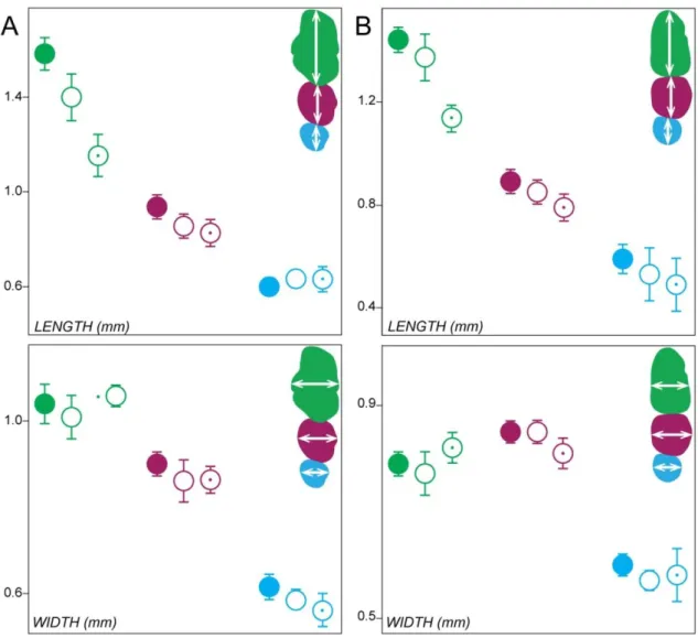
Fig. 2: Molar tooth proportions in the Spry1-/-, Spry2-/-, Spry4-/- and Rsk2-/Y mutant mice. First molars are coloured in green, second molars in pink, and third molars in blue. Filled forms represent the WT for each background, squares represent the Spry1-/- mutants, Spry2 -/-mutants without (blanked) or with (dot) ST, Spry4-/- mutants without (blanked) or with (dot) ST and Rsk2-/Y mutants without (blanked) or with (dot) ST.
Fig. 3: Abnormal phenotype in the Spry1-/-, Spry2-/-, Spry4-/- and Rsk2-/Y mutant mice. (A) WT dental rows; (B) Spry1-/- dental rows; (C) Spry2-/-dental rows; (D) Spry4-/- dental rows; (E) Rsk2-/Y dental rows. Coloured arrowheads correspond to the features displayed in the Fig. 2.
Similarities and differences of Rsk2 dental phenotype as compared to Sprouty dentitions
We next examined a cohort of 45 Rsk2-/Y mice. The postcanine occlusal surface area in Rsk2-/Y mice is smaller than in WT mice (M1 and M1,2 being significantly smaller, p-value<0.05, Fig. 2). Although many Rsk2-/Y specimens display relatively severe dental defects, 45% of the examined dental rows display a WT-like phenotype. The main defects of the Rsk2-/Y postcanine dentition are: (c1) the occurrence of a upper ST (14%); (c2-3-4) the presence of many defects on the first chevron of the M1 (71%); (c11) the occurrence of a lower ST (14%); (c12+14+16) an abnormal number and/or interconnection of the mesial cusps of the M1 (19%); (c17) abnormal mesio-distal connections between the second and the third lophs of the M2 (9%) (Fig. 1, Fig. 3E). The frequency of ST occurrence is lower than what has been previously reported (Laugel-Haushalter et al., 2014), and this may be explained by the examination of a larger sample or by shifts in the genetic background over time.
The comparison of the postcanine dental phenotypes between Rsk2-/Y and the 3 Sprouty mutant mice shows that Rsk2-/Y, Spry2-/- and Spry4-/- mice all develop ST, at varying frequencies. Spry2-/- mice never have an upper ST, but they frequently have a lower ST (27%). Spry4-/- mice frequently have an upper ST (17%), but they rarely have a lower ST (3%). Rsk2-/Y mice develop both upper and lower ST relatively frequently (14%). In addition to developing lower and upper ST, Rsk2-/Y and Spry4-/- postcanine dentitions share many similar defects affecting the mesial parts of both M1 and M1. The occurrence of defects on mesial parts of the M1 is much more frequent than the occurrence of an upper ST, but the defects in the mesial parts of the M1 and M1 are also associated with a shortening in length of both teeth (Fig. 2). Finally, Rsk2-/Y mutants do not develop the supplementary distal cusp of the M1 common to all Sprouty mutants, nor do they share with Spry1-/- and Spry4-/- mutants the trend towards having larger teeth than the WT mice.
The presence of ST impacts both the shape and size of the other teeth in the row
The size and shape of ST range from small rounded monocuspid teeth to large complex multicuspid teeth that can comprise up to five cusps (Fig. 4). In complex upper ST, a large central cusp is always present surrounded by a variable number of cusps linked by an almost circular crest. The most complex upper ST tend to have a mesial chevron pointing mesially (especially in Spry4-/-) whereas the lower ST mainly have a bicuspid shape (especially in Spry2-/-). These features show that ST have a clear murine shape identity.
The larger the ST is, the more the mesial parts of the neighbouring M1 and M1 are impacted. The occurrence of a ST leads to compression and flattening of the mesial part of the following tooth. This is particularly visible on M1 of Spry4-/-, and on both M1 and M1 of Rsk2
-/Y
mutants. As a consequence, the overall occlusal area of the molar row is smaller in mutants Fig. 4: ST phenotype and surface in Spry2-/-, Spry4-/- and Rsk2-/- mutants. ST range from small rounded monocuspid teeth to large multicuspid teeth. Right column indicates the mean surface in all the ST-displaying mutant mice, error bars represent the standard deviation. V and M respectively point towards the vestibular and mesial directions. Scale bar: 0.4mm.
displaying a ST, with the exception of the Spry4-/- upper row in which the area of the M3 seems to increase in compensation for the decrease of M1 area (Fig. 2).
In the most severely impacted phenotypes, the mesial contour of the M1 becomes rounded, whereas it is triangular in WT mice (e.g. Fig. 3E without ST). In addition, the two mesial-most cusps of the first chevron become extremely reduced into a simple crest (53% Spry4-/- and in Rsk2-/Y), and the mesio-vestibular cusp may completely disappear (20% Spry4 -/-and 4% in Rsk2-/Y). Interestingly, the mesial shrinkage of the M1 is associated with a change in the tilt angle of both cusps and roots of the first chevron (Suppl Fig. 1B). In WT mice, the slope of the M1 mesial root is in continuity with the tilt of 50° that makes the central cusp of the first chevron with the dental neck. In mutants having a ST, as well as in some mutants that do not display any ST, the tilt between the root axis and the tooth neck tends to be more vertical (about 70°), as does the slope of the first chevron central cusp (about 60°). The same type of defects can be seen when M1 is preceded by a ST in the three mutants: the first loph of the M1 is shortened and cusps appear as crushed by the presence of the ST.
Suppl. Fig. 1: Details of some shared mutant features depicted in the Rsk2-/Y mice. (A) The disconnection of the lingual-most cusp of the M1 1st chevron. (B) Variation of the M1 mesial root inclination in mutant mice. Note the straightening of the slope in Rsk2-/- mutants. Yellow angle is 50° in WT mice, and 60° in mutant mice. The root orientation itself is also modified, as depicted in orange.