DOI 10.1007/s10072-008-1060-9
O R I G I N A L A R T I C L E
Candidate genes for temporal lobe epilepsy: a replication study
Annick Salzmann · Nader Perroud · Arielle Crespel Carmen Lambercy · Alain Malafosse
Received: 1 July 2008 / Accepted in revised form: 14 November 2008 / Published online: 6 December 2008 © Springer-Verlag 2008
A. Salzmann · C. Lambercy · A. Malafosse (쾷) Division of Medical Genetics
University Hospitals of Geneva Geneva, Switzerland
e-mail: [email protected] A. Crespel
Epilepsy Laboratory
University Hospital of Montpellier Montpellier, France
N. Perroud · A. Malafosse Department of Psychiatry University Hospitals of Geneva Geneva, Switzerland
Introduction
Temporal lobe epilepsy (TLE) is the most common form of partial epilepsy [1] and this disorder is considered to be polygenic and complex. Various susceptibility genes and environmental factors are believed to be involved in the aetiology of TLE and concordance between genotype and phenotype is relatively weak. To understand the genetic background of complex diseases, association studies have been proposed as a method of choice [2]. However, this approach remains controversial and, in order to avoid false-positive association, replication of the first case-control study in independent groups of patients is recommended [3]. Recently a number of groups, including our own, have reported non-replications between several putative suscep-tibility genes and TLE [1, 4].
Since several other studies have reported an associa-tion between common variants in specific genes and TLE, our aim, in the present study, is to replicate them:
apolipoprotein E (ApoE) [5], interleukin 1α (IL-1α), interleukin 1β (IL-1β), interleukin 1RA (IL-1RA) [6] and prodynorphin (PDYN) [7].
Methods
Subjects
Our study group consisted of 109 unrelated patients with a diagnosis of non-lesional TLE. They were admitted under partial epilepsy criteria in the epilepsy unit of the university hospital of Montpellier (France). These patients suffered from a severe form of epilepsy with poor control of their seizures. Poor control is defined as a failure to respond to two or three single drugs or a
com-Abstract The objective of this study is to replicate
previ-ously published results regarding the involvement of sev-eral susceptibility genes in temporal lobe epilepsy (TLE):
interleukin 1β (IL-1β), interleukin 1α (IL-1α), interleukin 1RA (IL-1RA), apolipoprotein E (ApoE) and prodynor-phin (PDYN). We used a case-control approach
compar-ing several polymorphisms within these candidate genes between unrelated TLE patients and matched controls. We were thus able to confirm the role of ApoE, IL-1α and
IL-1RA genes in TLE disease, but failed to confirm the
involvement of IL-1β and PDYN. This failure should be interpreted with caution, as this may be due to the small size of our study groups and the resultant lack of statisti-cal power.
Keywords Temporal lobe epilepsy · Prodynorphin ·
bination of them. Diagnosis was based on patient history, clinical examination, interictal and ictal EEG analysis carried out with monitoring video-EEG, and MR evalua-tion. Demographic and clinical characteristics of our cohort of TLE patients have been reported in detail in a previous study [4].
The healthy control group was recruited from blood donors at the hospital in Geneva. To minimise morbidity among subjects in this group, only blood donors older than 35 years and without personal and/or family history of epilepsy and seizure were included. Patients and con-trols were European Caucasian for at least two genera-tions. Written informed consent was obtained from all participants. The Research Ethics Board of the Department of Clinical Neurosciences of Geneva reviewed and approved this study.
Molecular methods
DNA was extracted from peripheral blood leukocytes by use of the Nucleon BACC 2 kit (Amersham). Genotyping reactions were carried out as described in the different studies [6, 8, 9].
Statistical analysis
For our statistical analysis, in addition to the total cohort of patients, we divided the TLE group into subgroups, according to the existence or not of familial risk and hip-pocampal sclerosis. This was done in order to facilitate comparison between our data and that from analogous published work. In order to perform case-control genetic comparisons, differences in genotype and allele frequen-cies between all TLE, TLE subgroups and healthy con-trols were analysed by using the Chi-square test: 3 by 2 tables for PDYN, IL-1α and 1β; 4 by 2 tables for
IL-1RA. As age at onset displayed a non-parametric curve,
the Mann–Whitney U-test was used in place of the t-test for the comparison of age at onset and the presence or absence of the ApoE ε4 allele. We used the statistical package SPSS V.11.0.
As we compared genotypic and allelic distributions of various polymorphic markers between healthy and dis-eased subjects, a Bonferroni correction should be applied to correct for multiple testing. Adjustments for multiple com-parisons are recommended to avoid excessively easy rejec-tion of the null hypothesis. However, reducing the type I error increases the type II error and therefore increases the frequency of incorrect statements of no relationship between two factors. This could thus lead to missing an association in data that is not the result of chance [10]. The
rejection of the null hypothesis should be considered by taking into account both the evidence from the data and the relevance of other explanations. Moreover, our argument is based on previous findings and should be seen as a confir-mation of research in the field of TLE. In this present study, choosing not to penalise ourselves by missing possibly important findings, we decided not to correct our results for multiple testing.
Ethnicity was recorded using a self-reporting ques-tionnaire including perceived nationality, mother tongue and ethnicity of the subject together with all four grand-parents. To reduce the possibility of stratification bias, we used an average FSTbetween pairs of ethnic popula-tions between 0.0009 and 0.0048 (table 5.5.1 in ref. [11]).
Power calculation
Statistical power to detect associations was estimated using the Genetic Power Calculator (http://pngu.mgh.harvard.edu/purcell/gpc/). For age at onset we aimed to detect genetic effects explaining at least 2% of variance in the trait under an additive genet-ic model for a polymorphism with a minor allele frequen-cy of 0.12. Power for detecting such effect was calculat-ed for the nominal significance level α=0.05. For discrete traits, power for each study was calculated separately using an additive model with genetic relative risk of 2 and an α level=0.05.
Results
Throughout our study, genotypic distributions in both patients and controls were in Hardy–Weinberg equilibri-um.
Apolipoprotein E
Five studies have been published on ApoE and TLE age at onset [1, 5, 12–14]. In one of them, a positive associa-tion was found between early age at onset of the disease and the presence of ApoEε4 allele (ε4+: 5±5 years vs. ε4–: 14.9±10 years; p=0.005) [5]; however, the four other studies were unable to replicate this positive association. Our results confirmed a significant association between an early age at onset of epilepsy and the presence of allele ε4 (ε4+: 10.54±6.36 years vs. ε4–: 16.51±9.90 years; p=0.003) (Table 1). The study had 99% power to detect a gene effect, explaining 2% of the variance in the trait (Table 1).
Interleukin 1α
Based on pathophysiological hypotheses, several associ-ation studies have been carried out between interleukin-related genes and TLE. Two were performed with IL-1α and both showed no association [6, 15].
We partitioned our cohort of patients, like Kanemoto’s group, based on the existence or non-exis-tence of a hippocampal sclerosis (TLE-HS+) [6], and like Ozkara’s team, based on the absence of antecedent of febrile convulsion (TLE-FC–) [15]. We found a sig-nificant positive genotypic association between the whole TLE sample and the promoter IL-1α–889 SNP, but this was observed in TLE-HS+ patients (Table 2). However, as shown in Table 2, we did not have enough power to detect any significant association between TLE-HS– and controls (power=43%). Given the high frequency of the 1 allele in this sample (80.4%), an association could be suspected and further study with higher number of individuals is warranted. Interestingly, we also found a positive association between IL-1α–889 SNP and TLE-FC–(Table 2), but no statistical difference between TLE-HS+-FC– vs. con-trols (data not shown).
Interleukin 1RA
IL-1RA is the second interleukin-related gene studied by
Kanemoto et al. [6]. Once again they failed to show any association.
We observed significant differences in the allelic and genotypic frequencies between patients and controls, but here this concerns TLE-HS–(Table 3).
Interleukin 1β
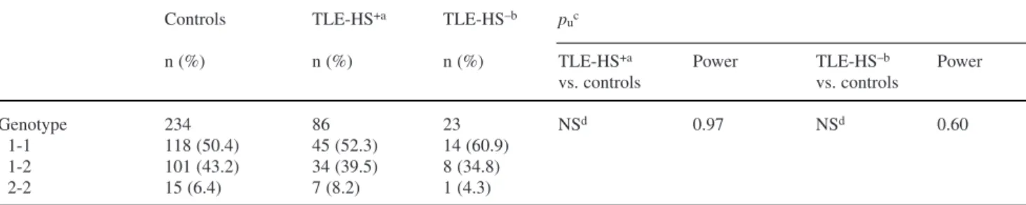
In this case, Kanemoto et al. [6] and Ozkara et al. [15] did not find an association between IL-1β–3953 and TLE. We were also unable to find any significant statis-tical difference (Table 4).
In contrast, Kanemoto et al. found a statistically higher frequency of the IL-1β–511 2-2 genotype in TLE-HS+ subjects compared to control subjects [6], and confirmed this result using a larger study group [16]. However this association was not observed in five other ethnically variable populations [1, 15, 17–19] or in our cohort of patients (Table 5).
In addition, we analysed the two IL-1β SNP haplo-types for the two subgroups of TLE (data not shown) and again we found no significant association. However, and as previously stated, negative results involving the TLE-HS– group should be kept with cau-tion as power to detect an associacau-tion is limited.
Prodynorphin
Stogmann et al. reported that PDYN promoter L allele confers an increased risk for TLE in patients with a family history of seizures (OR=2.25 (CI 1.41–3.62); p=0.0006) [7]. This result remained uncon-firmed in three independent Caucasian populations [1, 20, 21].
In the present study, we find a non-significant trend of excess of the L allele in those patients with a family history of epilepsy (OR=1.60 (CI 0.82–3.31); p=0.163) (Table 6).
Table 1 ApoE allele and genotype distributions and association between ε4 allele and age at onset
Controls Patients p Age at onset p (Mann–Whitney U-test) Power
of seizures n (%) n (%) Presence Absence of ε4 allele of ε4 allele Genotype 227 109 NS* ε2–2 0 (0.0) 0 (0.0) ε2–3 25 (11.0) 9 (8.3) ε2–4 5 (2.2) 1 (0.9) n=26 n=80 ε3–3 151 (66.5) 72 (66.1) Age: 10.54±6.36 Age: 16.51±9.90 p=0.003 0.99 ε3–4 43 (18.9) 27 (24.8) ε4–4 3 (1.3) 0 (0.0) Allele 454 218 NS* ε2 30 (6.6) 10 (4.6) ε3 370 (81.5) 180 (82.6) ε4 54 (11.9) 28 (12.8) *NS, non-significant
T
able 2
IL-1
α
–889
allele and genotype distrib
utions Controls TLE TLE-TLE pu d HS +a HS –b FC –c n (%) n (%) n (%) n (%) n (%) TLE-HS +a Po wer TLE-HS –b Po wer TLE Po wer TLE-FC –c Po wer vs. vs. vs. vs. controls controls controls controls Allele 470 218 172 46 108 0.059 0.89 0.066 0.43 0.018 0.95 0.079 0.74 1 316 166 129 37 82.0 (67.2) (76.1) (75.0) (80.4) (75.9) 2 154 52 43 9 26.0 (32.8) (23.9) (25.0) (19.6) (24.1) Genotype 235 109 86 23 54 0.027 0.104 0.0078 0.022 1-1 99 65 50 15 33 (42.1) (59.6) (58.1) (65.2) (61.1) 1-2 118 36 29 7 16 (50.2) (33.1) (33.7) (30.4) (29.6) 2-2 8 8 7 1 5 (7.7) (7.3) (8.1) (4.4) (9.3) aTLE-HS +, TLE with hippocampal sclerosis; bTLE-HS –, TLE without hippocampal sclerosis; cTLE-FC –, TLE without antecedents of febrile con
vulsion; dp u , p uncorrected T able 3 IL-1RA
allele and genotype distrib
utions Controls TLE-HS +a TLE-HS –b pu c n (%) n (%) n (%) TLE-HS +a Po wer TLE-HS –b Po wer TLE-HS –b Po wer vs. controls vs. controls vs. TLE-HS +a Genotype 242 86 23 0.768 0.95 0.001 0.51 0.036 0.64 1-1 128 43 5 (52.9) (50.0) (21.7) 1-2 90 36 13 (37.2) (41.9) (56.5) 1-4 5 1 0 (2.1) (1.2) (0.0) 1-5 0 (0.0) 0 (0.0) 1 (4.3) 2-2 16 (6.6) 6 (7.0) 4 (17.4) 2-4 3 (1.2) 0 (0.0) 0 (0.0) Allele 484 172 46 0.525 <0.0001 0.022 1 351 (72.5) 123 (71.5) 24 (52.2) 2 125 (25.8) 48 (27.9) 21 (45.7) 4 8 (1.7) 1 (1.4) 0 (0.0) 5 0 (0.0) 0 (0.0) 1 (2.2) aTLE-HS +, TLE with hippocampal sclerosis; bTLE-HS –, TLE without hippocampal sclerosis; cp
u
,
p
Discussion
In this present paper, we attempt to replicate published association studies between TLE, and subtypes of this disease, and several candidate genes.
Some evidence has implicated ApoE in the hip-pocampal response to injury. Indeed, astrocytes reply to neuronal damage by synthesising and releasing ApoE. Three major isoforms of ApoE have been identified, ε2, ε3 and ε4 [8]. The ApoE ε4 allele has often been asso-ciated with neurological diseases and/or their early-age
onset in carriers: Alzheimer’s disease [22], amyotroph-ic lateral sclerosis [23] and Parkinson’s disease [24]. Although three other TLE association studies were unable to find such effect [1, 13, 14] and the present sample size is small, we have enough power to detect a statistically significant result between an early age at onset of TLE and the presence of the ε4 allele. Moreover, the present study is the second one that reports an early age at onset in ε4 TLE carriers. These results suggest that TLE is one of the multiple neuro-logical diseases for which ApoE has a general role in
Table 4 IL-1β+3953 allele and genotype distributions
Controls TLE-HS+a TLE-HS–b p
uc
n (%) n (%) n (%) TLE-HS+a Power TLE-HS–b Power
vs. controls vs. controls
Genotype 234 86 23 NSd 0.97 NSd 0.60
1-1 118 (50.4) 45 (52.3) 14 (60.9)
1-2 101 (43.2) 34 (39.5) 8 (34.8)
2-2 15 (6.4) 7 (8.2) 1 (4.3)
aTLE-HS+, TLE with hippocampal sclerosis;bTLE-HS–, TLE without hippocampal sclerosis; cp
u, p uncorrected; dNS, non-significant
Table 5 IL-1β–511 allele and genotype distributions
Controls TLE-HS+a TLE-HS–b p
uc
n (%) n (%) n (%) TLE-HS+a Power TLE-HS–b Power
vs. controls vs. controls
Genotype 227 86 23 NSd 0.98 NSd 0.51
1-1 99 (43.6) 35 (40.7) 12 (52.2)
1-2 108 (47.6) 45 (52.3) 9 (39.1)
2-2 20 (8.8) 6 (7.0) 2 (8.7)
aTLE-HS+, TLE with hippocampal sclerosis; bTLE-HS–, TLE without hippocampal sclerosis; cp
u, p uncorrected; dNS, non-significant
Table 6 PDYN allele and genotype distributions
Controls Familial- OR pua Power Nonfamilial- OR pua Power
risk (95% CI) risk (95% CI)
TLE TLE n (%) n (%) n (%) Allele 412 42 0.163 0.68 166 NSb 0.98 L 106 15 1.60 (25.7) (35.7) (0.82– 50 3.31) (30.1) 1.24 (0.84–1.85) H 306 27 0.62 116 0.80 (74.3) (64.3) (0.32–1.21) (69.9) (0.54– 1.20) Genotype 206 21 0.32 83 NSb LL 14 (6.8) 2 (9.5) 1.44 7 (8.4) 1.26 (0.30–6.83) (0.49–3.25) LH 78 (37.9) 11 (52.4) 1.81 36 (43.4) 1.26 (0.73–4.45) (0.75–2.11) HH 114 (55.3) 8 (38.1) 0.50 (0.20–1.25) 40 (48.2) 0.75 (0.45–1.25) ap u, p uncorrected; bNS, non-significant
neuronal degeneration or regeneration, rather than a specific role in their aetiopathogenesis.
IL-1 shows two structurally distinct forms, IL-1α and IL-1β, that act on the IL-1 receptor. Interleukin 1 tor antagonist protein (IL-1RA) acts on the same recep-tor and inhibits IL-1α and 1β binding. 1α and IL-1β are involved in various immune responses, inflamma-tory processes, apoptosis and haematopoiesis. They are produced by monocytes and macrophages. Moreover, IL-1α and IL-1β are also synthesised by glial and neuronal cells [25]. Their implication in the central nervous sys-tem was demonstrated by the presence of high-density IL-1 receptors in molecular and granular layers of den-tate gyrus [26], suggesting a physiological role for IL-1 in the hippocampus.
In contrast to Kanemoto et al. [6], we found a positive association between TLE and IL-1α–889, which is a vari-ation in the 5’ regulatory region. This result is in agree-ment with the work of Peltola et al. These authors also found a significantly higher frequency for IL-1α allele 1 in patients with localisation-related epilepsy, such as TLE, lobe temporal epilepsy, parieto-occipital epilepsy and multifocal epilepsy [27]. Despite higher frequencies of IL-1α allele 1 and genotype 1-1 in all the sub-groups compared to the controls, only some of them reach sig-nificance (the whole TLE population and the TLE-HS+ and TLE-FC–sub-groups). This is probably due to a lack of statistical power and larger groups of patients will be needed to confirm this association and determine whether it is subgroup specific. It is noteworthy that the
IL-1α–889 polymorphism is functional: 2-2
homozy-gotes showed an increased transcriptional activity com-pared to 1-1 homozygotes and heterozygotes [28]. This suggests that vulnerability to TLE may be related to deregulation of the immune system, a hypothesis further supported by the finding that three immune-related pro-teins are down- or up-regulated – specifically the com-plement factor 3, C3 – in brain of TLE patients [29]. Interestingly, the promoter region of C3 was found to be very responsive to IL-1 [30].
Discrepancies have been reported in the association studies between various epileptic syndromes and the
IL-1RA VNTR, which is described as a putative
protein-binding site and may influence gene expression [31]. For the first time, in contrast to the research of Kanemoto et al. [6] and despite weak statistical power, we observed quite a strong positive association between this variant and TLE: when compared to controls, TLE-HS– patients displayed lower frequencies of allele 1 and genotype 1-1, and higher frequencies of allele 2 and genotype 1-2 and 2-2. Tsai et al. reported an association between the same polymorphism and FS, but in the opposite sense: the
IL-1RA allele 1 was significantly more frequent in FS
chil-dren than in controls [32]. Due to the small sample size, we were unable to test the associations between IL-1RA VNTR and the TLE-HS–-FC+ and TLE-HS–-FC– sub-groups. Before proposing pathophysiological hypotheses for these different associations, replication using larger study groups is required.
Similarly, conflicting results were also reported for
IL-1β–511 SNP, which is an AP-2 binding site [33]. The
positive association with IL-1β–511 SNP observed in a Japanese population [6] may be restricted to this ethnic population. However IL-1β–511 allele 2 was signifi-cantly more present in Caucasian patients with localisa-tion-related epilepsy, such as TLE, lobe temporal epilepsy, parieto-occipital epilepsy and multifocal epilepsy [27]. Finally, the IL-1β–511 SNP was associat-ed with FS only in Finnish [34] and German cohorts of patients [35]. Very recently, Kauffman et al. assessed a meta-analysis by pooling together all published studies up to March 2007, regardless of the cohorts’ ethnicity. They found a modest association between IL-1β–511 allele 2 and TLE-HS+ [36]. It is noteworthy that our results also showed no significant association for the
1L-1β+3953 SNP, in accordance with previously
pub-lished studies [6, 15]. In contrast, the present negative results for TLE-HS– and these two SNP must be inter-preted with caution due to the weak power of associa-tion detecassocia-tion.
Finally, this present study, as well as some previous related studies [1, 20, 21], were unable to replicate the initial association between PDYN and TLE with a family history for seizures [7]. However, a recently stratified analysis, which pools four previous studies [1, 7, 20, 21], showed a significant association [37]. Our non-obser-vance of this association is possibly due to a lack of sta-tistical power. Moreover, a study reported an association between 32 autosomal dominant lateral temporal epilep-sy index cases and PDYN [38].
Conclusion
The present study adds further data to the search for susceptibility genes involved in TLE. Despite failure to replicate most of the previously reported positive associations, the publishing of our results is important for future meta-analyses. Similarly, the positive asso-ciation we report for the first time with IL-1α and
IL-1RA requires further analysis for confirmation of this
result.
Acknowledgements This study was supported by the University
Hospitals of Geneva. We thank Dr. Roger Snowden, Firmenich S.A., for correcting the manuscript
Conflict of Interest statement The Authors certify that there is no
actual or potential conflict of interest in relation to this article
References
1. Cavalleri GL, Lynch JM, Depondt C et al (2005) Failure to repli-cate previously reported genetic associations with sporadic tem-poral lobe epilepsy: where to from here? Brain 128:1832–1840 2. Cardon LR, Bell JI (2001) Association study designs for complex
diseases. Nat Rev Genet 2:91–99
3. Tan NC, Mulley JC, Berkovic SF (2004) Genetic association stu-dies in epilepsy: “the truth is out there”. Epilepsia 45:1429–1442 4. Salzmann A, Moulard B, Crespel A et al (2005) GABA receptor 1 polymorphism (G1465A) and temporal lobe epilepsy. Epilepsia 46:931–933
5. Briellmann RS, Torn-Broers Y, Busuttil BE et al (2000) APOE epsilon4 genotype is associated with an earlier onset of chronic temporal lobe epilepsy. Neurology 55:435–437
6. Kanemoto K, Kawasaki J, Miyamoto T et al (2000) Interleukin (IL)1beta, IL-1alpha, and IL-1 receptor antagonist gene polymor-phisms in patients with temporal lobe epilepsy. Ann Neurol 47:571–574
7. Stogmann E, Zimprich A, Baumgartner C et al (2002) A functio-nal polymorphism in the prodynorphin gene promotor is associa-ted with temporal lobe epilepsy. Ann Neurol 51:260–263 8. Blumcke I, Brockhaus A, Scheiwe C et al (1997) The
apolipopro-tein E epsilon 4 allele is not associated with early onset temporal lobe epilepsy. Neuroreport 8:1235–1237
9. Zimprich A, Kraus J, Woltje M et al (2000) An allelic variation in the human prodynorphin gene promoter alters stimulus-induced expression. J Neurochem 74:472–477
10. Rothman KJ (1990) No adjustments are needed for multiple com-parisons. Epidemiology 1:43–46
11. Cavalli-Sforza L, Menozzi P, Piazza A (1994) History and geo-graphy of human genes. Princeton University Press, New Jersey 12. Gambardella A, Aguglia U, Cittadella R et al (1999)
Apolipoprotein E polymorphisms and the risk of non-lesional temporal lobe epilepsy. Epilepsia 40:1804–1807
13. Yeni SN, Ozkara C, Buyru N et al (2005) Association between APOE polymorphisms and mesial temporal lobe epilepsy with hippocampal sclerosis. Eur J Neurol 12:103–107
14. Gambardella A, Aguglia U, Chifari R et al (2005) ApoE epsilon4 allele and disease duration affect verbal learning in mild tempo-ral lobe epilepsy. Epilepsia 46:110–117
15. Ozkara C, Uzan M, Tanriverdi T et al (2006) Lack of associa-tion between IL-1beta/alpha gene polymorphisms and tempo-ral lobe epilepsy with hippocampal sclerosis. Seizure 15:288–291
16. Kanemoto K, Kawasaki J, Yuasa S et al (2003) Increased fre-quency of interleukin-1beta-511T allele in patients with temporal lobe epilepsy, hippocampal sclerosis, and prolonged febrile con-vulsion. Epilepsia 44:796–799
17. Heils A, Haug K, Kunz WS et al (2000) Interleukin-1beta gene polymorphism and susceptibility to temporal lobe epilepsy with hippocampal sclerosis. Ann Neurol 48:948–950
18. Buono RJ, Ferraro TN, O’Connor MJ et al (2001) Lack of asso-ciation between an interleukin 1 beta (IL-1beta) gene variation and refractory temporal lobe epilepsy. Epilepsia 42:782–784 19. Jin L, Jia Y, Zhang B et al (2003) Association analysis of a
polymorphism of interleukin 1 beta (IL-1 beta) gene with tem-poral lobe epilepsy in a Chinese population. Epilepsia 44:1306–1309
20. Tilgen N, Rebstock J, Horvath S et al (2003) Prodynorphin gene promoter polymorphism and temporal lobe epilepsy. Ann Neurol 53:280–281
21. Gambardella A, Manna I, Labate A et al (2003) Prodynorphin gene promoter polymorphism and temporal lobe epilepsy. Epilepsia 44:1255–1256
22. Schellenberg GD (1995) Genetic dissection of Alzheimer disease, a heterogeneous disorder. Proc Natl Acad Sci U S A 92:8552–8559
23. Moulard B, Sefiani A, Laamri A et al (1996) Apolipoprotein E genotyping in sporadic amyotrophic lateral sclerosis: evidence for a major influence on the clinical presentation and prognosis. J Neurol Sci 139[Suppl]:34–37
24. Li YJ, Hauser MA, Scott WK et al (2004) Apolipoprotein E con-trols the risk and age at onset of Parkinson disease. Neurology 62:2005–2009
25. Hopkins SJ, Rothwell NJ (1995) Cytokines and the nervous system. I: Expression and recognition. Trends Neurosci 18:83–88 26. Ban E, Milon G, Prudhomme N et al (1991) Receptors for inter-leukin-1 (alpha and beta) in mouse brain: mapping and neuronal localization in hippocampus. Neuroscience 43:21–30
27. Peltola J, Keranen T, Rainesalo S, Hurme M (2001) Polymorphism of the interleukin-1 gene complex in localization-related epilepsy. Ann Neurol 50:275–276
28. Dominici R, Cattaneo M, Malferrari G et al (2002) Cloning and functional analysis of the allelic polymorphism in the transcrip-tion regulatory region of interleukin-1 alpha. Immunogenetics 54:82–86
29. Jamali S, Bartolomei F, Robaglia-Schlupp A et al (2006) Large-scale expression study of human mesial temporal lobe epilepsy: evidence for dysregulation of the neurotransmission and comple-ment systems in the entorhinal cortex. Brain 129:625–641 30. Wilson DR, Juan TS, Wilde MD et al (1990) A 58-base-pair
region of the human C3 gene confers synergistic inducibility by interleukin-1 and interleukin-6. Mol Cell Biol 10:6181–6191
31. Vamvakopoulos JE, Taylor CJ, Morris-Stiff GJ et al (2002) The interleukin-1 receptor antagonist gene: a single-copy variant of the intron 2 variable number tandem repeat (VNTR) polymor-phism. Eur J Immunogenet 29:337–340
32. Tsai FJ, Hsieh YY, Chang CC et al (2002) Polymorphisms for interleukin 1 beta exon 5 and interleukin 1 receptor antagonist in Taiwanese children with febrile convulsions. Arch Pediatr Adolesc Med 156:545–548
33. Dominici R, Malferrari G, Mariani C et al (2002) The Interleukin 1-beta exonic (+3953) polymorphism does not alter in vitro pro-tein secretion. Exp Mol Pathol 73:139–141
34. Virta M, Hurme M, Helminen M (2002) Increased frequency of interleukin-1beta (-511) allele 2 in febrile seizures. Pediatr Neurol 26:192–195
35. Tilgen N, Pfeiffer H, Cobilanschi J et al (2002) Association analysis between the human interleukin 1beta (-511) gene poly-morphism and susceptibility to febrile convulsions. Neurosci Lett 334:68–70
36. Kauffman MA, Moron DG, Consalvo D et al (2008) Association study between interleukin 1 beta gene and epileptic disorders: a HuGe review and meta-analysis. Genet Med 10:83–88
37. Makoff A (2007) Genetics of epilepsy: Epilepsy Research Foundation workshop report – What is the role of replication? Epileptic Disord 9:206–209
38. Bovo G, Diani E, Bisulli F et al (2008) Analysis of LGI1 promo-ter sequence, PDYN and GABBR1 polymorphisms in sporadic and familial lateral temporal lobe epilepsy. Neurosci Lett 2:436:23–26