Exp Brain Res (2003) 153: 467–476 DOI 10.1007/s00221-003-1606-2
R E S E A R C H A RT I C L E
A.-V. Jacomme . F. R. Nodal . V. M. Bajo . Y. Manunta .
J.-M. Edeline . A. Babalian . E. M. Rouiller
The projection from auditory cortex to cochlear nucleus
in guinea pigs: an in vivo anatomical
and in vitro electrophysiological study
Received: 8 November 2002 / Accepted: 11 April 2003 / Published online: 18 September 2003 # Springer-Verlag 2003
Abstract Previous anatomical experiments have
demon-strated the existence of a direct, bilateral projection from
the auditory cortex (AC) to the cochlear nucleus (CN).
However, the precise relationship between the origin of the
projection in the AC and the distribution of axon terminals
in the CN is not known. Moreover, the influence of this
projection on CN principal cells has not been studied
before. The aim of the present study was two-fold. First, to
extend the anatomical data by tracing anterogradely the
distribution of cortical axons in the CN by means of
restricted injections of biotinylated dextran amine (BDA)
in physiologically characterized sites in the AC. Second,
in an in vitro isolated whole brain preparation (IWB), to
assess the effect of electrical stimulation of the AC on CN
principal cells from which intracellular recordings were
derived. BDA injections in the tonotopically organized
primary auditory cortex and dorsocaudal auditory field at
high and low best frequency (BF) sites resulted in a
consistent axonal labeling in the ipsilateral CN of all
injected animals. In addition, fewer labeled terminals were
observed in the contralateral CN, but only in the animals
subjected to injections in low BF region. The axon
terminal fields consisting of boutons en passant or
terminaux were found in the superficial granule cell
layer and, to a smaller extent, in the three CN
subdivi-sions. No axonal labeling was seen in the CN as result of
BDA injection in the secondary auditory area (dorsocaudal
belt). In the IWB, the effects of ipsilateral AC stimulation
were tested in a population of 52 intracellulary recorded
and stained CN principal neurons, distributed in the three
CN subdivisions. Stimulation of the AC evoked slow late
excitatory postsynaptic potentials (EPSPs) in only two
cells located in the dorsal CN. The EPSPs were induced in
a giant and a pyramidal cell at latencies of 20 ms and
33 ms, respectively, suggesting involvement of
polysyn-aptic circuits. These findings are consistent with
anatom-ical data showing sparse projections from the AC to the
CN and indicate a limited modulatory action of the AC on
CN principal cells.
Keywords Biotinylated dextran amine (BDA) .
Intracellular recording . Intracellular staining .
Corticobulbar axons
Introduction
The cochlear nucleus (CN) occupies a strategic position in
the auditory system. Being the first relay in the auditory
pathways, it receives stereotyped inputs from the auditory
nerve (AN), modifies this information, and then sends
output signals to multiple nuclei of the brainstem and
midbrain. In addition, it receives reciprocal, descending
auditory inputs which constitute a substantial proportion
of connections in the auditory system (Huffman and
Henson 1990; Romand and Avan 1997; Rouiller 1997).
The sources of the descending auditory projections to the
CN have been shown to arise from the contralateral CN
(Cant and Gaston 1982; Wenthold 1987; Shore et al. 1992;
Schofield and Cant 1996), the superior olivary complex
(Adams 1983; Spangler et al. 1987; Brown et al. 1988;
Schofield and Cant 1999), the dorsal nucleus of the lateral
lemniscus (DLL) (Caicedo and Herbert 1993), and the
inferior colliculus (IC) (Van Noort 1969; Carey and
Webster 1971; Adams and Warr 1976; Kane and Finn
1977; Andersen et al. 1980; Conlee and Kane 1982;
Hashikawa 1983; Faye-Lund 1986, 1988; Coleman and
Clerici 1987; Herbert et al. 1991; Malmierca et al. 1996;
A. Jacomme . A. Babalian . E. M. Rouiller (*)
Division of Physiology, Department of Medicine, University of Fribourg, Rue du Musée 5, 1700 Fribourg, Switzerland e-mail: [email protected] Tel.: +41-26-3008609 Fax: +41-26-3009675 F. R. Nodal . V. M. Bajo
University Laboratory of Physiology, University of Oxford, Oxford, UK
Y. Manunta . J. Edeline
NAMC, UMR CNRS 8620, University Paris Sud, Orsay, France
Schofield 2001). The auditory cortex (AC) is the most
remote auditory region giving rise to descending auditory
projections to the CN. For a long time, it was believed that
the AC might influence the CN only via indirect pathways
involving the midbrain. However, recent anatomical
experiments (Feliciano et al. 1995; Weedman and Ryugo
1996a, 1996b; Saldaña et al. 1996; Schofield et al. 2001;
Doucet et al. 2002) have demonstrated the existence of
direct, bilateral projection from the AC to the CN. It has
been shown that projections from AC to CN in the rat and
the guinea pig arise from layer V neurons of the AC and
terminate mainly in the granule cell domain of the CN
(Weedman and Ryugo 1996a, 1996b; Schofield et al.
2001). However, the precise relationship between the
origin of the projection in the AC and the distribution of
axon terminals in the CN remains largely unknown.
Although the existence of descending auditory inputs to
CN suggests a possible mechanism for cortical feedback
control of afferent information at peripheral levels, the
functional nature of the influence exerted by direct
descending projections from the AC to CN remains
unknown.
Therefore, the aim of the present study was two-fold.
The first aim was to extend the available anatomical data,
by establishing the distribution of the corticobulbar axons
in the CN. The second objective was to assess the effects
of activating the descending projections from the AC on
CN neurons. Two types of experiments were conducted in
the guinea pig to address these questions. First, the
distribution of corticobulbar axons in the CN was
established neuroanatomically, based on restricted
injec-tions of the anterograde tracer biotinylated dextran amine
(BDA) in physiologically characterized sites in the AC.
Second, an in vitro isolated whole brain (IWB) preparation
was used to perform intracellular recording and staining of
single neurons in the CN in order to test the presence and
the physiological properties of synaptic responses to
electrical stimulation of the AC. Since, in the IWB
preparation, it was impossible to record intracellularly
from small neurons, the effects of AC stimulation could be
assessed only in CN principal cells.
Materials and methods
In vivo experimentsBDA injections into the AC were performed in five adult guinea pigs (body weight 530–850 g). Under general anesthesia (diazepam 8 mg/kg, followed 15 min later by pentobarbital 20 mg/kg; see Evans 1979), a craniotomy (about 5×8 mm) was made in the temporal bone. Small punctures were made in the dura under microscopy control to insert low impedance glass pipettes (<3 MΩ) that allowed multiunit recordings (3–5 neurons) at a depth of 500– 1000 µm. The frequency tuning at each recording site was tested between 20 and 80 dB SPL using a previously described sound delivery system (Manunta and Edeline 1997, 1999). Briefly, ascending sequences of pure tones (frequency 0.1–35 kHz, 100 ms duration, 1 s intertone interval) were delivered to the contralateral ear. Online displays of the peri-stimulus time histograms allowed determination of the frequency tuning at each intensity. A regular progression of the best frequency (BF) from low
to high when moving from rostral to caudal was interpreted as a location in the primary auditory cortex (AI), whereas a regular progression of the BF from high to low was assumed to be typical of the dorsocaudal field (DC) (Redies et al. 1989; Wallace et al. 2000). Locations in secondary auditory fields (DCB dorsocaudal belt, VCB ventrocaudal belt; Wallace et al. 2000) were characterized by (1) long-latency responses, (2) very broad tuning, and (3) a lack of tonotopy. At a previously characterized locus, a pipette (tip 25– 50 µm) filled with BDA (10%; Molecular Probes, Eugene, OR, USA) was lowered 1500 µm below the pial surface and the tracer was iontophoretically ejected for 45 min (8 µA positive current, 7 s on/off). After a survival time of 7–10 days, the animals were deeply anesthetized (pentobarbital, 120 mg/kg) and perfused with a 4% paraformaldehyde (PFA) solution in 0.1 M phosphate buffer (PB, pH 7.4). After 24 h postfixation, the brain was placed in a solution of 30% sucrose in 0.1 M PB until it sank. Serial coronal sections (40 µm) were prepared with a freezing microtome and collected in 0.05 M Tris (pH 7.6). BDA labeling was vizualized using previously described protocols (see for details Bajo et al. 1999). Individual sections were examined under brightfield illumination (×4, ×10, ×20, and ×40), and reconstruction of the corticobulbar axons in the CN was made using a drawing tube.
The parcellation of the CN and the corresponding nomenclature used in the present study were derived from the original description of the CN in the guinea pig by Hackney et al. (1990) in Nissl material. Two main divisions of the CN are clearly distinguishable. Caudally, the dorsal cochlear nucleus (DCN) has been subdivided in four layers. Rostrally, the ventral cochlear nucleus (VCN) contains two distinct regions, the anteroventral (AVCN) and posteroventral (PVCN) subdivisions separated by the cochlear nerve root. AVCN comprises mainly the spherical cell area (sca), whereas PVCN includes mainly the globular and octopus cell areas (gca and oca, respectively). In addition, several territories formed by small neurons surround the CN. They consist of the superficial granular layer (sgrl) covering the lateral and dorsal surface of VCN, the granular lamina (lam) separating the DCN and PVCN, and the small cell cap region (cap) located medially between the dorsal edge of PVCN and the sgrl. For simplification, lam and sgrl were grouped in the present report and referred to as the granule cell domain (GCD).
In vitro experiments
The physiological experiments were performed on guinea pigs of both genders, weighing 200–600 g. The methods used to isolate the brain and maintain it in vitro were similar to those described previously (Babalian et al. 1997, 1999, 2002a). Briefly, the animals were deeply anesthetized with pentobarbital (150–300 mg/kg i.p.) and perfused through the ascending aorta with a cold (10–13°C) Ringers solution for 5–6 min. Then the animal was decapitated and the skull bones covering the brain were removed. The exposed brain was transected rostrally at the level of olfactory bulbs and caudally at the cervical segments C1–C2. The isolation of the brain from the skull was completed by gradual cutting of cranial nerves and arteries on the ventral surface of the brain. The brain was transferred to the incubation/recording chamber filled with saline at 13°C, and secured in the ventral side up position. The brain was cannulated through one of the vertebral arteries with a metallic tube (external diameter 0.4 mm) that was connected to the perfusion system. Following elimination of all major leaks from the brains arterial system by ligating the arteries severed during the dissection (second vertebral artery, carotids, hypophyseal and labyrinthine arteries), the temper-ature of the chamber and perfusate was gradually increased to 29°C. The perfusion rate was progressively increased during rewarming to reach 4.5–5 ml/min. The perfusion solution had the following composition (in mM): NaCl 126; NaHCO326; MgSO41.3; KH2PO4
1.2; KCl 3; CaCl2 2.4; glucose 15; dextran 1.5% (MW 70000,
Macrodex 70; Braun Medical AG, Sempach, Switzerland). The solution was continuously bubbled with a mixture of 95% O2and
The auditory nerves and the ipsilateral auditory cortex were stimulated using bipolar metallic (stainless steel or tungsten) electrodes with interpolar distance of 0.5 and 3–4 mm for AN and AC stimulation, respectively. The electrodes were inserted in the stump of the AN and 1–1.5 mm below the surface of the AC, at a location where a field potential was present in response to AN stimulation. Intracellular recordings from CN neurons were made by micropipettes filled with a solution of 1–2% neurobiotin in 2 M potassium acetate (resistance 120–200 MΩ). At the end of the recording session, neurons were stained by passing positive currents of 0.5–1.5 nA for 5–30 min in a 200 ms on/off duty cycle. The recorded signals were displayed and stored using a computer-based data acquisition system (PowerLab; ADInstruments, Castle Hill, Australia). After each successful experiment, the brain was fixed with a solution of 4% PFA in 0.1 M PB delivered through the perfusion system for 30–45 min. The brain was then postfixed overnight and placed in 30% sucrose solution until further processing. Injected cells were visualized on coronal brain sections (100μm) using standard ABC histochemistry with some modifica-tions (Wan et al. 1992).
Results
In vivo experiments
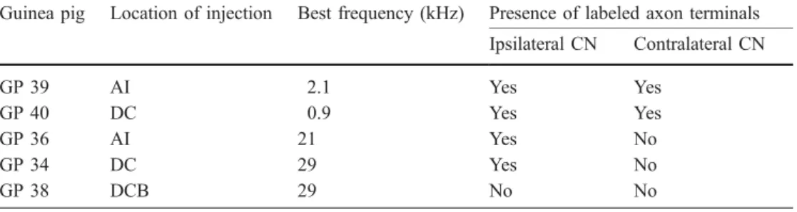
BDA injection sites are shown in Fig. 1. Two guinea pigs
received an injection into the area AI at sites where the BF
was 21 kHz and 2.1 kHz, respectively. In two other guinea
pigs, BDA injections were placed in the area DC, where
the BFs were 29 kHz and 0.9 kHz, respectively. Finally,
one guinea pig received an injection into the area DCB at a
site where the BF was 29 kHz (Table 1). In all animals
injected in AI or DC (n=4), BDA-labeled terminal fields,
including axons, terminal endings and boutons en passant,
were present in the CN either bilaterally or ipsilaterally. In
contrast, no labeled axons were found in the CN either
ipsilaterally or contralaterally as a result of injection into
the area DCB (animal GP 38, Table 1).
Concerning the trajectory of the BDA-labeled axons,
these were seen to travel from AC in the most dorsal part
of cerebral peduncle and reach the lateral lemniscus in its
rostral corner. From the lateral lemniscus, AC fibers
traveled in the dorsal acoustic stria and entered the DCN.
In the DCN, they ran in layers III and IV, from dorsal to
ventral and from medial to lateral. More rostrally, the
majority of axons were seen in the GCD and small cell cap
surrounding the CN, whereas few of them reached the
PVCN. Very few BDA-labeled axons traveled in the
trapezoid body.
BDA-labeled axon terminals were observed in restricted
zones of the ipsilateral CN in four animals. In two of these
guinea pigs, some axon terminals were also found in the
contralateral CN, but they were much less numerous than
in the CN ipsilateral to the cortical injection. Interestingly,
BDA-labeled axon terminals in the contralateral CN were
seen only in the two animals subjected to injection in low
BF region (Table 1). An example of the distribution of
BDA-labeled terminal fields in the ipsilateral and
contra-lateral CN is shown in Fig. 2 for two animals. Figure 2A
illustrates that, following an injection at BF 21 kHz in the
area AI, BDA-labeled axons were present only in the
Fig. 1A–E Photomicrographs showing the injection sites of biotinylated dextran amine. A Guinea pig (GP) 39 at a best frequency (BF) site of 2.1 kHz in the area AI (primary auditory cortex). B GP 36 at a BF site of 21 kHz in the area AI. C GP 40 at a BF site of 0.9 kHz in the area DC (dorsocaudal auditory field). D GP 34 at a BF site of 29 kHz in the area DC; E GP 38 at a BF site of 29 kHz in the area DCB (dorsocaudal belt) (D dorsal, M medial)
Fig. 2A,B Distribution of biotinylated dextran amine (BDA)-labeled axons in the ipsilateral cochlear nucleus (CN) of guinea pig (GP) 36 and bilaterally in the CN of GP 40, as seen on coronal sections. A GP 36, BDA was injected in primary auditory cortex (AI) at a best frequency (BF) site of 21 kHz. B GP 40, BDA was injected in dorsocaudal auditory field (DC) at a BF site of 0.9 kHz. The sections of the CN were arranged from caudal (left) to rostral
(right). Percentages indicate the caudorostral level of each section, where 0% is the caudal limit of the CN and 100% its rostral limit. CN parcellation: GCD granule cell domain, PVCN posteroventral cochlear nucleus, AVCN anteroventral cochlear nucleus, AN auditory nerve, cap small cell cap region; 1, 2, 3, 4 are the four laminae of the dorsal cochlear nucleus
Table 1 Properties of biotinyla-ted dextran amine injection sites and presence of labeled axons in the cochlear nucleus (CN). AI Primary auditory cortex, DC dorsocaudal auditory field, DCB dorsocaudal belt
Guinea pig Location of injection Best frequency (kHz) Presence of labeled axon terminals Ipsilateral CN Contralateral CN GP 39 AI 2.1 Yes Yes GP 40 DC 0.9 Yes Yes GP 36 AI 21 Yes No GP 34 DC 29 Yes No GP 38 DCB 29 No No
ipsilateral CN. Figure 2B shows the location of labeled
terminal fields in the CN bilaterally, as a result of BDA
injection in the area DC at BF 0.9 kHz. As a general
finding, labeled terminal fields were present
predomi-nantly in the GCD of the CN and in the small cell cap.
Fewer labeled axons were seen in the CN subdivisions,
with the density decreasing from DCN to PVCN and to
AVCN. After BDA injection in high BF sites in the AC,
axonal labeling tended to be located more caudally in the
CN (Fig. 2A) than after injection in low BF sites (Fig. 2B
upper row). Confirming previous observations (Schofield
et al. 2001), the axon terminals consisted of spherical
boutons en passant or terminaux, as illustrated in Fig. 3
for axon terminal fields in DCN and GCD. The diameter
of the boutons was less than 1 µm.
In vitro experiments
Taking into account that the AC projections to CN are
predominantly ipsilateral (see tracing data above), the
physiological effects in CN cells were tested by electrical
stimulation of the ipsilateral AC. We tested the effects of
AC stimulation on 52 CN principal cells in 28 IWB
preparations. Tested neurons were located in the three
subdivisions of the CN: AVCN, PVCN and DCN. In the
AVCN, the activity of eleven neurons was recorded. Seven
of them were recovered on histological sections and
identified as stellate (n=6) or bushy (n=1) cells. The other
four neurons were not recovered, being only partially
stained or unable to be stained after physiological
characterization. Among 24 neurons recorded in the
PVCN, seven were characterized as stellate cells, and
two as octopus cells, whereas 15 were not identified.
Finally, the population of 17 cells recorded in DCN
comprised three giant, three pyramidal, two vertical, two
stellate and seven unidentified neurons. The location in the
CN of the cell body of all identified neurons is illustrated
in Fig. 4 as a result of the transposition of the IWB
sections onto a CN of reference (Hackney et al. 1990; see
Methods section).
Single-pulse electrical stimulation of the AC induced
responses in only 2 of the 52 tested cells (4%). No
response to AC stimulation was observed in the great
Fig. 3A–D Photomicrographs illustrating examples of axons anterogradely labeled with biotinylated dextran amine in the cochlear nuclus (CN) of guinea pig 36. A,B Labeled axon terminal fields in the dorsal cochlear nucleus (DCN) of the section at 14% of
caudorostral level shown in Fig. 2A. C,D Labeled axon terminal fields in the DCN and granule cell domain (GCD) of the section at 24% of caudorostral level shown in Fig. 2A
majority of neurons even though many of them responded
to stimulation of other auditory and non-auditory inputs
(e.g., contralateral AN, dorsal column nuclei, and
trigem-inal nerve). Electrical stimulation of AC with trains of 3
–6
pulses (100
–250 Hz), tested in about 50% of cells, did not
result in any effect different from that observed with
single-pulse stimulation. The two CN cells responsive to
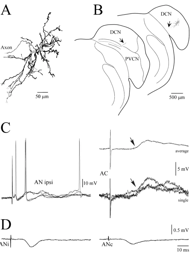
AC stimulation were located in the DCN (arrows in
Fig. 4), and had morphological properties typical of a giant
and a pyramidal cells, respectively. The morphology of the
giant cell is illustrated in Fig. 5A, together with its location
in the CN (Fig. 5B). The axon of the cell left the CN in the
direction of the dorsal acoustic stria (Fig. 5B, arrows),
without emitting recurrent collaterals in the CN itself.
The two cells had similar physiological properties,
which are illustrated in Fig. 5C for the giant cell. Both
cells responded to stimulation of the ipsilateral AN with a
sequence of excitatory (EPSPs) and inhibitory
postsynap-tic potentials (IPSPs), and exhibited long latency EPSPs to
stimulation of AC. Stimulation of the AC evoked an EPSP
of 2.9 mV at a latency of 20 ms in the giant cell (Fig. 5C).
For the pyramidal cell, the EPSP had an amplitude of
7 mV and a latency of 33 ms (not shown). These long
latencies suggest that responses of CN neurons to AC
stimulation were induced polysynaptically.
In order to ensure stimulation of adequate sites in the
AC, field potentials induced by stimulation of auditory
nerve were systematically recorded through the
stimulat-ing electrode in the AC. Figure 5D illustrates an example
of responses in the AC to stimulation of the ipsilateral or
contralateral AN in the same animal. Stimulation of
auditory nerves evoked a monophasic field potential in the
AC. The latency of the field potentials was in the range of
10
–15 ms and their amplitudes varied from 0.1 to 0.5 mV
across the experiments. These data confirm that the
location of the electrical stimulations was in the AC.
Discussion
The original contribution of the present study is to extend
previous tracing data on the projection of the AC to the
Fig. 4 Distribution of all identified neurons in the cochlear nucleus (CN) derived from the in vitro experiments. The brain of one guinea pig was sectioned in the coronal plane and Nissl-stained in order to establish a CN of reference (atlas). Individual sections derived from the isolated whole brain preparations were superimposed to the corresponding rostrocaudal section of the atlas in order to position the intracellularly stained neurons. Percentages indicate caudoros-tral levels as in Fig. 2. Arrows indicate the two cells that responded
to the stimulation of the auditory cortex; asterisk a bushy cell, open circles stellate cells, solid diamonds giant cells, solid triangles pyramidal cells, solid squares vertical cells, solid circles octopus cells. CN parcellation: GCD granule cell domain, PVCN poster-oventral cochlear nucleus, AVCN anterposter-oventral cochlear nucleus, AN auditory nerve, VN vestibular nerve, cap small cell cap region; 1, 2, 3, 4, are the four laminae of the dorsal cochlear nucleus
Fig. 5A–D Physiological and morphological characteristics of a giant cell in the dorsal cochlear nucleus (DCN). A High magnifi-cation reconstruction of the neuron whose lomagnifi-cation is shown on B a transverse section of the cochlear nucleus. Arrows indicate the cell axon (PVCN posteroventral cochlear nucleus). C Intracellular recordings from the cell at a membrane potential of −60 mV. Responses induced by stimulation of the ipsilateral auditory nerve
(AN ipsi, left panel, individual traces) or auditory cortex (AC, right panel, averaged and individual records). Arrows indicate the response onset. D Field potentials in AC in response to contralateral (ANc) or ipsilateral auditory nerve (ANi) stimulations recorded in the same experiment. Averages of 10–20 individual responses. Same time scale is used in C and D
CN, by introducing physiological characterization of the
AC injection sites, and an investigation of corticobulbar
effects in the IWB preparation. The anterograde transport
of BDA was used to localize the axon terminal fields
formed by the direct projection from the AC to the CN.
The corticobulbar axons terminate mainly in the GCD of
the ipsilateral CN and, to a smaller extent, in the DCN and
the PVCN. This result is consistent with previous
retrograde and anterograde tracing studies in the rat and
guinea pig showing direct projections from the AC to the
CN (Weedman and Ryugo 1996a, 1996b; Schofield et al.
2001). We found some axon terminals in the contralateral
CN but they were much less numerous than in the
ipsilateral CN. This observation is also in accordance with
a recent anterograde tracing study in the guinea pig
demonstrating bilateral projections from the AC to the CN,
with predominance of the ipsilateral ones (Schofield et al.
2001). A new finding of the present study is that
contralateral AC projections were not observed in the
two animals subjected to BDA injection in the high BF
regions of AI or DC. This result is not due to a particularly
small injection site (see Fig. 1B,D) and/or poor labeling
(see Fig. 3). Nevertheless, the observed difference between
projections originating from high versus low BF regions is
based on a small number of animals (n=4) and needs
further confirmation.
As mentioned above, in our physiological experiments
using the IWB preparation, the effects produced by AC
stimulation could be tested only in CN principal cells. The
AC-induced responses observed in two DCN cells had
very long latencies suggesting their polysynaptic origin.
Thus it can be concluded that AC projections to the three
CN subdivisions and GCD most likely do not establish
direct contacts with somata or dendrites of CN principal
cells. Moreover, even the polysynaptic effects were
observed in a very low percentage of recorded CN
principal neurons (4%). To some extent, this is consistent
with the relatively sparse corticobulbar projection
ob-served in AVCN, PVCN and DCN as a result of BDA
injection in the AC. In contrast, a much stronger projection
was seen in the GCD and small cell cap. The GCD
consists of a continuous sheet of small cells that surrounds
the magnocellular VCN and penetrates into the DCN as
layer II (Mugnaini et al. 1980). These small cells project to
some CN principal cells and other CN cells, which in turn
can contact CN principal cells. Therefore, one may wonder
why so few polysynaptic responses induced by AC
stimulation were observed in the present study. One
possible explanation could be that the stimulation of AC
and/or position of stimulating electrodes in the AC were
not optimal. This argument can be ruled out for at least
two reasons. First, the position of the stimulating
electrodes in the AC was confirmed by recording, through
the same electrodes, of field potentials in response to AN
stimulation. Second, a large interpolar distance of the
stimulating electrodes and high stimulation currents
suggest that a large area of AC and, consequently, a
large number of projecting AC cells was activated in our
experiments. The observation of rare AC-induced effects
in the CN principal cells is probably not due to the
impaired GCD and other polysynaptic circuits in the IWB
preparation. Indeed, in the same set of experiments, a large
proportion of CN principal cells responded to stimulation
of other sources of inputs to the CN (e.g., dorsal column
nuclei, and trigeminal nerve; see Babalian et al. 2001)
known also to operate mainly through the GCD (Itoh et al.
1987; Wright and Ryugo 1996; Shore et al. 2000;
Haenggeli et al. 2002; Ryugo et al. 2003). This
compar-ison under the same experimental conditions thus supports
the interpretation that the AC projection exerts indeed a
limited influence on CN principal cells as compared to the
other two mentioned sources of non-auditory inputs. The
reasons for that are not known. One may, however,
speculate that the density of projections and/or the
reliability of synaptic transmission in the GCD and other
polysynaptic circuits are lower for the projections
originating from the AC. For example, AC stimulation
might produce predominantly subthreshold modulation of
the membrane potential in small interneurons, which
would not result in any visible effects on CN principal
cells. Possible failure of AC inputs to generate action
potential in small relay cells would thus account for the
rare observation of responses at the level of CN principal
cells. This explanation somewhat contradicts our
experi-ments using train stimulation of AC intended to produce
temporal summation of synaptic responses and discharges
in relay neurons between AC and CN principal cells. Yet it
is possible that parameters of train stimulation were not
optimal in our study. Thus, influences of AC on granule
cells and other CN interneurons remain to be elucidated.
The polysynaptic nature of the observed excitatory
responses in two DCN cells suggests an alternative
possibility that these effects were due to activation of
AC projections to the IC and/or superior olivary complex,
which in turn project to the CN. The existence of these
pathways is well documented (Herbert et al. 1991;
Feliciano and Potashner 1995; Saldaña et al. 1996;
Malmierca et al. 1996; Games and Winer 1998; Schofield
and Cant 1999; Budinger et al. 2000; Schofield 2001;
Coomes and Schofield 2001). However, this possibility
seems to be unlikely for the following reasons. Our
previous results in the IWB preparation indicate that
electrical stimulation of IC and various auditory and
non-auditory brainstem inputs to the CN produce
predomi-nantly inhibitory effects in the vast majority of CN cells
(Babalian et al. 2002b; Jacomme et al. 2002). In contrast,
the two responses observed in the present study were
excitatory. Moreover, stimulation of the IC and brainstem
sources of inputs to CN induces synaptic responses in CN
cells, usually at relatively short latencies (below 10 ms).
Even if we take into account propagation and synaptic
delay time related to the additional activation of AC
projections to IC and/or brainstem, the resulting values
will be hardly compatible with response latencies in the
present study (20 and 33 ms). Therefore, we think that the
observed long-latency responses are mediated through the
AC projections to CN and slow signal processing
compatible with the granule cell circuits or other
interneuronal circuits in the CN. The fact that AC
stimulation effects were observed only in DCN cells
may indicate that the AC preferentially influence, though
weakly, output activity of the DCN. Such a conclusion
would be consistent with our observation of higher density
of AC projections in the DCN relative to other CN
subdivisions.
In conclusion, the anatomical data show sparse
projec-tions from the AC to the three CN subdivisions and more
dense projections to the GCD. Physiological results in the
IWB preparation indicate the existence of a limited
modulatory action of the AC on CN principal cells.
Thus, in the guinea pig, the auditory cortex might
modulate the CN activity essentially at the level of
granule cells and CN interneurons.
Acknowledgements This work was supported by Swiss National Science Foundation grants No. 31-55836.98 and 31-66731.01, and the National Center of Competence in Research (NCCR) Neural plasticity and repair.
References
Adams JC (1983) Cytology of periolivary cells and the organization of their projections in the cat. J Comp Neurol 215:275–289 Adams JC, Warr WB (1976) Origins of axons in the cats acoustic
striae determined by injection of horseradish peroxidase into severed tracts. J Comp Neurol 170:107–122
Andersen RA, Roth GL, Aitkin LM, Merzenich MM (1980) The efferent projections of the central nucleus and the pericentral nucleus of the inferior colliculus in the cat. J Comp Neurol 194:649–662
Babalian AL, Vibert N, Assie G, Serafin M, Mühlethaler M, Vidal PP (1997) Central vestibular networks in the guinea-pig: functional characterization in the isolated whole brain in vitro. Neuroscience 81:405–426
Babalian AL, Ryugo DK, Vischer MW, Rouiller EM (1999) Inhibitory synaptic interactions between cochlear nuclei: evi-dence from an in vitro whole brain study. Neuroreport 10:1913–1917
Babalian AL, Jacomme AV, Doucet JR, Ryugo DK, Rouiller EM (2001) Auditory and non-auditory inputs to the cochlear nucleus neurons: an in vitro whole brain study. ARO Abstr 24:197
Babalian AL, Jacomme AV, Doucet JR, Ryugo DK, Rouiller EM (2002a) Commissural glycinergic inhibition of bushy and stellate cells in the anteroventral cochlear nucleus. Neuroreport 13:555–558
Babalian AL, Jacomme AV, Ryugo DK, Rouiller EM (2002b) Functional input from the inferior colliculus to cochlear nucleus neurons: an in vitro whole brain study. ARO Abstr 25:8 Bajo VM, Merchán MA, Malmierca MS, Nodal FR, Bjaalie JG
(1999) Topographic organization of the dorsal nucleus of the lateral lemniscus in the cat. J Comp Neurol 407:349–366 Brown MC, Liberman MC, Benson TE, Ryugo DK (1988)
Brainstem branches from olivocochlear axons in cats and rodents. J Comp Neurol 278:591–603
Budinger E, Heil P, Scheuch (2000) Functional organization of auditory cortex in the Mongolian gerbil (Merionis unguicula-tus). IV. Connections with anatomically characterized subcor-tical structures. Eur J Neurosci 12:2452–74
Caicedo A, Herbert H (1993) Topography of descending projections from the inferior colliculus to auditory brainstem nuclei in the rat. J Comp Neurol 328:377–392
Cant NB, Gaston KC (1982) Pathways connecting the right and left cochlear nuclei. J Comp Neurol 212:313–326
Carey CL, Webster DB (1971) Ascending and descending projections of the inferior colliculus in the kangoroo rat (Dipodomys merriami). Brain Behav Evol 4:401–412 Coleman JR, Clerici WJ (1987) Sources of projections to
subdivisions of the inferior colliculus in the rat. J Comp Neurol 262:215–226
Conlee JW, Kane ES (1982) Descending projections from the inferior colliculus to the dorsal cochlear nucleus in the cat: an autoradiographic study. Neuroscience 7:161–172
Coomes DL, Schofield BR (2001) Cortical projections to the superior olivary complex contact cells that project to the cochlear nucleus in guinea pigs. ARO Abstr 24:45
Doucet JR, Weedman DL, Ryugo DK (2002) The projections of auditory cortex to the cochlear nucleus. In: Proceedings of International Symposium: Central Auditory Processing. Inte-gration of Auditory and Nonauditory Information, Monte Verità, Switzerland, 12-15 May 2002, Abstract P28
Evans EF (1979) Neuroleptanesthesia for guinea pig. Arch Otolar-yngol 105:185–186
Faye-Lund H (1986) Projection from the inferior colliculus to the superior olivary complex in the albino rat. Anat Embryol 175:35–52
Faye-Lund H (1988) Inferior colliculus and related descending pathways in rat. Upsala J Med Sci 93:1–17
Feliciano M, Potashner SJ (1995) Evidence for a glutamatergic pathway from the guinea pig auditory cortex to the inferior colliculus. J Neurochem 65:1348–1357
Feliciano M, Saldaña E, Mugnaini E (1995) Direct projection from the rat primary auditory neocortex to the nucleus sagulum, paralemniscal regions, superior olivary complex and cochlear nuclei. Aud Neurosci 1:287–308
Games KD, Winer JA (1988) Layer V in rat auditory cortex: projections to the inferior colliculus and contralateral cortex. Hear Res 34:1–26
Hackney CM, Osen KK, Kolston J (1990) Anatomy of the cochlear nuclear complex of guinea pig. Anat Embryol 182:123–149 Haenggeli CA, Doucet JR, Ryugo DK (2002) Trigeminal
projec-tions to the cochlear nucleus in rats. ARO Abstr 25:7 Hashikawa T, Kawamura K (1983) Retrograde labeling of ascending
and descending neurons in the inferior colliculus: a fluorescent double labeling study in the cat. Exp Brain Res 49:457–461 Herbert H, Aschoff A, Ostwald J (1991) Topography of projections
from the auditory cortex to the inferior colliculus in the rat. J Comp Neurol 304:103–122
Huffman RF, Henson OW, Jr. (1990) The descending auditory pathway and acousticomotor systems: connections with the inferior colliculus. Brain Res Rev 15:295–323
Itoh K, Kamiya H, Mitani A, Yasui Y, Takada M, Mizuno N (1987) Direct projections from the dorsal column nuclei and the spinal trigeminal nuclei to the cochlear nuclei in the cat. Brain Res 400:145–150
Jacomme AV, Rouiller EM, Ryugo DK, Babalian AL (2002) Physiological inputs from the inferior colliculus to the cochlear nucleus in the guinea pig studied in in vitro whole brain preparation. FENS Abstr 1:A184.9
Kane ES, Finn RC (1977) Descending and intrinsic inputs to dorsal cochlear nucleus of cats: a horseradish peroxidase study. Neuroscience 2:897–912
Malmierca MS, Le Beau FEN, Rees A (1996) The topographical organization of descending projections from the central nucleus of the inferior colliculus in guinea pig. Hear Res 93:167–180 Manunta Y, Edeline JM (1997) Effects of noradrenaline on
frequency tuning of rat auditory cortex neurons. Eur J Neurosci 9:833–847
Manunta Y, Edeline JM (1999) Effects of noradrenaline on frequency tuning of auditory cortex neurons during wakeful-ness and slow-wave sleep. Eur J Neurosci 11:2134–2150 Mugnaini E, Warr WB, Osen KK (1980) Distribution and light
microscopic features of granule cells in the cochlear nuclei of cat, rat, and mouse. J Comp Neurol 581–606
Redies H, Brandner S, Creutzfeldt OD (1989) Functional subdivi-sions in the auditory cortex of the guinea pig. J Comp Neurol 282:473–488
Romand R, Avan P (1997) Anatomical and functionnal aspects of the cochlear nucleus. In: Ehret G, Romand R (eds) The central auditory sytem. Oxford University Press, New York, pp 97–191 Rouiller EM (1997) Functionnal organization of the auditory pathways. In: Ehret G, Romand R, (eds) The central auditory system. Oxford University Press, New York, pp 3–96 Ryugo DK, Haenggeli C-A, Doucet JR (2003) Multimodal inputs to
the granule cell domain of the cochlear nucleus. Exp Brain Res DOI: 10.1007/s00221-003-1605-3
Saldaña E, Feliciano M, Mugnaini E (1996) Distribution of descending projections from primary auditory neocortex to inferior colliculus mimics the topography of intracollicular projections. J Comp Neurol 371:15–40
Schofield BR (2001) Origins of projections from the inferior colliculus to the cochlear nucleus in guinea pigs. J Comp Neurol 429:206–220
Schofield BR, Cant NB (1996) Projections from the ventral cochlear nucleus to the inferior colliculus and the contralateral cochlear nucleus in guinea pigs. Hear Res 102:1–14
Schofield BR, Cant NB (1999) Descending auditory pathways: Projections from the inferior colliculus contact superior olivary cells that project bilaterally to the cochlear nuclei. J Comp Neurol 409:210–223
Schofield BR, Coomes DL, Schofield R (2001) Projections from the auditory cortex to the cochlear nucleus in guinea pigs. Assoc Res Otolaryngol, Abs. 24:44
Shore SE, Godfrey DA, Helfert RH, Altschuler RA, Bledsoe SC Jr (1992) Connections between the cochlear nuclei in guinea pig. Hear Res 62:16–26
Shore SE, Vass Z, Wys NL, Altschuler RA (2000) Trigeminal ganglion innervates the auditory brainstem. J Comp Neurol 419:271–285
Spangler KM, Cant NB, Henkel CK, Farley GR, Warr WB (1987) Descending projections from the superior olivary complex to the cochlear nucleus of the cat. J Comp Neurol. 259:452–465 Van Noort J (1969) The structure and connections of the inferior
colliculus. Van Gorcum, Assen
Wallace MN, Rutkowski RG, Palmer AR (2000) Identification and localisation of auditory areas in guinea pig cortex. Exp Brain Res 132:445–456
Wan XST, Liang F, Moret V, Wiesendanger M, Rouiller EM (1992) Mapping of the motor pathways in rats: c-fos induction by intracortical microstimulation of the motor cortex correlated with efferent connectivity of the site of cortical stimulation. Neuroscience 49:749–761
Weedman DL, Ryugo DK (1996a) Pyramidal cells in primary auditory cortex project to cochlear nucleus in rat. Brain Res 706:97–102
Weedman DL, Ryugo DK (1996b) Projections from auditory cortex to the cochlear nucleus in rats: synapses on granule cell dendrites. J Comp Neurol 371:311–324
Wenthold RJ (1987) Evidence for a glycinergic pathway connecting the two cochlear nuclei: an immunocytochemical and retro-grade transport study. Brain Res 415:183–187
Wright DD, Ryugo DK (1996) Mossy fiber projections from the cuneate nucleus to the cochlear nucleus in the rat. J Comp Neurol 365:159–172