M. Tomaske P. Harpes R. Prêtre
A. Dodge-Khatami U. Bauersfeld
Evolution of paced QRS and QTc intervals
in children with epicardial pacing leads
Received: 11 December 2006 Accepted: 21 May 2007
Published online: 13 August 2007
Maren Tomaske, MD (
)
) Urs Bauersfeld, MDDivision of Pediatric Cardiology University Children’s Hospital Steinwiesstrasse 75 8032 Zurich, Switzerland Tel.: +41-44 / 2 66 75 19 Fax: +41-44 / 2 66 79 81 E-Mail: [email protected] Paul Harpes, PhD Biostatistics Unit
University Zurich, Switzerland Rene Prêtre, MD
Ali Dodge-Khatami, MD PhD Division of Congenital Cardiovascular Surgery
University Children’s Hospital Zurich, Switzerland
n Abstract Aims Permanent
ven-tricular pacing in children is as-sociated with ventricular dysfunc-tion due to asynchronous activa-tion. It is unclear whether paced QRS intervals increase dispropor-tionately over time, which could potentially cause ventricular dys-function. Methods A total of 52 children, with bipolar steroid-eluting epicardial leads implanted at a median age of 5.6 years (0.0– 17.4), was analyzed and followed up to 12.2 years (median 3.7). Pa-tients were subdivided in two groups: right (RV, n = 21) and left (LV, n = 31) ventricular pacing. To correct for age, standard deviation scores (Z-scores) for paced QRS and QTc intervals were calculated from published standard-ECG norm-values. As a measure for dividual paced QRS and QTc in-terval changes, a regression slope coefficient (inclinei) was calculated
for each patient’s course. Results
Mean Z-scores for paced QRS in-tervals at first and last follow-up were 4.7±1.2 and 4.9±0.9 for group RV, 4.4±1.1 and 4.8±1.1 for group LV. Inclinei of paced QRS (group
RV: 0.038 [–0.27–0.12], group LV: 0.147 [–0.05–0.30]; p = 0.07) and QTc intervals (group RV: 0.026 [–0.08–0.06], group LV: 0.023 [–0.04–0.09]; p = 0.63) did not dif-fer between both groups and in-dicated limited interval changes over time. Conclusion Neither epi-cardial pacing of the right nor left ventricle caused disproportionate paced QRS or QTc interval in-creases over time. An age-related prolongation of the electrical acti-vation unlikely causes ventricular dysfunction.
n Key words permanent pacing –
epicardial lead – children – ECG – paced QRS interval –
cardiac asynchrony
Introduction
Ventricular pacing in children is known to alter inter-and intraventricular conduction delay [1]. The paced ventricle is depolarized first, followed by a slow elec-trical activation through the myocardium of the un-paced ventricle. Therefore, the un-paced QRS complex re-sembles that of a right or left ventricular conduction delay, depending on the pacing site. On surface ECG, this produces a prolongation of the paced QRS, and
indirectly of the QTc interval. Thus, uncoordinated contraction of the ventricles results in asynchrony, de-creased stroke volume, and adverse myocyte remodel-ing. Over time, this may contribute to structural and functional changes [2–5]. Theoretically, these struc-tural changes, along with fibrosis surrounding the pa-cing lead, and a physiologic increase of the ventricular mass in a growing child, may result in further slowing of the electrical activation, and therefore increased in-traventricular conduction delay.
There is no information whether permanent epi-cardial pacing in children results in a possible dis-proportionate increase of the paced QRS intervals on surface electrocardiogram (ECG) over time. There-fore, we evaluated the evolution of paced QRS and QTc intervals in children with permanent epicardial ventricular pacing.
Patients and methods
n Patient population
With hospital ethical committee approval and in-formed consent, we retrospectively reviewed charts and electronic pacemaker databases of children who received a permanent epicardial ventricular pacing system. Bipolar steroid-eluting epicardial leads (Med-tronic CapSureEpi 10 366 or 4968, Med(Med-tronic, Inc, Minneapolis, MN, USA) were implanted at the right ventricular apex or the left ventricular free wall. They were connected to various pulse generators. In case of a lead exchange, follow-up was completed at that point. From the date of exchange, the child was in-cluded as a new patient (n = 3). Children with transve-nous pacing systems, those with less than 90% ventri-cular pacing, as well as children with less than three ECGs during follow-up were excluded. Patients were subdivided into two groups: those with right (group
RV) and those with left ventricular epicardial pacing
leads (group LV).
n Paced QRS and QTc interval
ECG documentation directly after implantation and at various follow up times consisted of hard copies. Surface 12-channel ECGs were recorded with a Mac®5000 System (GE Medical Systems, Milwaukee, WI, USA) either in our institution or at outpatient visits by the referring cardiologist. Measurements of paced QRS and QTc intervals automatically were per-formed by computed electrocardiographic system analysis of intervals, as well as manually measured from lead V5 by one author (M.T.). In case of incon-sistent results, intervals were double-checked (U.B.). The paced QRS interval was defined as the length of time from the beginning of the pacing spike to the end of the QRS complex. No pronounced latency from the pacemaker stimulus to the onset of the ear-liest paced QRS complex was seen (<40 ms). No drugs known to prolong QRS or QTc intervals were given to any patient included into the study.
The standardized deviation-score (Z-score) for paced QRS and QTc intervals were calculated from
published standard-ECG values of healthy children [6] to correct for age. For calculation of the age cor-rected paced QRS the following formula was used:
paced QRSmeasured norm mean QRSad
norm standard deviation of QRSad
where QRSad is the age-dependent QRS in healthy
children.
For each patient’s measurement (paced QRS or QTc-interval), the corresponding Z-score is therefore the difference of the measurement from the age-de-pendent norm mean, expressed in units of norm standard deviations.
n Global left ventricular function
Echocardiographic data were assessed by experi-enced cardiologists and retrospectively derived from electronic patient charts. To determine global left ventricular function, the fractional shortening (para-sternal long axis) and ejection fraction (apical 4-chamber view, Simpson equation) at first and last follow-up were noted.
n Statistical analysis
Data are expressed as either median ([interquartile range], range) or mean (± standard deviation) de-pending on distribution pattern of the data evaluated by the Kolmogorov-Smirnov test. A p-value < 0.05 was considered statistically significant.
To determine an individual persistent change of paced QRS and QTc intervals over time, slope coeffi-cients from a regression over individual repeated measurements (inclinei) were calculated for each
pa-tient’s course. Mann-Whitney U tests were used for analyzing differences in continuous variables be-tween independent groups. For group differences in categorical variables, the chi-squared test was used. Using the Wilcoxon signed rank test, intra-individu-al changes in continuous variables between the two follow-up intervals were evaluated. To determine a possible influence of the age at implantation and total time of follow-up on the inclinei of paced
QRS- and QTc-intervals a regression analysis was performed. All statistical analyses were performed using the Statistical Package for Social Sciences (SPSS for Windows, Version 14.0.1, Inc., Chicago, IL, USA).
Results
n Demographic and surgical data,
clinical characteristics
Demographic data, clinical characteristics, pace-maker data and underlying cardiac anatomy of group RV (n = 21) and group LV (n = 31) are given in Tables 1 and 2. All children with a congenital heart defect underwent prior cardiac surgery. In a
total of 47 children (90.4%) ventricular pacing was seen in 100% of the time. Even though there was a difference between median ages at lead implantation for right or left ventricular pacing, it was not statis-tically significant (p = 0.095). No significant dif-ference between group RV and group LV was seen in the distribution of children with a structural nor-mal heart or congenital heart disease (p = 0.85) and the underlying indication for permanent pacing (p = 0.89).
Table 1 Demographic data, clinical characteristics, and pacemaker data at implant
All Patients(n = 52) Group RV (n = 21) Group LV (n = 31)
Gender (male/female), n = 27/25 8/13 18/13 At lead implantationa Age (y) Weight (kg) 5.6 (0.0–17.4) 18 (1.0–61.6) 3.3 (0.0–16.4) 15.5 (2.8–61.6) 7.0 (0.0–17.4) 20.0 (1.0–61.0)
Total time of follow up (y)a 3.7 (0.7–12.2) 4.0 (0.9–12.2) 3.2 (0.7–11.1)
Epicardial leads, n = (%) Medtronic 10366 Medtronic 4968 18 (35) 34 (65) 10 (48) 11 (52) 8 (26) 23 (74)
Ventricular pacing mode, n=(%)
Unipolar pacing mode Bipolar pacing mode
40 (77) 12 (23) 18 (86) 3 (14) 22 (71) 9 (29) Pacing mode, n=(%) VVI/VVIR DDD 10 (19) 42 (81) 5 (24) 16 (76) 5 (16) 26 (84) Indications, n=(%) Congenital AVB Post-operative AVB Acquired AVB SSS Post-operative SSS Sinus node dysfunction
14 (27) 25 (48) 5 (10) 1 (2) 4 (8) 2 (4) 6 (29) 10 (48) 3 (14) – 2 (10) – 8 (26) 15 (48) 3 (10) 1 (3) 2 (7) 2 (7)
aData are given as medians (range)
AVB atrioventricular block; SSS sick sinus syndrome
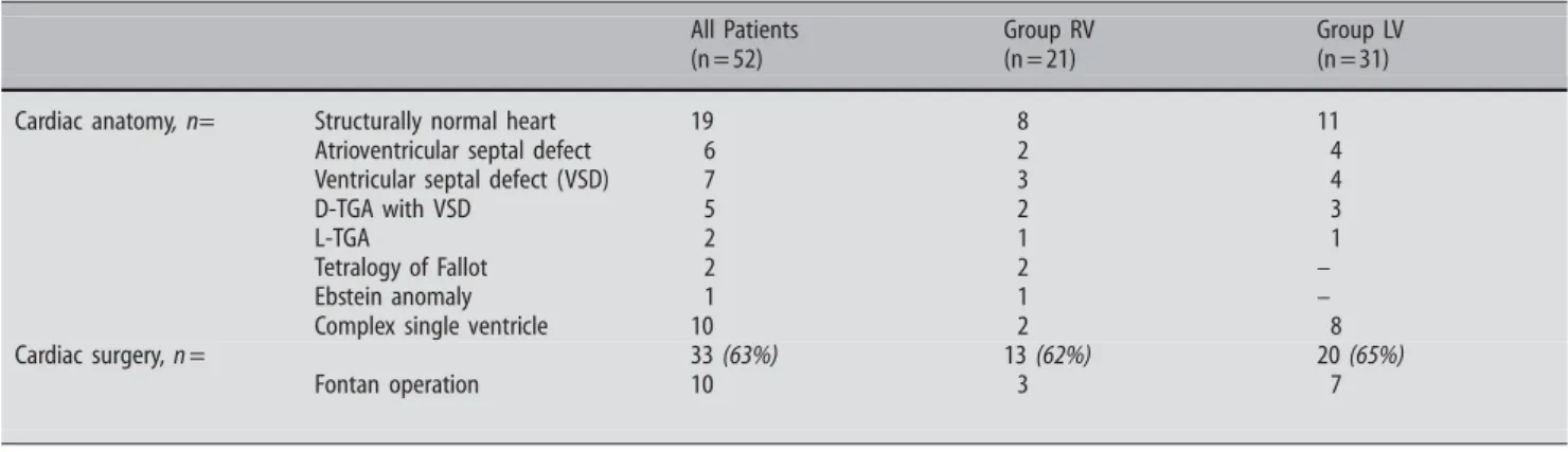
Table 2 Underlying cardiac anatomy and concomitant or previous surgery of the study cohort
All Patients (n = 52) Group RV (n = 21) Group LV (n = 31) Cardiac anatomy, n= Structurally normal heart
Atrioventricular septal defect Ventricular septal defect (VSD) D-TGA with VSD
L-TGA
Tetralogy of Fallot Ebstein anomaly Complex single ventricle
19 6 7 5 2 2 1 10 8 2 3 2 1 2 1 2 11 4 4 3 1 – – 8 Cardiac surgery, n = Fontan operation 33 (63%) 10 13 (62%) 3 20 (65%) 7
n Paced QRS and QTc intervals on surface ECG
at various follow-up times
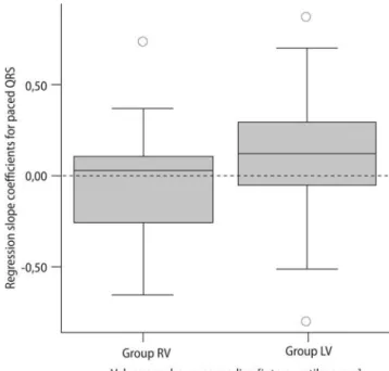
During follow up, ECGs of the children were available at median interval of 1.3 years (0.3–2.2). At first and last follow-up, absolute values as well as Z-scores for paced QRS and QTc intervals did not differ signifi-cantly for group RV and group LV. Furthermore, the inclineiof paced QRS and QTc intervals did not differ
significantly between both groups (Table 3). Low val-ues for the inclineiof paced QRS and QTc intervals
in-dicated little individual interval changes during the whole observation period (Fig. 1).
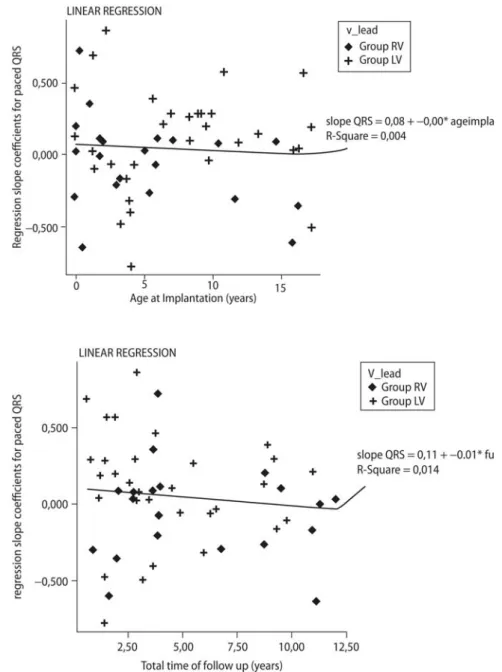
Regression analysis revealed no significant impact of the child’s’ age at epicardial lead implantation or of the total time of follow up on the inclinei of QRS
intervals (lead implant: R2= 0.004, p = 0.65; follow up: R2= 0.014, p = 0.41) (Figs. 2 and 3). Furthermore, no significant impact on the inclineiof QTc intervals
was observed (lead implant: R2= 0.041, p = 0.15; fol-low-up: R2= 0.004, p = 0.21). Additionally, differences in the inclinei of paced QRS and QTc intervals with
regard to the underlying heart defect or indication for permanent pacing were analyzed. No significant difference was observed for the presence or absence of a congenital heart defect (QRS interval: p = 0.63 and QTc interval: p = 0.49), nor the underlying indi-cation, especially congenital or post-operative atrio-ventricular block (QRS interval: p = 0.56 and QTc in-terval: p = 0.85).
Separate analysis between single or dual chamber pacing modes revealed no difference for absolute paced QRS values at first (143±21 ms vs 156±26 ms,
p = 0.09) or last follow-up (156±21 ms vs 167±24 ms, p = 0.18). Likewise, no difference was seen for ab-solute paced QTc values at first (482±39 ms vs 494±32 ms, p = 0.39) or last follow-up (485±38 ms vs 497±35 ms, p = 0.25). Moreover, no difference was seen for the inclinei of paced QRS and QTc
intervals between single or dual chamber pacing modes (QRS interval: p = 0.63 and QTc interval: p = 0.39).
n Global left ventricular function
In 39 of the 52 children (75%), global left ventricular function was analyzed. Fractional shortening at a first and last follow-up did not differ for group RV (37±6% vs 34±6%; p = 0.14) or group LV (35±6% vs 37±5%; p = 0.14). Similarly, ejection fraction did not differ at a first and last follow-up for group RV (58±5% vs 54±7%; p = 0.15) or group LV (58±6% vs 58±7%; p = 0.75). In the remaining 13 children, glo-bal systemic ventricular function could not be as-sessed due to the underlying cardiac anatomy (com-plex single ventricle (n=10), L-transposition of the great arteries (n=2), D-transposition of the great ar-teries after Senning repair (n=1)). The echocardio-graphic aspect of the systemic ventricular function was judged as good by experienced cardiologists in all 13 children.
Table 3 Absolute values, Z-scores and regression slope coefficients (inclinei)
for paced QRS and QTC intervals for group RV and group LV
Variable Group RV (n = 21) Group LV (n = 31) p-value Paced QRS (ms) First follow-up Last follow-up 148 (108–228) 158 (138–212) 150 (100–210) 160 (120–224) 0.84c 0.99c Paced QTc (ms) First follow-up Last follow-up 495 (424–535) 488 (439–564) 490 (421–588) 489 (421–588) 0.39c 0.63c Z-score for paced QRSa
First follow-up Last follow-up 4.6 (2.5–7.9) 4.7 (3.9–6.9) 4.2 (1.7–6.9) 4.5 (3.1–7.2) 0.59c 0.63c Z-score for paced QTca
First follow-up Last follow-up 1.8 (1.0–2.0) 1.7 (1.0–2.8) 1.7 (1.0–3.0) 1.7 (1.0–3.0) 0.37c 0.55c Inclineifor paced
QRSb
0.038 [–0.27–0.12] 0.147 [–0.05–0.30] 0.07c Inclineifor paced
QTcb 0.026 [–0.08–0.06] 0.023 [–0.04–0.09] 0.63 c
Data are given asamedians (range) orbmedians [interquartile range];cnot significant
Fig. 1 Boxplot of individual regression slope coefficients (inclinei) of paced
QRS intervals indicating little interval changes during follow-up. Inclinei of
paced QRS for group RV were centered near 0 (median = 0.038), but showed a tendency to be skewed towards negative values
Discussion
Permanent ventricular pacing provides increasing evidence of asynchronous ventricular electrical acti-vation, leading to conduction delay and induction of ventricular dysfunction. The risk of heart failure due to impaired ventricular pump function increases over time [7, 8]. There is no information whether paced QRS intervals increase disproportionately in the growing heart of pediatric patients, which could be an additional cause for long-term cardiac mor-bidity.
The right ventricular apex used to be the gold standard for ventricular pacing. Recognition of an
abnormal electrical activation and the resulting im-paired ventricular pump function has led to contro-versies about the optimal ventricular pacing site [9, 10]. Recent literature favors the left ventricle, as it seems to result in a more synchronous activation of the left ventricle and improved hemodynamics [11, 12]. Different abnormalities have been described to be associated with deterioration of the ventricular pump function. In children with chronic right ven-tricular apex pacing, histopathologic abnormalities by endomyocardial biopsy have been found that point to diminished function, as observed clinically [13]. Moreover, an asymmetric hypertrophy with in-creased wall thickness due to higher workload in the Fig. 2 Correlation between age at epicardial
lead implantation and individual regression slope coefficients (inclinei) for paced QRS intervals did
not show a significantly influence (p = 0.65)
Fig. 3 Correlation between the total time of
follow-up and individual regression slope coefficients (inclinei) for paced QRS intervals did
late activated area of the left ventricle has been ob-served after chronic right ventricular pacing [14, 15].
However, there is little knowledge whether these abnormalities cause an increasing prolongation of the paced QRS interval on surface electrocardio-gram. An animal model has examined the effect of workload on electrophysiology [16]. Conduction ve-locity was significantly reduced in the late-activated areas. As the mechanism of this change remained unclear, the authors suggested a theory of remodeled Purkinje fiber electrophysiology. Even though tissue fibrosis as a possible cause of reduced conduction velocity was not found, it is a main determinant. Changes in the amount and type of fibrous tissue in the interstitium can alter conduction velocity [17], and therefore lead to conduction delay. Marked tis-sue fibrosis has been observed in the use of transve-nous electrodes [18, 19]. Since the introduction of steroid eluting leads, the inflammatory response at the electrode-tissue interface has been diminished. This has consistently led to low pacing thresholds in transvenous as well as epicardial leads [20–22]. The electrode-epimyocardial tissue reaction has hereby been limited, but not totally eliminated.
A relationship between conduction delay and im-paired left ventricular function was demonstrated in clinical trials. In children with long-term pacing at the right ventricular apex, left ventricular short axis fractional area of change decreased by 0.2% for every 1-ms increase in QRS duration [7]. In adults, a progressive prolongation of the paced QRS intervals during follow-up has been shown to be a predictor for congestive heart failure [23], whereas corrected electrical asynchrony with narrowed QRS intervals by simultaneous right and left ventricular pacing improved cardiac output [24, 25].
As discussed above, all the processes and changes of the ventricles themselves, including asymmetrical hypertrophy, reduced conduction velocity, and tissue fibrosis can alter electrical activation, and as a con-sequence, lead to conduction delay. A further dete-rioration of these processes due to chronic ventricu-lar pacing over time, and the physiologic increase of the ventricular mass in a growing child can be an-ticipated. Theoretically, this would result in an in-creasingly delayed electrical activation through the myocardium and therefore disproportional prolonga-tion of the paced QRS intervals on surface ECG in children with permanent pacing.
In our study, we analyzed a total of 52 children with permanent epicardial ventricular pacing fol-lowed up to 12.2 years. We subdivided our popula-tion into two groups for further analysis: those with right and left ventricular epicardial pacing. The main finding was that there was no significant
pro-longation of age-correlated paced QRS or QTc inter-vals over time in children with permanent epicardial pacing, regardless of the pacing site. No significant impact of a congenital heart disease or the underly-ing indication for permanent pacunderly-ing was detected. Remarkably, a slight even though not significant trend of regression slope coefficients for paced QRS skewed towards negative values in patients with right ventricular pacing was observed, and remains un-clear.
Furthermore, no significant impact on global left ventricular function was observed between the first and last follow-up in children with two-chamber anatomy and either right or left ventricular epicar-dial pacing. However, regional ventricular dyssyn-chrony is rather obtained by tissue Doppler imaging or strain echocardiography [26–28]. Thus, potential right or left ventricular dyssynchrony might have evolved even though fractional shortening and ejec-tion fracejec-tion of the left ventricle remained un-changed.
Considering all the arguments for a possible in-crease in intraventricular conduction delay over time, it seems comforting that permanent epicardial pacing in a growing child is associated with a rather homogenous age-dependent prolongation of paced QRS intervals. A disproportionate increase is an un-likely factor for the asynchronous electrical activa-tion in permanent epicardial pacing. However, the pathophysiologic changes and regional ventricular dyssynchrony might not strongly correlate with a widening of QRS intervals on surface ECG [29].
n Limitation of the study
A main limitation of this study is its retrospective design with regard to completeness and reproduc-ibility of the collected variables. In order to avoid potential selection bias, every child with epicardial pacing leads implanted in our institution was in-cluded in the cohort of our study. Those children with less than three ECGs during follow-up due to missing documentation were excluded. The main variables paced QRS and QTc intervals were objec-tive measurements performed by computed electro-cardiographic system analysis of intervals, as well as manually measured by a single cardiologist from lead V5. This causes a low probability for misclassi-fication, and allows for a precise statement.
A further limitation is the assessment of global left ventricular function by conventional measure-ments. An analysis with advanced techniques, such as tissue Doppler imaging or strain echocardiogra-phy, might have revealed regional ventricular dys-synchrony.
Conclusion
Permanent epicardial ventricular pacing results in a prolongation of the paced QRS intervals. However, it is associated with a homogenous age-dependent pro-longation of paced QRS and QTc intervals up to a maximum observation period of 12.2 years. An age-related prolongation of the electrical activation
un-likely aggravates or causes ventricular dysfunction. However, regional ventricular dyssynchrony might not alter ECG measurements. Further prospective studies are required with documentation of the evo-lution of paced QRS and QTc intervals as well as si-multaneous measurement of ventricular dyssyn-chrony by advanced technique echocardiography.
References
1. Bedetto JB, Grayburn PA, Black WH et al (1990) Alterations in left ventri-cular relaxation during atrioventricu-lar pacing in humans. J Am Coll Car-diol 15:658–664
2. Adomian GE, Beazell J (1986) Myofi-brillar disarray produced in normal hearts by chronic electrical pacing. Am Heart J 112:79–83
3. Prinzen FW, Augustijn CH, Allessie MA, Reneman RS (1990) Redistribu-tion of myocardial fiber strain and blood flow by asynchronous activa-tion. Am J Physiol 259: 300–308 4. Karpawich PP (2004) Chronic right
ventricular pacing and cardiac per-formance: The pediatric perspective. Pacing Clin Electrophysiol 27:844–849 5. Vernooy K, Verbeek XA, Peschar M et al (2005) Left bundle branch block induces ventricular remodeling and functional septal hypoperfusion. Eur Heart J 26:91–98
6. Garson A Jr (1998) Electrocardiogra-phy. In: Garson A Jr, Bricker JT, Fisher DJ, Neish SR (eds) The Science of Pe-diatric Cardiology, 2nd edn. MD Balti-more, Williams & Wilkins, pp 713–788 7. Tantengco MV, Thomas RL, Karpa-wich PP (2001) Left ventricular dys-function after long-term right ventri-cular apical pacing in the young. J Am Coll Cardiol 37:2093–2100 8. Thambo JB, Bordachar P, Garrigue S
et al (2004) Detrimental ventricular remodeling in patients with congeni-tal heart block and chronic right ven-tricular apical pacing. Circulation 110:3766–3772
9. Karpawich PP, Mital S (1997) Com-parative left ventricular function fol-lowing atrial, septal and apical single chamber heart pacing in the young. Pa-cing Clin Electrophysiol 20:1983–1988 10. Tse HF, Yu C, Wong KK et al (2002) Functional abnormalities in patients with permanent right ventricular pa-cing: the effect of sites of electrical stimulation. J Am Coll Cardiol 40: 1451–1458
11. Vanagt WY, Verbeek XA, Delhaas T, Mertens L, Daenen WJ, Prinzen FW (2004) The left ventricular apex is the
optimal site for pediatric pacing: cor-relation with animal experience. Pa-cing Clin Electrophysiol 27:837–843 12. Vanagt WY, Verbeek XA, Delhaas T, et
al (2005) Acute hemodynamic benefit of left ventricular apex pacing in chil-dren. Ann Thorac Surg 79:932–936 13. Karpawich PP, Rabah R, Haas JE
(1999) Altered cardiac histology fol-lowing apical right ventricular pacing in patients with congenital atrioven-tricular block. Pacing Clin Electro-physiol 22:1372–1377
14. Van Oosterhout MFM, Prinzen FW, Arts T et al (1998) Asynchronous electrical activation induces asymme-trical hypertrophy of left ventricular wall. Circulation 98:588–595
15. Prinzen FW, Hunter WC, Wyman BT, Mc Veigh ER (1999) Mapping of re-gional myocardial strain and work during ventricular pacing: experi-mental study using magnetic reso-nance imaging tagging. J Am Coll Cardiol 33:1735–1742
16. Spragg DD, Akar FG, Helm RH, Tu-nin RS, Tomaselli GF, Kass DA (2005) Abnormal conduction and repolariza-tion in late-activated myocardium of dyssynchronously contracting hearts. Cardiovasc Research 67:77–86 17. Akar FG, Spragg DD, Tunin RS, Kass
DA, Tomaselli GF (2004) Mechanisms underlying conduction slowing and arrythmogenesis in nonischemic di-lated cardiomyopathy. Circulation Re-search 95:717–725
18. Epstein AE, Kay NK, Plump VG, Dai-ley SM, Anderson PG (1998) Gross and microscopic pathological changes associated with nonthoracotomy im-plantable defibrillator leads. Circula-tion 98:1517–1524
19. Higashi Y, Sato T, Shimojima H et al (2003) Mechanism of decrease in the atrial potential after implantation of a single-lead VDD pacemaker: Atrial his-tological changes after implantation of a VDD pacemaker lead in dogs. Pacing Clin Electrophysiol 26:685–691 20. Radovsky AS, Van Vleet JF (1989)
Ef-fects of dexamethasone elution on tis-sue reaction around stimulating
elec-trodes of endocardial pacing leads in dogs. Am Heart Journal 117:1288–1298 21. Hamilton R, Gow R, Bahoric B, Grif-fiths J, Freedom R, Williams W (1991) Steroid-eluting epicardial leads in pediatrics: improved epicardial thresholds in the first year Pacing Clin Electrophysiol 14:2066–2072 22. Cohen MI, Bush DM, Vetter VL et al
(2001) Permanent epicardial pacing in pediatric patients: seventeen years of experience and 1200 outpatients. Circulation 103:2585–2590
23. Miyoshi F, Kobayashi Y, Itou H et al (2005) Prolonged paced QRS dura-tion as a predictor for congestive heart failure in patients with right ventricular apex pacing. Pacing Clin Electrophysiol 28:1182–1188
24. Bax JJ, Abraham T, Barold SS et al (2005) Cardiac resynchronization therapy. Part 2-Issues during and after device im-plantation and unresolved questions. J Am Coll Cardiol 46:2168–2182 25. Riedlbauchova L, Cihak R, Bytesnik J
et al (2006) Optimization of right ventricular lead position in cardiac resynchronization therapy. Eur J Heart Fail 8:609–614
26. Cazeau S, Bordachar P, Jauvert G, et al (2003) Echocardiographic model-ing of cardiac dyssynchrony before and during multisite stimulation: a prospective study. Pacing Clin Elec-trophysiol 26:137–143
27. Pitzalis MV, Iacoviello M, Romito, R et al (2005) Ventricular asynchrony predicts better outcome in patients with chronic heart failure receiving cardiac resynchronization therapy. J Am Coll Cardiol 45:65–69
28. Goetze S, Butter C, Fleck (2006) Car-diac resynchronization therapy for heart failure – From experimental pa-cing to evidence-based therapy. Clin Res Cardiol (Suppl 4) 95:18–35 29. Bleeker GB, Schalij MJ, Molhoek SG,
et al (2004) Relationship between QRS duration and left ventricular dyssynchrony in patients with end-stage heart failure. J Cardiovasc Elec-trophysiol 15:544–549