ORIGINAL ARTICLE
Clinical and microbiological results following nonsurgical
periodontal therapy with or without local administration
of piperacillin/tazobactam
Marc Lauenstein&Marion Kaufmann&
G. Rutger Persson
Received: 15 November 2011 / Accepted: 27 September 2012 / Published online: 20 January 2013 # Springer-Verlag Berlin Heidelberg 2013
Abstract
Objectives We assessed if adjunct administration of piper-acillin/tazobactam added clinical and microbiological treat-ment benefits.
Materials and methods Thirty-six subjects (mean age 52.1 years (SD±10.3)) (NS by group) with chronic periodontitis were randomly enrolled receiving subgingival debridement and the local administration of piperacillin/tazobactam (test group) or debridement alone (control group). Bleeding on probing (BOP), probing pocket depth (PPD), and microbi-ological counts of 74 species were studied by checkerboard DNA-DNA hybridization up to month 6 after treatment. Results >Mean PPD changes between baseline and month 6 in the test and control groups were 1.5 and 1.8 mm, respec-tively (NS between groups). BOP in both groups decreased from about 80 to 40 %. At 4 and 12 weeks, lower counts of the following bacteria were found in the test group (site level): Fusobacterium species, Parvimonas micra, Pseudomonas
aeruginosa, Staphylococcus aureus, Tannerella forsythia, Treponema denticola, and a composite load of nine pathogens (p<0.001). At week 26, subjects receiving local antibiotics had a lower prevalence at tested sites for Fusobacterium nucleatum sp. polymorphum, Fusobacterium periodonticum, P. micra, and T. denticola.
Conclusions At 26 weeks, treatment with or without piper-acillin/tazobactam resulted in similar BOP and PPD improvements. At week 26 and at the subject level, the prevalence of 4/74 pathogens was found at lower counts in the group receiving local antibiotics.
Clinical relevance Administration of piperacillin/tazobac-tam reduces the prevalence of Fusobacterium, P. micra, and T. denticola to a greater extent than debridement alone but with no short-term differences in PPD or BOP.
Keywords Periodontitis . Debridement . Local antibiotics . Piperacillin/tazobactam . Microbiota . Checkerboard DNA-DNA
Introduction
The routine initial treatment of both chronic and aggressive periodontitis includes supra- and subgingival debridement with the objective to remove or at least reduce the infectious burden such that clinical evidence of inflammation is re-duced to a clinically acceptable level. The literature on short- and long-term results of nonsurgical mechanical
peri-odontal therapies is extensive (i.e., [1–7]). Supportive
peri-odontal therapy is considered as critical in maintaining
initial reductions of the subgingival microbiota [8]. Data
suggest that comprehensive periodontal debridement can achieve important reductions of bacterial counts that may
last for up to 8 months after initial treatment [9]. The
progression of site-specific periodontitis can be predicted
Electronic supplementary material The online version of this article (doi:10.1007/s00784-012-0856-4) contains supplementary material, which is available to authorized users.
M. Lauenstein
:
M. Kaufmann:
G. R. Persson (*) Department of Periodontology, University of Bern, Freiburgstrasse 7,CH 3010 Bern, Switzerland e-mail: rutger.persson@hkr.se G. R. Persson
Department of Periodontics, University of Washington, Seattle, WA, USA
G. R. Persson
Department of Oral Medicine, University of Washington, Seattle, WA, USA
G. R. Persson
Department of Health Sciences, Kristianstad University, Kristianstad, Sweden
by monitoring counts of bacteria associated with
periodon-titis [10]. It is also known that subgingival debridement of
root surfaces cannot effectively eliminate all bacteria and some bacteria may also be present within the dentin layer
[11]. Although treatment often results in an immediate
re-duction of bacterial counts, recolonization has been reported
shortly after therapy [12].
Most studies on microbiological changes following the administration of local antibiotics have focused on peri-odontal site-based changes. This has been consistent with a perception of a site-specific periodontal disease progres-sion and that site-specific disease progresprogres-sion appears to
occur in clusters of patients [13–17].
Recent studies have also demonstrated that the microbial content of the periodontal pocket is a determinant of gene expression in the gingival tissues and this controls the differential ability of periodontal species to elicit a local
host response [18]. Thus, genetic factors may explain why
periodontitis-susceptible subjects carry a specific pathogen-ic mpathogen-icrobiota in their periodontal pockets. This concept is consistent with findings that within a periodontitis-susceptible patient, the presence of many bacterial species is similar within different sites of a patient with similar
probing pocket depths (PPDs) [19].
Both systemic and local antibiotics have been advocated as adjunct antimicrobial therapies. Chronic periodontitis often presents with deep periodontal pockets at a limited number of sites. Therefore, the administration of local anti-biotics with a high local concentration, and in combination with debridement, may be more effective in the manage-ment of localized periodontal infections than treatmanage-ment with debridement alone. Recent studies have documented the positive effects of using systemic antibiotics to control the periodontal infection in combination with nonsurgical
peri-odontal therapy [20–23].
Management of“refractory periodontitis” with reduction of
bacteria is also possible by using a combination of a local
antibiotic and subgingival debridement [24]. Others have
shown that local administration of doxycycline in periodontal pockets of subjects with a smoking habit results in a signifi-cantly greater reduction in the levels of Porphyromonas
gingivalis in comparison to debridement alone [25]. Recent
studies have, however, also shown that the combined use of local administration of doxycycline and debridement of molars with furcation involvement failed to show a significant difference in vertical probing pocket depth up to 12 months after treatment and in comparison to local debridement alone
[26]. A difference in horizontal furcation probing depth to the
benefit of drug administration was found [26].
Data suggest that treatment with minocycline HCl micro-spheres as an adjunct treatment to debridement alone results in a greater reduction in the levels of P. gingivalis, Tanner-ella forsythia, and Treponema denticola in combination than
by debridement alone [27–30]. Other studies have, however,
shown that slow release of local antibiotics may not provide sustainable control of the subgingival microbiota and may not
be different from that of subgingival debridement alone [31,
32]. Thus, probing pocket depth reduction correlated
signifi-cantly with a decrease in the numbers and proportions of red
complex bacteria 30 days after administration [31]. The other
study [32] showed that controlled release of doxycycline did
not significantly suppress several subgingival pathogenic microorganisms and the authors concluded that this treatment did not seem to possess no distinct advantage over broad spectra, safe and inexpensive antiseptics and that the rationale for its employment in periodontal therapy remains unclear
[32]. In one study using a 14 % doxycycline gel applied
subgingivally at furcation sites following supportive peri-odontal therapy, the results showed that when applied only once at baseline, the administration failed to reduce the fre-quency of the need for re-instrumentation at furcation sites
during supportive therapy for a period of 12 months [26]. In
the most recent study on topical administration of doxycycline at periodontal sites in subjects with therapy-resistant peri-odontal pockets, the added use of doxycycline yielded on average 0.1 mm more reduction in probing pocket depth than
supportive therapy alone at 3 months [33]. At 26 weeks, this
study failed to demonstrate better odds of improved
periodon-tal conditions if treatment included the antibiotic or not [33].
This is consistent with the findings by others using locally administered doxycycline and that the benefits did not remain
beyond 3 months [34]. The long-term clinical value of adjunct
local antibiotics has been questioned [35,36].
Thus, local administration of either metronidazole, tet-racycline, doxycycline, or minocycline adjunct to support-ive periodontal therapy may not provide additional significant benefits in regard to probing depth reduction or gain in clinical attachment compared to results obtained
by mechanical debridement alone [35]. Recent data
sug-gest that treatment with local administration with azithro-mycin in a gel preparation may provide some clinical
benefits over scaling and root planning alone [37]. The
microbiological impact of such local antibiotic therapy remains unclear. Some of the tested local antibiotics are no longer available on the market.
Quorum sensing and plasmid transfer may inhibit efforts
with both systemic and local antibiotics [38, 39]. Data
suggest that piperacillin/tazobactam appears to be effective
against infections by gram-negative anaerobes [40, 41].
Antibiotic resistance to piperacillin/tazobactam seems to be
less than to that to amoxicillin/clavulanic acid [42].
Piper-acillin/tazobactam also appears to be effective against
Pseu-domonas aeruginosa [43,44]. P. aeruginosa is known to be
a key pathogen in transmitting antibiotic resistance through
plasmid transfer in biofilms [45,46]. P. aeruginosa has been
control of P. aeruginosa infection in periodontal pockets may be of importance to enhance the effects of local antibi-otic administration. Currently, there are no studies available assessing the effects on P. aeruginosa by local periodontal administration of antibiotics.
The objective of the present randomized clinical study was to assess the efficacy of local treatment with piperacil-lin/tazobactam in conjunction with subgingival debridement of periodontal pockets in comparison to local debridement alone using 26-week results as the study endpoint. We tested the null hypothesis that there are neither microbiological nor clinical differences in the treatment outcomes between the two study arms.
Materials and methods
The Ethics Committee at the University of Bern, Switzer-land (Kantonale Ethik Kommission, Bern, SwitzerSwitzer-land) approved the study. The study was conducted between 2007 and 2010. All participating subjects signed an in-formed consent. The flow chart for the study is presented
in Fig. 1.
Exclusion criteria Subjects were excluded
1. If they had received periodontal therapy within the preceding 6 months
2. If they had been treated with systemic or local anti-biotics within the preceding 6 months
3. If they were allergic to piperacillin/tazobactam or penicillin
4. If they were using anti-inflammatory medications or medications known to cause gingival overgrowth Inclusion criteria
Subjects were included
1. If they were 18 years of age or older
2. If they had four periodontal non-adjacent sites with a
PPD≥5 mm requiring therapy
3. If they had a diagnosis of chronic periodontitis Periofilm T®/Asbacare Clinic® (Medirel AS, Agno, Switzerland) is an antibiotic formulation (European Union Class III drug) which contains piperacillin and tazobactam in a formulation suitable for the treatment of periodontitis. Periofilm T® contains a powder (sodium piperacillin 100 mg, sodium tazobactam 12.5 mg) and a liquid (amino-alkyl-methacrylate copolymer, ammonium methacrylate co-polymer, ethanol 95 %, and purified water). The liquid and
powder are mixed immediately before administration with a supplied syringe.
Clinical measurements and treatment procedures
Subjects were allocated to the treatment group through ran-domization using the PASW, statistics 18.0 software (IBM/ SPSS, Armonk, NY, USA). Subjects were given coded numb-ers and then through the software program allocated to either the test group or control group. Study subjects and therapist were strictly instructed not to inform the examiner if they had received treatment with the test drug or not. The clinical examiner had no access to dental records and was not present in the clinic when initial treatment or administration of anti-biotics was performed. The allocation to the intervention group was not revealed until the data set had been locked. Bacterial samples were collected at the four selected sites from which clinical data were collected. The same brand of peri-odontal probes with 1-2-3-5-7-8-9-10 mm markings (SE4 Pocket probe, Deppeler SA, Rolle, Switzerland) was used to measure PPD at six sites per tooth. Bleeding on probing (BOP) of the pockets was defined approximately 10 s after the measurement of PPD. Only non-adjacent sites from dif-ferent teeth were studied.
Each subject provided four teeth with a PPD≥5 mm, and
these teeth were included in the study and treated with de-bridement as deemed necessary with hand instruments and/or ultrasound. The treatment was performed without time restrains and continued until the clinician was convinced that subgingival plaque and calculus had been removed at all the selected test teeth and neighboring teeth. In subjects with more than four sites presenting with a PPD≥5 mm, the four sites with the deepest PPD were chosen with consideration to risk for saliva contamination when performing bacterial sampling. Thus, if a test tooth had more than one site with the same (deepest) PPD, a buccal and preferably a mesio-buccal site was chosen. Third molars were excluded. In addition, no site represented sites associated with furcation involvement. All teeth were treated with nonsurgical debridement according to clinical protocol. During the study period, systemic antibiotics were not prescribed.
Oral hygiene instructions and information about the eti-ology of periodontitis and various treatment options were given to each subject prior to the enrollment in the study and before the initial therapy was given. Following the comple-tion of this therapy, the selected test teeth were then treated with the adjunct local antibiotics according to what study group the subject had been assigned to by computer-based randomization. The assignment to study group was not given until the debridement had been completed.
The local antibiotic drug was administered by a clinician who was unaware of the randomization schedule and other-wise also not associated with the study. The administration
of the local antibiotics was performed circumferentially in the pockets of the selected teeth. No additional treatment was performed before study endpoint at month 6. Subjects were given individualized post-treatment oral hygiene guidelines as required by routine clinical protocol at weeks 1, 2, 4, and 12.
Microbiological sampling
All microbiological samples were taken before probing pocket depth assessments had been performed. The se-lected sites representing the deepest non-adjacent sites within the subject had been defined at a previous visit. GCF was collected with sterile endodontic paper points (absorbent paper points size 50, Dentsply/Maillefer, Bal-laigues, Switzerland). The paper points remained in situ for 15 s and were then placed in dry Eppendorf tubes (1.5 ml natural flat cap microcentrifuge tubes, Starlab, Ahrensburg, Germany). Bacterial samples were taken
before treatment and at 2, 4, 12, and 26 weeks after intervention.
Analysis of subgingival bacterial samples
The vials were stored at−20 °C and processed after a standard
storage period of 3 months. To each sample, 0.15 ml TE
(10 mM Tris–HCl, 1 mM EDTA, pH 7.6) and 0.5 ml NaOH
were added. All samples were analyzed by checkerboard DNA-DNA hybridization technique. The 74 species assessed
are presented in Table1. The checkerboard DNA-DNA
hy-bridization was performed as described elsewhere [48,49].
Briefly, bacterial DNA was extracted, concentrated on nylon membranes (Roche Diagnostics GmbH, Mannheim, Ger-many), and fixed by cross-linking using ultraviolet light (Stratalinker 1800, Stratagene, La Jolla, CA, USA). The mem-branes with fixed DNA were placed in a Miniblotter 45 (Immunetics, Cambridge, MA, USA). Signals were detected by using the Storm Fluor-Imager (Storm 840, Amersham
Biosciences, Piscataway, NJ, USA) with a setup of 200μm and 600 V. The digitized information was analyzed by a software program (ImageQuant, Amersham Pharmacia), allowing com-parisons of the density of the 19 sample lanes against the two
standard lanes (105or 106cells). Signals were converted to
absolute counts by comparisons with these standards [34].
Statistical methods
We assumed at 25 % difference in the proportion of positive test results at study sites for P. gingivalis (15 %/40 %) at 26 weeks after intervention. Using the four sites in 18 subjects from each group and anticipating that two subjects in each group would
Table 1 Reference bacteria strains included in the DNA-DNA checkerboard analysis
Species Collection Species Collection
Actinomyces israelii ATCC 12102 Lactobacillus jensenii GUH 160339 Actinomyces naeslundii (type I + II) ATCC 43146 Lactobacillus vaginalis GUH 078092
Actinomyces neuii GUH 550898 Leptotrichia buccalis ATCC14201
Actinomyces odontolyticus ATCC 17929 Mobiluncis curtisii GUH 070927 Aggregatibacter actinomycetemcomitans (a) ATCC29523 Mobiluncus mulieris GUH 070926 Aggregatibacter actinomycetemcomitans (Y4) ATCC 43718 Neisseria mucosa ATCC 33270
Aerococcus christensenii GUH 070938 Parvimonas micra ATCC 19696
Aanaerococcus vaginalis GUH 290486 Peptoniphilus sp. GUH 55097
Atopbium parvulum GUH 160323 Porphyromonas endodontalis ATCC 35406
Atopobium vaginae GUH 010535 Porphyromonas gingivalis ATCC 33277
Bacteroides ureolyticus GUH 080189 Prevotella bivia GUH 450429
Bifidobacterium biavatii GUH 071026 Prevotella disiens GUH 190184 Bifidobacterium bifidum GUH 070962 Prevotella intermedia ATCC 25611 Bifidobacterium breve GUH 080484 Prevotella melaninogenica ATCC 25845 Bifidobacterioum longum GUH 180689 Propionibacterium acnes ATCC 11727/2
Campyobacter gracilis ATCC 33236 Proteus mirabilis GUH 07092
Campylobacter rectus ATCC 33286 Pseudomonas aeruginosa DSMZ 50071
Campylovacter showae ATCC 51146 Selenomonas noxia ATCC 43541
Capnocytophaga gingivalis ATCC 33612 Staphylococcus anaerobius DSMZ 20714 Capnocytophaga ochraceae ATCC 335945 Staphylococcus aureus ATCC 25923 Capnocytophaga sputigena ASTCC 33612 Staphylococcus aureus (yellow) GUH 070921 Corynebacterium nigricans GUH450453 Staphylococcus aureus (white) GUH 070922 Corynerbacterium aurimucosum GUH 071035 Staphylococcus epidermis GUH 130381
Dialister sp. GUH 071045 Staphylococcus haemolyticus DSMZ 20263
Escherichia coli GUH 070903 Streptococcus agalactiae GUH 230282
Eikenella corrodens ATCC 23834 Streptococcus anginosus ATCC 33397 Enterococcus faecalis GUH 170812 Streptococcus constellatus ATCC 27823 Enterococcus faecalis ATCC 29212 Streptococcus gordonii ATCC 10558 Fusobacterium nucleatum
nucleatum
ATCC 25586 Streptococcus intermedius ATCC 27335 Fusobacterium nucleatum polymorphum ATCC 10953 Streptococcus mitis ATCC 49456 Fusobacterium nucleatum naviforme ATCC 49256 Streptococcus oralis ATCC 35037 Fusobacterium periodonticum ATCC 33693 Streptococcus pneumoniae DSMZ 11866 Gardnerella vaginalis GUH 080585 Streptococcus sanguinis ATCC 10556 Haemophilus influenzae ATCC 49247 Streptococcus mutans ATCC 25175
Helicobacter pylori ATCC 43504 Tannerella forsythia ATCC 43037
Lactobacillus acidophilus ATCC 11975 Treponema denticola ATCC 35405
Lactobacillus crispatus GUH 160342 Treponema socranskii D40DR2
Lactobacillus gasseri GUH 17085 Varibaculum cambriense GUH 070917
Lactobacillus iners GUH 160334 Veillonella parvula ATCC 10790
ATCC American Type Culture Collection; D sample from Forsyth Institute, Boston, MA, USA; GUH Ghent University Hospital Collection, Ghent, Belgium
not complete the study (n064 per group), the power would be 88 %. The Kolmogorov–Smirnov test was used to assess whether the data set had a normal distribution pattern or not. Independent t tests (equal variance not assumed) and Mann– Whitney U tests to screen for bacteria with significant differ-ences in bacterial counts were performed. Subject-based bacte-rial counts were calculated based on the number of sites with bacteria present at week 26. General linear model multivariate ANOVA using Sidak’s correction for multiple observations and with smoking, age, and gender as covariates was used to assess differences by bacterial presence at the subject level. The PASW, statistics 18.0 software for MAC OS X version 10.6.7 was used for the analysis of the data (IBM SPSS).
Results
A total of four subjects were lost to follow-up before the first visit after the intervention. Due to the fact that they never returned for the bacterial samplings or clinical follow-up, no data after enrollment could be collected from these four subjects in the control group. The data from these subjects were exclud-ed from the analysis. Data were studiexclud-ed in 18 subjects (4/18, 22.2 % smokers) from the test group including 72 test sites and in 14 (2/14, 14.3 % smokers) subjects from the control group including 56 test sites. The mean age of the study population was 52.1 years (SD±10.3) in both groups and thus with no statistically significant difference by study groups. During the study period, no adverse events were identified.
Clinical data: probing pocket depth and bleeding on probing at sites from which bacterial samples were taken
The study included 41.0 % incisors and cuspids, 20.1 % premolars, and 38.8 % molars. Baseline overall PPD values at sites from which bacterial samples were collected varied between 5 and 11 mm (overall mean 6.8 mm, SD±1.3 mm). At baseline, the mean PPD in the control and test groups were 7.0 mm (SD±1.2) and 6.8 mm (SD±1.3), respectively (NS). At week 26, the corresponding PPD values were 5.2 mm (SD ±1.5) and 5.1 (SD ±1.4), respectively (NS). At baseline, all PPD values were >5 mm. At week 26, 42.9 % of the sites in the control group had a PPD value >5 mm, whereas 35.5 % of the sites in the test group had a PPD value >5 mm. The mean difference (decrease) in PPD between baseline and week 26 in the control group was 1.8 mm (SE±0.3; 95 % CI 1.2, 2.3; p<0.001). The mean difference (decrease) in PPD between baseline and week 26 in the test group was 1.5 mm (SE±0.2; 95 % CI 1.1, 2.0; p<0.001). Both at baseline and at week 26, general linear model univariate analysis with smoking status and sample site as covariates, statistical analysis failed to demonstrate study group differ-ences in PPD values both at baseline and at week 26.
At baseline and at sites from which bacterial samples were taken, BOP was present at 85.1 % in the control group and at 82.1 % in the test group (NS). At week 26, BOP was found at 44.9 % in the control group and at 38.2 % sites in the test group. Statistical analysis failed to demonstrate group differences in BOP scores at either time point.
Considering smoking status, statistical analysis failed to demonstrate differences in PPD values. At baseline, the mean difference in PPD by smoking status was 0.3 mm
(SE±0.3; 95 % CI −0.2, 0.8; p00.24). The mean PPDs in
smokers and nonsmokers at baseline were 6.5 mm (SD±1.3) and 6.8 mm (SD±1.3) (p00.24). The mean PPDs in smok-ers and nonsmoksmok-ers at week 26 were 4.9 mm (SD±1.2) and
5.2 mm (SD±1.5), respectively (p00.38).
Microbiology
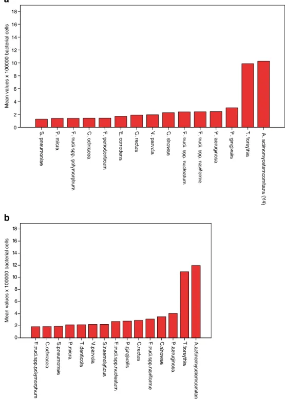
Analysis by Kolmogorov–Smirnov test failed to identify a normal distribution pattern by bacterial counts. Based on the screening of the 74 bacterial species included in the check-erboard DNA-DNA hybridization method, the 15 species with the highest prevalence rates at baseline and 2, 4, 12, and 26 weeks after treatment in the test and control groups were identified. The mean values of each of these species by
study group are presented in descending order (Figs.2a, b;
3a, b; 4a, b; 5a, b; and 6a, b). It is noteworthy that at
baseline, the mean values of these bacteria were similar by study group. With few exceptions, the same bacteria were identified including Aggregatibacter actinomycetemcomi-tans (Y4), Fusobacterium nucleatum spp. naviforme, Fuso-bacterium nucleatum spp. nucleatum, Parvimonas micra, P. gingivalis, P. aeruginosa, and T. forsythia. The total count of a composite pathogen group including these species and also including T. denticola and Staphylococcus aureus was computed. The load of this composite bacterial group was further assessed (see below).
At 2, 4, and 12 weeks, it is obvious that shifts in the prevalence ranking order of species had occurred in both study groups. Consistently, A. actinomycetemcomitans (Y4) was present at much higher levels in the control group. Other species were also present at higher counts in the control group. At 26 weeks, most differences between the two groups had disappeared. The prevalence rates (percent) at the bacterial
level≥1.0×10⁵cells for each of the bacteria included in the
composite pathogen group are shown in Table2. In the control
group and at 4 weeks, a relevant decrease in the prevalence
rate based on the≥1.0×10⁵cells detection level was not found
for any of the species. At week 4 in the test group, P.
gingi-valis, P. aeruginosa, and T. forsythia decreased ≥20 % in
detection prevalence rates. At 26 weeks, the decrease remained in the test group for P. aeruginosa and T. forsythia. At baseline, smokers had a higher bacterial count of T.
not exist at week 26. Analysis by general linear model multi-variate analysis with smoking status, and sample site as cova-riates, with the above listed species, and the composite bacterial counts as dependent variables and adjustment for multiple comparisons by Sidak correction for multiple obser-vation found significantly higher bacterial counts in the con-trol group at baseline for the following species: P. micra, S.
aureus (p<0.01), and with the largest difference for T.
denti-cola (mean difference−1.3×10⁵cells; 95 % CI−2.1, −0.4×
105; p00.005). The corresponding results from the analysis at
weeks 2, 4, 12, and 24 are presented in Table 3. Thus,
specifically at weeks 4 and 12, significantly lower counts of the following bacteria were found in the test group: A. actino-mycetemcomitans (Y4), F. nucleatum species, P. micra, P.
Mean values x 100000 bacterial cells
18 16 14 12 10 8 6 4 2
0 S. pneumoniae P. micra F. nucl spp. polymorphum C. ochracea F. periodonticum E. corrodens C. rectus V. parvula C. showae F. nucl. spp. nucleatum F. nucl. spp. naviforme P. aeruginosa P. gingivalis T. forsythia A. actinomycetemcomitans (Y4)
A.actinomycetemcomitans (Y4 ) T.forsythia P.aeruginosa C.showae F.nucl.spp.naviform e C.rectus P.gingivalis F.nucl.spp.nucleatum S.haemolyticus V.parvula T.denticola P.micra S.pneumonaie C.ochracea F.nucl.spp.polymorphu m
Mean values x 100000 bacterial cells
18 16 14 12 10 8 6 4 2 0
a
b
Fig. 2 a Baseline meanbacterial counts in descending order for the 15 most prevalent species in the control group. b Baseline mean bacterial counts in descending order for the 15 most prevalent species in the test group
A.actinomycetemcomitans (Y4) T.forsythia C.showa e F.nucl.spp.nucleatu m F.nucl.spp.naviforme N.mucos a V.parvula C.rectus E.corrodens P.micra F.nucl.spp.polymorphum F.periodonticu m L.buccali s C.ochrace a S.pneumonai e Mean 18 16 14 12 10 8 6 4 2 0 A.actinomycetemcomitans (Y4 ) V.parvula T.forsythia F.nucl.spp.naviforme C.showae N.mucosa F.nucl.spp.nucleatu m S.haemolyticus S.haemolyticus P.micra F.nucl.spp.polymorphum C.ochracea F.periodonticu m E.corrodens L.buccalis C.rectus
Mean values x 100000 bacterial cells
18 16 14 12 10 8 6 4 2 0
a
b
Fig. 3 a Week 2 mean bacterial counts in descending order for the 15 most prevalent species in the control group. b Week 2 mean bacterial counts in descending order for the 15 most prevalent species in the test group
A.actinomycetemcomitans (Y4) T.forsythia C.showae F.nucl.spp.nucleatum F.nucl.spp.naviforme N.mucosa V.parvula C.rectus E.corrodens P.micra F.nucl.spp.polymorphum S.haemolyticus F.periodonticum L.buccalis C.ochracea
Mean values x 100000 bacterial cells
18 16 14 12 10 8 6 4 2 0 A.actinomycetemcomitans (Y4 ) T.forsythia C.showae V.parvula F.nucl.spp.nucleatu m F.nucl.spp.naviforme S.haemolyticus P.micra F.nucl.spp.polymorphum C.rectus C.ochracea F.periodonticum L.buccalis N.mucosa E.corrodens
Mean values x 100000 bacterial cells
18 16 14 12 10 8 6 4 2 0
a
b
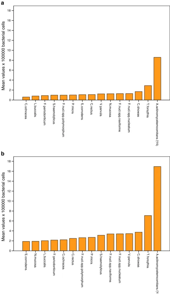
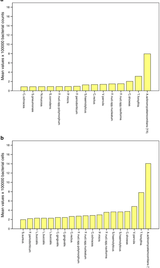
Fig. 4 a Week 4 mean bacterial counts in descending order for the 15 most prevalent species in the control group. b Week 4 mean bacterial counts in descending order for the 15 most prevalent species in the test groupA.actinomycetemcomitans (Y4) T.forsythia C.showae F.nucl.spp.naviforme F.nucl.spp.nucleatum V.parvula C.rectus S.haemolyticus F.periodonticum P.micra F.nucl.spp.polymorphum E.corrodens N.mucosa S.pneumonaie C.ochracea
Mean values x 100000 bacterial counts
18 16 14 12 10 8 6 4 2 0 A.actinomycetemcomitans (Y4 ) T.forsythia V.parvula C.showae S.haemolyticus S.haemolyticus F.nucl.spp.naviforme P.micra C.ochracea F.nucl.spp.nucleatum F.nucl.spp.polymorphum C.rectus C.gingivalis C.gingivalis L.buccalis L.buccalis L.buccalis F.periodonticum S.aureus
Mean values x 100000 bacterial cells
18 16 14 12 10 8 6 4 2 0
a
b
Fig. 5 a Week 12 meanbacterial counts in descending order for the 15 most prevalent species in the control group. b Week 12 mean bacterial counts in descending order for the 15 most prevalent species in the test group
gingivalis, P. aeruginosa, S. aureus, and T. denticola were found in the test group. At week 26, only F. nucleatum spp.
polymorphum, F. periodonticum, P. micra, P. aeruginosa, and T. denticola were found at lower counts in the test group.
A.actinomycetemcomitans (Y4) T.forsythia V.parvula C.showae C.rectus S.haemolyticus F.nucl.spp.naviforme F.nucl.spp.nucleatum F.nucl.spp.polymorphum F.periodonticum E.corrodens S.pneumonaie L.buccalis P.micra C.ochracea
Mean values x 100000 bacterial cells
18 16 14 12 10 8 6 4 2 0 A.actinomycetemcomitans (Y4 ) T.forsythia F.nucl.spp.naviforme F.nucl.spp.nucleatum C.showae F.nucl.spp.polymorphum P.micra C.rectus F.periodonticum S.haemolyticus L.buccalis V.parvula C.ochracea P.gingivalis T.denticola
Mean values x 1000000 bacterial cells
18 16 14 12 10 8 6 4 2 0
a
b
Fig. 6 a Week 26 mean bacterial counts in descending order for the 15 most prevalent species in the control group. b Week 26 mean bacterial counts in descending order for the 15 most prevalent species in the test group
Subject-based microbiological analysis
The five microbiological species that were found to differ by the site-specific analysis were further investigated. At baseline and at week 26, the number of sites that were defined as being positive for these five species identified as being present at a
bacterial level≥1.0×10⁵cells (primary detection level for the
checkerboard method) were provided a subject-based number. Analysis by general linear model multivariate ANOVA in-cluding smoking habit, subject age, and gender as covariates, and using Sidak’s correction for multiple observations, the analysis failed to identify baseline differences in a number of positive sites for the five species studied. At week 26, how-ever, the same analysis identified that significantly fewer positive sites were found in subjects who had been treated with the antibiotics for the following species: T. denticola (p<0.001), F. nucleatum spp. polymorphum (p00.001), P.
micra (p00.006), and F. periodonticum (p00.013).
Discussion
There are currently no other clinical data on the efficacy to reduce bacterial counts in periodontal pockets by a single administration of piperacillin/tazobactam in subjects with moderate to advanced periodontitis. P. gingivalis was cho-sen as the target pathogen and as the primary outcome measure because it has been studied extensively in
associa-tion with periodontitis [8,23–25,30,35,37]. Several other
bacterial species demonstrated a greater susceptibility to the intervention in the test group and this effect remained also at week 26 for some species but not for P. gingivalis, T. forsythia, or A. actinomycetemcomitans.
The limitation of the present study is that the control subjects were not treated with a placebo drug administration. Neverthe-less, the clinical examiner and the laboratory staff members
were blinded to the protocol assignment to control for bias. Another limitation is that the evidence of bacterial changes following periodontal interventions from other studies does not easily provide information that can be utilized for statistical power analysis. Thus, we assumed based on our laboratory experiences that a 20–25 % difference could be anticipated. A decrease amounting to approximately 20–25 % was obtained in the test group at weeks 2 and 4 for P. gingivalis and P. aeruginosa. At week 26, this remained the case for P. aerugi-nosa, suggesting that the administration of piperacillin/tazobac-tam has a relevant effect but limited to the control of P. aeruginosa subgingival colonization. One of the reasons why the reduction in bacterial counts was limited may be the result of less than optimal control of gingival inflammation as noticed by the rather high proportion of BOP at study endpoint.
The decreases in PPD and BOP obtained in the present study are consistent with the other studies on subgingival
debridement not using antibiotics [1–7]. The extent of PPD
reduction and decrease in BOP in the present study suggested a clinical effective outcome of therapy provided in both groups. Furthermore, the extent of PPD reduction in both study groups in the present study was comparable, or greater to PPD reduc-tions after combined local debridement and local antibiotics in
other studies [30,50]. In the present study, we treated subjects
who were diagnosed with moderate to severe chronic perio-dontitis, and we only assessed interproximal conditions. In other similar studies, periodontal sites with more shallow PPDs
have been studied [18, 21, 38]. It is well known that the
reduction of PPD in the range of 1.5 to 2.0 mm can be obtained
by debridement alone in deep periodontal pockets [1–3]. The
extent of possible probing depth reduction may also be limited by anatomical factors such as the extent and topography of alveolar bone loss and attachment loss.
Some data have shown that local administration of doxycy-cline or minocydoxycy-cline in addition to debridement in subjects who smoke results in greater reduction in the frequency of P.
Table 2 Distributions of selected bacteria at different sample times in the test and control groups by the detection level for a positive test result (≥×1.05bacterial cells) that is the standard detection level for the checkerboard DNA-DNA hybridization method
Composite species Baseline 2 weeks 4 weeks 12 weeks 24 weeks
Test Control Test Control Test Control Test Control Test Control A. actinomycetemcomitans 88.0 80.4 65.2 78.3 71.4 83.0 71.1 76.0 71.6 73.0 F. nucleatum naviforme 54.3 70.4 44.9 60.9 38.1 55.3 47.0 68.0 49.4 59.5 P. micra 51.1 66.7 28.1 63.0 34.5 68.1 61.0 64.5 39.5 63.2 P. gingivalis 38.0 58.5 13.5 21.7 16.7 29.8 16.9 30.0 24.7 29.7 P. aeruginosa 43.7 61.5 24.5 61.8 13.4 74.3 26.2 61.5 17.8 50.0 S. aureus 28.3 51.0 14.6 52.2 15.1 53.2 27.7 52.0 23.5 35.1 T. forsythia 68.5 76.5 42.7 67.4 41.7 61.7 41.0 60.0 48.1 57.8 T. denticola 47.8 68.6 30.3 58.7 32.1 66.0 37.3 50.0 37.0 73.0
gingivalis [22,25]. In the present study, smoking did not seem to have an impact on the study outcomes neither on PPD nor BOP changes or microbiological changes. This may be explained by the low prevalence of smokers in the study. Smoking, subject age, and gender were included as covariates in the subject-based analysis but did not significantly influence the results.
Although subject-based factors must be considered, it is generally perceived that chronic periodontitis is
tooth/site-specific [13–17]. In the present study, each subject
contrib-uted four individual test sites representing the sites with the most advanced periodontitis. Thus, no subject was overrep-resented providing more data than any other subject.
Several studies have used site-based analysis and performed microbiological sampling only from mesio-buccal surfaces
[8,22,24,50–56]. There appears to be a defined order in
bacterial species succession in early supragingival and sub-gingival biofilm redevelopment after professional cleaning. The site-specific development of periodontitis may be the result of the symbiotic effects due to co-aggregation in subgingival biofilms including P. gingivalis, T. denticola,
and T. forsythia [53].
Thus, the presence and counts of P. gingivalis, T. denticola, and T. forsythia may suggest the stability of periodontal con-ditions at individual sites at teeth. The observations that local treatment with antibiotics can reduce the counts of these
Table 3 Microbiological differences by study groups after intervention at weeks 2, 4, 12, and 26
Time point Species Mean differences×105cells 95 % CI×105cells p values
Week 2 A. actinomycetemcomitans Y4 8.8 1.6, 16.0 0.001 F. nucleatum naviforme 1.3 0.5, 2.1 0.002 F. nucleatum polymorphum 1.3 0.5, 2.1 0.002 F. periodonticum 0.9 0.1, 1.7 0.02 S. aureus 1.0 0.6, 1.4 0.001 T. denticola 0.7 0.4, 1.1 0.001 Composite bacteria 12.3 0.4, 24.1 0.042 Week 4 A. actinomycetemcomitans Y4 7.9 2.5, 13.3 0.005 F. nucleatum naviforme 2.2 1.0, 3.4 0.000 F. nucleatum polymorphum 1.7 1.0, 2.5 0.000 F. periodonticum 1.3 0.7, 1.9 0.000 P. micra 1.8 1.0, 2.5 0.000 P. gingivalis 1.1 0.3, 1.9 0.006 P. aeruginosa 1.5 1.0, 2.1 0.000 S. aureus 1.0 0.7, 1.2 0.000 T. forsythia 4.5 0.8, 8.2 0.017 T. denticola 1.3 0.7, 1.9 0.000 Composite bacteria 21.3 12.1, 30.5 0.000
Week 12 F. nucleatum naviforme 2.2 1.0, 3.4 0.001
F. nucleatum nucleatum 1.4 0.6, 2.3 0.001 F. nucleatum polymorphum 1.9 1.3, 2.5 0.000 F. periodonticum 1.3 0.8, 1.8 0.000 P. micra 2.1 1.2, 3.0 0.000 P. gingivalis 0.4 0.1, 1.8 0.032 P. aeruginosa 1.1 0.5, 1.6 0.000 S. aureus 1.4 0.8, 1.9 0.000 T. forsythia 5.6 1.5, 9.8 0.009 T. denticola 1.2 0.6, 1.8 0.000 Composite bacteria 18.3 9.8, 26.8 0.000
Week 26 F. nucleatum polymorphum 1.1 0.1, 1.9 0.029
F. periodonticum 0.8 0.1, 1.5 0.019
P. micra 1.2 0.3, 2.0 0.008
P. aeruginosa 0.5 0.1, 1.0 0.046
species are important [31,50]. In the present study, the adjunct administration of piperacillin/tazobactam resulted in more reduction of not only P. gingivalis, T. denticola, and T. for-sythia but also other species associated with co-aggregation in biofilms (i.e., F. nucleatum) and other bacteria that are asso-ciated with several diseases (P. aeruginosa and S. aureus) and identified not only in periodontitis but also in subjects with
peri-implantitis [48,57–60].
It should also be noticed that the changes in bacterial counts over time were not consistently the same by different species. This may reflect the fluctuating state of bacterial growth and changes in the development of biofilms at different sites from which samples were taken. To some extent, it may also reflect measurement errors in sampling which might be the greatest error and by the laboratory procedures. The fact that the bacterial counts of P. aerugi-nosa and S. aureus at study endpoint did not differ by study group could be viewed as a positive finding in that these two species did not show evidence in counts that might suggest antibiotic resistance or other advantages by the medication. The present study identified that without the use of the antibiotic, limited changes were found after debridement among the target bacteria. Recolonization of bacteria also oc-curred in the test group, and this is consistent with other studies
[35]. Recolonization of bacteria following periodontal surgery
in newly established shallow periodontal pockets also occurs
soon after surgery [60]. This is consistent with the general
concept that mechanical elimination of bacteria in a biofilm is not possible. Oral bacteria in biofilm comprise a complex community depending on the interface between the host and
the microbial community as a whole [61]. Elimination of
bac-teria associated with periodontitis may therefore not be possible
using local administration of antibiotics [32]. In addition to
plasmid transfer and antibiotic resistance, there is a mechanical protective glycolax layer that protects the biofilm and prevents penetration of antibiotics, and debridement may not effectively eliminate this glycolax in deep periodontal pockets.
In the present study, high counts of P. aeruginosa were found in the post-treatment findings in subjects in the con-trol group. While piperacillin/tazobactam appears to be
ef-fective against P. aeruginosa [43,44], this may explain why
lower counts of P. aeruginosa were found in the test groups. In the present study, high counts of A. actinomycetemcomi-tans were found both at baseline and especially throughout the study in the control group, suggesting that subgingival debridement alone cannot significantly reduce or eliminate this microorganism. This observation is consistent with other studies suggesting that A. actinomycetemcomitans is difficult to manage through mechanical debridement alone
[32, 62, 63]. The reduction of A. actinomycetemcomitans
was, however, also limited in the test group. Although bacteria commonly viewed as putative pathogens in perio-dontitis, i.e., T. forsythia and P. gingivalis, were similarly
affected by study procedures, the pathogenic capacities of P. micra, Fusobacterium species, and T. denticola should not be minimized. The lower prevalence of these species in the test group should be considered as having a beneficial impact on periodontal status. There are many studies to suggest that P. micra, Fusobacterium species, and T. denti-cola are present at high counts in cases with periodontitis
(i.e., [8–10]).
In conclusion, the present study identified similar improve-ments in clinical periodontal outcomes at week 26 in subjects treated with nonsurgical debridement with or without a one-time administration of a local antibiotic (piperacillin/tazobac-tam). At the subject level, the local antibiotic therapy controlled the colonization of T. denticola, F. nucleatum polymorphum, F. periodonticum, and P. micra.
Acknowledgments We appreciate and thank Medirel AS Agno Swit-zerland who funded the study. We would also like to thank Ms. Marianne Weibel and Ms. Regula Hirschi-Imfeld for the laboratory work and support of this study. We appreciate the work by the dental staff at the University of Bern, Switzerland.
Conflict of interest None of the authors have a conflict of interest. All authors met the authorship requirements listed by the ICJME guidelines. Medirel AS Agno, Switzerland sponsored the study. The sponsor also provided antibiotics in prepared commercial packages with instructions.
References
1. Badersten A, Niveus R, Egelberg J (1987) 4-year observations of basic periodontal therapy. J Clin Periodontol 14:438–444 2. Ramfjord SP, Caffesse RG, Morrison EC, Hill RW, Kerry GJ,
Apple-berry EA, Nissle RR, Stults DL (1987) 4 modalities of periodontal treatment compared over 5 years. J Clin Periodontol 14:445–452 3. Rosén B, Olavi G, Badersten A, Rönström A, Söderholm G,
Egelberg J (1999) Effect of different frequencies of preventive maintenance treatment on periodontal conditions. 5-Year observa-tions in general dentistry patients. J Clin Periodontol 26:225–233 4. Wennström JL, Tomasi C, Bertelle A, Dellasega E (2005)
Full-mouth ultrasonic debridement versus quadrant scaling and root planing as an initial approach in the treatment of chronic perio-dontitis. J Clin Periodontol 32:851–859
5. Feres M, Gursky LC, Faveri M, Tsuzuki CO, Figueiredo LC (2009) Clinical and microbiological benefits of strict supragingival plaque control as part of the active phase of periodontal therapy. J Clin Periodontol 36:857–867
6. Santos VR, Lima JA, De Mendonça AC, Braz Maximo MB, Faveri M, Duarte PM (2009) Effectiveness of full-mouth and partial-mouth scaling and root planing in treating chronic periodontitis in subjects with type 2 diabetes. J Periodontol 80:1237–1245
7. Saito A, Hosaka Y, Kikuchi M, Akamatsu M, Fukaya C, Matsumoto S, Ueshima F, Hayakawa H, Fujinami K, Nakagawa T (2010) Effect of initial periodontal therapy on oral health-related quality of life in patients with periodontitis in Japan. J Periodontol 81:1001–1009 8. Cugini MA, Haffajee AD, Smith C, Kent RL Jr, Socransky SS
(2000) The effect of scaling and root planing on the clinical and microbiological parameters of periodontal diseases: 12-month results. J Clin Periodontol 27:30–36
9. De Soete M, Mongardini C, Peuwels M, Haffajee A, Socransky S, van Steenberghe D, Quirynen M (2001) One-stage full-mouth disinfection. Long-term microbiological results analyzed by checkerboard DNA-DNA hybridization. J Periodontol 72:374– 382
10. Byrne SJ, Dashper SG, Darby IB, Adams GG, Hoffmann B, Reynolds EC (2009) Progression of chronic periodontitis can be predicted by the levels of Porphyromonas gingivalis and Trepone-ma denticola in subgingival plaque. Oral Microbiol Immunol 24:469–477
11. Adriaens PA, De Boever JA, Loesche WJ (1988) Bacterial inva-sion in root cementum and radicular dentin of periodontally dis-eased teeth in humans. A reservoir of periodontopathic bacteria. J Periodontol 59:222–230
12. Zijnge V, Meijer HF, Lie MA, Tromp JA, Degener JE, Harmsen HJ, Abbas F (2010) The recolonization hypothesis in a full-mouth or multiple-session treatment protocol: a blinded, randomized clin-ical trial. J Clin Periodontol 37:518–525
13. Haffajee AD, Socransky SS, Goodson JM (1983) Comparison of different data analyses for detecting changes in attachment level. J Clin Periodontol 10:298–310
14. Lindhe J, Okamoto H, Yoneyama T, Haffajee A, Socransky SS (1989) Periodontal loser sites in untreated adult subjects. J Clin Periodontol 16:671–678
15. Persson GR, Page RC (1992) Diagnostic characteristics of crevicular fluid aspartate aminotransferase (AST) levels asso-ciated with periodontal disease activity. J Clin Periodontol 19:43–48
16. Page RC (1992) Host response tests for diagnosing periodontal diseases. J Periodontol 63(4 Suppl):356–366
17. Bader HI, Boyd RL (1995) Long-term monitoring of adult perio-dontitis patients in supportive periodontal therapy: correlation of gingival crevicular fluid proteases with probing attachment loss. J Clin Periodontol 26:99–105
18. Papapanou PN, Behle JH, Kebschull M, Celenti R, Wolf DL, Handfield M, Pavlidis P, Demmer RT (2009) Subgingival bacterial colonization profiles correlate with gingival tissue gene expres-sion. BMC Microbiol 9:221. doi:10.1186/1471-2180-9-221
19. Persson GR, Weibel M, Hirschi R, Katsoulis J (2008) Simi-larities in the subgingival microbiota assessed by a curet sampling method at sites with chronic periodontitis. J Perio-dontol 79:2290–2296
20. Cionca N, Giannopoulou C, Ugolotti G, Mombelli A (2009) Amox-icillin and metronidazole as an adjunct to full-mouth scaling and root planing of chronic periodontitis. J Periodontol 80:364–271 21. Mestnik MJ, Feres M, Figueiredo LC, Duarte PM, Lira EA, Faveri
M (2010) Short-term benefits of the adjunctive use of metronida-zole plus amoxicillin in the microbial profile and in the clinical parameters of subjects with generalized aggressive periodontitis. J Clin Periodontol 37:353–365
22. Ardila CM, Fernández N, Guzmán IC (2010) Antimicrobial sus-ceptibility of moxifloxacin against gram-negative enteric rods from Colombian patients with chronic periodontitis. J Periodontol 81:292–299
23. Oteo A, Herrera D, Figuero E, O'Connor A, González I, Sanz M (2010) Azithromycin as an adjunct to scaling and root planing in the treatment of Porphyromonas gingivalis-associated periodonti-tis: a pilot study. J Clin Periodontol 37:1005–1015
24. Haffajee AD, Uzel NG, Arguello EI, Torresyap G, Guerrero DM, Socransky SS (2004) Clinical and microbiological changes asso-ciated with the use of combined antimicrobial therapies to treat "refractory" periodontitis. J Clin Periodontol 31:869–877 25. Shaddox LM, Andia DC, Casati MZ, Nociti FH Jr, Sallum EA,
Gollwitzer J, Walker CB (2007) Microbiologic changes following administration of locally delivered doxycycline in smokers: a 15-month follow-up. J Periodontol 78:2143–2149
26. Dannewitz B, Lippert K, Lang NP, Tonetti MS, Eickholz P (2009) Supportive periodontal therapy of furcation sites: non-surgical instru-mentation with or without topical doxycycline. J Clin Periodontol 36:514–522
27. Oringer RJ, Al-Shammari KF, Aldredge WA, Iacono VJ, Eber RM, Wang HL, Berwald B, Nejat R, Giannobile WV (2002) Effect of locally delivered minocycline microspheres on markers of bone resorption. J Periodontol 73:835–842
28. Lu HK, Chei CJ (2005) Efficacy of subgingivally applied minocy-cline in the treatment of chronic periodontitis. J Periodontal Res 40:20–27
29. Cortelli JR, Querido SM, Aquino DR, Ricardo LH, Pallos D (2006) Longitudinal clinical evaluation of adjunct minocycline in the treatment of chronic periodontitis. J Periodontol 77:161–166 30. Goodson JM, Gunsolley JC, Grossi SG, Bland PS, Otomo-Corgel
J, Doherty F, Comiskey J (2007) Minocycline HCl microspheres reduce red-complex bacteria in periodontal disease therapy. J Periodontol 78:1568–1579
31. Bland PS, Goodson JM, Gunsolley JC, Grossi SG, Otomo-Corgel J, Doherty F, Comiskey JL (2010) Association of antimicrobial and clinical efficacy: periodontitis therapy with minocycline micro-spheres. J Int Acad Periodontol 12:11–19
32. Jorgensen MG, Safarian A, Daneshmand N, Keim RJ, Slots J (2004) Initial antimicrobial effect of controlled-release doxycy-cline in subgingival sites. J Periodontal Res 39:315–319 33. Tonetti MS, Lang NP, Cortellini P, Suvan JE, Eickholz P,
Fourmousis I, Topoll H, Vangsted T, Wallkamm B (2012) Effects of a single topical doxycycline administration adjunctive to mechanical debridement in patients with persistent/recurrent periodontitis but acceptable oral hygiene during supportive periodontal therapy. J Clin Periodontol 39(5):475–482. doi:10.1111/j.1600-051X.2012.01864.x
34. Bogren A, Teles RP, Torresyap G, Haffajee AD, Socransky SS, Wennström JL (2008) Locally delivered doxycycline during support-ive periodontal therapy: a 3-year study. J Periodontol 79:827–835 35. McColl E, Patel K, Dahlen G, Tonetti M, Graziani F, Suvan J, Laurell
L (2006) Supportive periodontal therapy using mechanical instru-mentation or 2 % minocycline gel: a 12 month randomized, con-trolled, single masked pilot study. J Clin Periodontol 33:141–150 36. Bonito AJ, Lux L, Lohr KN (2005) Impact of local adjuncts to
scaling and root planing in periodontal disease therapy: a system-atic review. J Periodontol 76:1227–1236
37. Pradeep AR, Sagar SV, Daisy H (2008) Clinical and microbiologic effects of subgingivally delivered 0.5 % azithromycin in the treat-ment of chronic periodontitis. J Periodontol 79:2125–2135 38. Lazar V (2011) Quorum sensing in biofilms—how to destroy the
bacterial citadels or their cohesion/power? Anaerobe 17:280–285 39. Tauch A, Schlüter A, Bischoff N, Goesmann A, Meyer F, Pühler A
(2003) The 79,370-bp conjugative plasmid pB4 consists of an IncP-1beta backbone loaded with a chromate resistance transpo-son, the strA–strB streptomycin resistance gene pair, the oxacilli-nase gene bla (NPS-1), and a tripartite antibiotic efflux system of the resistance-nodulation-division family. Mol Genet Genomics 268:570–584
40. Roberts SA, Shore KP, Paviour SD, Holland D, Morris AJ (2006) Antimicrobial susceptibility of anaerobic bacteria in New Zealand: 1999–2003. J Antimicrob Chemother 57:992–998
41. Glupczynski Y, Berhin C, Nizet H (2009) Antimicrobial suscepti-bility of anaerobic bacteria in Belgium as determined by E-test methodology. Eur J Clin Microbiol Infect Dis 28:261–267 42. Nagy E, Urbán E, CE N, on behalf of the ESCMID Study Group on
Antimicrobial Resistance in Anaerobic Bacteria (2011) Antimicrobi-al susceptibility of Bacteroides fragilis group isolates in Europe: 20 years of experience. Clin Microbiol Infect 17(3):371–379 43. Joly-Guillou ML, Kempf M, Cavallo JD, Chomarat M, Dubreuil L,
Comparative in vitro activity of meropenem, imipenem and pipera-cillin/tazobactam against 1071 clinical isolates using 2 different methods: a French multicentre study. BMC Infect Dis 10:72 44. Sadovskaya I, Vinogradov E, Li J, Hachani A, Kowalska K,
Filloux A (2010) High-level antibiotic resistance in Pseudomonas aeruginosa biofilm: the ndvB gene is involved in the production of highly glycerol-phosphorylated beta-(1->3)-glucans, which bind aminoglycosides. Glycobiol 20:895–904
45. Zhanel GG, Adam HJ, Low DE, Blondeau J, Decorby M, Karlowsky JA, Weshnoweski B, Vashisht R, Wierzbowski A, Hoban DJ, Canadian Antimicrobial Resistance Alliance (CARA) (2011) Antimicrobial susceptibility of 15,644 pathogens from Canadian hospitals: results of the CANWARD 2007–2009 study. Diagn Microbiol Infect Dis 69:291–306
46. Barbosa FC, Mayer MP, Saba-Chujfi E, Cai S (2001) Subgingival occurrence and antimicrobial susceptibility of enteric rods and pseudomonads from Brazilian periodontitis patients. Oral Micro-biol Immunol 16:306–310
47. Gupta R, Schuster M (2012) Quorum sensing modulates colony morphology through alkyl quinolones in Pseudomonas aerugi-nosa. BMC Microbiol 12, 9
48. Persson GR, Hitti J, Paul K, Hirschi R, Weibel M, Rothen M, Persson RE (2008) Tannerella forsythia and Pseudomonas aeru-ginosa in subgingival bacterial samples from parous women. J Periodontol 79:508–516
49. Socransky SS, Haffajee AD, Smith C, Martin L, Haffajee JA, Uzel NG, Goodson JM (2004) Use of checkerboard DNA-DNA hybrid-ization to study complex microbial ecosystems. Oral Microbiol Immunol 19:352–362
50. Flemmig TF, Petersilka G, Völp A, Gravemeier M, Zilly M, Mross D, Prior K, Yamamoto J, Beikler T (2011) Efficacy and safety of adjunctive local moxifloxacin delivery in the treatment of perio-dontitis. J Periodontol 82:96–105
51. Rao SK, Setty S, Acharya AB, Thakur SL (2012) Efficacy of locally-delivered doxycycline microspheres in chronic localized periodontitis and on Porphyromonas gingivalis. J Investig Clin Dent. 3(2):128–134. doi:10.1111/j.2041-1626.2011.00110.x
52. Feres M, Cortelli SC, Figueiredo LC, Haffajee AD, Socransky SS (2004) Microbiological basis for periodontal therapy. J Appl Oral Sci 12:256–266
53. Teles FR, Teles RP, Uzel NG, Song XQ, Torresyap G, Socransky SS, Haffajee AD (2012) Early microbial succession in
redeveloping dental biofilms in periodontal health and disease. J Periodontal Res 47:95–104
54. Uzel NG, Teles FR, Teles RP, Song XQ, Torresyap G, Socransky SS, Haffajee AD (2011) Microbial shifts during dental biofilm re-development in the absence of oral hygiene in periodontal health and disease. J Clin Periodontol 38:612–620
55. Mineoka T, Awano S, Rikimaru T, Kurata H, Yoshida A, Ansai T, Takehara T (2008) Site- specific development of periodontal disease is associated with increased levels of Porphyromonas gingivalis, Treponema denticola, and Tannerella forsythia in sub-gingival plaque. J Periodontol 79:670–676
56. da Silva-Boghossian CM, do Souto RM, Luiz RR, Colombo AP (2011) Association of red complex, A. actinomycetemcomitans and non-oral bacteria with periodontal diseases. Arch Oral Biol 56:899–906
57. Fritschi BZ, Albert-Kiszely A, Persson GR (2008) Staphylococcus aureus and other bacteria in untreated periodontitis. J Dent Res 87:589–593
58. Cuesta AI, Jewtuchowicz VM, Brusca MI, Mujica MT, Rosa AC (2011) Antibiotic susceptibility of Staphylococcus aureus isolates in oral mucosa and pockets of patients with gingivitis-periodontitis. Acta Odontol Latinoam 24:35–40
59. Persson GR, Roos-Jansåker AM, Lindahl C, Renvert S (2011) Mi-crobiologic results after non-surgical erbium-doped:yttrium, alumi-num, and garnet laser or air-abrasive treatment of peri-implantitis: a randomized clinical trial. J Periodontol 82:1267–1278
60. Duss C, Lang NP, Cosyn J, Persson GR (2010) A randomized, controlled clinical trial on the clinical, microbiological, and stain-ing effects of a novel 0.05 % chlorhexidine/herbal extract and a 0.1 % chlorhexidine mouthrinse adjunct to periodontal surgery. J Clin Periodontol 37:988–997
61. Jenkinson HF, Lamont RJ (2005) Oral microbial communities in sickness and in health. Trends Microbiol 13:589–595
62. Renvert S, Wikström M, Dahlén G, Slots J, Egelberg J (1990) On the inability of root debridement and periodontal surgery to elim-inate Actinobacillus actinomycetemcomitans from periodontal pockets. J Clin Periodontol 17:351–355
63. Ioannou I, Dimitriadis N, Papadimitriou K, Sakellari D, Vouros I, Konstantinidis A (2009) Hand instrumentation versus ultrasonic debridement in the treatment of chronic periodontitis: a random-ized clinical and microbiological trial. J Clin Periodontol 36:132– 241