HAL Id: hal-02982506
https://hal.archives-ouvertes.fr/hal-02982506
Submitted on 21 Dec 2020
HAL is a multi-disciplinary open access
archive for the deposit and dissemination of
sci-entific research documents, whether they are
pub-lished or not. The documents may come from
teaching and research institutions in France or
abroad, or from public or private research centers.
L’archive ouverte pluridisciplinaire HAL, est
destinée au dépôt et à la diffusion de documents
scientifiques de niveau recherche, publiés ou non,
émanant des établissements d’enseignement et de
recherche français ou étrangers, des laboratoires
publics ou privés.
Lymph Node Stromal Cells Generate Antigen-Specific
Regulatory T Cells and Control Autoreactive T and B
Cell Responses
Reza Nadafi, Catarina Gago de Graça, Eelco Keuning, Jasper Koning, Sander
de Kivit, Tanja Konijn, Sandrine Henri, Jannie Borst, Rogier Reijmers, Lisa
G.M. van Baarsen, et al.
To cite this version:
Reza Nadafi, Catarina Gago de Graça, Eelco Keuning, Jasper Koning, Sander de Kivit, et al..
Lymph Node Stromal Cells Generate Antigen-Specific Regulatory T Cells and Control
Autore-active T and B Cell Responses.
Cell Reports, Elsevier Inc, 2020, 30 (12), pp.4110-4123.e4.
Article
Lymph Node Stromal Cells Generate
Antigen-Specific Regulatory T Cells and Control Autoreactive
T and B Cell Responses
Graphical Abstract
Highlights
d
Lymph node stromal cells convert naive CD4
+T cells into
T
REGcells
d
Conversion of TREG
cells by lymph node stromal cells is IL-2
dependent
d
Autoreactive TFH
cells and B cells are controlled by lymph
node stromal cells
Authors
Reza Nadafi, Catarina Gago de Grac¸a,
Eelco D. Keuning, ..., Rogier M. Reijmers,
Lisa G.M. van Baarsen, Reina E. Mebius
Correspondence
[email protected]
In Brief
Lymph node stromal cells influence
adaptive immune cells in various ways.
Nadafi et al. show that by presenting
self-antigens on MHC class II, lymph node
stromal cells promote the differentiation
of CD4
+T cells into regulatory T cells,
which are able to prevent the generation
of follicular T helper cells and germinal
center B cells directed against the same
self-antigen.
Nadafi et al., 2020, Cell Reports30, 4110–4123 March 24, 2020ª 2020 The Author(s).
Cell Reports
Article
Lymph Node Stromal Cells Generate
Antigen-Specific Regulatory T Cells and
Control Autoreactive T and B Cell Responses
Reza Nadafi,1,2Catarina Gago de Grac¸a,1Eelco D. Keuning,1Jasper J. Koning,1Sander de Kivit,2,3Tanja Konijn,1
Sandrine Henri,4Jannie Borst,2,3Rogier M. Reijmers,1,7Lisa G.M. van Baarsen,5,6and Reina E. Mebius1,8,*
1Department of Molecular Cell Biology and Immunology, Amsterdam UMC, Vrije Universiteit Amsterdam, Amsterdam Infection and Immunity
Institute, Amsterdam, the Netherlands
2Department of Immunohematology and Blood Transfusion, Leiden University Medical Center, Leiden, the Netherlands 3Oncode Institute, Leiden University Medical Center, Leiden, the Netherlands
4Centre d’Immunologie de Marseille-Luminy, Aix Marseille Universite, INSERM, CNRS, 13288 Marseille, France
5Department of Rheumatology and Clinical Immunology and Department of Experimental Immunology, Amsterdam Infection and Immunity
Institute, Amsterdam UMC and University of Amsterdam, Amsterdam, the Netherlands
6Amsterdam Rheumatology and Immunology Center (ARC), Academic Medical Center, Amsterdam, the Netherlands 7Present address: LUMICKS, Pilotenstraat 41, Amsterdam, the Netherlands
8Lead Contact
*Correspondence:[email protected] https://doi.org/10.1016/j.celrep.2020.03.007
SUMMARY
Within lymph nodes (LNs), T follicular helper (TFH)
cells help B cells to produce antibodies, which can
either be protective or autoreactive. Here, we
demonstrate that murine LN stromal cells (LNSCs)
suppress the formation of autoreactive TFH
cells in
an antigen-specific manner, thereby significantly
reducing germinal center B cell responses directed
against the same self-antigen. Mechanistically,
LNSCs express and present self-antigens in major
histocompatibility complex (MHC) class II, leading
to the conversion of naive CD4
+T cells into T
regula-tory (TREG) cells in an interleukin-2 (IL-2)-dependent
manner. Upon blockade of TREG
cells, using
neutral-izing IL-2 antibodies, autoreactive TFH
cells are
allowed to develop. We conclude that the continuous
presentation of self-antigens by LNSCs is critical to
generate antigen-specific TREG
cells, thereby
repres-sing the formation of TFH
cells and germinal center B
cell responses. Our findings uncover the ability of
LNSCs to suppress the early activation of
autoreac-tive immune cells and maintain peripheral tolerance.
INTRODUCTIONAutoimmunity emerges when immune tolerance against self-an-tigens is lost. As a result, the recognition of self-anself-an-tigens by cells of the adaptive immune system leads to the formation and devel-opment of autoreactive B cells in germinal centers (GCs) and autoantibody production (Yanaba et al., 2008). T follicular helper (TFH) cells are speculated to be the central players in the
pathogenesis of autoimmune diseases by steering a selective expansion of high-affinity autoreactive B cells in GCs (Craft, 2012). Accordingly, alterations in TFHcells are associated with
the development of a broad range of autoimmune diseases such as juvenile dermatomyositis (Morita et al., 2011), Sjogren’s syndrome (Simpson et al., 2010; Szabo et al., 2013), systemic lupus erythematous (He et al., 2013; Simpson et al., 2010), and rheumatoid arthritis (Arroyo-Villa et al., 2014; Ma et al., 2012; Rao et al., 2017; Wang et al., 2013; Zhang et al., 2015). TFHcells
differentiate within the T cell area of lymph nodes (LNs) and are identified by the expression of Cxcr5, PD-1, and Bcl6 (Crotty, 2014). Bcl6 is an essential transcription factor for their develop-ment, while expression of chemokine receptor Cxcr5 guides TFH
cell migration to B cell follicles, where they contribute to the maturation and differentiation of B cells (Crotty, 2014).
Regulatory pathways need to be in place to control the forma-tion and development of autoreactive TFHcells. Among diverse
regulatory mechanisms, it is particularly important to mention the fundamental role of T regulatory (TREG) cells in preventing
autoreactive T cell formation in the thymus and periphery ( Jose-fowicz et al., 2012). Generation of TREGcells in the thymus is the
result of the recognition of self-antigens by T cells bearing a high-affinity T cell receptor (TCR) (Jordan et al., 2001). Expression of the vast majority of self-antigens in the thymus is restricted to a unique non-hematopoietic stromal cell population known as medullary thymic epithelial cells (mTECs), which are able to steer the differentiation of CD4+T cells with high-affinity TCRs into FoxP3+T
REGcells (Anderson et al., 2002; Aschenbrenner et al., 2007; Derbinski et al., 2001; Hinterberger et al., 2010). The expression and presentation of self-antigens, however, is not limited to mTECs in the thymus, as several LN stromal cell (LNSC) subsets— including fibroblastic reticular cells (FRCs), lymphatic endothelial cells (LECs), blood endothelial cells (BECs), and double-negative (DN) cells—also display unique expression patterns of self-antigens (Cohen et al., 2010; Fletcher et al., 2010; Nichols et al., 2007). The effect of self-antigen pre-sentation by LNSCs on autoreactive T cells has been shown in various studies (Cohen et al., 2010; Dubrot et al., 2014; Fletcher et al., 2010; Lee et al., 2007; Magnusson et al., 2008; Nichols et al., 2007; Rouhani et al., 2015), suggesting that the process
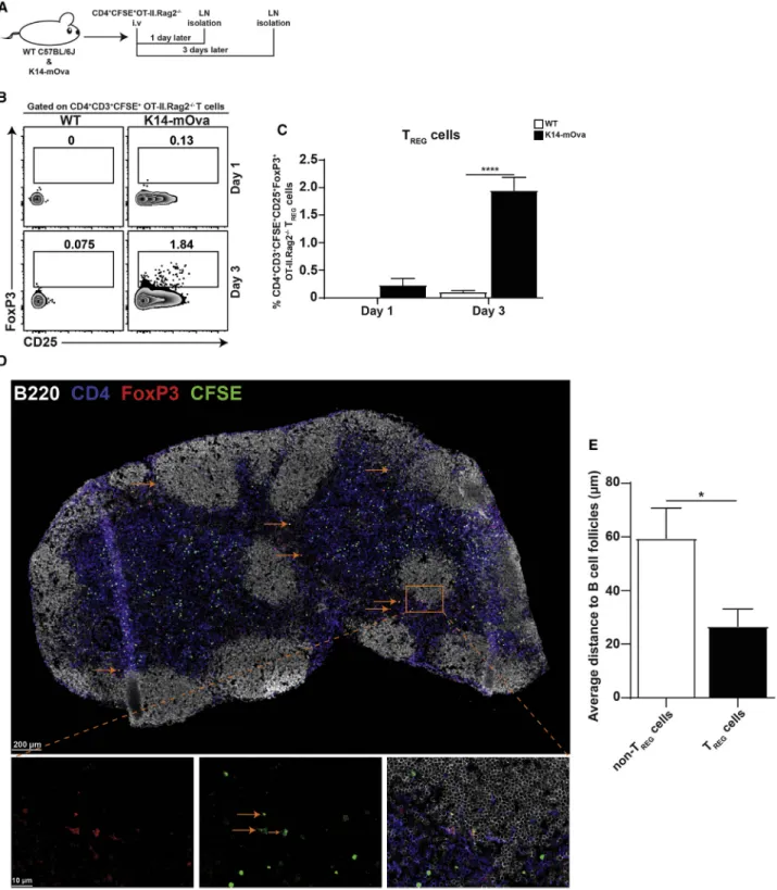
Figure 1. Antigen-Specific TREGCell Conversion Occurs in the Lymph Nodes
(A) In vivo experimental design to study TREGcell conversion in LNs. WT C57L/6J and K14-mOva mice were intravenously injected with 5–83 10 6
CFSE-labeled CD4+OT-II.Rag2 / T cells. 1 and 3 days after CD4+T cell transfer, mice were sacrificed, and LNs were isolated for analysis of transferred CD4+OT-II.Rag2 / T cells by flow cytometry (B and C) and immunofluorescence (D and E).
(B) Representative contour plots of live CFSE-labeled CD4+
CD3+
OT-II.Rag2 /
T cells in peripheral LNs, in which the numbers in the plots display the frequency of CD25+
FoxP3+
TREGcells.
of peripheral tolerance is necessary to further complete TREGcell
generation in the thymus.
Recently, we have demonstrated that LNSCs constrain immune reactivity via presentation of self-antigens in major his-tocompatibility complex (MHC) class II, which results in the selective maintenance of antigen-specific TREGcells (Baptista et al., 2014). The expression of MHC class II by LNSCs and not dendritic cells (DCs) was shown to be critical to preventing the activation of both CD4+and CD8+cells (Baptista et al., 2014; Dubrot et al., 2018). Nevertheless, the question of whether self-antigen presentation by LNSCs may in fact induce anti-gen-specific TREGcell formation, analogous to the function of
non-hematopoietic stromal cells in the thymus, remains unsolved.
Using antigen-specific CD4+ T cells from TCR transgenic mice, we showed, with in vitro and in vivo systems, that antigen presentation by LNSCs resulted in the conversion of naive CD4+ T cells into antigen-specific CD25+FoxP3+TREGcells, a process
that depended on interleukin-2 (IL-2). Converted TREGcells were
preferentially present in the area close to the B cell follicles. These TREGcells were necessary for the LNSC-mediated
sup-pression of autoreactive TFHcells, which supported the
genera-tion of autoreactive GC B cells. Altogether, our data reveal that LNSCs critically participate in maintaining tolerance by express-ing and presentexpress-ing self-antigens necessary for the conversion of naive T cells into antigen-specific TREGcells.
RESULTS
Naive CD4+T Cells Can Convert into T
REGCells in the
LNs
We have previously shown that under steady-state conditions, LNSCs are essential for the maintenance of antigen-specific TREGcells via the presentation of self-antigens in MHC class II
(Baptista et al., 2014). However, it is not clear whether self-anti-gen presentation by LNSCs leads to the induction of TREGcells,
or if it only provides survival signals to existing TREGcells. To
directly address this, we used K14-mOva transgenic mice, where membrane-bound ovalbumin (Ova) is expressed as a skin antigen by keratinocytes under the human keratin 14 pro-moter (Baptista et al., 2014; Bianchi et al., 2009). Notably, Ova mRNA is expressed by LNSCs and not by any other antigen-pre-senting cell such as B cells, macrophages, or DCs at steady state within the LN (Figure S1A). The expression of Ova as a skin self-antigen is at levels equivalent to those of Mlana and Tryosinase, two skin-restricted endogenous antigens previously shown to be expressed by LNSCs (Figure S1A) (Fletcher et al., 2010). This indicates that in K14-mOva LNs, stromal cells are the only cell type that express Ova as a self-antigen. To explore
the contribution of LNSCs in TREGcell conversion in vivo, naive
carboxyfluorescein succinimidyl ester (CFSE)-labeled CD4+ T cells from OT-II.Rag2 / , which lack endogenous thymic
CD25+FoxP3+TREGcells (Figure S1B), were intravenously
in-jected into wild-type (WT) C57BL/6J and K14-mOva mice. LNs were isolated 1 and 3 days after the CFSE-labeled CD4+T cell transfer (Figure 1A). Analysis of peripheral (axillary, brachial, inguinal, and popliteal) LNs at day 3 showed that Ova-specific TCR-transgenic CD4+CD3+ OT-II.Rag2 / T cells converted into CD25+FoxP3+T
REGcells solely in K14-mOva LNs, wherein
Ova is exclusively expressed as a self-antigen by LNSCs. Con-version of CD4+T cells to T
REGcells required at least 3 days,
as the conversion was not observed at day 1 of the CD4+ T cells’ transfer (Figures 1B and 1C). Furthermore, the frequency of endogenous CD25+FoxP3+TREGcells was comparable
be-tween WT and K14-mOva LNs (Figures S2A and S2B). To explore the location of converted TREGin the LNs under
homeo-static conditions, we injected CFSE-labeled CD4+T cells from OT-II.Rag2 / to K14-mOva mice. Three days later, peripheral
LNs were isolated, and LN sections were stained with a multi-plexed antibody panel. Injected CFSE-labeled CD4+ T cells
from OT-II.Rag2 / appeared to be broadly distributed throughout the LN (Figure 1D). Nevertheless, quantitative anal-ysis revealed that converted TREGcells were mainly
concen-trated at the follicle-T-zone interface and interfollicular regions close to the B cell area (Figures 1E andS2C). These data show that antigen-specific naive T cells can be converted into TREG
cells within LNs upon self-antigen encounter and that they pref-erentially localize in the T cell area close to the B cell follicles.
Antigen Presentation by LNSCs Results in the
Conversion of Naive CD4+T Cells into Antigen-Specific TREGCells
To study the mechanism of TREGcell conversion by LNSCs,
co-cultures of Ova-expressing LNSC lines (isolated from K14-mOva mice) and Ova-specific TCR transgenic CD4+ T cells from OT-II.Rag2 / transgenic mice were started. In this in vitro
model, Ova is expressed as a self-antigen by the K14-mOva LEC and FRC lines and is absent in the control cell lines, while all cell lines are devoid of hematopoietic antigen-presenting cells (Figures S3A and S3B). In these co-culture experiments, T cell activation could be observed by the upregulation of CD25 at 48 h of culture only when T cells were cultured together with K14-mOva LECs. Subsequently, by 72 h of culture, a distinct population of CD25+FoxP3+TREGcells was present, and the
fre-quency of these cells further increased by 96 h of culture (Figures 2A–2C). This conversion into FoxP3+cells was not observed in the presence of the WT LEC line or in the absence of stromal cells (medium) (Figures 2A–2C). Similarly, the K14-mOva FRC line was
(C) Frequency of CD25+
FoxP3+
TREGcells within CD4+CD3+CFSE+OT-II.Rag2 / T cells in peripheral LNs of WT and K14-mOva mice. The data represent the
mean± SEM for one (n = 4 mice per group for day 1) and two (n = 3 mice per group for day 3) independent experiments analyzed by two-way ANOVA followed by Turkey’s multiple comparison test. ****p < 0.0001.
(D) Representative immunofluorescence staining of an entire brachial LN section from K14-mOva mice. Arrows point to representative converted TREGcells.
Images are representative of four LNs (inguinal, brachial, axillary, and popliteal) from a total of three mice. Three sections, representing three different locations within each LN, were analyzed.
(E) Bar graph representing the average distance of CFSE+
CD4+
FoxP3+
TREGcells to the B cell area. n = mean of 4 LNs/3 mice. Data analyzed by unpaired
able to convert CD4+OT-II.Rag2 / T cells into CD25+FoxP3+ TREGcells. Hereto, we used early cell line passages, as FRCs
tend to lose the expression of Ova upon long-term culture ( Fig-ures S3A–S3C). In order to determine whether the conversion of naive CD4+T cells toward T
REGcells was solely dependent
on LNSCs, sorted naive CD4+OT-II.Rag2 / T cells (99% pure) versus magnetic bead-enriched naive CD4+ OT-II.Rag2 / T cells were co-cultured with LNSCs for 3 days. These experi-ments showed that the presence of a minor impurity of DCs within the bead-enriched fraction of CD4+T cells was necessary
for the conversion toward CD25+FoxP3+TREGcells, while these
were absent within the sorted CD4+T cells (Figures S3D–S3H). In
the absence of these DCs, CD25-FoxP3+TREGcells, which can
still be classified as TREG cells (Owen et al., 2019; Zelenay et al., 2005), were generated, while CD25+FoxP3+TREG cells
were absent (Figures S3D–S3H). These data suggest that both FRCs and LECs can present self-antigens to naive CD4+ T cells, which converted—with the assistance of DCs—into CD25+FoxP3+ T
REG cells upon antigen recognition. Although
mRNA for the self-antigen Ova was only expressed by LNSCs (Figure S1A), skin-derived DCs could have picked up Ova in the skin and, as such, have contributed to antigen presentation in the LN. Therefore, to determine the potential contribution of LNSCs in TREG cell conversion, we transplanted (Tx)
K14-mOva LNs into WT hosts and analyzed the fate of the transferred naive CFSE-labeled CD4+OT-II.Rag2 / T cells (Figure 2D). In our transplantation model, popliteal LNs of WT recipient mice were surgically removed and replaced by LNs from either WT (WT Tx) or K14-mOva (K14-mOva Tx) mice. After 4 weeks, lymphatic and blood vasculature are reconnected to the Tx LNs. While LNSCs within the transplant are of donor origin, nearly all immune cells of the donor animal are replaced by host-derived cells (Baptista et al., 2014; Hammerschmidt et al., 2008; Molenaar et al., 2009; Wolvers et al., 1999). Consequently, Ova expression as self-antigens is confined to the LNSCs within K14-mOva Tx LNs. Four days after the intravenous injection of naive CFSE-labeled CD4+OT-II.Rag2 / T cells, both Tx and
endogenous (host inguinal) LNs were analyzed. The frequency of CFSE+CD4+ T cells in transplanted and endogenous LNs
was comparable between K14-mOva Tx LNs and WT Tx LNs. However, CFSE+CD4+ OT-II.Rag2 / T cells converted into CD25+FoxP3+T
REGcells when Ova was expressed as a
self-an-tigen by LNSCs, as seen in K14-mOva Tx LNs (Figures 2E–2G). Further characterization of the converted CD25+FoxP3+ T
REG
cells in K14-mOva Tx LNs showed a negligible expression of Cxcr5, a lack of PD-1, and a high expression of CD62L when compared to endogenous TFHand TREGcells (Figure S4A).
More-over, conversion of CD4+T cells was not observed in WT Tx LNs or endogenous LNs (Figures 2E–2G). There were no differences between the two groups in the frequencies of endogenous (host-derived) CD4+T cells and T
REGcells within the transplanted and
the endogenous LNs (Figures S4B–S4D). In thymic TREGcells,
the TREG-specific demethylated region (TSDR) in the FoxP3
gene locus is significantly demethylated, which is required for stable FoxP3 expression (Baron et al., 2007; Floess et al., 2007). The TSDR of OT-II.Rag2 / TREGcells induced by LNSCs
was methylated to the same degree as in activated non-TREG
cells of the OT-II.Rag2 / and in contrast to the demethylated
TSDR in thymic TREGcells (Figure 2H). This result corroborated
that the OT-II.Rag2 / T
REGcells induced by LNSCs originated
from the activated conventional OT-II.Rag2 / T cells. Alto-gether, these data show that in steady state, the presentation of self-antigens by LNSCs leads to the conversion of naive CD4+T cells into CD25+FoxP3+TREGcells upon antigen
recogni-tion by the TCR.
TREGConversion by LNSCs Requires IL-2 and
Co-stimulation
To assess the specific requirements for TREGcell conversion by
LNSCs, we addressed whether IL-2 was involved in this process, as IL-2 is crucial in TREGcell maintenance and function (Abbas et al., 2018; Cheng et al., 2011; D’Cruz and Klein, 2005; Fontenot et al., 2005) and implicated in TREGcell generation in the thymus
(Malek et al., 2002). Hereto, neutralizing IL-2 antibody was added to the co-culture of naive Ova-specific TCR-transgenic CD4+ T cells from OT-II.Rag2 / with WT LEC or K14-mOva LEC lines.
Blocking of IL-2 in the culture completely prevented the conver-sion of naive CD4+ T cells into T
REG cells (Figures 3A–3C).
Figure 2. LNSCs Convert Antigen-Specific Naive CD4+T Cells into T REGCells
(A) CD4+T cells obtained from spleens and LNs of OT-II.Rag2 / mice were either cultured alone (medium) (n = 2) or co-cultured with WT LEC (n = 2) or K14-mOva LEC (n = 2) stromal cell lines, and CD4+
T cells were harvested at the indicated time points and analyzed by flow cytometry. Representative density plots are shown; the numbers in the plots correspond to the frequency of the cells within drawn gates of the live CD4+
OT-II.Rag2 /
T cell population. (B and C) Bar graphs represent the mean frequency of (B) CD25+
FoxP3+
TREGcells and (C) CD25+FoxP3 non-TREGcells. The graphs show mean± SEM of
triplicate co-cultures and are representative of two independent experiments (24 h, 48 h, and 72 h) and one independent experiment (96-h time point). Data analyzed by two-way ANOVA followed by Turkey’s multiple comparison test. *p < 0.05; ****p < 0.0001.
(D) Experimental design to study TREGcell conversion by LNSCs in vivo. LNs, either derived from WT or K14-mOVA mice, were transplanted into WT C57L/6J
mice, and after 4 weeks of recovery, WT Tx and K14-mOva Tx mice were intravenously injected with 83 106
CFSE-labeled CD4+
OT-II.Rag2/
T cells. 4 days after CD4+
T cell transfer, mice were sacrificed, and transferred CD4+
OT-II.Rag2 /
T cells in both transplanted and endogenous (inguinal) LNs were examined using flow cytometry.
(E) Representative contour plots of live CFSE-labeled CD4+OT-II.Rag2/ T cells within transplanted (top) and endogenous (bottom) LNs, in which the numbers in the plots display the frequency of CD25+
FoxP3+
TREGcells.
(F and G) Frequency of CD4+
CFSE+
OT-II.Rag2/
T cells in endogenous and transplanted LNs (F) and frequency of CD25+
FoxP3+
TREGcells within the live
CFSE-labeled CD4+
OT-II.Rag2 /
T cell population (G). The data represent the mean± SEM; n = 5 mice per group and analyzed by two-way ANOVA followed by Turkey’s multiple comparison test. ****p < 0.0001.
(H) CD4+T cells were obtained from spleens and LNs of OT-II.Rag2 / mice and co-cultured with K14-mOva LEC lines. After 72 h, CD4+T cells were harvested, and converted TREG cells and non-TREG cells were sorted and compared to sorted naive OT-II.Rag2 / (CD4+CD3+CD62L+), sorted naive WT
(CD4+
CD3+
CD62L+
), and sorted thymic TREGWT (CD4+CD3+CD25hi). Heatmap represents the percentage of TREG-specific demethylated region (TSDR) DNA
Similarly, blockage of MHC class II also prevented TREGcell
con-version (Figures 3A–3C). In addition, blocking co-stimulatory ligand CD80 alone, or together with co-stimulatory ligand
CD86, diminished the generation of TREGcells (Figures 3A–3C).
Interestingly, the activation of non-TREGcells, as addressed by
the expression of CD25 and lack of FoxP3, required MHC class
Figure 3. IL-2 and CD80/86 Are Necessary for TREGCell Conversion by LNSCs (A) CD4+
T cells were obtained from spleens and LNs of OT-II.Rag2 /
mice and either cultured alone (medium) (n > 4) or co-incubated with WT LEC (n = 2 for all conditions) or K14-mOva LEC stromal cell lines in the presence or absence of neutralizing antibodies, as indicated in the figure (n > 4 for anti-IL-2, anti-MHC class II; n = 2 for anti-CD80, anti-CD86, anti-CD80/CD86, anti-PD-L1, anti-TGF-b). After 72 h, CD4+
T cells were harvested and analyzed by flow cytometry. Repre-sentative density plots are shown. The numbers in the plots represent the frequency of the cells within the quadrants of the live CD4+
OT-II.Rag2 /
T cell population.
(B and C) Bar graphs represent the mean frequency of (B) CD25+
FoxP3+
TREGcells and (C) CD25 +
FoxP3 non-TREGcells. The graphs show mean± SEM of
triplicate co-cultures and are representative of two independent experiments.
(D) Peripheral LNs from WT C57BL/6J mice were isolated and digested and stained for CD45, 119, CD31, and gp38. Stromal cells were gated as CD45 Ter-119 cells, and the expression of co-stimulatory and inhibitory molecules on different subclasses of LNSCs, as defined by CD31 and gp38 expression, were assessed using flow cytometry. Histograms show the expression of indicated receptors on different stromal cell subsets in comparison to DCs (CD45+
CD11c+
). (E) Mean fluorescence intensity (MFI) of MHC class II, CD80,CD86, and PD-L1 on LNSC subsets and DCs (CD45+
CD11c+
). The graphs depict the mean± SEM (n = 3 mice per experiment) and are representative of three independent experiments.
II and co-stimulatory molecules but not IL-2 (Figure 3C). As pro-grammed death-ligand 1 (PD-L1) expression by LECs has been implicated as essential for the deletional tolerance of autoreac-tive CD8+T cells (Tewalt et al., 2012), we also added PD-L1 blocking antibody in our co-culture. Notably, no significant ef-fects on TREGcell conversion were observed when using the
PD-L1 blocking antibody (Figures 3A–3C). Several studies re-ported that IL-2 together with transforming growth factor b (TGF-b) has a significant effect on the induction, maintenance, and function of TREGcells (Chen et al., 2003; Park et al., 2004; Zheng et al., 2007). However, TGF-b did not seem to be involved in LNSC-mediated conversion of naive CD4+T cells, as T
REG
cells were still present when cultured together with the K14-mOva LEC line in the presence of neutralizing anti-TGF-b ( Fig-ures 3A–3C), as seen in a recent study (Akamatsu et al., 2019). These data further support an essential role of IL-2 and co-stim-ulatory molecules in the conversion of naive CD4+T cells into CD25+FoxP3+TREGcells upon the recognition of self-antigens,
presented by LNSCs in the context of MHC class II molecules. In accordance with these findings, low levels of MHC class II, compared to expression levels on DCs, were detected on all LNSC subsets when freshly isolated, as shown before (Baptista et al., 2014; Dubrot et al., 2018, 2014; Malhotra et al., 2012) (Figures 3D and 3E). Furthermore, CD80 was mainly expressed by FRCs and LECs derived from steady-state peripheral LNs and LNSC lines (Figures 3D, 3E, and S5A). CD86 expression was barely detectable on either freshly isolated LNSCs or cell lines, whereas PD-L1 expression was detected at low levels on some FRCs and at higher levels on endothelial cells (Figures 3D, 3E, andS5A). To further visualize the expression of MHC class II and other co-stimulatory molecules, we applied t-distributed stochastic neighbor embedding (t-SNE) unsuper-vised clustering (Amir et al., 2013) to the manually gated CD45 Ter-119 LNSCs using the online analysis platform Cy-tobank (Figures S5B and S5C for gating strategy). The tSNE maps indicate the existence of LNSCs, particularly FRCs, that co-expressed MHC class II and co-stimulatory molecules at low levels, making them suitable as antigen-presenting stromal cells involved in converting the naive CD4+T cells into T
REG
cells (Figure 3F). Altogether, these results indicate that steady-state LNSCs express low levels of MHC class II and co-stimulatory molecules, which are required for the conversion of naive CD4+ T cells into TREG cells in an IL-2 dependent
manner.
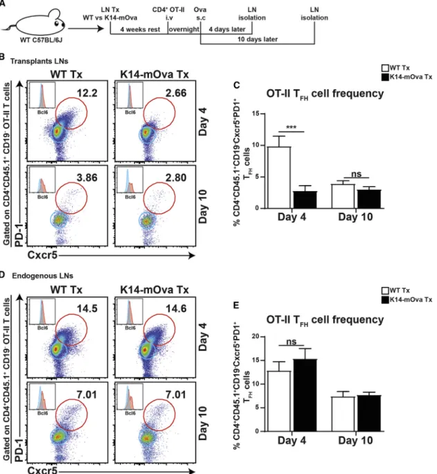
LNSCs Control Autoreactive TFHCells
As TFHcells are known to be important players in establishing
autoimmune diseases (Craft, 2012), the potential role of LNSCs in controlling autoreactive TFHcells in vivo was addressed by
transplanting LNs of WT and K14-mOva mice into WT C57BL/6J hosts, followed by the transfer of Ova-specific TCR-transgenic naive CD4+ OT-II T cells and immunization with Ova in incomplete Freund’s adjuvant (IFA) for 4 and 10 days (Figure 4A). Analysis at day 4 showed a significant reduction in Ova-specific Cxcr5+PD1+Bcl6+ T
FH cells (OT-II
TFH) within K14-mOva Tx LNs when compared to WT Tx LNs,
which were no longer apparent at day 10 (Figures 4B and 4C). However, no effect on OT-II TFH cells was seen within
endogenous LNs in mice that received K14-mOva Tx LNs (Figures 4D and 4E). Furthermore, transplantation and immuni-zation did not affect the endogenous pools of TFH(endo-TFH)
and TREG(endo-TREG) cells (Figures S6A–S6D). The transferred
OT-II TREGcells were similar in frequency at day 4 after transfer
in both K14-mOva and WT Tx LNs, while an increase in OT-II TREG cells was seen in WT Tx LNs at day 10 (Figure S6E).
The frequencies of endo-TFH, endo-TREG, and OT-II TREGcells
were not influenced in endogenous LNs (Figures S6F–S6I). Collectively, our data suggest a critical function of LNSCs in controlling the formation of autoreactive TFHcells.
IL-2 Is Required for LNSC-Dependent Control of Autoreactive TFHCells
TREGcells have been implicated to control antigen-specific TFH
cells’ expansion (Wing et al., 2014). Our data suggest that the con-version of naive CD4+T cells toward TREGcells controls the
forma-tion of autoreactive TFHcells within LNs. To investigate this
possi-bility, the function of TREG cells was blocked by providing
neutralizing IL-2 antibodies when WT Tx and K14-mOva Tx mice were immunized with Ova in IFA. Neutralizing IL-2 antibodies or an isotype control were provided daily for 4 days. Next, the fate of the transferred CD4+OT-II T cells was analyzed (Figure 5A).
As expected, autoreactive OT-II TFHcells were strongly reduced
in K14-mOva Tx LNs, compared to WT Tx LNs, when treated with an isotype control antibody (Figures 5B and 5C). However, the frequency of autoreactive OT-II TFHcells in K14-mOva Tx
LNs treated with anti-IL-2 was similar to that of WT Tx LNs (Figures 5B and 5C). Importantly, both OT-II TREG and endo-TREG cells
were considerably depleted in anti-IL-2 treated mice when compared to isotype-control-treated mice (Figures 5D and 5E). Moreover, no remarkable changes in the frequency of TFHcells
within CD4+OT-II T in the endogenous LNs were observed ( Fig-ures 5F and 5G). Similar to the Tx LNs, OT-II TREG and
endo-TREG cell frequencies in the endogenous LNs were strongly
reduced in anti-IL-2-treated animals when compared to isotype control animals (Figures 5H and 5I). Collectively, our data indicate that upon IL-2 blocking, TREGcells no longer control the
genera-tion of TFHcells that react to self-antigens expressed and
pre-sented by LNSC and are thus autoreactive.
Self-Antigen Presentation by LNSCs Controls Autoreactive B Cell Formation
Since TFHcells are critical for the development and maintenance
of GC B cells (Breitfeld et al., 2000; Schaerli et al., 2000; Victora and Nussenzweig, 2012; Walker et al., 1999), the formation of Ova-specific GC B cells within Tx LNs was determined. For this purpose, WT or K14-mOva LNs were transplanted into WT C57BL/6J mice, followed by an intravenous injection of Ova-specific TCR-transgenic naive CD4+ OT-II T cells 4 weeks post-transplantation. After 18 h, the mice received a subcutaneous injection of Ova in IFA, and 10 days later, CD19+CD38-GL7+Ova+GC B cells in Tx LNs were analyzed ( Fig-ure 6A). The frequencies of total B cells and GC B cells were comparable between Tx LNs from WT and K14-mOva mice ( Fig-ures 6B–6D). However, the Ova-specific B cells were signifi-cantly reduced in K14-mOva Tx LNs, as compared to WT Tx LNs (Figures 6B and 6E). Moreover, the reduction of
Ova-specific B cells was restricted to K14-mOva Tx LNs, as the endogenous LNs showed similar frequencies of B cells, GC B cells, and Ova-specific GC B cells (Figures 6F–6I). Altogether, these results support a model in which self-antigen-presenting
LNSCs promote the differentiation of autoreactive CD4+T cells
into TREG cells while preventing the generation of TFH cells,
thereby reducing GC B cells’ formation directed against the same self-antigen.
Figure 4. Formation of Autoreactive TFHCells Is Controlled by LNSCs
(A) In vivo experimental design to study the effect of self-antigen-presenting LNSCs on TFHcell formation. WT Tx and K14-mOva Tx mice were intravenously
injected with 103 106
transgenic CD45.1+
CD4+
OT-II T cells. After 18 h, mice were injected with Ova/IFA in the ankles of both hind legs. 4 days and 10 days later, mice were sacrificed, transferred CD45.1+
CD4+
OT-II T cells were transplanted, and endogenous LNs were examined using flow cytometry. (B and D) Representative density plots of CD45.1+
CD4+
CD19 OT-II T cells are shown. The numbers in the plots display the percentage of Cxcr5+
PD-1+
TFH(OT-II
TFH) cells of all live CD45.1 +
CD4+T cells within (B) the transplanted and (D) the endogenous LNs. Histograms represent the expression of Bcl6 in Cxcr5 PD-1 (blue; non-TFH) and Cxcr5+PD-1+(red; TFH) T cells within the live CD45.1+CD4+T cell population.
(C and E) The graphs represent the percentage of OT-II TFHcells of all live CD45.1+CD4+T cells within (C) the transplanted and (E) the endogenous LNs. Data show
the mean± SEM of a combination of two independent experiments for day 4 (WT Tx, n = 10 mice; K14-mOva Tx, n = 10 mice in total) and one independent experiment for day 10 (WT Tx, n = 5 mice; K14-mOva Tx, n = 5 mice in total). Statistical analyses were done by two-way ANOVA followed by Turkey’s multiple comparison test. ***p < 0.001; ns, not significant.
DISCUSSION
The repertoires of naive T cells as well as TREGcells are generated
in the thymus, and after completion of their thymic differentiation, naive T cells enter the periphery. Through the expression of LN homing receptors, naive T cells migrate with great efficiency to the LNs, where they may encounter the antigen that they can spe-cifically recognize with their TCR when presented by hematopoi-etic antigen-presenting cells. This migration is also important for the interaction with LNSCs to complete the control of autoreac-tive T cell development (Dubrot et al., 2014; Lee et al., 2007; Mag-nusson et al., 2008; Rouhani et al., 2015; Tewalt et al., 2012). Our data showed that the presentation of self-antigens in the context of MHC class II molecules by LNSCs, together with the assis-tance of DCs, resulted in the conversion of naive CD4+T cells into TREG cells. Furthermore, the converted TREG cells were
methylated to a similar degree as seen in naive CD4+T cells, un-like the demethylated TSDR in thymic TREGcells, providing further
evidence that the converted cells are derived from naive T cells. These TREGcells lacked Cxcr5 and PD-1 expression, which are
classical markers of TFHcells, suggesting that converted TREG
cells do not have follicular regulatory T cell (Tfr) properties ( Fon-seca et al., 2019; Sage and Sharpe, 2016), at least at day 3 after T cell transfer. This conversion was antigen specific and required CD80 co-stimulation, as well as IL-2. Both LECs and FRCs have the capacity to mediate this conversion in vitro, while converted TREGcells are preferentially found within the T cell zone close to
the B cell follicles at 3 days after the T cell transfer. Since FRCs, as well as LECs, can be found around the B cell follicles, both populations may be responsible for this process within our sys-tem (Cohen et al., 2014). Blocking the function of TREGcells by
anti-IL-2 allowed the development of antigen-specific TFHcells. Figure 5. Blockade of IL-2In Vivo Prevents TREGDevelopment and Favors Autoreactive TFHCell Formation
(A) In vivo experimental design to study the role of IL-2 in LNSC-mediated TFHcell suppression. WT Tx and K14-mOva Tx mice were intravenously injected with
103 106
transgenic CD45.1+
CD4+
OT-II T cells. After 18 h, mice were injected with Ova/IFA in the ankles of both hind legs. Mice were injected intraperitoneally with isotype control or anti-IL2-neutralizing antibodies once a day for 4 days. At day 5, mice were sacrificed, and transferred CD45.1+
CD4+
OT-II T cells in both transplanted and endogenous LNs were examined using flow cytometry.
(B and F) Representative density plots of live CD45.1+
CD4+
Cxcr5+
PD-1+
TFHcells are shown. The numbers in the plot display the frequency of Cxcr5+PD-1+TFH
cells within the live CD45.1+
CD4+
T cell population of (B) the transplanted and (F) the endogenous LNs. (C and G) The graphs represent the frequency of OT-II Cxcr5+
PD-1+
TFH(OT-II TFH) cells within the live CD45.1+CD4+T cell population of the (C) transplanted and
(G) endogenous LNs.
(D, E, H, and I) The graphs represent the frequency of (D and H) CD45.1+CD4+CD25+FoxP3+OT-II TREGcells and (E and I) CD45.1 CD4 +
CD25+FoxP3+ endogenous (endo) TREGcells within the live CD4+T cell population of the (D and E) transplanted and ( H and I) endogenous LNs. Data shown are the mean± SEM
(WT Tx, n = 6 mice; K14-mOva Tx, n = 6 mice) and are representative of two independent experiments. Statistical analyses were done by two-way ANOVA followed by Turkey’s multiple comparison test. *p < 0.05; **p < 0.001; ***p < 0.0001; ****p < 0.0001; ns, not significant.
Our data indicate that LNSCs can inhibit the development of au-toreactive B cells by promoting TREG cell formation at the
expense of TFHcells’ generation from autoreactive CD4+T cells.
Analysis of freshly isolated LNSCs showed that both FRCs and LECs expressed low levels of MHC class II and co-stimulatory
CD80, thereby marking both subsets as potential
Figure 6. LNSCs Control the Formation of Autoreactive GC B Cells
(A) In vivo experimental design to study the effect of self-antigen-presenting LNSCs on GC B cell formation. WT Tx and K14-mOva Tx mice were intravenously injected with 103 106transgenic CD45.1+CD4+OT-II T cells. After 18 h, mice were injected subcutaneously with Ova/IFA in the ankles of both hind legs. 10 days later, mice were sacrificed, and B cells in both transplanted and endogenous LNs were examined using flow cytometry.
(B and F) Representative density plots of CD4 CD19+
B cells are shown. The numbers in the top plots display the frequency of CD38 GL7+
GC B cells, and bottom plots display Ova+
B cells within the live CD4 CD19+
CD38 GL7+
GCs B cell population of (B) the transplanted and (F) the endogenous LNs. (C–E and H–I) The graphs represent the frequencies of (C and G) CD4 CD19+
B cells, (D and H) CD38 GL7+
GC B cells, and (E and I) Ova+
-specific GC B cells within the live CD4 CD19+B cell population of the transplanted and endogenous LNs. Data shown represent the mean± SEM (n = 5 mice per group) and are representative of two independent experiments. Data are analyzed by unpaired Student’s t test. **p < 0.001; ns, not significant.
antigen-presenting LNSCs involved in the conversion of naive T cells into TREGcells. In our model, Ova expression was confined
to the peripheral LNSCs, as other antigen-presenting cells such as B cells, macrophages, or DCs lacked Ova expression. While both DCs and LNSCs could be the source of co-stimulatory cells, LNSCs are the obligatory source of self-antigens in the context of MHC class II molecules (Baptista et al., 2014; Dubrot et al., 2018). We propose that DCs may assist in this process, as DCs have the capacity to pick up antigen from other cells (Koble and Kyewski, 2009) and can thus participate in presenting self-antigen derived from LNSCs, leading to T cell conversion. In our transplan-tation model, tissue necrosis may result in such an uptake of self-antigens by DCs, leading to the conversion of endogenous Ova-specific T cells toward TREGcells. Self-antigen presentation
by LNSCs, when assisted by DCs, induced CD25 expression on naive CD4+T cells, suggesting an initial activation of T cells upon encountering MHC class II/peptide and CD80, potentially leading to low levels of IL-2 production. Indeed, blockade of MHC class II prevented the induction of CD25 expression, as well as the induc-tion of FoxP3 expression. The produced levels of IL-2 were not sufficient to induce T cell proliferation (Baptista et al., 2014) but were necessary to induce the conversion of naive CD4+T cells into CD25+FoxP3+T
REGcells in our in vitro and in vivo models.
These results are in agreement with the essential role of IL-2 as a central player in the development, maintenance, and function of TREGcells (Bayer et al., 2005; Cheng et al., 2011; D’Cruz and Klein, 2005; Fontenot et al., 2005; Malek and Bayer, 2004).
The site within the LN where converted T cells preferentially locate is within the T cell area close to the boundary of the B cell follicles. Supporting the possibility that conversion is taking place at this location is the report that clusters of pStat5-ex-pressing FoxP3+T cells with few IL-2 producing CD4+T cells are present within LNs in steady state at the outer T cell areas close to the B cell follicles. This specific localization required TCR triggering, suggesting antigen encounter by T cells at the B-T cell border (Liu et al., 2015). It is therefore likely that within this area subsets of sessile LNSCs that present self-antigens are localized. The recently described population of CCL19lo LNSCs is specifically assigned to reside within this area at the T-B cell border. Interestingly, these cells potentially co-express the necessary molecules MHC class II and CD80 (Rodda et al., 2018). Furthermore, CCL19lo LNSCs express all molecules
required to attract immune cells to such regulatory hotspots, as mRNA transcripts for CCL21 and Ch25h—described to attract DCs as well as B and T cells—are detected in these cells (Rodda et al., 2018). These locations could be the same as those in which conventional DC2 (cDC2), a DC subset, together with naive CD4+T cells were described to localize, promoting CD4+ T cell help for CD8+T cell responses (Baptista et al., 2019). Thus, by attracting incoming naive T cells to these self-anti-gen-presenting stromal cells, autoreactive T cells can be converted into TREGcells and come into contact with DCs in
or-der to further control potential autoreactive T cells.
The subclass of IL-2-producing CD4+T cells that were shown to be present in steady-state LNs was not further specified (Liu et al., 2015). However, a recent report byDiToro et al. (2018)
showed IL-2-producing T cells to be precursors for TFHcells.
Thus, autoreactive naive CD4+ T cells that have not been
depleted in the thymus and reach peripheral LNs can differentiate into TFHcells upon self-antigen encounter, thereby providing IL-2
for the generation of TREGcells, as we show here. When access to
self-antigen recognition is denied, larger quantities of IL-2 are produced and clustering of pStat5+cells is lost, leading to the
loss of control of T effector cells by TREGcells (Liu et al., 2015).
Similarly, within our experiments, blockade of TREGcells resulted
in an increase in TFH cells. As these TFH cells were directed
against self-antigen expressed by LNSCs, we could follow the consequences of these autoreactive TFHcells for the
differentia-tion of antigen-specific B cells. Indeed, increased numbers of TFH
cells resulted in an increase in B cells specific for the same anti-gen. Thus, our data strongly suggest that the generation of autor-eactive B and T cells can be controlled at the level of LNSCs, and dysregulation of LNSCs (e.g., as a result of an ongoing infection) may result in autoantibody production, leading to autoimmunity. Interestingly, we have shown that human LNSCs are functionally altered during the earliest phases of rheumatoid arthritis ( Ha¨hn-lein et al., 2018a, 2018b), potentially allowing unwanted activa-tion of autoreactive lymphocytes. Since elevated levels of TFH
cells and B cells are seen in patients that are at high risk of a broad range of autoimmune diseases (Crotty, 2014; Gensous et al., 2018; van Baarsen et al., 2013), these observations are also clin-ically relevant.
In conclusion, our in vitro and in vivo data show the existence of a unique population of LNSCs that express MHC class II and possess co-stimulatory properties in steady state. Thereby, with the support of DCs, they provide essential signals to naive CD4+
T cells to facilitate the generation of antigen-specific TREGcells in
an IL-2-dependent manner. By doing so, LNSCs limit autoreactive TFHcell formation and control the autoreactive humoral immune
response. The function of these LNSCs need to be further studied in humans during health and autoimmunity to explore whether we can use their properties for therapeutic strategies.
STAR+METHODS
Detailed methods are provided in the online version of this paper and include the following:
d KEY RESOURCES TABLE
d LEAD CONTACT AND MATERIALS AVAILABILITY d EXPERIMENTAL MODEL AND SUBJECT DETAILS
B Mice d METHOD DETAILS B LN transplantation B Adoptive transfer B Immunization B IL-2 neutralization B Flow cytometry B Immunofluorescence staining B LNSC lines preparation B T cell co-culture assays B Quantitative RT-PCR
B Methylation analysis of the TSDR
d QUANTIFICATION AND STATISTICAL ANALYSIS
B Statistical analysis
SUPPLEMENTAL INFORMATION
Supplemental Information can be found online athttps://doi.org/10.1016/j. celrep.2020.03.007.
ACKNOWLEDGMENTS
We thank M.M.J. van Gool, M.N. Erkelens, A.C. Breedveld, A.V. Bos, T. Smeekes, I. Licht, and H. van der Laan for excellent technical support. This work was financially supported by grants from ReumaFonds (14-2-403 to R.E.M.), NWO-ALW (ALWOP.271 to J.J.K.), and NWO-ZonMW (VENI 916.13.011 to R.M.R.; VIDI 917183711 to L.G.M.v.B.; TOP 01217014 to R.E.M. and L.G.M.v.B.).
AUTHOR CONTRIBUTIONS
R.N. and R.E.M. designed the experiments, discussed the data, and wrote the manuscript. R.N., C.G.d.G., E.D.K., J.J.K., S.d.K., T.K., and R.M.R. performed the experiments. S.H., J.B., L.G.M.v.B., and R.M.R. revised the manuscript.
DECLARATION OF INTERESTS
The authors declare no competing interests. Received: November 7, 2019
Revised: January 13, 2020 Accepted: February 28, 2020 Published: March 24, 2020
REFERENCES
Abbas, A.K., Trotta, E., R Simeonov, D., Marson, A., and Bluestone, J.A. (2018). Revisiting IL-2: Biology and therapeutic prospects. Sci. Immunol. 3, eaat1482.
Akamatsu, M., Mikami, N., Ohkura, N., Kawakami, R., Kitagawa, Y., Sugimoto, A., Hirota, K., Nakamura, N., Ujihara, S., Kurosaki, T., et al. (2019). Conversion of antigen-specific effector/memory T cells into Foxp3-expressing Tregcells by
inhibition of CDK8/19. Sci. Immunol. 4, eaaw2707.
Amir, el-A.D., Davis, K.L., Tadmor, M.D., Simonds, E.F., Levine, J.H., Bendall, S.C., Shenfeld, D.K., Krishnaswamy, S., Nolan, G.P., and Pe’er, D. (2013). viSNE enables visualization of high dimensional single-cell data and reveals phenotypic heterogeneity of leukemia. Nat. Biotechnol. 31, 545–552. Anderson, M.S., Venanzi, E.S., Klein, L., Chen, Z., Berzins, S.P., Turley, S.J., von Boehmer, H., Bronson, R., Dierich, A., Benoist, C., and Mathis, D. (2002). Projection of an immunological self shadow within the thymus by the aire protein. Science 298, 1395–1401.
Arroyo-Villa, I., Bautista-Caro, M.B., Balsa, A., Aguado-Acı´n, P., Bonilla-Herna´n, M.G., Plasencia, C., Villalba, A., Nun˜o, L., Puig-Kro¨ger, A., Martı´n-Mola, E., and Miranda-Caru´s, M.E. (2014). Constitutively altered frequencies of circulating follicullar helper T cell counterparts and their subsets in rheuma-toid arthritis. Arthritis Res. Ther. 16, 500.
Aschenbrenner, K., D’Cruz, L.M., Vollmann, E.H., Hinterberger, M., Emmerich, J., Swee, L.K., Rolink, A., and Klein, L. (2007). Selection of Foxp3+ regulatory T cells specific for self antigen expressed and presented by Aire+ medullary thymic epithelial cells. Nat. Immunol. 8, 351–358.
Baptista, A.P., Roozendaal, R., Reijmers, R.M., Koning, J.J., Unger, W.W., Greuter, M., Keuning, E.D., Molenaar, R., Goverse, G., Sneeboer, M.M., et al. (2014). Lymph node stromal cells constrain immunity via MHC class II self-antigen presentation. eLife 3.
Baptista, A.P., Gola, A., Huang, Y., Milanez-Almeida, P., Torabi-Parizi, P., Urban, J.F., Jr., Shapiro, V.S., Gerner, M.Y., and Germain, R.N. (2019). The Chemoattractant Receptor Ebi2 Drives Intranodal Naive CD4(+) T Cell Periph-eralization to Promote Effective Adaptive Immunity. Immunity 50,
1188–1201.e1186.
Baron, U., Floess, S., Wieczorek, G., Baumann, K., Gr€utzkau, A., Dong, J., Thiel, A., Boeld, T.J., Hoffmann, P., Edinger, M., et al. (2007). DNA demethyla-tion in the human FOXP3 locus discriminates regulatory T cells from activated FOXP3(+) conventional T cells. Eur. J. Immunol. 37, 2378–2389.
Bayer, A.L., Yu, A., Adeegbe, D., and Malek, T.R. (2005). Essential role for interleukin-2 for CD4(+)CD25(+) T regulatory cell development during the neonatal period. J. Exp. Med. 201, 769–777.
Bianchi, T., Pincus, L.B., Wurbel, M.A., Rich, B.E., Kupper, T.S., Fuhlbrigge, R.C., and Boes, M. (2009). Maintenance of peripheral tolerance through controlled tissue homing of antigen-specific T cells in K14-mOVA mice. J. Immunol. 182, 4665–4674.
Breitfeld, D., Ohl, L., Kremmer, E., Ellwart, J., Sallusto, F., Lipp, M., and Fo¨r-ster, R. (2000). Follicular B helper T cells express CXC chemokine receptor 5, localize to B cell follicles, and support immunoglobulin production. J. Exp. Med. 192, 1545–1552.
Chen, W., Jin, W., Hardegen, N., Lei, K.J., Li, L., Marinos, N., McGrady, G., and Wahl, S.M. (2003). Conversion of peripheral CD4+CD25- naive T cells to CD4+CD25+ regulatory T cells by TGF-beta induction of transcription factor Foxp3. J. Exp. Med. 198, 1875–1886.
Cheng, G., Yu, A., and Malek, T.R. (2011). T-cell tolerance and the multi-func-tional role of IL-2R signaling in T-regulatory cells. Immunol. Rev. 241, 63–76. Cohen, J.N., Guidi, C.J., Tewalt, E.F., Qiao, H., Rouhani, S.J., Ruddell, A., Farr, A.G., Tung, K.S., and Engelhard, V.H. (2010). Lymph node-resident lymphatic endothelial cells mediate peripheral tolerance via Aire-independent direct an-tigen presentation. J. Exp. Med. 207, 681–688.
Cohen, J.N., Tewalt, E.F., Rouhani, S.J., Buonomo, E.L., Bruce, A.N., Xu, X., Bekiranov, S., Fu, Y.X., and Engelhard, V.H. (2014). Tolerogenic properties of lymphatic endothelial cells are controlled by the lymph node microenviron-ment. PLoS ONE 9, e87740.
Craft, J.E. (2012). Follicular helper T cells in immunity and systemic autoimmu-nity. Nat. Rev. Rheumatol. 8, 337–347.
Crotty, S. (2014). T follicular helper cell differentiation, function, and roles in disease. Immunity 41, 529–542.
Cuadrado, E., van den Biggelaar, M., de Kivit, S., Chen, Y.Y., Slot, M., Doubal, I., Meijer, A., van Lier, R.A.W., Borst, J., and Amsen, D. (2018). ). Proteomic An-alyses of Human Regulatory T Cells Reveal Adaptations in Signaling Pathways that Protect Cellular Identity. Immunity 48, 1046–1059.e1046.
D’Cruz, L.M., and Klein, L. (2005). Development and function of agonist-induced CD25+Foxp3+ regulatory T cells in the absence of interleukin 2 signaling. Nat. Immunol. 6, 1152–1159.
Derbinski, J., Schulte, A., Kyewski, B., and Klein, L. (2001). Promiscuous gene expression in medullary thymic epithelial cells mirrors the peripheral self. Nat. Immunol. 2, 1032–1039.
DiToro, D., Winstead, C.J., Pham, D., Witte, S., Andargachew, R., Singer, J.R., Wilson, C.G., Zindl, C.L., Luther, R.J., Silberger, D.J., et al. (2018). Differential IL-2 expression defines developmental fates of follicular versus nonfollicular helper T cells. Science 361, eaao2933.
Dubrot, J., Duraes, F.V., Potin, L., Capotosti, F., Brighouse, D., Suter, T., Lei-bundGut-Landmann, S., Garbi, N., Reith, W., Swartz, M.A., and Hugues, S. (2014). Lymph node stromal cells acquire peptide-MHCII complexes from dendritic cells and induce antigen-specific CD4+
T cell tolerance. J. Exp. Med. 211, 1153–1166.
Dubrot, J., Duraes, F.V., Harle´, G., Schlaeppi, A., Brighouse, D., Madelon, N., Go¨pfert, C., Stokar-Regenscheit, N., Acha-Orbea, H., Reith, W., et al. (2018). Absence of MHC-II expression by lymph node stromal cells results in autoim-munity. Life Sci. Alliance 1, e201800164.
Fletcher, A.L., Lukacs-Kornek, V., Reynoso, E.D., Pinner, S.E., Bellemare-Pel-letier, A., Curry, M.S., Collier, A.R., Boyd, R.L., and Turley, S.J. (2010). Lymph node fibroblastic reticular cells directly present peripheral tissue antigen under steady-state and inflammatory conditions. J. Exp. Med. 207, 689–697. Fletcher, A.L., Malhotra, D., Acton, S.E., Lukacs-Kornek, V., Bellemare-Pellet-ier, A., Curry, M., Armant, M., and Turley, S.J. (2011). Reproducible isolation of
lymph node stromal cells reveals site-dependent differences in fibroblastic reticular cells. Front. Immunol. 2, 35.
Floess, S., Freyer, J., Siewert, C., Baron, U., Olek, S., Polansky, J., Schlawe, K., Chang, H.D., Bopp, T., Schmitt, E., et al. (2007). Epigenetic control of the foxp3 locus in regulatory T cells. PLoS Biol. 5, e38.
Fonseca, V.R., Ribeiro, F., and Graca, L. (2019). T follicular regulatory (Tfr) cells: Dissecting the complexity of Tfr-cell compartments. Immunol. Rev.
288, 112–127.
Fontenot, J.D., Rasmussen, J.P., Gavin, M.A., and Rudensky, A.Y. (2005). A function for interleukin 2 in Foxp3-expressing regulatory T cells. Nat. Immunol.
6, 1142–1151.
Gensous, N., Charrier, M., Duluc, D., Contin-Bordes, C., Truchetet, M.E., Laz-aro, E., Duffau, P., Blanco, P., and Richez, C. (2018). T Follicular Helper Cells in Autoimmune Disorders. Front. Immunol. 9, 1637.
Ha¨hnlein, J.S., Nadafi, R., de Jong, T., Ramwadhdoebe, T.H., Semmelink, J.F., Maijer, K.I., Zijlstra, I.A., Maas, M., Gerlag, D.M., Geijtenbeek, T.B.H., et al. (2018a). Impaired lymph node stromal cell function during the earliest phases of rheumatoid arthritis. Arthritis Res. Ther. 20, 35.
Ha¨hnlein, J.S., Ramwadhdoebe, T.H., Semmelink, J.F., Choi, I.Y., Berger, F.H., Maas, M., Gerlag, D.M., Tak, P.P., Geijtenbeek, T.B.H., and van Baarsen, L.G.M. (2018b). Distinctive expression of T cell guiding molecules in human autoimmune lymph node stromal cells upon TLR3 triggering. Sci. Rep. 8, 1736. Hammerschmidt, S.I., Ahrendt, M., Bode, U., Wahl, B., Kremmer, E., Fo¨rster, R., and Pabst, O. (2008). Stromal mesenteric lymph node cells are essential for the generation of gut-homing T cells in vivo. J. Exp. Med. 205, 2483–2490. He, J., Tsai, L.M., Leong, Y.A., Hu, X., Ma, C.S., Chevalier, N., Sun, X., Vanden-berg, K., Rockman, S., Ding, Y., et al. (2013). Circulating precursor CCR7(lo) PD-1(hi) CXCR5+
CD4+
T cells indicate Tfh cell activity and promote antibody responses upon antigen reexposure. Immunity 39, 770–781.
Hinterberger, M., Aichinger, M., Prazeres da Costa, O., Voehringer, D., Hoff-mann, R., and Klein, L. (2010). Autonomous role of medullary thymic epithelial cells in central CD4(+) T cell tolerance. Nat. Immunol. 11, 512–519. Jordan, M.S., Boesteanu, A., Reed, A.J., Petrone, A.L., Holenbeck, A.E., Ler-man, M.A., Naji, A., and Caton, A.J. (2001). Thymic selection of CD4+CD25+ regulatory T cells induced by an agonist self-peptide. Nat. Immunol. 2, 301–306.
Josefowicz, S.Z., Lu, L.F., and Rudensky, A.Y. (2012). Regulatory T cells: mechanisms of differentiation and function. Annu. Rev. Immunol. 30, 531–564. Koble, C., and Kyewski, B. (2009). The thymic medulla: a unique microenviron-ment for intercellular self-antigen transfer. J. Exp. Med. 206, 1505–1513. Lee, J.W., Epardaud, M., Sun, J., Becker, J.E., Cheng, A.C., Yonekura, A.R., Heath, J.K., and Turley, S.J. (2007). Peripheral antigen display by lymph node stroma promotes T cell tolerance to intestinal self. Nat. Immunol. 8, 181–190.
Li, L.C., and Dahiya, R. (2002). MethPrimer: designing primers for methylation PCRs. Bioinformatics 18, 1427–1431.
Liu, Z., Gerner, M.Y., Van Panhuys, N., Levine, A.G., Rudensky, A.Y., and Ger-main, R.N. (2015). Immune homeostasis enforced by co-localized effector and regulatory T cells. Nature 528, 225–230.
Ma, J., Zhu, C., Ma, B., Tian, J., Baidoo, S.E., Mao, C., Wu, W., Chen, J., Tong, J., Yang, M., et al. (2012). Increased frequency of circulating follicular helper T cells in patients with rheumatoid arthritis. Clin. Dev. Immunol. 2012, 827480. Magnusson, F.C., Liblau, R.S., von Boehmer, H., Pittet, M.J., Lee, J.W., Turley, S.J., and Khazaie, K. (2008). Direct presentation of antigen by lymph node stro-mal cells protects against CD8 T-cell-mediated intestinal autoimmunity. Gastroenterology 134, 1028–1037.
Malek, T.R., and Bayer, A.L. (2004). Tolerance, not immunity, crucially de-pends on IL-2. Nat. Rev. Immunol. 4, 665–674.
Malek, T.R., Yu, A., Vincek, V., Scibelli, P., and Kong, L. (2002). CD4 regulatory T cells prevent lethal autoimmunity in IL-2Rbeta-deficient mice. Implications for the nonredundant function of IL-2. Immunity 17, 167–178.
Malhotra, D., Fletcher, A.L., Astarita, J., Lukacs-Kornek, V., Tayalia, P., Gonza-lez, S.F., Elpek, K.G., Chang, S.K., Knoblich, K., Hemler, M.E., et al.; Immuno-logical Genome Project Consortium (2012). Transcriptional profiling of stroma from inflamed and resting lymph nodes defines immunological hallmarks. Nat. Immunol. 13, 499–510.
Mebius, R.E., Breve´, J., Kraal, G., and Streeter, P.R. (1993). Developmental regulation of vascular addressin expression: a possible role for site-associated environments. Int. Immunol. 5, 443–449.
Molenaar, R., Greuter, M., van der Marel, A.P., Roozendaal, R., Martin, S.F., Edele, F., Huehn, J., Fo¨rster, R., O’Toole, T., Jansen, W., et al. (2009). Lymph node stromal cells support dendritic cell-induced gut-homing of T cells. J. Immunol. 183, 6395–6402.
Morita, R., Schmitt, N., Bentebibel, S.E., Ranganathan, R., Bourdery, L., Zur-awski, G., Foucat, E., Dullaers, M., Oh, S., Sabzghabaei, N., et al. (2011). Hu-man blood CXCR5(+)CD4(+) T cells are counterparts of T follicular cells and contain specific subsets that differentially support antibody secretion. Immu-nity 34, 108–121.
Nichols, L.A., Chen, Y., Colella, T.A., Bennett, C.L., Clausen, B.E., and Engel-hard, V.H. (2007). Deletional self-tolerance to a melanocyte/melanoma antigen derived from tyrosinase is mediated by a radio-resistant cell in peripheral and mesenteric lymph nodes. J. Immunol. 179, 993–1003.
Owen, D.L., Mahmud, S.A., Sjaastad, L.E., Williams, J.B., Spanier, J.A., Si-meonov, D.R., Ruscher, R., Huang, W., Proekt, I., Miller, C.N., et al. (2019). Thymic regulatory T cells arise via two distinct developmental programs. Nat. Immunol. 20, 195–205.
Park, H.B., Paik, D.J., Jang, E., Hong, S., and Youn, J. (2004). Acquisition of anergic and suppressive activities in transforming growth factor-beta-costi-mulated CD4+CD25- T cells. Int. Immunol. 16, 1203–1213.
Quah, B.J., Warren, H.S., and Parish, C.R. (2007). Monitoring lymphocyte pro-liferation in vitro and in vivo with the intracellular fluorescent dye carboxyfluor-escein diacetate succinimidyl ester. Nat. Protoc. 2, 2049–2056.
Rao, D.A., Gurish, M.F., Marshall, J.L., Slowikowski, K., Fonseka, C.Y., Liu, Y., Donlin, L.T., Henderson, L.A., Wei, K., Mizoguchi, F., et al. (2017). Pathologi-cally expanded peripheral T helper cell subset drives B cells in rheumatoid arthritis. Nature 542, 110–114.
Rodda, L.B., Lu, E., Bennett, M.L., Sokol, C.L., Wang, X., Luther, S.A., Barres, B.A., Luster, A.D., Ye, C.J., and Cyster, J.G. (2018). Single-Cell RNA Sequencing of Lymph Node Stromal Cells Reveals Niche-Associated Hetero-geneity. Immunity 48, 1014–1028.e1016.
Rouhani, S.J., Eccles, J.D., Riccardi, P., Peske, J.D., Tewalt, E.F., Cohen, J.N., Liblau, R., Ma¨kinen, T., and Engelhard, V.H. (2015). Roles of lymphatic endo-thelial cells expressing peripheral tissue antigens in CD4 T-cell tolerance in-duction. Nat. Commun. 6, 6771.
Sage, P.T., and Sharpe, A.H. (2016). T follicular regulatory cells. Immunol. Rev.
271, 246–259.
Schaerli, P., Willimann, K., Lang, A.B., Lipp, M., Loetscher, P., and Moser, B. (2000). CXC chemokine receptor 5 expression defines follicular homing T cells with B cell helper function. J. Exp. Med. 192, 1553–1562.
Simpson, N., Gatenby, P.A., Wilson, A., Malik, S., Fulcher, D.A., Tangye, S.G., Manku, H., Vyse, T.J., Roncador, G., Huttley, G.A., et al. (2010). Expansion of circulating T cells resembling follicular helper T cells is a fixed phenotype that identifies a subset of severe systemic lupus erythematosus. Arthritis Rheum.
62, 234–244.
Szabo, K., Papp, G., Barath, S., Gyimesi, E., Szanto, A., and Zeher, M. (2013). Follicular helper T cells may play an important role in the severity of primary Sjo¨gren’s syndrome. Clin. Immunol. 147, 95–104.
Tewalt, E.F., Cohen, J.N., Rouhani, S.J., Guidi, C.J., Qiao, H., Fahl, S.P., Con-away, M.R., Bender, T.P., Tung, K.S., Vella, A.T., et al. (2012). Lymphatic endo-thelial cells induce tolerance via PD-L1 and lack of costimulation leading to high-level PD-1 expression on CD8 T cells. Blood 120, 4772–4782. van Baarsen, L.G., de Hair, M.J., Ramwadhdoebe, T.H., Zijlstra, I.J., Maas, M., Gerlag, D.M., and Tak, P.P. (2013). The cellular composition of lymph nodes in the earliest phase of inflammatory arthritis. Ann. Rheum. Dis. 72, 1420–1424.
Victora, G.D., and Nussenzweig, M.C. (2012). Germinal centers. Annu. Rev. Immunol. 30, 429–457.
Walker, L.S., Gulbranson-Judge, A., Flynn, S., Brocker, T., Raykundalia, C., Goodall, M., Fo¨rster, R., Lipp, M., and Lane, P. (1999). Compromised OX40 function in CD28-deficient mice is linked with failure to develop CXC chemo-kine receptor 5-positive CD4 cells and germinal centers. J. Exp. Med. 190, 1115–1122.
Wang, J., Shan, Y., Jiang, Z., Feng, J., Li, C., Ma, L., and Jiang, Y. (2013). High frequencies of activated B cells and T follicular helper cells are correlated with disease activity in patients with new-onset rheumatoid arthritis. Clin. Exp. Im-munol. 174, 212–220.
Wing, J.B., Ise, W., Kurosaki, T., and Sakaguchi, S. (2014). Regulatory T cells control antigen-specific expansion of Tfh cell number and humoral immune re-sponses via the coreceptor CTLA-4. Immunity 41, 1013–1025.
Wolvers, D.A., Coenen-de Roo, C.J., Mebius, R.E., van der Cammen, M.J., Ti-rion, F., Miltenburg, A.M., and Kraal, G. (1999). Intranasally induced
immuno-logical tolerance is determined by characteristics of the draining lymph nodes: studies with OVA and human cartilage gp-39. J. Immunol. 162, 1994–1998. Yanaba, K., Bouaziz, J.D., Matsushita, T., Magro, C.M., St Clair, E.W., and Tedder, T.F. (2008). B-lymphocyte contributions to human autoimmune dis-ease. Immunol. Rev. 223, 284–299.
Zelenay, S., Lopes-Carvalho, T., Caramalho, I., Moraes-Fontes, M.F., Rebelo, M., and Demengeot, J. (2005). Foxp3+ CD25- CD4 T cells constitute a reser-voir of committed regulatory cells that regain CD25 expression upon homeo-static expansion. Proc. Natl. Acad. Sci. USA 102, 4091–4096.
Zhang, Y., Li, Y., Lv, T.T., Yin, Z.J., and Wang, X.B. (2015). Elevated circulating Th17 and follicular helper CD4(+) T cells in patients with rheumatoid arthritis. APMIS 123, 659–666.
Zheng, S.G., Wang, J., Wang, P., Gray, J.D., and Horwitz, D.A. (2007). IL-2 is essential for TGF-beta to convert naive CD4+CD25- cells to CD25+Foxp3+ regulatory T cells and for expansion of these cells. J. Immunol. 178, 2018–2027.
STAR
+METHODS
KEY RESOURCES TABLE
REAGENT or RESOURCE SOURCE IDENTIFIER
Antibodies
Anti-mouse CD45.1-eFlour 450 ThermoFisher Cat #48-0453-82; RRID:AB_1272189
Anti-mouse CD4-Brilliant violet 786 BD-Biosciences Cat #563331; RRID:AB_2738140
Anti-mouse CD4-Brilliant violet 785 BioLegend Cat #100453; RRID:AB_2565843
Anti-mouse Cxcr5-Brilliant violet 605 BioLegend Cat #145513; RRID:AB_2562208
Anti-mouse PD-1-PE-Cyanine7 ThermoFisher Cat #25-9985-82; RRID:AB_10853805
Anti-mouse FoxP3-PE ThermoFisher Cat #12-5773-82; RRID:AB_465936
Anti-mouse Bcl-6-Alexa fluor 647 BD-Biosciences Cat #561525; RRID:AB_10898007
Anti-mouse CD25-Alexa fluor 700 BioLegend Cat #102024; RRID:AB_493709
Anti-mouse CD25-Alexa fluor 488 ThermoFisher Cat #53-0251-82; RRID:AB_763472
Anti-mouse CD45-Pacific Blue ThermoFisher Cat #MCD4528; RRID:AB_10373710
Anti-mouse CD19- PE-Cyanine7 BioLegend Cat #25-0193-82 RRID:AB_657663
Anti-mouse CD38-PE BioLegend Cat #102708; RRID:AB_312929
Anti-mouse GL7-Percp/cy5.5 BioLegend Cat #144610; RRID:AB_2562979
Anti-mouse CD31-PE-Cyanine7 ThermoFisher Cat #25-0311-82; RRID:AB_2716949
Anti-mouse GP38 (PDPN)-Alexa fluor 488 BioLegend Cat #127406; RRID:AB_2161930
Anti-mouse CD80 (B7-1)-PE-Cyanine5 ThermoFisher Cat #15-0801-82; RRID:AB_468774
Anti-mouse CD86 (B7-2)-PE ThermoFisher Cat #12-0861-82; RRID:AB_465765
Anti-mouse CD247 (PDL-1)-Pe/dazzle 594 BioLegend Cat #124324; RRID:AB_2565639
Anti-mouse MHC-II-Alexa fluor 647 MO2Ab facility Cat # N/A; Clone:M5/114
Anti-mouse CD11c-Alexa fluor 700 BioLegend Cat #117320; RRID:AB_528736
Anti-mouse CD11c-eFluor 450 ThermoFisher Cat #48-0114-82; RRID:AB_1548654
Anti-mouse CD3-PE-Cyanine7 ThermoFisher Cat #25-0031-82; RRID:AB_469572
Anti-mouse CD11b-PE ThermoFisher Cat #12-0112-82; RRID:AB_2734869
Anti-mouse CD62L-Brilliant violet 711 BioLegend Cat #104445; RRID:AB_2564215
Anti-mouse TER-119-Brilliant violet 605 BioLegend Cat #116239; RRID:AB_2562447
Anti-mouse CD4-Alexa fluor 555 MO2Ab facility Cat #N/A; Clone:GK1.5
Anti-mouse B220-Alexa fluor 700 BioLegend Cat #103232; RRID:AB_493717
Anti-mouse FoxP3-eFlour 615 ThermoFisher Cat #42-5773-82; RRID:AB_10804396
Anti-mouse Sytox blue-Alexa fluor 405 ThermoFisher Cat #S11348
Anti-mouse IL-2 BioXcell Cat #BE0043-1; RRID:AB_1107705
Anti-mouse IL-2 BioXcell Cat #BE0043 RRID:AB_1107702
Anti-mouse MHC-II BioXcell Cat #BE0108; RRID:AB_10949298
Anti-mouse PD-L1 BioXcell Cat #BE0101; RRID:AB_10949073
Anti-mouse CD80 BioXcell Cat #BE0024; RRID:AB_1107676
Anti-mouse CD86 BioXcell Cat #BE0025; RRID:AB_1107678
Anti-mouse TGF-b BioXcell Cat #BE0057; RRID:AB_1107757
Chemicals, Peptides, and Recombinant Proteins
Collagenase P Sigma-Aldrich Cat #11213857001
Dispase II Sigma-Aldrich Cat #04942078001
DNase I Sigma-Aldrich Cat #11284932001
Collagen from calf skin Sigma-Aldrich Cat #C9791-50MG
Trizol Sigma-Aldrich Cat #15596018
Freund’s Adjuvant, Incomplete Sigma-Aldrich F5506-10ML
LEAD CONTACT AND MATERIALS AVAILABILITY
Further information and requests for resources and reagents should be directed to and will be fulfilled by the Lead Contact, Reina E. Mebius ([email protected]). The primary cell lines that are generated in this study have a limited lifespan and can therefore only be provided in limited numbers upon request from the Lead Contact with a completed Materials Transfer Agreement.
EXPERIMENTAL MODEL AND SUBJECT DETAILS Mice
C57BL/6J (wild-type), human keratin 14 membrane-bound ovalbumin (K14-mOva) (on the C57BL/6 genetic background) and F1 gen-eration of C57BL/6J-Tg (TcraTcrb) 425Cbn/J (OT-II) crossed to C57BL/6-CD45.1 (Charles River, Italy) transgenic mice were used between six to twelve weeks of age. For transplantation experiments, host and donor LNs were obtained from male mice between six to eight weeks of age. To specifically study TREGcell conversion by LNSCs, we generated OT-II.Rag2 / by crossing Rag2 / to
OT-II mice. All the mice were bred and maintained in our colony at VU university in Amsterdam. The mice were kept under specific pathogen-free conditions. All animal experiments were reviewed and approved by the VU University Scientific and Ethics Committees.
METHOD DETAILS LN transplantation
LN transplantations were performed as described previously (Mebius et al., 1993). In brief, wild-type recipient mice were anesthe-tized by intraperitoneal injection of xylazine and ketamine. The popliteal fossa was chosen as the peripheral site of transplantation, after removal of the host popliteal LN. Each male recipient received two peripheral LNs (axillary, brachial or inguinal), one in each
Continued
REAGENT or RESOURCE SOURCE IDENTIFIER
Critical Commercial Assays
Fixable viability dye ThermoFisher Cat #65-0865-14
MagniSort mouse CD4+ T cell enrichment kit ThermoFisher Cat #8804-6821-74; RRID:AB_2575263
CFSE ThermoFisher Cat #C1157
Ovalbumin, Alexa Fluor 488 Conjugate ThermoFisher Cat #O34781
Ovalbumin CalBiochem Cat #32467 Lot #B76074-1
Intracellular Fixation and Permeabilization Buffer ThermoFisher Cat #00-5521-00 cDNA synthesis using RevertAid First Strand cDNA
Synthesis Kit
ThermoFisher Cat #K1622
EZ DNA Methylation-Direct kit Zymo Research Cat #D5021
Experimental Models: Cell Lines
Mouse primary cell line: WT LEC This paper N/A
Mouse primary cell line: K14-mOva LEC This paper N/A
Mouse primary cell line: WT FRC This paper N/A
Mouse primary cell line: K14-mOva FRC This paper N/A
Experimental Models: Organisms/Strains
WT C57BL/6J mice Our colony N/A
OT-II CD45.1 Our colony N/A
OT-II.Rag2 / mice Our colony N/A
K14-mOva mice Our colony N/A
Oligonucleotides
For primers see Table S2 This paper N/A
Software and Algorithms
FlowJo TreeStar Inc https://www.flowjo.com/
Imaris Bitplane https://imaris.oxinst.com/
Graph Pad Prism 7 Graph Pad Software https://www.graphpad.com/