HAL Id: inserm-00146818
https://www.hal.inserm.fr/inserm-00146818 Submitted on 15 May 2007
HAL is a multi-disciplinary open access
archive for the deposit and dissemination of sci-entific research documents, whether they are pub-lished or not. The documents may come from teaching and research institutions in France or abroad, or from public or private research centers.
L’archive ouverte pluridisciplinaire HAL, est destinée au dépôt et à la diffusion de documents scientifiques de niveau recherche, publiés ou non, émanant des établissements d’enseignement et de recherche français ou étrangers, des laboratoires publics ou privés.
Characterization of a novel Dp71 dystrophin-associated
protein complex (DAPC) present in the nucleus of HeLa
cells: members of the nuclear DAPC associate with the
nuclear matrix.
Lizeth Fuentes-Mera, Rafael Rodríguez-Muñoz, Ricardo González-Ramírez, Francisco García-Sierra, Everardo González, Dominique Mornet, Bulmaro
Cisneros
To cite this version:
Lizeth Fuentes-Mera, Rafael Rodríguez-Muñoz, Ricardo González-Ramírez, Francisco García-Sierra, Everardo González, et al.. Characterization of a novel Dp71 dystrophin-associated protein com-plex (DAPC) present in the nucleus of HeLa cells: members of the nuclear DAPC associate with the nuclear matrix.. Experimental Cell Research, Elsevier, 2006, 312 (16), pp.3023-35. �10.1016/j.yexcr.2006.06.002�. �inserm-00146818�
Characterization of a novel Dp71 dystrophin-associated protein complex
(DAPC) present in the nucleus of HeLa cells: Members of the nuclear
DAPC associate with the nuclear matrix
Lizeth Fuentes-Meraa,1, Rafael Rodríguez-Muñoza,1, Ricardo González-Ramíreza, Francisco García-Sierrab, Everardo Gonzáleza, Dominique Mornetc, Bulmaro Cisnerosa,*
a
Department of Genetics and Molecular Biology, Centro de Investigación y de Estudios Avanzados del IPN, Avenida Instituto Politécnico Nacional 2508, Apartado Postal 14-740, C.P. 07000, México D.F., México
b
Department of Cell Biology, Centro de Investigación y de Estudios Avanzados del IPN, Avenida Instituto Politécnico Nacional 2508, Apartado Postal 14-740, C.P. 07000,
México D.F., México c
Departement de Physiologie des Interactions (EA701), Institut de Biologie, Bvd Henry IV, 34060 Montpellier, France
* Corresponding author. Department of Genetics and Molecular Biology, Centro de Investigación y Estudios Avanzados del IPN. Av. I.P.N. 2508, Col. San Pedro Zacatenco, México D.F., CP 07360, México. Fax: +52 55 5061 3931. E-mail address: bcisnero@cinvestav.mx (B. Cisneros). 1 These authors made equal contributions to this study.
Keywords:
Dystrophin Dp71 DAPC Nuclear matrix HeLa cell Lamin B1 Actin
HAL author manuscript inserm-00146818, version 1
HAL author manuscript
ABSTRACT: Dystrophin is an essential component in the assembly and
maintenance of the dystrophinassociated protein complex (DAPC), which includes members of the dystroglycan, syntrophin, sarcoglycan and dystrobrevin protein families. Distinctive complexes have been described in the cell membrane of different tissues and cultured cells. In this work, we report the identification and characterization of a novel DAPC present in the nuclei of HeLa cells, which contains dystrophin Dp71 as a key component. Using confocal microscopy and cell fractionation analyses, we found the presence of Dp71, β-sarcoglycan, β-dystroglycan, α-and β-syntrophin, α1-and β-dystrobrevin and nNOS in the nuclei of HeLa cells. Furthermore, we demonstrated by co-immunoprecipitation experiments that most of these proteins form a complex in the nuclear compartment. Next, we analyze the possible association of the nuclear DAPC with the nuclear matrix. We found the presence of Dp71, dystroglycan, nNOS, sarcoglycan, α/β syntrophin, α1-dystrobrevin and β-dystrobrevin in the nuclear matrix protein fractions and in situ nuclear matrix preparations from HeLa cells. Moreover, we found that Dp71, β-dystroglycan and β-dystrobrevin co-immunoprecipitated with the nuclear matrix proteins lamin B1 and actin. The association of members of the nuclear DAPC with the nuclear matrix indicates that they may work as scaffolding proteins involved in nuclear architecture.
Introduction
Dystrophin, the product of the Duchenne muscular dystrophy gene (DMD) [1], is an essential component of a multi-protein complex collectively termed the dystrophin-associated protein complex (DAPC) [2]. Proteins that comprise the DAPC are structurally organized into three distinct subcomplexes: the cytoskeletal proteins dystrophin, dystrobrevins (α and β subunits) and syntrophins (α, β and γ subunits); the dystroglycans (α and β subunits); and the sarcoglycans (α, β, γ, δ and ε subunits). In skeletal muscle, the DAPC is assembled around dystrophin; this scaffold links the actin cytoskeleton to the basement membrane via the transmembrane protein βdystroglycan and anchors the syntrophins and dystrobrevins to the muscle membrane [2,3]. Loss of dystrophin leads to disassembly of the complex and muscle degeneration [3].It has been shown that neuronal nitric oxide synthase (nNOS) binds to skeletal muscle syntrophin through PDZ domain interactions [4]; thus, the DAPC may also regulate signal
transduction.
Many of the DAPC components found in skeletal muscle, including β-and ε-sarcoglycans, dystroglycans and syntrophins, are also expressed in other tissues [5–8]. Likewise, several dystrophin isoforms are ubiquitously expressed [9,10]. Therefore, distinct DAPC may form in different tissues implying that they may have important roles outside skeletal muscle. In fact, the characterization of different DAPCs in non-muscle tissues as well as cultured cells has been recently documented [11–13].
Dystrophin Dp71 is a DMD gene isoform produced from an internal promoter located on intron 62 [14]. As dystrophin Dp71 is the most widely expressed DMD gene product, it is expected that a number of non-muscle DAPC have this protein as a key component. In fact, the association of Dp71 with proteins of the DAPC such as β-dystroglycan, dystrobrevins and syntrophins has been reported [15–17]. Moreover, it has been evidenced that Dp71 is necessary for the anchorage and/ or organization of the brain DAPC [18].
An additional complication in understanding the diversity of DAPC functions has emerged from the evidence showing that several DAPC components are present in the nuclear compartment, such as α-and β-dystrobrevins [17,19] and α-, β-and γ1-syntrophins [20]. Moreover, by using green fluorescence protein-Dp71 protein fusions (GFP-Dp71), we have previously shown that Dp71 splicing variants containing amino acids encoded by exon 71 and/or 78 distribute predominantly in the nuclei of HeLa cells [21].
Although the evidence mentioned above raises the possibility that a Dp71-containing DAPC (Dp71-DAPC) exists in the nucleus, the biochemical characterization of such protein complex remains to be approached. To address this issue, we analyzed the expression and subcellular distribution of Dp71 splicing isoforms and DAPC components in HeLa cells, a human cell line widely used to characterize the molecular mechanisms underlying the function of nuclear proteins. Using confocal microscopy and cell fractionation analyses we showed the presence of Dp71, β-sarcoglycan, βdystroglycan, α-and β-syntrophins, α1-and β-dystrobrevins and nNOS in the nuclei of HeLa cells. Moreover, we demonstrated by co-immunoprecipitation experiments that most of these proteins form a complex in the nuclear compartment. Nuclear fractionation analyses together with in situ nuclear matrix preparations showed that Dp71, β-dystroglycan, α1-dystrobrevin, β-dystrobrevin, α/β syntrophin, β-sarcoglycan and nNOS, components of the nuclear DAPC, are present in the HeLa nuclear matrix. Furthermore, we found that Dp71, β-dystroglycan and β-dystrobrevin copurified with the nuclear matrix proteins lamin B1 and actin. Our findings suggest the presence of a DAPC in the nuclei of HeLa cells, and the association of several members of this complex with the nuclear matrix may indicate their participation as nuclear scaffolding proteins.
Materials and methods
Cell culture
HeLa cells were grown in Dulbecco's modified Eagle medium (DMEM), (Invitrogen, Carlsbad, CA, USA) supplemented with 10% (v/v) neonatal serum, 25 U/ml penicillin and 25 μg/ml streptomycin (Invitrogen), plated on 100-mm-diameter dishes and maintained at 37°C in a humidified incubator with CO2 atmosphere.
RNA extraction and RT-PCR
Total RNA was isolated from confluent cell cultures using a High Pure RNA Isolation Kit (Roche Applied Science, Indianapolis, IN, USA). RNA concentration was determined spectrophotometrically at OD 260 nm. For cDNA synthesis, 1 μgof total RNA was primed with random hexadeoxynucleotides and reverse transcribed using 2 U of MuLV reverse transcriptase (Invitrogen), according to manufacturer's instructions. PCR reactions, containing 2.5 μl of cDNA/20 mM Tris–HCl (pH 8.4)/50 mM KCl/1.5 mM MgCl2/2.5 μM of each dNTP/200 ng of each oligonucleotide, and 1U of Taq DNA polymerase (Invitro-gen) were performed in a Gene Amp PCR System 2400 Thermal Cycler (Perkin Elmer, Boston, MA, USA). The sequence of primers and the expected size for the amplified products are given in Table 1.
Immunofluorescence and confocal microscopy analysis
Cells, plated on coverslips, were washed with cytoskeleton buffer (10mM MES/150mM NaCl/5mM EGTA/5mM MgCl2/ 5mM glucose), fixed in 2% (w/v) paraformaldehyde in PBS for 15min at room temperature and permeabilized with 0.1% (v/v) Triton X-100 in PBS for 5min. Fixed cells were blocked 20min with 0.5% (w/v) gelatin in PBS and incubated overnight at 4°C with primary antibodies (Table 2). The day after, cells were washed with PBS and incubated for 1h with a Cy3-conjugated secondary anti-rabbit antibody, a fluorescein-conjugated (FITC) secondary anti-mouse IgG antibody (Amersham Biosciences, UK, England) or FITC secondary anti-rabbit IgG antibody (Amersham Biosciences). For counterstaining, cells were incubated for 20min at room temperature with Texas Red-phalloidin (Molecular Probes) or during 5 min with propidium iodide (PI) (Sigma, St. Louis, MO, USA). After washing, coverslips were mounted on microscope slides with VectaShield (Vector Laboratories Inc. Burlingame, CA, USA) and viewed throughout a confocal laser scanning microscope (TCP-SP2, Leica, Heidelberg Germany) using 63× and 100× oil-immersion plan apochromat objectives (NA 1.32 and 1.4, respectively). Sixty-eight consecutive single sections were obtained simultaneously for one or two channels throughout the Z axis of the sample. The resulting stack of images were
projected and analyzed onto the two-dimensional plane using a pseudocolor display green (FITC) and red (Texas Red and Cy3) for double labeling experiments. Fluorochromes were excited at 488nm (for FITC) wavelength for single labeling, and additionally at 560nm (for Texas Red and Cy3) wavelength in double labeling experiments. Single optical sections of the stacks were selected to analyze the colocalization patterns between two markers.
Isolation of total, cytosolic and nuclear protein extracts
To obtain total extracts, we washed cell dishes with cold PBS and suspend in complete protease inhibitor mixture (Roche Applied Sciences) containing 50mM Tris–HCl (pH 8.0) and 0.1% (w/v) DNase I. Homogenates were sonicated for 30s and protein concentrations were determined by the Bradford protein dye binding method (Bio-Rad, Hercules, CA, USA) using bovine serum albumin as standard. To obtain nuclear and cytosolic protein extracts, we harvested cells in cold buffer I [0.32M sucrose/10mM Tris–HCl (pH 8.0)/3mM CaCl2/ 2mM magnesium acetate/0.1mM EDTA/1mM DTT/0.5mM PMSF/0.5% (v/v) NP-40/complete protease inhibitor mixture], Dounce homogenized and spun at 200×g for 10min at 4°C. The supernatant was saved as a cytosolic fraction, and the pellet was suspended in buffer I, mixed with buffer II [2M sucrose/ 10mM Tris–HCl (pH 8.0)/5mM magnesium acetate/0.1mM EDTA/1mM DTT/0.5mM PMSF/complete
protease inhibitor mixture], to adjust sucrose concentration to 1M, and centrifuged through a 2-M sucrose cushion at 30,000 × g for 50min. Finally, to obtain nuclear extract, purified nuclei were lysated in complete inhibitor mixture with 3% (v/v) Triton X-100, 3% (w/v) SDS and 0.5% (w/v) DNase I and sonicated twice for 30s.
Gel electrophoresis and Western blot
Equal amounts of protein extracts (80μg) were mixed with an equalvolumeof 2×samplebuffer[50mMTris–HCl (pH 6.8)/15% (w/v) SDS/5% (v/v) β-mercaptoethanol/20% (v/v) glycerol/0.1% (w/v) bromophenol blue] and boiled for 3min. Proteins were resolved by 10% (v/v) sodium dodecyl sulfate-polyacrylamide gel electrophoresis (SDS–PAGE) and electrotransferred onto nitrocellulose membrane. Membranes were blocked for 1h in TBS-T [150mM NaCl/100mM Tris–HCl (pH 8.0)/0.5% (v/v) Tween-20] with 6% (w/v) low-fat dried milk and then incubated overnight at 4°C with the respective primary antibody (Table 2). When anti-rabbit antibodies were employed, a further incubation step in TBS [150mM NaCl/ 100mM Tris–HCl (pH 8.0)] with 2.5% (v/v) β-mercaptoethanol was carried out. Following three washes in TBS-T, membranes were incubated with the appropriate horseradish peroxidase-conjugated secondary antibody (Amersham Biosciences) and developed using the ECL Western blotting analysis system (Amersham Biosciences).
Immunoprecipitation
Nuclear extracts obtained in a final volume of 250μl were incubated for 1h at 4°C with 5μg of the appropriate polyclonal antibody, previously bound to protein A-agarose. As control for non-specific interactions, parallel incubations with no polyclonal antibodies were performed. The immune complexes were precipitated by centrifuging for 2min at 500 × g and washed twice in NET-BSA [150mM NaCl/50mM Tris–HCl (pH 7.5)/1mM EDTA/0.1% (v/v) NP-40/0.5mM PMSF/0.25% (w/v) BSA]. Precipitated proteins were eluted by boiling in sample buffer for 10min, separated on a 10% (v/v) SDS–PAGE and immunodetected as described above.
Nuclear matrix preparations
High salt isolation of nuclear matrix was carried out essentially as previously reported, with some modifications [22,23]. Nuclei, isolated by centrifugation in a 2-M sucrose cushion, as described above, were stripped of membranes by resuspension in 150μl of Triton 100 extraction buffer [0.5% (v/v) Triton X-100/10mM Tris–HCl (pH 7.4)/2.5mM MgCl2], followed by incubation on ice for 15min. Membrane-depleted nuclei were collected by centrifugation at 5000 × g for 10min at 4°C. Chromatin and soluble proteins were removed by suspension in 300μl of DNase I digestion buffer [10mM Tris–HCl (pH 7.4)/ 0.1mM DTT/10mM MgCl2] containing 200U/ml RNase-free DNase I and incubated 2h at
37°C, addition of 100μl stop buffer [10mM Tris–HCl(pH7.5)/200mMEDTA(pH 8.0)] and400μlof2× high salt buffer [20mM Tris–HCl (pH 7.4)/1.3M (NH4)2SO4] and further 5min incubation at 4°C. Sample was centrifuged at 10,000 × g for 10min at 4°C and the nuclear matrix pellet was resuspended in 150μl of RIPA lysis buffer [50mM Tris–HCl (pH 7.4)/5mM DTT/150mM NaCl/1% (v/v) NP-40/0.5% (w/v) sodium deoxycholate/0.1% (w/v) SDS/0.5mM PMSF]. Nuclear matrix proteins were separated on a 10% (v/v) SDS–PAGE and immunodetected as described above.
In situ nuclear matrix was prepared essentially as previously reported [24,25]. Briefly, HeLa cells grown on coverslips were washed twice with ice-cold PBS followed by incubation with CSK buffer [10mM Pipes (pH 6.8)/100mM NaCl/30mM sucrose/3mM MgCl2/1mM EGTA/0.5% (v/v) Triton X-100], supplemented with complete protease inhibitor mixture to remove cytosolic soluble proteins. After 10min at 4°C, the chromatin was eliminated by incubating with DNase I digestion buffer [CSK buffer containing 200U/ml of RNase-free DNase I] for 2h at 37°C. Next, to remove the remaining DNA and associated proteins, the nuclei were incubated with extraction buffer [CSK buffer containing 650mM (NH4)2SO4] for 20min at 4°C. Finally, samples were washed with the CSK buffer and then, fixed with CSK and 4% (w/v) paraformaldehyde for 40min in CSK. No detectable staining of nuclei with propidium iodide was observed, demonstrating the effective removal of DNA by this procedure. Immunofluorescence analysis on in situ nuclear matrix was performed as described above for whole cells.
Results
Expression and subcellular localization of endogenous Dp71 protein isoforms in HeLa cells
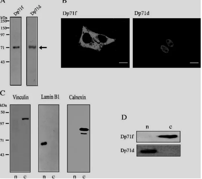
Using GFP-Dp71 gene fusions, we have previously demonstrated that alternative splicing determines the nuclear or cytoplasmic localization of exogenously expressed Dp71 protein isoforms in HeLa cells [21]. Hence, to corroborate these results, we decided to analyze the expression and distribution patterns of endogenous Dp71 protein isoforms in this cell line. HeLa cells are commonly used to study the function of nuclear proteins. To differentiate between Dp71 protein variants, we utilized different antibodies raised against the Dp71 C-terminal domain (Table 2): Dys2 and 2166, which recognize specifically the protein isoform containing the domain encoded by exon 78 (Dp71d), and 5F3, which detects exclusively the protein variant bearing 31 new amino acids that are incorporated into the molecule due to the alternative splicing of exon 78 (Dp71f). A typical immunoblot for the detection of Dp71 isoforms is shown in Fig. 1A; a band with an apparent molecular mass of 71kDa was observed when total protein extracts from HeLa cells were probed with either Dys2 or 5F3 antibodies. These results indicate that both Dp71d and Dp71f are expressed in HeLa cells. We did not observed any immunoreactive band above the 70-kDa protein marker, which indicates that besides Dp71, HeLa cells do not express any other DMD gene product at detectable levels.
Next, to study the subcellular distribution of each Dp71 variant, immunofluorescence and confocal microscopy analyses were carried out.
Immunostaining of Dp71f revealed a scattered signal distributed throughout the cytoplasm and excluded from nuclei. In contrast, although Dp71d staining was also present in the cytoplasm, it accumulated mainly in the nuclei, leaving the nucleoli clear of signal (Fig. 1B). To confirm the distinctive subcellular distribution of each Dp71 isoform, we carried out immunoblotting analyses of nuclear and cytosolic protein extracts. To verify the fidelity of our fractionation procedure, we employed vinculin, a focal adhesion protein [26], and calnexin, a membrane protein from the endoplasmic reticulum, as cytoplasmic markers. On
Fig. 1 – Expression and subcellular localization of endogenous Dp71 protein isoforms in HeLa cells. (A) Total protein extracts from HeLa cells were separated on 10% (v/v) SDS–PAGE, transferred to nitrocellulose membranes and probed with 5F3 and Dys2 monoclonal antibodies for the detection of Dp71f and Dp71d isoforms, respectively. Migration of Dp71 protein isoforms is indicated on the right by an arrow. Positions of molecular weight standards are shown on the left. (B) Cells, cultured on glass coverslips for confocal microscopy analysis, were immunostained with the primary monoclonal antibodies described above, and the specific signal was developed using a fluorescein-conjugated secondary antibody. Representative optical Z sections were selected to show the subcellular distribution of Dp71 isoforms. Scale bar = 16 μm. (C) Cellular fractionation was performed as described in Materials and methods. Nuclear (n) and cytosolic (c) protein extracts were subjected to immunoblot analysis using vinculin and calnexin antibodies as cytoplasmic markers and lamin B1 antibody as nuclear marker. (D) Subcellular fractions were analyzed with 5F3 and 2166 antibodies for the detection of Dp71f and Dp71d protein isoforms, respectively. Dp71d and Dp71f protein isoforms are expressed in HeLa cells; Dp71f distributes
exclusively in the cytoplasm, whereas Dp71d accumulates mainly in the nuclear compartment.
the other hand, lamin B1, a nuclear matrix-associated protein [27], was used as nuclear marker. As expected, the vinculin (135kDa) and calnexin (90kDa) bands were found exclusively in the cytosolic fraction whereas the lamin B1 band (68kDa) was absent in the cytosolic fraction but present in the nuclear extract (Fig. 1C). These results indicate that nuclear preparations were free of cytosolic proteins and membrane proteins from cytoplasm. Regarding the distribution of Dp71 isoforms, the Dp71f immunoreactive band was undetectable in the nuclear fraction but prominent in the cytosolic preparation; in contrast, the Dp71d immunoreactive band, clearly observed in the nuclear extract, was absent in the cytosolic fraction (Fig. 1D).
Expression of DAPC members in HeLa cells
To examine the expression profile of the dystrophin-associated protein complex (DAPC) components in HeLa cells, we performed RT-PCR and immunoblot analyses. We designed different pairs of primers to amplify specifically the cDNA of each DAPC member. Positive controls were carried out on human muscle total RNA or rat brain total RNA and the identity of the RT-PCR products was determined by DNA sequencing (data not shown). In summary, we determined that β-dystroglycan, β-sarcoglycan, α-syntrophin, β2-syntrophin, α1-dystrobrevin, β-dystrobrevin and nNOS are expressed at mRNA level in HeLa cells (Table 1). On the basis of these results, we further investigated the presence of the DAPC proteins in total extracts of HeLa cells using antibodies directed specifically to each member (Table 2). Representative immunoblots of DAPC members are shown in Fig. 2 (see lanes corresponding to total extracts). By using a monoclonal antibody anti-β-dystroglycan (NCL-b-DG), a robust band migrating at 43kDa was observed, which corresponds to the expected molecular weight of β-dystroglycan; in addition, a faint band migrating between the 43-and 59-kDa markers was observed. The expected band for β-sarcoglycan positioned above the 43-kDa protein marker was obtained after using a monoclonal antibody anti-β-sarcoglycan (NCL-b-SARC). By using the pan-specific antibody α-CTFP, two bands with apparent molecular weights of 70 and 94kDa were revealed, which must correspond to β-and α1-dystrobrevin, respectively. A band of approximately 60kDa, which must correspond to α-and/or β-syntrophin, was revealed after using a pan-specific antibody for α-and β-syntrophins. Finally, a band of 150kDa that corresponds to nNOS expression was obtained after using a commercial anti-nNOS antibody.
Subcellular distribution of DAPC components in HeLa cells
To examine the subcellular localization of DAPC members, we subjected HeLa cells, grown on coverslips, to immunofluorescence and confocal microscopy analysis using the appropriate fluorescein-conjugated secondary antibodies. As
shown in Fig. 3, β-dystroglycan displayed a predominant cytoplasmic staining with a perinuclear distribution that could correspond to endoplasmic reticulum or nuclear membrane labeling; in addition, we observed a scarce diffuse punctuate labeling inside nuclei (viewed specifically with propidium iodide), which was excluded from nucleoli. On the other hand, α1dystrobrevin and β-sarcoglycan immunostaining spanned the whole cell body but accumulated mainly in the nuclei (Fig. 3). Finally, as indicated by the phalloidin labeling of cytoplasmic actin, the immunostaining of β-dystrobrevin, nNOS and α/β syntrophin appeared to be mostly nuclear but, excluded from nucleoli. To distinguish between α-and β-syntrophin, we employed specific antibodies for α-or β2-syntrophin (Table 2) in the immunofluorescence assays. We observed that β2syntrophin displayed a predominant nuclear localization whereas α-syntrophin accumulated evenly between cytoplasm and nucleus (data not shown).
To support the immunofluorescence results with biochemical evidence, we carried out subcellular fractionation of HeLa cells into cytoplasmic and nuclear extracts and we subjected
Fig. 2 – Subcellular distribution of DAPC components in HeLa cells. Total (t), nuclear (n) and cytoplasmic (c) extracts, obtained as indicated in Materials and methods, were separated on 10% (v/v) SDS–PAGE, transferred to nitrocellulose membranes and probed with antibodies directed specifically against dystroglycan (NCL-b-DG), β-sarcoglycan (NCL-b-SARC), α1-dystrobrevin and β-dystrobrevin (β-CTFP), α/β-syntrophin, nNOS and actin (Table 2). For comparison, equal amounts of cytoplasmic and nuclear proteins were loaded in the corresponding lanes. Immunodetected DAPC members are indicated at the top of the panels. Position of protein markers is shown at the right. Arrows at the left denote the migration of the expected protein bands. All of the DAPC components analyzed by cellular fractionation were recovered in the nuclear fraction.
Fig. 3 – Immunofluorescence analysis of DAPC components in HeLa cells. Cells immunostained with the following antibodies: anti-β-dystroglycan (JAF), anti-α1-dystrobrevin (α-CTFP) and anti-β-sarcoglycan (NCL-b-SARC) (green color) were counterstained with propidium iodide (PI, red color) to visualize nuclei. Cells immunolabeled with anti-β-dystrobrevin antibody (β-521), anti-nNOS and anti-α/β-syntrophin (Pan-syntrophins) (green color) were counterstained with Texas red phalloidin (red color) to visualize cytoplasmic actin. After double staining, cell preparations were subjected to confocal microscopy analysis and single optical Z sections were selected in each case to show the distribution of DAPC components. The perinuclear localization of β-dystroglycan is denoted by arrows. All merged images are shown in the right panels. DAPC members displayed different nuclear distribution patterns. Scale bar = 20 μm.
the resulting protein extracts to SDS–PAGE and immunoblotting analyses. The β-dystroglycan immunoreactive band (43kDa) was prominent in the cytosolic fraction but was practically absent in the nuclear extract; in contrast, a second slower migrating band was observed predominantly in the nuclear fraction showing a weak signal in the cytosolic extract (Fig. 2). The same pattern of immunoreactive bands for β-dystroglycan was obtained with two different antibodies (JAF and NCL-b-DG). It has been observed that phosphorylation of β-dystroglycan causes an upward mobility shift in HeLa cells [28]; therefore, the retarded band might correspond to a phosphorylated isoform of this protein that accumulates in the nucleus of HeLa cells. The β-sarcoglycan immunoreactive band seemed to accumulate in the cytosolic extract showing a comparative weaker intensity in the nuclei. In contrast, the nNOS protein signal was observed mostly in the nuclear fraction (Fig. 2). In regard to the distribution of dystrobrevins, it was observed that both β-dystrobrevin and α1-dystrobrevin protein levels are enriched in the nuclear preparation being higher the cytoplasmic protein levels of the latter protein (Fig. 2). Finally, the immunoreactive band corresponding to α/β-syntrophin was distributed evenly between the two cellular compartments (Fig. 2). Based on the immunofluorescence data obtained with antibodies directed specifically against α-syntrophin or β-syntrophin, it could be proposed that the immunoreactive band revealed in the nucleus with the pan-syntrophin antibody corresponds mainly to β-pan-syntrophin. In addition, because actin represents a nuclear/cytoplasmic protein that has been found associated with Dp71 in cultured cells [29], we decided to analyze its subcellular distribution as well. We observed that cytoplasmic protein levels of actin were similar to those found in the nuclear fraction (Fig. 2).
Overall, the immunofluorescence and cellular fractionation studies evidenced the presence of DAPC components in the HeLa cell nuclei; however, certain differences were observed in the nuclear abundance of the DAPC members by comparing these two methodologies: For instance, immunostaining of α/β-syntrophin appeared to be mainly nuclear but the fractionation analysis indicated that protein levels of α/βsyntrophin are portioned similarly between cytoplasm and nucleus. These inconsistencies could be due to the principle of each methodology; in the confocal images, mid-optical Z-sections were selected to emphasize the nuclear presence of the DAPC members whereas in the
Fig. 4 – Dp71 forms a DAPC in the HeLa nuclei. Detergent-soluble nuclear extracts from HeLa cells were immunoprecipitated with antibodies specific to Dp71 (mDp71), β-dystrobrevin (β-521), α1-dystrobrevin (α-CTFP) and β-dystroglycan (JAF). Co-immunoprecipitated proteins were analyzed by immunoblotting with antibodies directed against Dp71 (mDp71), β-dystroglycan (NCL-b-DG), nNOS and α1-and β-dystrobrevin (β-CTFP). As negative control, an antibody directed against the transcription factor Sp3 was employed. Representative results from three independent immunoprecipitation assays are shown. β-DG, β-dystroglycan; α1-DB, α1-dystrobrevin; β-DB, β-dystrobrevin; IP, immunoprecipitation; WB, western blotting. Input, soluble nuclear extracts subjected to immunoblotting. The unspecific binding lane refers to immunoprecipitation experiments where the respective primary antibody was omitted. Dp71, β-dystroglycan, nNOS, α1-dystrobrevin and β-dystrobrevin, copurified in the nuclear fraction.
fractionation studies a more accurate determination of the cytoplasm–nucleus ratio of these proteins was provided.
Dystrophin Dp71 forms a DAPC in the nuclei of HeLa cells
To explore the possibility that Dp71 interacts with DAPC components within HeLa nuclei to form a DAPC complex in this organelle, we immunoprecipitated nuclear protein extracts with an anti-Dp71 antibody raised against the N-terminus domain of Dp71 (mDp71) and analyzed by Western blotting with a panel of antibodies directed against different DAPC components. It was observed that β-dystroglycan, nNOS, α1-dystrobrevin and β-dystrobrevin immunoprecipitated with Dp71. In reciprocal experiments, Dp71 was immunoprecipitated by antibodies directed against β-dystroglycan (JAF), α1-dystrobrevin (α-CTFP) and β-dystrobrevin (β-521), confirming the association of Dp71 with these proteins (Fig. 4). Moreover, Dp71, nNOS, α1-dystrobrevin and β-dystrobrevin were found in the immunoprecipitates of β-dystroglycan; reciprocal experiments showed that β-dystroglycan immunoprecipitated with α1-and β-dystrobrevin (Fig. 4). In addition to Dp71 and dystroglycan, immunoprecipitates of α1-and β-dystrobrevin contained nNOS. Interestingly, α1-and β-β-dystrobrevin copurified, which suggest that they are part of the same protein complex within HeLa nuclei. We were unable to detect β-sarcoglycan in the Dp71, β-dystroglycan and α1-and βdystrobrevin immunoprecipitates (data not shown). Specificity of the
immunoprecipitation assays was established by the following negative controls: (a) Assays where the immunoprecipitating antibodies were omitted showed that none of the proteins examined precipitated in a unspecific way (Fig. 4, lane “unspecific binding”); and (b) Western blotting experiments with an antibody directed against the transcription factor Sp3 showed that the DAPC immunoprecipitates excluded an unrelated protein (Fig. 4). Overall, our data indicate that Dp71 forms a DAPC in the nuclei of HeLa cells, which contains, at least, β-dystroglycan, α1-/β-dystrobrevin and nNOS.
Components of the nuclear DAPC associate with the nuclear matrix of HeLa cells Because nuclear matrix has been proposed to be the underlying structural support for subnuclear organization of diverse protein complexes [30,31], we examined whether or not the members of the nuclear Dp71-containing DAPC complex bind to the HeLa nuclear matrix. We obtained nuclear and nuclear matrix protein extracts and in situ nuclear matrix preparations by a sequential high salt extraction procedure previously described [22–25]. To verify the authenticity of the nuclear matrix preparations, we analyzed the distribution of the nuclear matrix proteins lamin B1 and actin as well as the distribution of the soluble transcription factor Sp3. Fig. 5A shows optical sections of nuclear matrix preparations stained with antibodies directed against Dp71 or the different DAPC components. We obtained the typical staining of lamin B1 localized at the nuclear periphery with a dotted organization in the inner nuclear matrix (Fig. 5A). As expected for a soluble nuclear protein, the Sp3 immunolabeling was excluded from the nuclear matrix preparation (Fig. 5A). Interestingly, the immunostaining of Dp71 as well as β-dystroglycan, βdystrobrevin, α1-dystrobrevin and nNOS was retained within the nucleoskeletal structure displaying specific labeling patterns. The staining of Dp71 and β-dystroglycan distributed mainly in the nuclear matrix periphery forming a ringlike arrangement; besides, the signal of the latter protein produced some diffuse labeling in the inner nucleoskeletal structure that may correspond to invaginations of the nuclear envelope in the nucleus (Fig. 5A). The α1-dystrobre-vin immunostaining displayed a punctate labeling throughout the nuclear matrix structure that left nucleoli clear of signal. On the other hand, β-dystrobrevin labeling distributed through the nucleoskeletal structure showing patches with more intensive labeling. Finally, nNOS immunodetection displayed a discontinuous staining throughout the nucleoskeletal structure that forms a net-like arrangement (Fig. 5A). It should be noted that fluorescence pattern of Dp71, βdystroglycan and lamin B1 colocalized at certain extent in the nuclear matrix periphery, which may indicate their physical interaction.
Fig. 5 – Association of Dp71 and DAPC components with the nuclear matrix of HeLa cells. (A) In situ nuclear matrix preparations were obtained as described in Materials and methods. Immunofluorescence confocal microscopy images of representative nuclear matrices stained with antibodies directed against Sp3, lamin B1, Dp71d (2166), β-dystroglycan (JAF), β-dystrobrevin (β-521), α1-dystrobrevin (α-CTFP) and nNOS are shown. As control, a phase contrast micrograph of the in situ nuclear matrix preparation used for Sp3 immunostaining is also shown. Immunostaining of Dp71, β-dystroglycan, β-dystrobrevin, α1-dystrobrevin and nNOS was retained within the nuclear matrix preparation whereas that of Sp3 was not. Immunostaining of lamin B1, Dp71d and β-dystroglycan in the nuclear matrix periphery is denoted by arrows. (B) Equal amounts of nuclear (N) and nuclear matrix (NM) protein extracts, obtained as described in Materials and methods, were subjected to immunoblotting analysis. Proteins were visualized by using specific antibodies against Sp3 (negative control of the nuclear matrix
extracts), lamin B1, actin, Dp71 (mDp71), β-dystroglycan (NCL-b-DG), α1-and β-dystrobrevin (β-CTFP), nNOS, β-sarcoglycan (NCL-b-SARC) and α/β-syntrophin. Representative results from three independent assays are shown. The position of the protein markers is shown at the left. Arrows point to the specific bands of Sp3. The slower-migrating band of β-dystroglycan is denoted by an arrow. The expected protein bands for lamin B1, actin, Dp71, β-dystroglycan, α1-and β-dystrobrevin, nNOS, β-sarcoglycan and α/β-syntrophin were recovered in the nuclear matrix fraction.
To obtain protein extracts from the nuclear matrices of HeLa cells, previously isolated nuclei were digested with DNase I and extracted with 1.3M (NH4)2SO4, and the nuclear material remaining after treatment was electrophoresed and immunoblotted with antibodies directed against Dp71 or to the different DAPC components. As expected, the immunoreactive bands for lamin B1 (68kDa) and actin (43kDa) were present in both the nuclear and the nuclear matrix fractions whereas those of Sp3 protein (115, 80 and 70kDa) were present in the nuclear extract but excluded from the nuclear matrix fraction (Fig. 5B). Fig. 5B also shows that the Dp71, dystroglycan, α1-and βdystrobrevin, nNOS, β-sarcoglycan and α/β-syntrophin immunoreactive bands were recovered in both the nuclear and nuclear matrix fractions after using the antibodies mDp71, NCL-b-DG, β-CTFP, anti-nNOS, NCL-b-SARC and pan-syntrophins, respectively. The additional slower-migrating band of β-dystroglycan, noted in the nuclear fraction, was also revealed in the nuclear matrix extract (indicated by black arrow, Fig. 5B). It was also observed that only α1-dystrobrevin displayed enrichment in the nuclear matrix fraction, compared with the nuclear extract (Fig. 5B). Altogether, confocal microscopy and immunoblotting analyses of HeLa nuclear matrix preparations evidenced that some members of the nuclear DAPC are nuclear matrix-associated proteins.
Fig. 6 – Interaction of Dp71 and DAPC members with nuclear matrix proteins. Detergent-soluble nuclear extracts from HeLa cells were immunoprecipitated with antibodies specific to Dp71 (mDp71), β-dystroglycan (JAF), α1-dystrobrevin (α-CTFP) and β-α1-dystrobrevin (β-521). Co-immunoprecipitated proteins were analyzed by Western blotting with antibodies directed against actin and lamin B1. Representative results from three independent immunoprecipitation assays are shown. IP, immunoprecipitation. WB, Western blotting. Input, soluble nuclear extracts subjected to immunoblotting analysis. The unspecific binding lane refers to immunoprecipitation experiments where the respective primary antibody was omitted. β-DG, β-dystroglycan; α1-DB, α1-dystrobrevin; β-DB, β-dystrobrevin.
Interaction of nuclear DAPC components with the nuclear matrix proteins actin and lamin B1
To verify the experimental evidence concerning the association of nuclear DAPC components with the nuclear matrix, we examined whether any of the DAPC members establishes physical interactions with the nuclear matrix proteins lamin B1 and actin. Therefore, nuclear protein extracts prepared from HeLa cells were immunoprecipitated with specific antibodies for Dp71, α1-dystrobrevin, β-dystrobrevin or βdystroglycan and the resulting immunoprecipitates were immunoblotted with antibodies directed against lamin B1 and actin. Fig. 6 shows that the amount of actin and lamin B1 recovered in the immunoprecipitates of Dp71, β-dystroglycan and β-dystrobrevin was clearly higher than that obtained in the negative control experiment. In contrast, the intensity of the actin and lamin B1 immunoreactive bands was undistinguishable between the α1-dystrobrevin immunoprecipitate and the negative control experiment. Western blotting assays using an anti-Sp3 antibody demonstrated that immunoprecipitates of DAPC members excluded an unrelated protein (see lower panel of Fig. 4). Altogether, these results indicate that actin and lamin B1 copurified specifically with Dp71, β-dystroglycan and β-dystrobrevin, but not with α1-dystrobrevin.
Discussion
The characterization of different dystrophin-associated protein complexes (DAPCs), such as those localized in the skeletal muscle, neuromuscular junction, brain, lung, retina and kidney, has been reported [3,11–13,17,32]. In addition, an increasing number of cellular functions associated with these multiple protein complexes have been proposed, such as membrane stability, cellular signaling and force transduction [7,33,34]. The specific function of each DAPC seems to be determined by the cellular environment where it resides as well as by the unique combination of proteins that conforms the complex.
In this work, we report for the first time the existence of a DAPC complex in a cellular compartment different from cell membrane, the nucleus. We identified and characterized a DAPC present in the nuclei of HeLa cells, which contains dystrophin Dp71 as a key component. Besides, Dp71, the complex is composed, at least, of β-dystroglycan, α1-and βdystrobrevin and nNOS. Previously, the presence of Dp71 and several DAPC components in the nuclei of different cell types has been described [17,19,20]. The nuclear distribution of Dp71 has been observed in cultured hippocampal neurons
[35] and the PC12 cell line [36,37]. Additionally, α-dystrobrevin was found in the nuclei of the human cell line HL-60
[19] whereas β-dystrobrevin was observed in the nuclear compartment of
hippocampal pyramidal neurons [17]. Furthermore, the nuclear localization of α-, β-and γ1syntrophin has been described in HeLa cells [20]. Finally, the nuclear localization of nNOS in the rat brain and MDCK cells has been also reported [38]; the authors proposed that nNOS may traffic nuclear proteins to the cytosol via its PDZ domain.
Although the evidence mentioned above point to the existence of a nuclear DAPC complex, to our knowledge, this is the first report establishing the formation of a Dp71contaning DAPC complex within nuclei. Our statements are based on the following experimental evidence: We have demonstrated by immunofluorescence and cell fractionation experiments that Dp71, β-dystroglycan, nNOS, β-sarcoglycan, α/β-syntrophin and α1-and β-dystrobrevin colocalize in the HeLa cell nuclei. In addition, we established by co-immuno-precipitation assays that Dp71, β-dystroglycan, nNOS, α1and β-dystrobrevin associate by themselves to form a protein complex within this organelle. Because two different dystrobrevins, α1 and β, colocated in the HeLa nuclei, it would be expected that at least two distinct Dp71-containing complexes are formed in this cellular compartment, each one including a specific dystrobrevin. However, we found that α1and β-dystrobrevin copurified together in the HeLa nuclear fraction suggesting that they are part of a single protein complex. This finding contrasts to the previously described cell membrane-associated complexes where a single dystrobrevin is included in each DAPC [11]. Further biochemical experiments are clearly needed to define the precise arrangement of dystrobrevins in the nuclear DAPC. We were unable to show any association of β-sarcoglycan with the nuclear DAPC; nevertheless, because this protein is distributed in the nuclear compartment of HeLa cells, the possibility that β-sarcoglycan binds to this novel protein complex still open.
The existence of a nuclear DAPC raises important questions about the molecular mechanism governing its nuclear transport, either as a protein complex or as independent components. Selective nuclear import occurs through the nuclear pore complex [39], which allows small molecules (under 45kDa) to diffuse freely into and out of the nucleus [39]. The targeting of larger proteins requires the presence of either a classical or a bipartite nuclear localization signal (NLS), both of which are composed of basic amino acids [40,41]. In this regard, a computational analysis of the entire amino acid sequence of each DAPC component showed that only the βdystroglycan contains a stretch of basic amino acids (RKKRKGK) that could correspond to an NLS (amino acids 776–782). Therefore, it is tempting to speculate that DAPC components gain access to the soluble transport machinery through its association with β-dystroglycan. Alternatively, it couldbepossiblethatDp71orany otherofthe DAPC components contains an atypical but functional NLS motif [42]. It has been recently reported that α-, β-and γ1syntrophin are recruited into HeLa cell nuclei via their interaction with the enzyme diacylglycerol kinase-ζ [20]. Hence, it seems that several alternative molecular mechanisms could be involved in the transport of DAPC components to HeLa nuclei.
The existence of a nuclear DAPC elicits interesting questions regarding its physiological function within this organelle; to start exploring this issue, we determined whether any of the nuclear DAPC members associates with the nuclear matrix. It is now becoming increasingly clear that the nuclear matrix provides the underlying support for nuclear compartmentalization; hence, nuclear factors localize to discrete subdomains within the nucleus to perform specialized functions, such as DNA organization and replication, gene transcription and RNA processing [43]. Rather than behave as soluble, easily extractable factors, we were surprised to find that Dp71 and various DAPC components, such as β-dystroglycan, β-sarcoglycan, βdystrobrevin, α1-dystrobrevin, nNOS and α/β-syntrophins, were resistant to nuclei extraction with detergent, nucleases and salt. Such treatments are normally used to extract chromatin components including transcription factors or histones. Thus, we have demonstrated that members of the Dp71-containing DAPC are not only localized in the nucleus; but also tightly associated with the nuclear matrix. Interestingly, the immunofluorescence-labeling pattern of Dp71 and β-dystroglycan colocalized at certain extent with that of lamin B1, especially at the nuclear matrix periphery. These results indicate that these three proteins might colocalize spatially. Supporting these data, we found that Dp71, βdystroglycan and β-dystrobrevin co-immunoprecipitated with the nuclear matrix proteins lamin B1 and actin. Because lamin B1 is a structural component of the nuclear matrix that facilitates the interaction between chromatin and the inner nuclear membrane [27] and actin is a component of several mammal chromatin-remodeling complexes [44], the association of Dp71 and some DAPC members with those proteins might indicate that they work as scaffolding proteins involved in nuclear architecture. Additional biochemical studies are necessary to establish whether the interaction between DAPC components and actin and lamin B1 is direct or due to an association within the context of a larger nuclear matrix complex.
Based on the results of this work and on the well-known protein–protein interactions occurring among DAPC members at the cell membrane, it is tempting to speculate that the arrangement of the nuclear DAPC is similar to that displayed by the DAPC at the cell membrane. We hypothesize that βdystroglycan and β-sarcoglycan are inserted in the inner nuclear membrane via their respective transmembrane domains. Therefore, the C-terminal domain of β-dystroglycan would interact with the WW domain of Dp71, which in turns might bind to β-dystrobrevin by their coiled-coil domains in the C-terminal region of each protein. Likewise, we propose that α1-and β-dystrobrevin bind each other via a coiled-coil interaction forming a dystrobrevin heterodimer. On the other hand, nNOS may be recruited to the nuclear DAPC by interacting with α/β-syntrophin [20] or with a PDZ-containing nuclear protein. We propose that the physical contact between the DAPC and the nuclear matrix might be mediated by the interaction of β-dystroglycan with lamin B1. In addition, Dp71 might contact nuclear actin via its N-terminal actin-binding domain [29].
In summary, we have identified and characterized a novel Dp71-containing
DAPC present in the nuclei of HeLa cells. In addition, we have revealed that several members of this novel complex are associated with the nuclear matrix. Altogether, our results suggest that the nuclear DAPC may work as a nuclear scaffolding complex involved in nuclear organization.
Acknowledgments
The authors would like to thank Dr. Manuel Hernández for supplying anti-actin monoclonal antibody and Dr. Dereck Blake for providing us with antibodies mDp71, 2166, β-CTFP, αCTFP and β-521. We also thank Victor Tapia for technical assistance and Dr. Raul Mena for the confocal microscopy facility. We are grateful to Dr. Angela L. Guillozet-Bongaarts for the critical revision of the manuscript. This work was supported by CONACyT, Mexico: Grant 43285-M.
REFERENCES
[1] E.P. Hoffman, R.H. Brown Jr., L.M. Kunkel, Dystrophin: the protein product of the Duchenne muscular dystrophy locus, Cell 51 (1987) 919–928.
[2] J.M. Ervasti, S.D. Kahl, K.P. Campbell, Purification of dystrophin from skeletal muscle, J. Biol. Chem. 266 (1991) 9161–9165.
[3] O. Ibraghimov-Beskrovnaya, J.M. Ervasti, C.J. Leveille, C.A. Slaughter, S.W. Sernett, K.P. Campbell, Primary structure of dystrophin-associated glycoproteins linking dystrophin to the extracellular matrix, Nature 355 (1992) 696–702.
[4] J.E. Brenman, D.S. Chao, H. Xia, K. Aldape, D.S. Bredt, Nitric oxide synthase complexed with dystrophin and absent from skeletal muscle sarcolemma in Duchenne muscular dystrophy, Cell 82 (1995) 743–752.
[5] L.E. Lim, F. Duclos, O. Broux, N. Bourg, Y. Sunada, V. Allamand,
J. Meyer, I. Richard, C. Moomaw, C. Slaughter, F.M.S. Tomé, M. Fardeau, C.E. Jackson, J.S. Beckmann, K.P. Campbell, Beta-sarcoglycan: characterization and role in limb-girdle muscular dystrophy linked to 4q12, Nat. Genet. 11 (1995) 257–265.
[6] A.J. Ettinger, G. Feng, J.R. Sanes, epsilon-Sarcoglycan, a broadly expressed homologue of the gene mutated in limb-girdle muscular dystrophy 2D, J. Biol. Chem. 272 (1997) 32534– 32538.
[7] J.M. Ervasti, K.P. Campbell, A role for the dystrophin-glycoprotein complex as a transmembrane linker between laminin and actin, J. Cell Biol. 122 (1993) 809–823.
[8] M.E. Adams, M.H. Butler, T.M. Dwyer, M.F. Peters, A.A. Murnane, S.C. Froehner, Two forms of mouse syntrophin, a 58 kd dystrophin-associated protein, differ in primary structure and tissue distribution, Neuron 11 (1993) 531–540.
[9] F.S. Walsh, J.A. Pizzey, G. Dickson, Tissue-specific isoforms of dystrophin, Trends Neurosci. 12 (1989) 235–238.
[10] D.J. Blake, D.R. Love, J. Tinsley, G.E. Morris, H. Turley, K. Gatter G. Dickson, Y.H. Edwards, K.E. Davies, Characterization of a 4.8 kb transcript from the Duchenne muscular dystrophy locus expressed in Schwannoma cells, Hum. Mol. Genet. 1 (1992) 103–109.
[11] N.Y. Loh, S.E. Newey, K.E. Davies, D.J. Blake, Assembly of multiple dystrobrevin-containing complexes in the kidney, J Cell Sci 113 (Pt. 15) (2000) 2715–2724.
[12] T. Claudepierre, C. Dalloz, D. Mornet, K. Matsumura, J. Sahel,
A. Rendon, Characterization of the intermolecular associations of the dystrophin-associated glycoprotein complex in retinal Muller glial cells, J. Cell Sci. 113 (Pt. 19) (2000) 3409– 3417.
[13] H. Moukhles, S. Carbonetto, Dystroglycan contributes to the formation of multiple dystrophin-like complexes in brain J. Neurochem. 78 (2001) 824–834. [14] J.P. Hugnot, H. Gilgenkrantz, N. Vincent, P. Chafey, G.E. Morris, A.P. Monaco, Y. Berwald-Netter, A. Koulakoff, J.C. Kaplan, A. Kahn, J. Chelly, Distal
transcript of the dystrophin gene initiated from an alternative first exon and encoding a 75-kDa protein widely distributed in nonmuscle tissues, Proc. Natl. Acad. Sci. U. S. A. 89 (1992) 7506–7510.
[15] N.R. Kramarcy, A. Vidal, S.C. Froehner, R. Sealock, Association of utrophin and multiple dystrophin short forms with the mammalian M(r) 58,000 dystrophin-associated protein (syntrophin), J. Biol. Chem. 269 (1994) 2870–2876.
[16] G. Rosa, M. Ceccarini, M. Cavaldesi, M. Zini, T.C. Petrucci, Localization of the dystrophin binding site at the carboxyl terminus of beta-dystroglycan, Biochem. Biophys. Res. Commun. 223 (1996) 272–277.
[17] D.J. Blake, R. Hawkes, M.A. Benson, P.W. Beesley, Different dystrophin-like complexes are expressed in neurons and glia,
J. Cell Biol. 147 (1999) 645–658.
[18] D.S. Greenberg, Y. Schatz, Z. Levy, P. Pizzo, D. Yaffe, U. Nudel, Reduced levels of dystrophin associated proteins in the brains of mice deficient for Dp71, Hum. Mol. Genet. 5 (1996) 1299–1303.
[19] A. Kulyte, R. Navakauskiene, G. Treigyte, A. Gineitis,
T. Bergman, K.E. Magnusson, Characterization of human alpha-dystrobrevin isoforms in HL-60 human promyelocytic leukemia cells undergoing granulocytic differentiation, Mol. Biol. Cell 13 (2002) 4195–4205.
[20] A. Hogan, L. Shepherd, J. Chabot, S. Quenneville, S.M. Prescott,
M.K. Topham, S.H. Gee, Interaction of gamma 1-syntrophin with diacylglycerol kinase-zeta. Regulation of nuclear localization by PDZ interactions, J. Biol. Chem. 276 (2001) 26526–26533.
[21] E. Gonzalez, C. Montanez, P.N. Ray, P.L. Howard, F. Garcia-Sierra, D. Mornet, B. Cisneros, Alternative splicing regulates the nuclear or cytoplasmic localization of dystrophin Dp71, FEBS Lett. 482 (2000) 209–214.
[22] P. Belgrader, A.J. Siegel, R. Berezney, A comprehensive study on the isolation and characterization of the HeLa S3 nuclear matrix, J. Cell Sci. 98 (Pt. 3) (1991) 281–291. [23] D.R. Stauffer, T.L. Howard, T. Nyun, S.M. Hollenberg, CHMP1 is a novel nuclear matrix
protein affecting chromatin structure and cell-cycle progression, J. Cell Sci. 114 (2001) 2383–2393.
[24] H. Ma, A.J. Siegel, R. Berezney, Association of chromosome territories with the nuclear matrix. Disruption of human chromosome territories correlates with the release of a subset of nuclear matrix proteins, J. Cell Biol. 146 (1999) 531–542.
[25] J.A. Nickerson, G. Krockmalnic, K.M. Wan, S. Penman, The nuclear matrix revealed by eluting chromatin from a cross-linked nucleus, Proc. Natl. Acad. Sci. U. S. A. 94 (1997) 4446–4450.
[26] A. Renaudin, M. Lehmann, J. Girault, L. McKerracher, Organization of point contacts in neuronal growth cones,
J. Neurosci. Res. 55 (1999) 458–471.
[27] Y. Gruenbaum, R.D. Goldman, R. Meyuhas, E. Mills, A. Margalit, A. Fridkin, Y. Dayani, M. Prokocimer, A. Enosh, The nuclear lamina and its functions in the nucleus, Int. Rev. Cytol. 226 (2003) 1–62.
[28] M. James, A. Nuttall, J.L. Ilsley, K. Ottersbach, J.M. Tinsley,
M. Sudol, S.J. Winder, Adhesion-dependent tyrosine phosphorylation of (beta)-dystroglycan regulates its interaction with utrophin, J. Cell Sci. 113 (Pt. 10) (2000) 1717–1726.
[29] P.L. Howard, H.J. Klamut, P.N. Ray, Identification of a novel actin binding site within the Dp71 dystrophin isoform, FEBS Lett. 441 (1998) 337–341.
[30] D.G. Capco, K.M. Wan, S. Penman, The nuclear matrix: three-dimensional architecture and protein composition, Cell 29 (1982) 847–858.
[31] M.J. Hendzel, F. Boisvert, D.P. Bazett-Jones, Direct visualization of a protein nuclear architecture, Mol. Biol. Cell 10 (1999) 2051–2062.
[32] K. Matsumura, J.M. Ervasti, K. Ohlendieck, S.D. Kahl, K.P. Campbell, Association of dystrophin-related protein with dystrophin-associated proteins in mdx mouse muscle, Nature 360 (1992) 588–591.
[33] M. Cavaldesi, G. Macchia, S. Barca, P. Defilippi, G. Tarone, T.C. Petrucci, Association of the dystroglycan complex isolated from bovine brain synaptosomes with proteins involved in signal transduction, J. Neurochem. 72 (1999) 1648–1655.
[34] S.A. Oak, K. Russo, T.C. Petrucci, H.W. Jarrett, Mouse alpha1-syntrophin binding to Grb2: further evidence of a role for syntrophin in cell signaling, Biochemistry 40 (2001) 11270–11278.
[35] V. Aleman, B. Osorio, O. Chavez, A. Rendon, D. Mornet, D. Martinez, Subcellular localization of Dp71 dystrophin isoforms in cultured hippocampal neurons and forebrain astrocytes, Histochem. Cell Biol. 115 (2001) 243–254.
[36] F.G. Marquez, B. Cisneros, F. Garcia, V. Ceja, F. Velazquez, F. Depardon, L. Cervantes, A. Rendon, D. Mornet, H. Rosas-vargas, M. Mustre, C. Montanez, Differential expression and subcellular distribution of dystrophin Dp71 isoforms during differentiation process, Neuroscience 118 (2003) 957–966.
[37] R. Acosta, C. Montanez, L. Fuentes-Mera, E. Gonzalez, P. Gomez, L. Quintero-Mora, D. Mornet, L.M. Alvarez-Salas, B. Cisneros, Dystrophin Dp71 is required for neurite outgrowth in PC12 cells, Exp. Cell Res. 296 (2004) 265–275.
[38] G.M. Riefler, B.L. Firestein, Binding of neuronal nitric-oxide synthase (nNOS) to carboxyl-terminal-binding protein (CtBP) changes the localization of CtBP from the nucleus to the cytosol: a novel function for targeting by the PDZ domain of nNOS, J. Biol. Chem. 276 (2001) 48262–48268.
[39] C.W. Akey, D.S. Goldfarb, Protein import through the nuclear pore complex is a multistep process, J. Cell Biol. 109 (1989) 971–982.
[40] D. Kalderon, B.L. Roberts, W.D. Richardson, A.E. Smith, A short amino acid sequence able to specify nuclear location, Cell 39 (1984) 499–509.
[41] J. Robbins, S.M. Dilworth, R.A. Laskey, C. Dingwall, Two interdependent basic domains in nucleoplasmin nuclear targeting sequence: identification of a class of bipartite nuclear targeting sequence, Cell 64 (1991) 615–623.
[42] E. Izaurralde, A. Jarmolowski, C. Beisel, I.W. Mattaj, G. Dreyfuss, U. Fischer, A role for the M9 transport signal of hnRNP A1 in mRNA nuclear export, J. Cell Biol. 137 (1997) 27– 35.
[43] T. Cremer, G. Kreth, H. Koester, R.H. Fink, R. Heintzmann,
M. Cremer, I. Solovei, D. Zink, C. Cremer, Chromosome territories, interchromatin domain compartment, and nuclear matrix: an integrated view of the functional nuclear architecture, Crit. Rev. Eukaryotic Gene Expression 10 (2000) 179–212.
[44] I.A. Olave, S.L. Reck-Peterson, G.R. Crabtree, Nuclear actin and actin-related proteins in chromatin remodeling, Annu. Rev. Biochem. 71 (2002) 755–781.
[45] E. Fabbrizio, U. Nudel, G. Hugon, A. Robert, F. Pons, D. Mornet, Characterization and localization of a 77 kDa protein related to the dystrophin gene family, Biochem. J. 299 (Pt. 2) (1994) 359–365.
[46] F. Rivier, A. Robert, G. Hugon, A. Bonet-Kerrache, V. Nigro, J.A. Fehrentz, J. Martinez,
D. Mornet, Dystrophin and utrophin complexed with different associated proteins in cardiac Purkinje fibres, Histochem. J. 31 (1999) 425–432.
[47] D.J. Blake, R. Nawrotzki, N.Y. Loh, D.C. Gorecki, K.E. Davies, Beta-dystrobrevin, a member of the dystrophin-related protein family, Proc. Natl. Acad. Sci. U. S. A. 95 (1998) 241–246.
[48] F. Rivier, A. Robert, M. Royuela, G. Hugon, A. Bonet-Kerrache,
D. Mornet, Utrophin and dystrophin-associated glycoproteins in normal and dystrophin deficient cardiac muscle, J. Muscle Res. Cell Motil. 20 (1999) 305–314.
[49] C.G. Garcia-Tovar, A. Perez, J. Luna, R. Mena, B. Osorio, V. Aleman, R. Mondragon, D. Mornet, A. Rendon, J.M. Hernandez, Biochemical and histochemical analysis of 71 (kDa dystrophin isoform (Dp71f) in rat brain, Acta Histochem. 103 (2001) 209–224