HAL Id: hal-01215737
https://hal.sorbonne-universite.fr/hal-01215737
Submitted on 14 Oct 2015
HAL is a multi-disciplinary open access
archive for the deposit and dissemination of
sci-entific research documents, whether they are
pub-lished or not. The documents may come from
teaching and research institutions in France or
abroad, or from public or private research centers.
L’archive ouverte pluridisciplinaire HAL, est
destinée au dépôt et à la diffusion de documents
scientifiques de niveau recherche, publiés ou non,
émanant des établissements d’enseignement et de
recherche français ou étrangers, des laboratoires
publics ou privés.
Distributed under a Creative Commons Attribution| 4.0 International License
DAMP-Associated Processes in Cancer
Jitka Fucikova, Irena Moserova, Linda Urbanova, Lucillia Bezu, Oliver Kepp,
Isabelle Cremer, Cyril Salek, Pavel Strnad, Guido Kroemer, Lorenzo Galluzzi,
et al.
To cite this version:
Jitka Fucikova, Irena Moserova, Linda Urbanova, Lucillia Bezu, Oliver Kepp, et al.. Prognostic and
Predictive Value of DAMPs and DAMP-Associated Processes in Cancer. Frontiers in Immunology,
Frontiers, 2015, 6, pp.402. �10.3389/fimmu.2015.00402�. �hal-01215737�
Edited by: Fabrizio Mattei, Istituto Superiore di Sanità, Italy Reviewed by: Stephan Gasser, National University of Singapore, Singapore Zong Sheng Guo, Harvard University, USA *Correspondence: Lorenzo Galluzzi, Equipe 11 - “Apoptose, Cancer et Immunité” Centre de Recherche des Cordeliers de Jussieu15, rue de l’École de Médecine, Paris 75006, France deadoc@vodafone.it; Radek Spisek, Sotio, Jankovcova 1518/2, 170 00 Prague 7, Czech Republic spisek@sotio.com †Lorenzo Galluzzi and Radek Spisek
share senior co-authorship. Specialty section: This article was submitted to Tumor Immunity, a section of the journal Frontiers in Immunology Received: 23 June 2015 Accepted: 22 July 2015 Published: 07 August 2015 Citation: Fucikova J, Moserova I, Urbanova L, Bezu L, Kepp O, Cremer I, Salek C, Strnad P, Kroemer G, Galluzzi L and Spisek R (2015) Prognostic and predictive value of DAMPs and DAMP-associated processes in cancer. Front. Immunol. 6:402. doi: 10.3389/fimmu.2015.00402
Prognostic and predictive value of
DAMPs and DAMP-associated
processes in cancer
Jitka Fucikova
1,2, Irena Moserova
1,2, Linda Urbanova
1,2, Lucillia Bezu
3,4,5,6,7,
Oliver Kepp
3,4,5,6,7, Isabelle Cremer
5,6,8, Cyril Salek
9, Pavel Strnad
10, Guido Kroemer
3,4,5,6,7,11,
Lorenzo Galluzzi
3,4,5,6,12*
†and Radek Spisek
1,2*
†1 Sotio, Prague, Czech Republic, 2 Department of Immunology, 2nd Faculty of Medicine, University Hospital Motol, Charles University, Prague, Czech Republic, 3 Equipe 11 labellisée par la Ligue Nationale contre le Cancer, Centre de Recherche des Cordeliers, Paris, France, 4 U1138, INSERM, Paris, France, 5 Sorbonne Paris Cité, Université Paris Descartes, Paris, France, 6 Université Pierre et Marie Curie, Paris, France, 7 Metabolomics and Cell Biology Platforms, Gustave Roussy Comprehensive Cancer Institute, Villejuif, France, 8 Equipe 13, Centre de Recherche des Cordeliers, Paris, France, 9 Institute of Hematology and Blood Transfusion, Prague, Czech Republic, 10 Department of Gynecology and Obsterics, 2nd Faculty of Medicine, University Hospital Motol, Charles University, Prague, Czech Republic, 11 Pôle de Biologie, Hopitâl Européen George Pompidou, AP-HP, Paris, France, 12 Gustave Roussy Comprehensive Cancer Institute, Villejuif, France
It is now clear that human neoplasms form, progress, and respond to therapy in the context
of an intimate crosstalk with the host immune system. In particular, accumulating evidence
demonstrates that the efficacy of most, if not all, chemo- and radiotherapeutic agents
commonly employed in the clinic critically depends on the (re)activation of tumor-targeting
immune responses. One of the mechanisms whereby conventional chemotherapeutics,
tar-geted anticancer agents, and radiotherapy can provoke a therapeutically relevant, adaptive
immune response against malignant cells is commonly known as “immunogenic cell death.”
Importantly, dying cancer cells are perceived as immunogenic only when they emit a set of
immunostimulatory signals upon the activation of intracellular stress response pathways. The
emission of these signals, which are generally referred to as “damage-associated molecular
patterns” (DAMPs), may therefore predict whether patients will respond to chemotherapy or
not, at least in some settings. Here, we review clinical data indicating that DAMPs and
DAMP-associated stress responses might have prognostic or predictive value for cancer patients.
Keywords: ATP, autophagy, calreticulin, eR stress response, HSPs, type i interferon
Abbreviations: AGER, advanced glycosylation end product-specific receptor; AML, acute myeloid leukemia; APC,
antigen-presenting cell; ATF6, activating transcription factor 6; BECN1, beclin 1; C1q, complement component 1, q subcomponent; CALR, calreticulin; CLEC9A, C-type lectin domain family 9, member A; CLL, chronic lymphocytic leukemia; CRC, colorectal carcinoma; CXCL10, chemokine (C-X-C motif) ligand 10; CXCR4, chemokine (C-X-C motif) receptor 4; DAMP, damage-associated molecular pattern; EIF2A, eukaryotic translation initiation factor 2A; EIF2AK2, eukaryotic translation initiation factor 2-alpha kinase 2; ENTPD1, ectonucleoside triphosphate diphosphohydrolase 1; FPR1, formyl peptide receptor 1; HCC, hepatocellular carcinoma; HMGB1, high mobility group box 1; HSP, heat-shock protein; HSP90AA1, heat shock protein 90 kDa alpha (cytosolic), class A member 1; HSPA1A, heat shock 70 kDa protein 1A; HSPA5, heat shock 70 kDa protein 5; HSP90B1, heat shock protein 90 kDa beta (Grp94), member 1; ICD, immunogenic cell death; IFN, interferon; IFNA8, interferon, alpha 8; IFNAR1, interferon (alpha, beta and omega) receptor 1; IL-6, interleukin-6; KLRD1, killer cell lectin-like receptor subfamily D; LMAN1, lectin, mannose-binding, 1; MAP1LC3, microtubule-associated protein 1 light chain 3; MX1, MX dynamin-like GTPase 1; MYD88, myeloid differentiation primary response gene 88; NSCLC, non-small cell lung carcinoma; NK, natural killer; NT5E, ecto 5′-nucleotidase; P2RY2, purinergic receptor P2Y, G-protein coupled, 2; PLSCR1, phospholipid scramblase 1; PS, phosphatidylserine; TAA, tumor-associated antigen; THBS1, thrombospondin 1; TICAM1, Toll-like receptor adaptor molecule 1; TLR, Toll-like receptor; TM173, transmembrane protein 173; TNFα, tumor necrosis factor α; UPR, unfolded protein response; XBP1, X-box binding protein 1.
introduction
For a long time, tumors were considered as highly homogenous
entities resulting from the clonal expansion of a single cell with
specific genetic or epigenetic defects (
1
). Now, it is clear that both
hematopoietic and solid neoplasms are highly heterogenous,
not only because malignant cells with distinct phenotypic and
behavioral features generally co-exist, but also because multiple
non-transformed cells are co-opted by growing cancers to
sup-port their needs. This is especially true for solid tumors, which
contain an abundant non-malignant cellular compartment
encompassing stromal, endothelial, and immune components
(
2
,
3
). The immune compartment of the tumor mass is per se
very heterogenous, varying not only with tumor type, stage, and
therapeutic regimen, but also on an inter-individual basis (
4
).
Evidence accumulating over the last decade indicates indeed that
human tumors form, progress, and respond to therapy in the
context of an intimate, bidirectional interaction with the immune
system (
5
,
6
). Thus, clinically manifest neoplasms can develop
only when they are able to escape immunosurveillance (
7
,
8
), and
they do so by evolving under the selective pressure imposed by
the immune system (
6
,
9
). Moreover, the composition, density,
and intratumoral localization of the immune infiltrate have been
ascribed with a robust prognostic or predictive value in several
cohorts of cancer patients (
10
–
12
). Finally, the efficacy of most,
if not all, therapeutic regimens commonly employed in cancer
patients has been etiologically linked to the (re)elicitation of an
adaptive immune response targeting malignant cells (
13
,
14
).
Conventional chemotherapeutics and targeted anticancer
agents can favor the (re)elicitation of anticancer immune
responses through several mechanisms (
13
–
15
). A precise
description of all these immunostimulatory pathways goes largely
beyond the scope of this review, and can be found in Ref. (
13
,
14
).
However, it is useful to note that anticancer therapy can boost
immunosurveillance by either of two mechanisms. First, it can
directly modulate the functions of immune cells, including
den-dritic cells (DCs), myeloid-derived suppressor cells (MDSCs),
tumor-associated macrophages (TAMs), CD8
+cytotoxic T
lymphocytes (CTLs), and CD4
+CD25
+FOXP3
+regulatory T
(T
REG) cells (
14
). Second, it can promote the immunogenicity or
adjuvanticity of cancer cells as it subjects them to a state of stress
(which sometimes leads to their death) (
14
,
16
). In particular,
some chemotherapeutic agents like anthracyclines, oxaliplatin,
and bortezomib, as well as specific forms of radiation therapy and
photodynamic therapy, are able to trigger a functionally peculiar
variant of caspase-dependent cell death that per se is perceived as
immunogenic by the immune system (
17
–
21
). This means that,
upon inoculation in immunocompetent hosts, cells
succumb-ing to such an immunogenic form of cell death are sufficient to
elicit an adaptive immune response against dead cell-associated
antigens associated with the establishment of immunological
memory (
22
,
23
).
Mechanistically, immunogenic cell death (ICD) relies on the
pre-mortem activation of several stress response pathways that
are associated with the emission of a well-defined set of danger
signals by dying cancer cells (
24
–
26
). When delivered in the
correct spatiotemporal order, such damage-associated molecular
patterns (DAMPs) recruit specific cellular components of the
innate and adaptive immune system to the tumor bed and activate
them, ultimately resulting in the elicitation of a tumor-targeting
immune response (
22
,
26
). Conversely, in physiological
condi-tions DAMPs are generally inaccessible to the immune system,
and serve metabolic, structural, or enzymatic functions (
26
–
28
).
Of note, DAMPs are not only involved in ICD-associated
antican-cer immunosurveillance, but also play a key role in the etiology
of shock conditions triggered by trauma and other non-microbial
stimuli (
29
,
30
).
So far, four DAMPs have been ascribed a non-redundant,
essential function in the context of anthracycline-induced ICD,
namely (1) the pre-apoptotic exposure of the endoplasmic
reticulum chaperone calreticulin (CALR) and various heat-shock
proteins (HSPs) on the outer leaflet of the plasma membrane,
which ensues the activation of an ER stress response
orches-trated around the phosphorylation of eukaryotic translation
initiation factor 2A, 65 kDa (EIF2A) and the overgeneration of
reactive oxygen species (ROS) (
31
–
36
); (2) the production of
type I interferons (IFNs), which depends on Toll-like receptor 3
(TLR3) signaling (
37
–
40
); (3) the secretion of ATP, which relies
on the activation of autophagy (
41
,
42
); and (4) the release of
the non-histone chromatin-binding protein high mobility group
box 1 (HMGB1) into the extracellular space, which correlates
with cell death induction (
43
,
44
). The role of other DAMPs
such as mitochondrial DNA (mtDNA), N-formylated peptides,
cardiolipin, and filamentous (F)-actin in ICD signaling has not
yet been investigated in detail (
30
,
45
).
Accumulating preclinical evidence indicates that monitoring
DAMPs or DAMP-associated stress responses in cancer patients
may have prognostic or predictive value. Here, we review clinical
data lending further support to this hypothesis.
Calreticulin, HSPs, and the eR Stress
Response
Cancer cells undergoing ICD exhibit several manifestations
of the so-called unfolded protein response (UPR) (
34
,
46
), i.e.,
the ensemble of mechanisms aimed at the re-establishment of
intracellular homeostasis following the accumulation of unfolded
proteins within the ER lumen (
47
). In particular, ICD is
etiologi-cally associated with the phosphorylation of EIF2A on S51 (
48
),
and this appears to be required for the exposure of CALR and
HSPs on the surface of dying cells (
34
). On the cell surface,
CALR, heat shock 70 kDa protein 1A (HSPA1A, best known as
HSP70) and heat shock protein 90 kDa alpha (cytosolic), class
A member 1 (HSP90AA1, best known as HSP90) play partially
overlapping (but not identical) immunostimulatory functions.
Indeed, CALR, HSP70 and HSP90 all bind to low density
lipo-protein receptor-related lipo-protein 1 (LRP1, best known as CD91)
on antigen-presenting cells (APCs), hence stimulating the uptake
of dead cell-associated antigens in the form of apoptotic bodies
(
32
,
33
). HSP70 and HSP90 favor CTL cross-priming by APCs
upon interaction with Toll-like receptor 4 (TLR4) and CD14
(
33
,
49
–
51
). In some settings, soluble HSPs and CALR
oper-ate as cytokines, stimulating the NF-κB-dependent secretion
of pro-inflammatory mediators like interleukin-6 (IL-6) and
tumor necrosis factor α (TNFα) (
52
,
53
). HSP70 boosts the
cytotoxic functions of natural killer (NK) cells by binding to
killer cell lectin-like receptor subfamily D, member 1 (KLRD1,
best known as CD94) (
54
,
55
). Moreover, ecto-HSP70 binds to
phosphatidylserine (PS), a phospholipid that is exposed in the
course of regulated cell death owing to the caspase-dependent
activation of phospholipid scramblase 1 (PLSCR1) (
56
). The
actual relevance of this interaction for ICD, however, has not been
determined yet. Along similar lines, it remains obscure whether
additional CALR receptors such as CD69; thrombospondin 1
(THBS1); complement component 1, q subcomponent (C1q);
lectin, mannose-binding, 1 (LMAN1); and various integrins of
the CD49 family are etiologically implicated in the perception
of ICD (
57
). Of note, ecto-CALR has been suggested to act as a
DC receptor for the tumor-associated antigen (TAA) NY-ESO-1,
hence facilitating the interaction between DCs and malignant
cells (
58
). To the best of our knowledge, however, this finding has
not been confirmed by independent investigators.
Accumulating clinical evidence indicates that various
param-eters linked to ICD-associated CALR and HSP signaling may
have prognostic or predictive value for cancer patients (Table 1).
In addition, the results of multiple clinical trials suggest that HSPs
can be harnessed as a means to boost the efficacy of anticancer
vaccines. High CALR levels in malignant cells have been shown
to correlate with favorable disease outcome in a cohort of 68
neuroblastoma patients (irrespective of treatment) (
59
), and in a
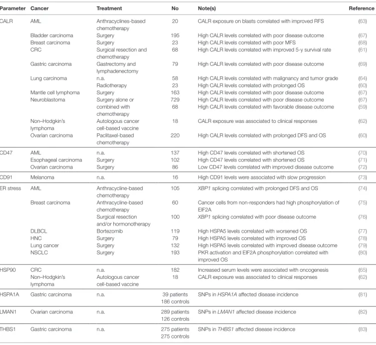
TABLe 1 | Clinical studies assessing the prognostic and predictive value of iCD-associated CALR and HSP signaling in cancer patients.
Parameter Cancer Treatment No Note(s) Reference
CALR AML Anthracyclines-based
chemotherapy
20 CALR exposure on blasts correlated with improved RFS (63) Bladder carcinoma Surgery 195 High CALR levels correlated with poor disease outcome (67) Breast carcinoma Surgery 23 High CALR levels correlated with poor MFS (68) CRC Surgical resection and
chemotherapy
68 High CALR levels correlated with improved 5-y survival rate (61) Gastric carcinoma Gastrectomy and
lymphadenectomy
79 High CALR levels correlated with poor disease outcome (69) Lung carcinoma n.a. 58 High CALR levels correlated with malignancy and tumor grade (64) Radiotherapy 23 High CALR levels correlated with prolonged OS (60) Mantle cell lymphoma Surgery 163 High CALR levels correlated with poor disease outcome (67) Neuroblastoma Surgery alone or
combined with chemotherapy
729 High CALR levels correlated with poor disease outcome (67) 68 High CALR levels correlated with favorable disease outcome (59) Non–Hodgkin’s
lymphoma
Autologous cancer cell-based vaccine
18 CALR exposure was associated to clinical responses (62) Ovarian carcinoma Paclitaxel-based
chemotherapy
220 High CALR levels correlated with prolonged DFS and OS (60)
CD47 AML n.a. 137 High CD47 levels correlated with shortened OS (70)
Esophageal carcinoma Surgery 102 High CD47 levels correlated with shortened OS (71) Ovarian carcinoma Surgery 86 Low CD47 levels correlated with improved disease outcome (72) CD91 Melanoma n.a. 16 High CD91 levels were associated with slow progression (73) ER stress AML Anthracycline-based
chemotherapy
105 XBP1 splicing correlated with prolonged DFS and OS (74) Breast carcinoma Anthracycline-based
chemotherapy
60 Cancer cells from non-responders had high phosphorylation of EIF2A
(75) Surgical resection
and/or hormonotherapy
100 XBP1 splicing correlated with poor disease outcome (76)
DLBCL Bortezomib 119 High HSPA5 levels correlated with worsened OS (77)
HNC Surgery 79 High HSPA5 levels correlated with improved OS (78)
Lung cancer Surgery 132 High HSPA5 levels correlated with improved disease outcome (79) NSCLC Surgery 193 PKR activation and EIF2A phosphorylation correlated with
improved OS
(80)
HSP90 CRC n.a. 182 Increased serum levels were associated with oncogenesis (65)
Non–Hodgkin’s lymphoma
Autologous cancer cell-based vaccine
18 CALR exposure was associated to clinical responses (62) HSPA1A Gastric carcinoma n.a. 39 patients SNPs in HSPA1A affected disease incidence (81)
186 controls
LMAN1 Ovarian carcinoma n.a. 289 patients SNPs in LMAN1 affected disease incidence (82) 126 controls
THBS1 Gastric carcinoma n.a. 275 patients SNPs in THBS1 affected disease incidence (83) 275 controls
AML, acute myeloid leukemia; CRC, colorectal carcinoma; DFS, disease-free survival; DLBCL, diffuse large B-cell lymphoma; ER, endoplasmic reticulum; HNC, head and neck cancer; ICD, immunogenic cell death; MFS, metastasis-free survival; NSCLC, non-small cell lung carcinoma; n.a., not applicable or not available; OS; overall survival; RFS, relapse-free survival; SNP, single nucleotide polymorphism.
cohort of 23 lung cancer patients and 220 ovarian cancer patients
treated with ICD inducers (i.e., radiotherapy and paclitaxel,
respectively) (
60
). Moreover, increased CALR expression by
can-cer cells has been associated with tumor infiltration by CD45RO
+memory T cells and improved 5-year overall survival amongst
68 subjects with Stage IIIB colorectal carcinoma (CRC) (
61
).
Elevated levels of HSP90 and CALR on the surface of neoplastic
cells have been associated with clinical responses amongst 18
patients with relapsed indolent non-Hodgkin‘s lymphoma treated
with an autologous cancer cell-based vaccine (
62
). Moreover,
CALR exposure by malignant blasts has been linked to prolonged
relapse-free (but not overall) survival in a cohort of 20 individuals
with acute myeloid leukemia (AML) (
63
). Of note, the blasts of
some of these patients exposed CALR spontaneously, and this
correlated not only with the degree of EIF2A phosphorylation
in malignant cells, but also with the ability of autologous T cells
to secrete IFNγ on stimulation (
63
). Along similar lines, healthy
individuals have been shown to differ from lung carcinoma
patients with respect to the circulating levels of soluble CALR,
as well as to the amount of CALR expressed on the surface of
pulmonary (normal versus malignant) cells (
64
). Moreover,
increased concentrations of soluble HSP90 have been detected
in the serum of CRC patients (n = 172) as compared to healthy
individuals (n = 10) (
65
). Interestingly, soluble HSP90 appears
to activate cancer cell-intrinsic signaling pathways that promote
disease progression (
65
,
66
). These data indicate that cancer cells
expose and/or shed CALR as well as HSPs even in the absence
of chemotherapy (at least to some degree), possibly as a result of
oncogenic stress and/or adverse microenvironmental conditions.
Moreover, they suggest that membrane-bound CALR and HSPs
have a different biological activity than their soluble counterparts.
Apparently at odds with the abovementioned clinical findings,
total CALR levels have been positively associated with accelerated
disease progression and poor outcome in a cohort of 79 gastric
cancer patients (
69
), in 23 women with breast carcinoma upon
surgery (
68
), as well in large cohorts of neuroblastoma (n = 729),
bladder carcinoma (n = 195) and mantle cell lymphoma (n = 163)
patients, irrespective of treatment type (
67
). Moreover, CALR
expression by malignant cells failed to affect overall survival in 88
patients with esophageal squamous cell carcinoma treated with
neo-adjuvant chemoradiotherapy and surgical resection (
84
).
These results may reflect the intracellular functions of CALR in
the preservation of reticular homeostasis, which is particularly
important for malignant cells owing to their highly accelerated
anabolic metabolism (
85
), or the fact that CALR exposure is
generally associated with an increased expression of CD47, a very
potent anti-phagocytic signal (
67
).
The phosphorylation of EIF2A as well as the activation
of eukaryotic translation initiation factor 2-alpha kinase 2
(EIF2AK2, best known as PKR) have been associated with
favorable disease outcome in a cohort of 193 non-small cell lung
carcinoma (NSCLC) patients (
80
). On the contrary, elevated
degrees of EIF2A phosphorylation in neoplastic cells have been
correlated with nuclear size (a surrogate marker of DNA content),
preferential tumor infiltration by T
REGcells, and poor disease
outcome in a cohort of 60 breast carcinoma patients treated with
anthracycline-based chemotherapy and tested longitudinally (
75
).
Other manifestations on an ongoing UPR have been ascribed with
prognostic or predictive value, including (but not limited to): (1)
the expression levels of the ER chaperone heat shock 70 kDa
pro-tein 5 (HSPA5, best known as GRP78), as demonstrated in cohorts
of 132 lung carcinoma patients (
79
), 79 individuals with head and
neck cancer (
78
) and 119 patients with diffuse large B-cell
lym-phoma treated with the proteasome inhibitor bortezomib (which
is a bona fide ICD inducer) (
77
); and (2) the splicing of X-box
binding protein 1 (XBP1) (
48
), as demonstrated in a cohort of
105 AML patients tested at diagnosis (
74
). Of note, both CALR
and GRP78 expression levels are also indirect manifestations of
the activation of another branch of the ER stress response, i.e., the
derepression of activating transcription factor 6 (ATF6) (
74
,
86
).
Finally, some studies have associated markers of an ongoing UPR
with dismal disease outcome. For instance, Davies and colleagues
have linked low levels of unspliced XBP1 as well as a high spliced/
unspliced XBP1 ratio with poor disease outcome in 100 primary
breast carcinoma patients treated with adjuvant hormonal therapy
(
76
). The apparent discrepancy in these observations may reflect
the differential reliance of distinct tumor types (or similar tumors
at distinct stages of progression) on the ER stress response for
survival in adverse microenvironment conditions (
87
).
Other processes and parameters linked to CALR and/or HSP
exposure and their immunostimulatory effects have been shown
to influence disease outcome in cancer patients. For instance, high
CD47 levels have been reported to constitute an independent
nega-tive prognostic factor in cohorts of 86 patients with ovarian clear
cell carcinoma (
72
), 102 individuals with esophageal squamous
cell carcinoma (
71
), and 137 subjects with karyotypically normal
AML (
70
). Along similar lines, the monocytes of 8 advanced
melanoma patients progressing in an unusually slow fashion have
been found to express increased amounts of CD91 as compared
to those of 8 patients progressing normally (
73
). Moreover,
single nucleotide polymorphisms (SNPs) affecting HSPA1A have
been linked to an increased incidence of gastric carcinoma (as
determined in a cohort of 39 patients and 186 controls) (
81
), a
SNP affecting THBS1 has been correlated with gastric cancer
occurrence and progression in a cohort of 275 patients and 275
healthy individuals (
83
), while a SNP in LMAN1 as well as the
consequent decrease in LMAN1 levels appear to be associated
with an increased risk for ovarian carcinoma (as determined in a
cohort of 289 women seen in gynecologic oncology practice and
126 healthy volunteers) (
82
).
The robust immunostimulatory activity of HSPs has been
har-nessed to develop various anticancer vaccines that are nowadays
in clinical development. These preparations generally consist in
HSP-enriched (autologous or heterologous) cancer cell lysates
that are administered directly to patients, in the presence of
adequate immunological adjuvants (
88
,
89
). The most common
of these approaches relies on heat shock protein 90 kDa beta
(Grp94), member 1 (HSP90B1, best known as GP96) and is often
referred to as HSPPC-96 (Oncophage
®or Vitespen
®) (
90
). So far,
the safety and clinical profile of HSPPC-96 have been tested in
cohorts of patients with metastatic melanoma (n = 36–322) (
91
–
94
), CRC (n = 29) (
95
), non-Hodgkin’s lymphoma (n = 20) (
96
);
pancreatic adenocarcinoma (n
= 10) (
97
), metastatic renal cell
carcinoma (n = 84–409) (
98
,
99
), glioma (n = 12) (
100
), recurrent
glioblastoma (n = 41) (
101
), and assorted advanced malignancies
(n = 16) (
102
). These studies demonstrate that the administration
of HSPPC-96 to cancer patients is safe and is generally associated
with markers of immunostimulation. However, most often such
effects are weak and unable to mediate long-term therapeutic
activity (
99
). Thus, further studies are required for translating
the well-established ability of HSPs to stimulate the priming of
TAA-specific immune responses into a therapeutic reality.
Taken together, these clinical observations suggest that CALR,
HSPs and various processes associated with their exposure,
secre-tion and signaling funcsecre-tions may have prognostic, predictive and
therapeutic value.
Type i iFN and TLR3 Signaling
Cancer cells responding to anthracyclines secrete type I IFNs
as a consequence of TLR3 activation (
39
), and this is required
for cell death to initiate adaptive immunity (
39
). By binding to
homodimeric or heterodimeric receptors expressed on several
immune effector cells, type I IFNs mediate multipronged
immunostimulatory effects (
40
). In particular, type I IFNs
promote cross-priming (
103
), boost the cytotoxic functions of
CTLs and NK cells (
104
), and increase the survival of memory
CTLs (
105
). Moreover, type I IFNs can protect antigen-activated
CD8
+CTLs from elimination by NK cells (
106
,
107
), trigger the
secretion of pro-inflammatory mediators by macrophages (
108
),
and counteract the immunosuppressive functions of T
REGcells
(
109
). Besides such immunostimulatory effects, type I IFNs can
ignite a cancer cell-intrinsic signal transduction pathway leading,
amongst various effects, to the synthesis of the chemotactic factor
chemokine (C–X–C motif) ligand 10 (CXCL10) (
39
). Indeed, at
odds with their wild-type counterparts, Ifnar1
−/
−cancer cells
suc-cumbing to anthracyclines are unable to prime adaptive immune
responses, even upon inoculation in wild-type hosts (
39
). Thus,
type I IFN signaling in cancer cells appears to be critical for
anthracycline-induced cell death to be perceived as
immuno-genic (
39
). Conversely, the efficacy of other immunotherapeutic
agents such as the TLR7 agonist imiquimod requires type I IFN
signaling in the host (
110
).
So far, only a few studies addressed the prognostic or predictive
value of parameters reflecting the proficiency or activation status
of TLR3 or type I IFN signaling (Table 2). High expression levels
of TLR3 and/or toll-like receptor adaptor molecule 1 (TICAM1,
a component of the TLR3 signaling apparatus best known as
TRIF) have been associated with improved disease outcome in
two cohorts of 85 and 172 subjects with hepatocellular carcinoma
(HCC) (
111
,
112
), as well as amongst 99 patients with
neuroblas-toma (
113
). Along similar lines, TLR3 expression levels have been
shown to predict the response of 194 breast carcinoma patients
treated with adjuvant radiotherapy plus a TLR3 agonist (
114
). SNPs
affecting TLR3 have been shown to influence prognosis in cohorts
of 582 patients with CRC, especially among untreated individuals
TABLe 2 | Clinical studies assessing the prognostic and predictive value of TLR3 status and type i iFN signaling in cancer patients.
Parameter Cancer Treatment No Note(s) Reference
IFNAR1 CRC n.a. 1327 patients A SNP in IFNAR1 was linked to increased risk for oncogenesis (122) 758 controls
Glioma n.a. 304 A SNP in IFNAR1 was shown to affect patient OS (123)
TLR3 Breast carcinoma n.a. 102 patients A SNP in TLR3 was linked to increased risk for oncogenesis (118) 72 controls
polyA:U plus radiotherapy 194 High TLR3 levels predicted clinical responses to therapy (114) Cervical carcinoma n.a. 130 patients A SNP in TLR3 was linked to increased risk for oncogenesis (117)
200 controls
CRC n.a. 582 SNPs in TLR3 were shown to influence disease outcome (115)
2309 patients SNPs in TLR3 were linked to increased disease incidence (121) 2915 controls
HCC n.a. 466 patients A SNP in TLR3 was linked to increased risk for oncogenesis (120) 482 controls
172 High TLR3 levels correlated with prolonged OS (111) Surgery 85 High TLR3 levels correlated with prolonged OS (112) Neuroblastoma n.a. 99 High TLR3 levels correlated with favorable disease outcome (113) NSCLC Surgery 568 SNPs in TLR3 were shown to influence disease outcome (116) Oral squamous cell
carcinoma
n.a. 93 patients SNPs in TLR3 were linked to increased risk for oncogenesis (119) 104 controls
240 patients A SNP in TLR4 was linked to increased risk for oncogenesis (124) 223 controls
TRIF HCC Surgery 85 High TRIF levels correlated with prolonged OS (112)
Type I IFN Breast carcinoma Anthracycline-based chemotherapy
50 A type I IFN-related signature predicted improved disease outcome
(39)
CRC n.a. 483 A SNP in IFNA7 was shown to affect patient OS (122)
Glioma n.a. 304 A SNP in IFNA8 was shown to affect patient OS (123)
CRC, colorectal carcinoma; HCC, hepatocellular carcinoma; NSCLC, non-small cell lung carcinoma; n.a., not applicable or not available; OS; overall survival; SNP, single nucleotide polymorphism.
with Stage II disease (
115
) and 568 NSCLC patients (
116
). Along
similar lines, TLR3 SNPs have been associated with an altered risk
for cervical cancer amongst 330 Tunisian women (
117
), breast
carcinoma amongst 174 African-American women (
118
), oral
squamous cell carcinoma amongst 197 individuals (
119
) HCC
amongst 948 subjects (
120
), and CRC amongst more than 5,000
individuals (
121
). A type I IFN-related transcription signature
centered around the expression of MX dynamin-like GTPase 1
(MX1) has been shown to predict the likelihood of 50 breast
car-cinoma patients to respond to neo-adjuvant anthracycline-based
chemotherapy (
39
). Moreover, SNPs affecting interferon (alpha,
beta and omega) receptor 1 (IFNAR1) have been associated with
an increased risk for the development of CRC amongst 2085
indi-viduals (
122
), as well as with significantly reduced overall survival
in a cohort of 304 glioma patients (
123
). Similar results have been
obtained for SNPs affecting the genes coding for two variants of
IFNα (i.e., IFNA7 and IFNA8) (
122
,
123
).
The results of these studies suggest that monitoring biomarkers
of TLR3 and type I IFN signaling may not only have prognostic/
predictive relevance for cancer patients, but also inform on the
risk for cancer development in healthy subjects. Of note,
recom-binant IFN-α2a (Roferon-A
®) is approved by the US Food and
Drug Administration and other regulatory agencies worldwide
for use in subjects with hairy cell leukemia and Philadelphia
chromosome-positive chronic myelogenous leukemia upon
minimal pretreatment, while recombinant IFN-α2b (Intron A
®)
is currently employed for the treatment of hairy cell leukemia,
AIDS-related Kaposi’s sarcoma, follicular lymphoma, multiple
myeloma, melanoma, condyloma acuminata and cervical
intraepithelial neoplasms.(
125
,
126
) It remains to be determined
to which extent, if any, the therapeutic efficacy of type I IFNs
reflects their ability to promote the initiation of adaptive immune
responses against dying cancer cells.
extracellular ATP and Autophagy
ATP is secreted during ICD through a mechanism that involves
pannexin 1 (PANX1) channels and lysosomal exocytosis (
127
,
128
). Importantly, autophagy is required for cancer cells
suc-cumbing to anthracyclines to release ATP in immunostimulatory
amounts (
42
,
129
,
130
). Thus, the ability of anthracyclines to cause
bona fide ICD is lost when cancer cells are rendered
autophagy-deficient by genetic manipulations or engineered to overexpress
ectonucleoside triphosphate diphosphohydrolase 1 (ENTPD1,
best known as CD39), an enzyme that degrades extracellular ATP
(
42
,
129
). In line with this notion, the administration of CD39
inhibitors or CD39-neutralizing monoclonal antibodies
report-edly relieves tumor-mediated immunosuppression (
131
), and (at
least in some models) allows autophagy-deficient cells treated
with anthracyclines to elicit normal immune responses upon
inoculation in immunocompetent mice (
42
,
129
). Extracellular
ATP exerts immunostimulatory functions via at least three
mechanistically distinct pathways: (1) by promoting the
recruit-ment of APCs or APC precursors to sites of cell death, upon
bind-ing to purinergic receptor P2Y, G-protein coupled, 2 (P2RY2)
(
132
–
134
); (2) by activating the so-called NLRP3 inflammasome
and hence triggering the secretion of pro-inflammatory IL-1β
(
135
,
136
), an effect that relies on purinergic receptor P2X, ligand
gated ion channel, 7 (
41
); and (3) by boosting the proliferation
and cytotoxic activity of NK cells (
26
). Notably, extracellular ATP
is sequentially metabolized by CD39 and 5′-nucleotidase, ecto
(NT5E, best known as CD73) into ADP, AMP and adenosine,
the latter of which has robust immunosuppressive effects (
137
).
Accumulating clinical evidence ascribes to parameters linked
to the capacity of cancer cells to recruit and activate immune
effectors (through extracellular ATP) a prognostic or predictive
value for cancer patients (Table 3). A SNP compromising the
function of P2RX7 has been associated with decreased
time-to-metastasis in a cohort of 225 breast carcinoma patients treated
with adjuvant anthracycline-based chemotherapy (
41
), with
worsened clinicopathological parameters amongst 121 subjects
with papillary thyroid cancer (
138
), and with an increased risk
for the development of chronic lymphocytic leukemia (CLL), as
determined in a cohort of 40 patients and 46 age-matched healthy
individuals (
139
). Contrasting with these latter findings, however,
the same SNP has been associated with increased overall survival
in a cohort of 170 subjects with CLL (
140
), or found to have
no correlation with disease incidence and/or outcome in
inde-pendent cohorts of 144 CLL patients and 348 healthy controls
(
141
), 121 individuals with CLL (
142
) 111 CLL patients and 97
controls (
143
), and 136 subjects with multiple myeloma (
144
).
These apparently discrepant observations may reflect the cancer
cell-intrinsic functions of P2RX7, which is known to control
proliferation and regulated cell death (
145
). Of note, increased
P2RY2 mRNA levels have also been detected in gastric cancer
biopsies from 14 patients (as compared to the adjacent healthy
mucosa) (
146
), but these findings do not allow to determine
whether gastric neoplasms were infiltrated by P2RY2
+immune
cells or whether they overexpressed P2RY2.
Further corroborating the advantage conferred to malignant
cells by an increased ability to convert immunostimulatory
extracellular ATP into immunosuppressive AMP and adenosine,
several studies ascribed a negative prognostic or predictive value
to increased CD39 or CD73 levels. For instance, elevated amounts
of CD39 and CD73 have been detected in 29 endometrial tumor
samples as compared to the adjacent non-malignant tissues, and
expression levels correlated with tumor grade (
152
). Along similar
lines, CD39 (but not CD73) levels on the surface of CD4
+and CD8
+T cells have been shown to positively correlate with disease stage in
two independent cohorts of 34 and 62 patients with CLL (
150
,
151
),
while CD73 downregulation has been associated with prolonged
disease-free survival amongst 500 individuals with glioblastoma
(
154
). At stark contrast with these findings, high levels of CD39
mRNA have been linked to improved disease outcome in a cohort
of 28 pancreatic cancer patients treated with surgery (
153
). The
reasons underlying this discrepancy have not yet been clarified.
Of note, quantifying functional autophagy in tissue biopsies
is rather complex, because most autophagic markers accumulate
both when the autophagic flux is increased and when lysosomal
degradation is blocked (
155
). Moreover, autophagy often serves
a dual role in the course of tumor progression: (1) on the one
hand it favors the survival of cancer cells exposed to adverse
microenvironmental conditions (including nutritional, metabolic
and therapeutic cues); (2) on the other hand, it is required for
ICD-associated ATP secretion and for the elicitation of robust
TAA-targeting immune responses (
130
,
156
,
157
). Notwithstanding
these caveats, immunohistochemistry has been employed to study
the prognostic or predictive value of autophagic markers such as
the expression and lipidation of microtubule-associated protein 1
light chain 3 (MAP1LC3, best known as LC3) (
158
), with mixed
results. For instance, LC3 expression has been associated with
prolonged overall survival in a cohort of 190 HCC patients (
148
),
but with lymph node involvement and high TNM score amongst
79 individuals with head and neck cancer (
78
). Along similar lines,
reduced expression of beclin 1 (BECN1), a key component of the
molecular machinery for autophagy, has been associated with poor
prognosis in two independent cohorts of 1067 and 1992 breast
carcinoma patients (
147
), but with improved disease outcome in a
cohort of 73 patients with pancreatic cancer (
149
). These are only
two examples of an abundant scientific literature correlating the
expression of autophagy proteins in biopsies from patients affected
with virtually all types of malignancies to clinicopathological
features and/or markers of disease progression. The development
of assays to monitor the functionality of the autophagic apparatus
in clinical samples is urgently awaited to properly assess the
prog-nostic and predictive value of autophagy for cancer patients.
HMGB1 and Cell Death
According to current models, HMGB1 gets released in the course
of cell death passively, upon the breakdown of the nuclear and
plasma membrane (
145
,
159
). Thus, besides differences in
expres-sion level, the extent of HMGB1 release generally correlates with
the degree of cell death (
160
). However, changes in the oxidation
status of extracellular HMGB1 have been suggested to
dramati-cally alter its biological activity (
161
–
163
). Indeed, while reduced
HMGB1 efficiently dimerizes with CXCL12 and mediate potent
chemotactic functions upon binding to chemokine (C–X–C
motif) receptor 4 (CXCR4) (
164
,
165
), its oxidized counterpart
fails to do so (
162
). Rather, oxidized HMGB1 signal via TLR2,
TLR4 and advanced glycosylation end product-specific
recep-tor (AGER, best known as RAGE) to stimulate the production
of pro-inflammatory cytokines (
162
,
166
–
168
). In addition,
TLR4 signaling promotes cross-priming by inhibiting the
fusion of antigen-containing endosomes with lysosomes (
169
).
Interestingly, HMGB1 also binds to TLR9 (
170
) and hepatitis
A virus cellular receptor 2 (HAVCR2, best known as TIM-3)
(
171
), in particular when complexed with DNA. However, while
TLR9 promotes cytokine secretion by plasmacytoid DCs and B
cells (
170
), TIM-3 signaling blunts the ability of DCs to respond
efficiently to inflammatory stimuli (
171
). Thus, extracellular
HMGB1 mediates multipronged and context-dependent
immu-nomodulatory functions.
Various clinical studies indicate that monitoring parameters
linked to HMGB1 release and signaling may convey prognostic
or predictive information for cancer patients (Table 4). High
expression levels of HMGB1 in malignant cells have been shown
to correlate with improved overall survival in 88 patients with
TABLe 3 | Clinical studies assessing the prognostic and predictive value of ATP release and extracellular ATP signaling in cancer patients.
Parameter Cancer Treatment No Note(s) Reference
Autophagy Breast carcinoma n.a. 1067 patients Low BECN1 levels correlated with worsened disease outcome (147) 1992 patients
HCC Surgery 190 High LC3 levels correlated with prolonged OS (148)
HNC Surgery 79 High LC3 levels correlated with node involvement and TNM score (78) Pancreatic carcinoma Surgery 73 High levels of BECN1 and other autophagy-related proteins
correlated with poor outcome
(149) CD39 CLL n.a. 34 patients High CD39 levels on T cells correlated with late disease (150)
31 controls
62 High CD39 levels on T cells correlated with late disease (151) Endometrial cancer Surgery 29 High CD39 levels correlated with tumor grade (152) Pancreatic carcinoma Surgery 28 High CD39 levels were linked to improved disease outcome (153) CD73 Endometrial cancer Surgery 29 High CD73 levels correlated with tumor grade (152) Glioblastoma n.a. 500 CD73 downregulation was associated with improved DFS (154) P2RX7 Breast carcinoma Anthracycline-based
chemotherapy
225 A SNP in P2RX7 was linked to shortened MFS (41) CLL n.a. 40 patients A SNP in P2RX7 was linked to increased risk for oncogenesis (139)
46 controls
144 patients Lack of correlation between P2RX7 status and disease incidence (141) 348 controls
111 patients Lack of correlation between P2RX7 status and disease incidence (143) 97 controls
170 A SNP in P2RX7 was associated to increased OS (140) 121 Lack of correlation between P2RX7 status and pathological features (142) Multiple myeloma n.a. 136 patients Lack of correlation between P2RX7 status and disease incidence (144)
95 controls
Papillary thyroid cancer n.a. 121 A SNP in P2RX7 was linked to poor clinicopathological features (138) P2RY2 Gastric cancer n.a. 14 patients Increased expression of P2RY2 in malignant cells (146) CLL, chronic lymphocytic leukemia; DFS, disease-free survival; HCC, hepatocellular carcinoma; HNC, head and neck cancer; MFS, metastasis-free survival; n.a., not applicable or not available; OS; overall survival; SNP, single nucleotide polymorphism.
TABLe 4 | Clinical studies assessing the prognostic and predictive value of HMGB1 release and extracellular HMGB1 signaling in cancer patients.
Parameter Cancer Treatment No Note(s) Reference
CASP3 Endometrial carcinoma
n.a. 1028 patients A SNP in CASP3 was linked to increased risk for oncogenesis (182) 1003 controls
CASP7 Endometrial carcinoma
n.a. 1028 patients SNPs in CASP7 were linked to increased risk for oncogenesis (182) 1003 controls
CASP9 CRC n.a. 402 patients SNPs in CASP9 were linked to decreased risk for oncogenesis and improved disease outcome
(183) 480 controls
HMGB1 Bladder carcinoma n.a. 164 High HMGB1 levels correlated to worsened disease outcome (175) Breast
carcinoma
Anthracycline-based chemotherapy
232 Loss of nuclear HMGB1 positively correlated with tumor size (173) 41 Increases in circulating HMGB1 were linked to clinical response (184) CRC n.a. 219 patients High levels of serum HMGB1 correlated with disease incidence (185)
75 controls
n.a. 192 High HMGB1 levels correlated with worsened disease outcome (177) Radioembolization
therapy
49 High levels of serum HMGB1 correlated with decreased OS (186) Surgery 72 Co-expression of HMGB1 in the nucleus and in the cytoplasm of
malignant cells was linked to worsened 5-year survival rate
(174) Esophageal
carcinoma
Chemoradiotherapy and surgery
88 High HMGB1 levels correlated with improved OS (84) Gastric
adenocarcinoma
Surgery 76 High HMGB1 levels in malignant cells correlated with improved OS (172) HCC n.a. 208 High HMGB1 levels correlated with worsened disease outcome (179) 161 High HMGB1 levels correlated with worsened disease outcome (178) HNC n.a. 71 patients High levels of serum HMGB1 correlated with disease progression (187)
50 controls
103 High HMGB1 levels correlated with worsened disease outcome (180) Malignant
mesothelioma
n.a. 61 patients High levels of serum HMGB1 correlated with disease incidence (188) 45 controls
Nasopharyngeal carcinoma
n.a. 166 High HMGB1 levels correlated with worsened disease outcome (176) Pancreatic
carcinoma
Multicomponent chemotherapy
78 High circulating HMGB1 correlated with poor therapy response (189) n.a. 70 High levels of serum HMGB1 correlated with decreased OS (190) Prostate
carcinoma
n.a. 85 High HMGB1 levels correlated with worsened disease outcome (181) Solid tumors Virotherapy 17 Increases in circulating HMGB1 levels were linked to clinical response (191) 202 Increases in circulating HMGB1 levels were linked to clinical response (192) MYD88 CRC Surgery 108 High MYD88 levels correlated with shortened DFS and OS (193)
Lymphoma Conventional chemotherapy
29 MYD88 mutations were involved in the pathogenesis of the disease (194) Ovarian carcinoma Surgery 123 High MYD88 levels correlated with worsened disease outcome (195) 109 High MYD88 levels correlated with shortened DFS and OS (196) RAGE Breast carcinoma n.a. 509 patients A SNP in AGER was linked to increased risk for oncogenesis (197)
504 controls
120 patients High levels of circulating RAGE correlated with advanced disease stage but improved outcome
(198) 92 controls
Gastric carcinoma Surgery 180 High RAGE levels were associated with worsened disease outcome (199) HCC Transarterial
chemoembolization
71 High levels of circulating RAGE correlated with clinical response (200) NSCLC Platinum-based
chemotherapy
562 patients SNPs in AGER were linked to increased risk for oncogenesis and differential clinical response
(201) 764 controls
Ovarian carcinoma n.a. 190 patients A SNP in AGER was linked to increased risk for oncogenesis (202) 210 controls
TLR2 CRC n.a. 2309 patients SNPs in TLR2 were associated with decreased 5-year survival rate (121) 2915 controls
Gastric carcinoma n.a. 289 patients A SNP in TLR2 was linked to increased risk for oncogenesis (203) 400 controls
HCC n.a. 211 patients SNPs in TLR2 were linked to increased risk for oncogenesis (204) 232 controls
Lymphoma n.a. 710 patients A SNP in TLR2 was linked to increased risk for oncogenesis (205) 710 controls
Prostate carcinoma
n.a. 195 patients A SNP in TLR2 was linked to increased risk for oncogenesis (206) 250 controls
Parameter Cancer Treatment No Note(s) Reference
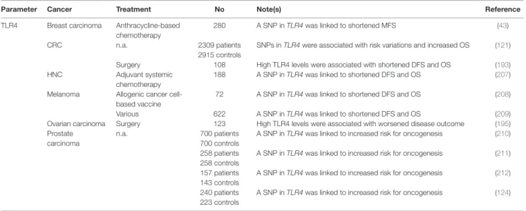
TLR4 Breast carcinoma Anthracycline-based chemotherapy
280 A SNP in TLR4 was linked to shortened MFS (43) CRC n.a. 2309 patients SNPs in TLR4 were associated with risk variations and increased OS (121)
2915 controls
Surgery 108 High TLR4 levels were associated with shortened DFS and OS (193) HNC Adjuvant systemic
chemotherapy
188 A SNP in TLR4 was linked to shortened DFS and OS (207) Melanoma Allogenic cancer
cell-based vaccine
72 A SNP in TLR4 was linked to shortened DFS and OS (208) Various 622 A SNP in TLR4 was linked to shortened DFS and OS (209) Ovarian carcinoma Surgery 123 High TLR4 levels were associated with worsened disease outcome (195) Prostate
carcinoma
n.a. 700 patients A SNP in TLR4 was linked to increased risk for oncogenesis (210) 700 controls
258 patients A SNP in TLR4 was linked to increased risk for oncogenesis (211) 258 controls
157 patients A SNP in TLR4 was linked to increased risk for oncogenesis (212) 143 controls
240 patients A SNP in TLR4 was linked to increased risk for oncogenesis (124) 223 controls
CRC, colorectal carcinoma; DFS, disease-free survival; HCC, hepatocellular carcinoma; HNC, head and neck cancer; MFS, metastasis-free survival; NSCLC, non-small cell lung carcinoma; n.a., not applicable or not available; OS; overall survival; RFS, relapse-free survival; SNP, single nucleotide polymorphism.
TABLe 4 | Continued
esophageal squamous cell carcinoma subjected to neo-adjuvant
chemoradiotherapy and surgical resection (
84
), as well as in 76
subjects with reseactable gastric adenocarcinoma (
172
). In a
cohort of 232 breast carcinoma patients treated with
anthracy-cline-based adjuvant chemotherapy, loss of nuclear HMGB1 has
been positively associated with tumor size (
173
). Along similar
lines, the co-expression of HMGB1 in the nucleus and in the
cytoplasm of malignant cells has been shown to inversely
cor-relate with tumor infiltration by CD45RO
+memory T cells and
5-year survival rate in 72 individuals with Stage IIIB CRC (
174
).
Finally, HMGB1 overexpression has been shown to correlate with
advanced clinical stage or decreased disease-free and/or overall
survival amongst 164 patients with bladder carcinoma (
175
),
166 individuals with nasopharyngeal carcinoma (
176
), 192 CRC
patients (
177
), 208 and 161 individuals with HCC (
178
,
179
), 103
subjets with head and neck squamous cell carcinoma (
180
), as
well as 85 patients with prostate cancer (
181
).
Notably, circulating HMGB1 and RAGE levels have been
intensively investigated for their predictive or prognostic value.
Elevations of HMGB1 in the serum have been correlated with
incidence, progression or unfavorable disease outcome in cohorts
of 49 individuals with CRC, or 219 CRC patients and 75 healthy
controls (
185
,
186
), 70 individuals with pancreatic
adenocarci-noma (
190
), 71 laryngeal squamous cell carcinoma patients and
50 healthy controls (
187
), 61 subjects with malignant pleural
mesothelioma (
188
), and 78 pancreatic carcinoma patients
(
189
). Conversely, a treatment-related increase in the circulating
levels of HGMB1 has been associated with pathological complete
response or partial remission amongst 41 breast carcinoma
patients treated with neo-adjuvant chemotherapy based on
epirubicin (an ICD inducer) (
184
), as well as amongst 17 and
202 subjects with chemotherapy-refractory tumors treated with
oncolytic virotherapy (
191
,
192
). High levels of RAGE in the
serum have been linked to advanced tumor stage but improved
clinical outcome amongst 120 patients with breast carcinoma
(
198
). Along similar lines, serum RAGE concentrations were
significantly higher in 32 individuals with HCC who favorably
responded to transarterial chemoembolization therapy than in
39 patients who progressed upon treatment (
200
).
Thus, in many (but not all) clinical settings high intratumoral
and circulating levels of HMGB1 have a negative prognostic or
predictive value. These findings may reflect the ability of some
tumors to retain HMGB1 in the course of stress response, the
intrinsic resistance of such tumors to the induction of cell death,
or the cancer cell-intrinsic functions of HMGB1 (
213
). In other
settings, however, circulating HMGB1 and RAGE levels appear
to reflect well the death of cancer cells exposed to immunogenic
treatment modalities (
184
,
191
,
192
). Possibly, the timing of
detec-tion plays a critical role in this setting, calling for the development
of optimized monitoring procedures.
SNPs in TLR2, TLR4 and AGER, as well as the circulating
levels of a soluble RAGE variant have been shown to affect cancer
susceptibility as well as disease outcome in several studies. In
par-ticular, TLR2 polymorphisms have been linked to an increased
risk for lymphoma (as determined in 710 patients and as many
healthy subjects) (
205
), gastric carcinoma (as assessed in 289
patients and more than 400 controls) (
203
), prostate carcinoma
(as investigated in 195 patients and 250 healthy individuals)
(
206
), HCC (as tested in 211 patients and 232 controls) (
204
), and
CRC (as assessed in 2,309 patients and 2,915 healthy individuals)
(
121
). Loss-of-function variants of TLR4 have been associated
with decreased time-to-metastasis amongst 280 women with
non-metastatic breast carcinoma treated with surgery followed
by anthracycline-based chemotherapy and local irradiation
(
43
), with reduced disease-free and overall survival amongst
188 head and neck cancer patients receiving adjuvant systemic
therapy (
207
), amongst 72 melanoma patients vaccinated with a
heat-shocked allogeneic melanoma cell line (
208
), and amongst
622 melanoma patients subjected to various treatment modalities
(
209
). Along similar lines, SNPs affecting TLR4 or AGER have
been linked to an increased risk for prostate cancer (as
deter-mined in multiple studies collectively testing more than 1,000
patients and as many age-matched controls) (
124
,
210
–
212
),
ovarian cancer (as assessed in a study testing 190 patients and 210
controls) (
202
), breast carcinoma (as investigated in 509 patients
and 504 healthy women) (
197
), CRC (as determined in a large
cohort encompassing 2,309 patients and 2,915 healthy
individu-als) (
121
), and NSCLC (as tested in 562 patients and 764 controls)
(
201
). Notably, this latter study also identified a specific AGER
SNP associated with a differential response of NSCLC patients to
chemotherapy (
201
).
Conversely, elevated expression levels of RAGE, TLR4 and/
or components of the TLR signaling machinery like myeloid
differentiation primary response gene 88 (MYD88) by malignant
tissues have been correlated with shortened disease-free and
overall survival in 2 cohorts of 109 and 123 ovarian carcinoma
patients subjected to surgery (
195
,
196
), in a cohort 108 subjects
with CRC (
193
), and amongst 180 individuals with gastric
carci-noma (
199
). Along similar lines, activating mutations in MYD88
have been linked to the pathogenesis of primary central nervous
system lymphomas (
194
). Most likely, these findings reflect the
advantage conferred to malignant cells by the expression of RAGE
and TLR4, which can activate robust pro-survival pathways via
NF-κB (
214
).
Finally, distinct SNPs affecting caspase-7 (CASP7) and one
affecting caspase-3 (CASP3) have been associated with an altered
risk for endometrial carcinoma (as investigated in a cohort of
1,028 patients and 1,003 healthy women) (
182
), whereas SNPs
affecting caspase-9 (CASP9) have been linked to reduced CRC
incidence or improved disease outcome (as determined in a
cohort of 402 patients and 480 healthy controls) (
183
). It remains
to be determined whether these SNPs truly compromise the
ability of cancer cells to emit DAMPs (and hence trigger
immu-nosurveillance mechanisms).
Other DAMPs
The abovementioned molecules and processes may constitute
only the tip of an iceberg, meaning that several other DAMPs
may contribute to the immunogenicity of cell death, at least in
some circumstances. These DAMPs include (but are not limited
to) various mitochondrial products like mtDNA, cardiolipin
and N-formylated peptides (
30
) as well as cytosolic proteins like
filamentous F-actin (
45
). Robust preclinical evidence implicates
mtDNA in the etiology of septic and non-septic shock as well as
in heart failure (
29
,
215
). Cytosolic, extra-cytosolic and
extracel-lular mtDNA molecules have indeed robust pro-inflammatory
effects as they trigger type I IFN synthesis via transmembrane
protein 173 (TM173, best known as STING) (
216
) or TLR9
activation (
215
). In line with this notion, circulating mtDNA
levels have been shown to reflect the degree of inflammation
and the extent of tissue damage in patients under maintenance
hemodialysis (
217
). Moreover, mtDNA concentrations in the
plasma of severe sepsis patients admitted to the emergency room
have been ascribed robust predictive value on disease outcome
(
218
). Upon binding to formyl peptide receptor 1 (FPR1),
N-formylated peptides reportedly attract neutrophils, stimulate
their degranulation, activate monocytes and favor the
produc-tion of pro-inflammatory cytokines (
219
–
223
). Cardiolipin, a
lipid that is specifically contained in the inner mitochondrial
membrane, binds CD1D on the surface of APC, thus endowing
them with the ability of priming CD1D-restricted γδ T cells
(
224
). Finally, F-actin becomes accessible upon disruption of
the plasma membrane and promotes the elicitation of adaptive
immune responses against dead cell-associated antigens by
bind-ing to C-type lectin domain family 9, member A (CLEC9A, best
known as DNGR1) on the surface of DCs (
45
). Studies
elucidat-ing the actual contribution of these DAMPs to ICD are urgently
awaited.
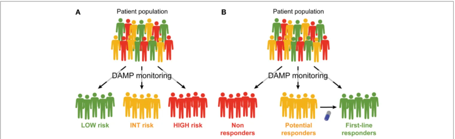
FiGURe 1 | Prognostic and predictive value of DAMPs and DAMP-associated processes. (A,B). Monitoring the emission of
damage-associated molecular patterns (DAMPs) or DAMP-damage-associated processes may have a multifaceted impact on the clinical management of cancer patients. First, it may allow for a prognostic assessment and permit the
stratification of patients in different risk groups (A). Second, it may allow for
the identification of patients who are intrinsically capable or uncapable to respond to a specific treatment, and amongst the latter, those who may benefit from combinatorial therapeutic approaches aimed at restoring normal DAMP signaling (B).