IX. COGNITIVE INFORMATION PROCESSING Prof. M. Eden Prof. J. Allen Prof. B. A. Blesser Prof. T. S. Huang Prof. F. F. Lee Prof. S. J. Mason J. Alba W. R. Bogan J. C. Borum J. E. Bowie B. E. Boyle R. M. Boza Becky J. Clark P. Coueignoux C. H. Cox J. L. Davis C. J. Drake J. R. Ellis, Jr. I. S. Englander A. M. Fakhr D. I. Falkenstein S. G. Finn
Academic and Research Staff Prof. W. F. Schreiber Prof. D. E. Troxel Prof. I. T. Young Dr. G. R. Granlund Dr. C. C. Jaffe Dr. D. M. Ozonoff Graduate Students T. Fukuda D. H. Harris D. W. Hartman M. E. Jernigan C. K. S. Kau J. W. Klovstad T. T. Kuklinski S-P. Kuo C. W. Lynn H. S. Magnuski L. C. Makowski P. L. Miller B. Minkow J. A. Myers D. O'Shaughnessy J. E. Ostrowski Dr. O. J. Tretiak F. X. Carroll M. Sharon Hunnicutt E. R. Jensen Mary J. Naus R. A. Piankian R. S. Putnam J. G. Richardson R. J. Shillman D. G. Sitler J. R. Sloan A. A. Smith R. D. Solomon H-m. D. Toong J. M. Tribolet B-K. Tye K. P. Wacks J. E. Walker J. A. Whealler, Jr. A. W. Wiegner H. M. Wolf son K. J. Wong
A. LOCALIZATION OF CELLULAR STRUCTURES
National Institutes of Health (Grant 5 P01 GM14940-07) I. T. Young, I. L. Paskowitz
1. Introduction
The localization of biological cells and their cellular components in a scanned image system is a problem of considerable interest in automated cytology. In this report we describe a technique that we have devised based on spectral transmission measurements and Boolean picture operations for the rapid identification of cell nuclei and white-cell cytoplasm in Wrights-Giemsa stained preparations. While its principal use is in the area of the automated white cell differential, we believe that the technique might have application to other areas of cytologic interest such as Pap smear screening and tissue analysis. In addition, a similar technique has been reported by Stark et al.1 for use in remote sensing of the Earth's resources.
2. Data Preparation
Blood samples are prepared by placing approximately 1 ml of either fresh or anti-coagulated blood, obtained through venipuncture, on a precleaned glass slide and
(IX. COGNITIVE INFORMATION PROCESSING)
spinning it for .75 s at 6000 rpm on a Platt Blood Centrifuge (Model 102M). While this technique of slide preparation is not required to implement the algorithm, it is a stan-dard part of our procedure, since it leads to slides with an evenly distributed monolayer of cells and a minimum of cell disruptions.2,3 The slide is then automatically stained on an Ames Hema-Tek slide stainer, by using a modified Wrights-Giemsa polychro-matic stain. After staining, the slide is placed in our television-microscope system for analysis. This system, diagrammed in Fig. IX-1, has the following components.
Leitz Ortholux microscope with multiple light sources, automated stage, microdensi-tometer, and multiple objectives/oculars.
High-resolution vidicon camera with 1029 lines per frame and 2:1 interlace. (Granger Associates system and RCA Vidicon Type 8507A).
Analog and digital circuitry to convert the video signal to a low rate (1 sample/hori-zontal line), quantized (6-bit) data stream.
Tempo-II 16-bit minicomputer assigned to control and process the video data. The Tempo is connected by a high-speed (1 mbit/s) serial link to a larger computer facility (PDP-9) so that the cell pictures obtained can be processed by our existing
soft-4
ware. The algorithms to be described here are, in fact, implemented on the PDP-9 facility. As indicated by the sampling density curve (Fig. IX-2) and the modulation trans-fer function curve (Fig. IX-3), the resolution of the system is limited only by the
resolution of light optics (approximately .25 4m in the visible spectrum). The 37 dB (measured) SNR of the video signal plus certain picture-averaging techniques that we have implemented provide the 6 bits of gray-level resolution.
3. Preliminary Cell Detection
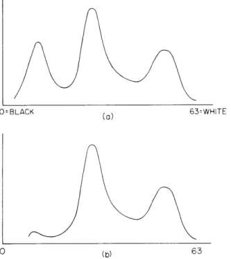
In the initial stage of processing we determine whether white cells are located in the current microscope field of view (100 X 100 Fm). This initial processing was devised by Bourk and has been discussed elsewhere5, 6; only a brief description will be given here. First, the specimen is illuminated with light that is spectrally filtered by an interfer-ence filter with a center wavelength of 570 nm and a bandwidth of 10 nm. The entire (100 Fm) field of view2 is then scanned and the histogram of brightness values calculated. A typical histogram for a field containing a white cell is shown in Fig. IX-4a, and for a field without a white cell in Fig. IX-4b. The difference in the histograms at this wave-length is the presence of dark nucleic material. Thus if no appreciable number of dark points occur in the picture the current field is rejected and a new one obtained. If, how-ever, there is dark material in the field (as represented by dark points), a clipping level is generated to separate all points of this darkness (or darker) from all other lighter points in the picture. Two special histograms, the row and column histograms, are then calculated to locate where the dark material in the picture occurred. These positional histograms are generated by looking along any given row (or column) and counting the
Fig. IX-1. Television-microscope scanning system. 0 0 200 400 600 800 MOG i400 16(
Fig. IX-2. Sample density curve of the television-microscope scanner.
63=WHITE
300 400 500 600 700 800 TV LINES
Fig. IX-3. Modulation transfer function of the television-microscope system.
Fig. IX-4.
(a) Histogram of field with white cell.
(b) Histogram of field without white
cell.
DRUMIORDS
(IX. COGNITIVE INFORMATION PROCESSING)
ROW
COLUMN
Fig. IX-5.
Row column histogram of cell
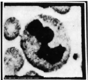
Fig. IX-6.
Typical field of blood
picture.
smear: located cell.
number of points that are fewer than the clipping level. As seen in Fig. IX-5, it is a
simple matter to determine the position of the nucleic material, given these two curves.
The coordinates of the nucleus are specified by a rectangular box (Xmin
,Xmax Ymin'
Ymax). We are not only interested in studying the nucleus (and its substructures); we are
interested in the cytoplasm and its substructures, too.
Therefore we need an algorithm
to locate all points in the picture that correspond to cytoplasm, as well as those points
that correspond to nucleus. The Wrights-Giemsa staining characteristics of the nucleus
cytoplasm and red cells when examined under light of various spectral contacts enable us
to secure this information.
4.
Algorithm for Component Location
The first stage in processing is to redefine the rectangular box that contains the
nucleus:
m
Xmin
(max
2min
3
1
x
min 2 = min 2 max
max
m
X
..
X
+(
max
min
X
X
max
m2
ax2
max
2
min
max
min
3
1
Y - -Y .i - Y Y
minm
m
2
min
2
max
mmax
mn
3
1
Y
- Y
+"
Y
=Ymax
-
Ym
max max 2 / max 2 min'
QPR No. 111
~---
~-
---
-~----~~hL
-~--~- ~ ~
(IX. COGNITIVE INFORMATION PROCESSING)
This effectively doubles the size of the box in both directions while the center of the box
is kept in the same location. We have found in practice that doubling the "field" size is
sufficient to guarantee the inclusion of all cytoplasmic material for human white blood
cells. An example of a typical field is shown in Fig. IX-6.
Next we record the picture and brightness histogram of all points in the new
rectan-gular box for three different spectral illuminations:
1.
570 nm - the "yellow" picture Y(x, y)
2.
530 nm -the "green" picture G(x, y)
3.
420 nm -the "blue" picture B(x,y).
At the sampling density at which this work is done (3 pels/±m) the storage requirements
for a typical 25 X 25 Lm picture and its histograms are 5827 words of storage in the
PDP-9 computer where pels (picture elements) are packed three to the 18-bit computer
word.
The characteristics of the three color pictures that are essential to the algorithm
are listed in Table IX-1. Our technique translates this linguistic description into a set of
Boolean operators on binary pictures.
Table IX-1.
Illumination
570 nm
530 nm
420 nm
Average intensities of different
wavelengths.
Nucleus
dark dark lightCytoplasm
gray
dark
light
cell structures at different
Red Cells
gray
dark
dark
Background
light
light
light
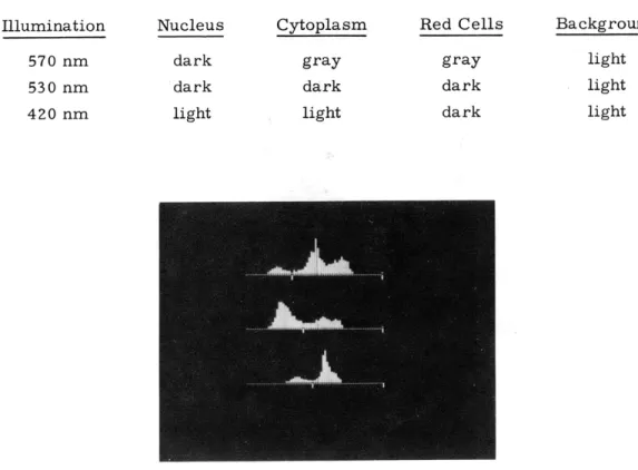
Fig. IX-7.
Typical set of histograms with clipping levels.
570 nm
530 nm
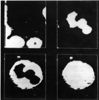
Fig. IX-8.
420 nm
Binary color pictures.Fig. IX-9.
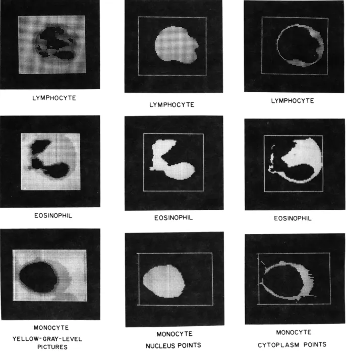
Four cells.LYMPHOCYTE LYMPHOCYTE EOSINOPHIL EOSINOPHIL MONOCYTE YELLOW-GRAY-LEVEL PICTURES MONOCYTE MONOCYTE
NUCLEUS POINTS CYTOPLASM POINTS
Fig. IX-10.
Cells with diverse staining characteristics.
EOSINOPHIL
(IX. COGNITIVE INFORMATION PROCESSING)
To first translate each 6-bit gray-level color picture into a binary color picture, a clipping level for that picture is extracted from the appropriate histogram. A typical
set of histograms for one cell is shown in Fig. IX-7, together with their attendant clipping levels. Thus, for example, in the yellow picture we set
Y(x,y) = 1 if Y(x, y) < Y-CLIP
Y(x,y) = 0 if Y(x,y) > Y-CLIP
and similarly for the green and blue pictures. Three resulting binary color pictures are shown in Fig. IX-8. Finally the three color pictures are combined by the following operators to generate cytoplasm points C(x, y), nucleus points N(x, y), red-cell
(erythro-cyte) points E(x, y), and total white-cell points W(x, y):
N(x, y) = Y(x, y)
C(x, y) = G(x, y) Y(x, y) B(x, y) E (x, y) =B (x, y)
W(x, y) = G(x, y) B(x, y) + Y(x, y) or
W(x, y) = G(x,y) B(x,y) (see Table IX-1).
Since the data are packed 3 pels per 18-bit computer word and the logical opera-tors operate on a bit-by-bit basis, it is convenient to define logical "1" to have octal value 77. This permits us to process 3 pels simultaneously, and considerably speeds up the computation. The final set of pictures produced by the algorithm is shown in Fig. IX-9.
To observe how this algorithm operates on cells whose staining characteristics are diverse, we have a set of cells (Fig. IX-10) with (a) the original gray-level (yellow) pic-ture, (b) the nucleus points, and (c) the cytoplasm points.
References
1. H. Stark, R. Barker, and D. Lee, "Some New Techniques for Processing Remotely Obtained Images by Self-Generated Spectral Masks," Appl. Opt. 11, 2540-2550
(1972).
2. M. Ingram and F. Minter, "Semiautomatic Preparation of Coverglass Blood Smears Using a Centrifugal Device," Am. J. Clin. Pathol. 51, 214 (1969).
3. I. T. Young, "The Measurement of Cell Adhesiveness in White Blood Cells," Proc. 24th ACEMB, Las Vegas, Nevada, October 31-November 4, 1971.
(IX. COGNITIVE INFORMATION PROCESSING)
4. J. E. Green and 0. J. Tretiak, "Modular Picture Processing Package (Mp3),"1 Quar-terly Progress Report No. 94, Research Laboratory of Electronics, M. I. T., July 15, 1969, pp. 261-279.
5. T. Bourk, "Automated Characterization of Leukocyte Nucleus Morphology," S. M. Thesis, Department of Electrical Engineering, M. I. T., 1970.
6. I. T. Young, "The Classification of White Blood Cells," IEEE Trans., Vol. BME-19, No. 4, pp. 291-298, July 1972.