HAL Id: hal-02094594
https://hal-amu.archives-ouvertes.fr/hal-02094594
Submitted on 22 May 2019
HAL is a multi-disciplinary open access
archive for the deposit and dissemination of sci-entific research documents, whether they are pub-lished or not. The documents may come from teaching and research institutions in France or abroad, or from public or private research centers.
L’archive ouverte pluridisciplinaire HAL, est destinée au dépôt et à la diffusion de documents scientifiques de niveau recherche, publiés ou non, émanant des établissements d’enseignement et de recherche français ou étrangers, des laboratoires publics ou privés.
Massive lateral transfer of genes encoding plant cell
wall-degrading enzymes to the mycoparasitic fungus
Trichoderma from its plant-associated hosts
Irina Druzhinina, Komal Chenthamara, Jian Zhang, Lea Atanasova, Dongqing
Yang, Youzhi Miao, Mohammad Rahimi, Marica Grujic, Feng Cai, Shadi
Pourmehdi, et al.
To cite this version:
Irina Druzhinina, Komal Chenthamara, Jian Zhang, Lea Atanasova, Dongqing Yang, et al.. Massive lateral transfer of genes encoding plant cell wall-degrading enzymes to the mycoparasitic fungus Tri-choderma from its plant-associated hosts. PLoS Genetics, Public Library of Science, 2018, 14 (4), pp.e1007322. �10.1371/journal.pgen.1007322�. �hal-02094594�
Massive lateral transfer of genes encoding
plant cell wall-degrading enzymes to the
mycoparasitic fungus Trichoderma from its
plant-associated hosts
Irina S. Druzhinina1*, Komal Chenthamara1, Jian Zhang2, Lea Atanasova1¤a, Dongqing Yang2, Youzhi Miao2, Mohammad J. Rahimi1, Marica Grujic1, Feng Cai1,2, Shadi Pourmehdi1, Kamariah Abu Salim3, Carina Pretzer1, Alexey G. Kopchinskiy1, Bernard Henrissat4,5,6, Alan Kuo7, Hope Hundley7, Mei Wang7, Andrea Aerts7, Asaf Salamov7, Anna Lipzen7, Kurt LaButti7, Kerrie Barry7, Igor V. Grigoriev7,8, Qirong Shen2
*, Christian P. Kubicek1¤b
1 Microbiology and Applied Genomics Group, Research Area Biochemical Technology, Institute of Chemical,
Environmental & Bioscience Engineering, TU Wien, Vienna, Austria, 2 Jiangsu Provincial Key Lab of Organic Solid Waste Utilization, Nanjing Agricultural University, Nanjing, China, 3 Environmental and Life Sciences, Universiti Brunei Darussalam, Bandar Seri Begawan, Brunei Darussalam, 4 Architecture et Fonction des Macromole´cules Biologiques, CNRS, Aix-Marseille Universite´, Marseille, France, 5 INRA, USC 1408 AFMB, Marseille, France, 6 Department of Biological Sciences, King Abdulaziz University, Jeddah, Saudi Arabia,
7 US Department of Energy Joint Genome Institute, Walnut Creek, CA, United States of America,
8 Department of Plant and Microbial Biology, University of California Berkeley, Berkeley, CA, United States of
America
¤a Current address: University of Natural Resources and Life Sciences–BOKU, Institute of Food Technology, Vienna, Austria
¤b Current address: Vienna, Austria
*irina.druzhinina@tuwien.ac.at(ISD);qirongshen@njau.edu.cn(QS)
Abstract
Unlike most other fungi, molds of the genus Trichoderma (Hypocreales, Ascomycota) are aggressive parasites of other fungi and efficient decomposers of plant biomass. Although nutritional shifts are common among hypocrealean fungi, there are no examples of such broad substrate versatility as that observed in Trichoderma. A phylogenomic analysis of 23 hypocrealean fungi (including nine Trichoderma spp. and the related Escovopsis weberi) revealed that the genus Trichoderma has evolved from an ancestor with limited cellulolytic capability that fed on either fungi or arthropods. The evolutionary analysis of Trichoderma genes encoding plant cell wall-degrading carbohydrate-active enzymes and auxiliary pro-teins (pcwdCAZome, 122 gene families) based on a gene tree / species tree reconciliation demonstrated that the formation of the genus was accompanied by an unprecedented extent of lateral gene transfer (LGT). Nearly one-half of the genes in Trichoderma pcwdCA-Zome (41%) were obtained via LGT from plant-associated filamentous fungi belonging to different classes of Ascomycota, while no LGT was observed from other potential donors. In addition to the ability to feed on unrelated fungi (such as Basidiomycota), we also showed that Trichoderma is capable of endoparasitism on a broad range of Ascomycota, including extant LGT donors. This phenomenon was not observed in E. weberi and rarely in other mycoparasitic hypocrealean fungi. Thus, our study suggests that LGT is linked to the ability a1111111111 a1111111111 a1111111111 a1111111111 a1111111111 OPEN ACCESS
Citation: Druzhinina IS, Chenthamara K, Zhang J,
Atanasova L, Yang D, Miao Y, et al. (2018) Massive lateral transfer of genes encoding plant cell wall-degrading enzymes to the mycoparasitic fungus
Trichoderma from its plant-associated hosts. PLoS
Genet 14(4): e1007322.https://doi.org/10.1371/ journal.pgen.1007322
Editor: Francis Martin, FRANCE Received: November 1, 2017 Accepted: March 20, 2018 Published: April 9, 2018
Copyright: This is an open access article, free of all
copyright, and may be freely reproduced, distributed, transmitted, modified, built upon, or otherwise used by anyone for any lawful purpose. The work is made available under theCreative Commons CC0public domain dedication.
Data Availability Statement: The whole genome
sequences used in this study have been deposited in NCBI GenBankhttps://www.ncbi.nlm.nih.gov/
database, accession numbers and URLs are listed inS1 Table. The NCBI or JGI accession numbers of individual genes analyzed in this study are listed in Supporting InformationS2 Table,S3 Table,S4 Table,S6 Table, andS1 Data. Single gene phylogenies produced in the phylogenomic analysis have been deposited to in iTOL database and are freely available athttp://itol.embl.de/ shared/druzhininaetal.
of Trichoderma to parasitize taxonomically related fungi (up to adelphoparasitism in strict sense). This may have allowed primarily mycotrophic Trichoderma fungi to evolve into decomposers of plant biomass.
Author summary
Individual fungi rely on particular host organisms or substrates for their nutrition. There-fore, the genomes of fungi feeding on plant biomass necessarily contain genes encoding plant cell wall-degrading enzymes, while animal parasites may depend on proteolytic activity. Molds in the genusTrichoderma (Ascomycota) display a unique nutritional
versa-tility. They can feed on other fungi, attack animals, and degrade plant debris. The later property is so efficient that one species (T. reesei) is commercially used for the production
of cellulolytic enzymes required for making biofuels and other industry. In this work, we have investigated the evolution of proteins required for plant cell wall degradation in nineTrichoderma genomes and found an unprecedented number of lateral gene transfer
(LGT) events for genes encoding these enzymes. Interestingly, the transfers specifically occurred from Ascomycota molds that feed on plants. We detected no cases of LGT from other fungi (e.g., mushrooms or wood-rotting fungi from Basidiomycota) that are fre-quent hosts ofTrichoderma. Therefore, we propose that LGT may be linked to the ability
ofTrichoderma to parasitize on related organisms. This is a characteristic ecological trait
that distinguishesTrichoderma from other mycoparasitic fungi. In this report, we
demon-strate that the lateral transfer of genes may result in a profound nutritional expansion and contribute to the emergence of a generalist capable of feeding on organic matter of any origin.
Introduction
Fungi are heterotrophs that live either inside or on the surface of their food. They feed by secreting cocktails of digestive enzymes that break down a diversity of biopolymers, such as cellulose, hemicellulose, lignin, chitin, lipids, and proteins. The resulting soluble products are subsequently absorbed into the fungal cells and metabolised. Many fungi form biotrophic interactions with other organisms (e.g. parasitism), while others decompose dead organic mat-ter (polyphagy, see Supporting InformationS1 Textfor terminology) [1]. Similar to other het-erotrophs, individual fungi usually rely on particular host organisms or substrates for their nutrition. This is reflected in the diverse composition of their genetically encoded digestive enzymes. Thus, fungi feeding on plant biomass (phytophags and plant parasites; Supporting InformationS1 Text) use mainly lignocellulolytic enzymes [2,3], while animal pathogens deploy proteolytic activities for this purpose [3].
Fungi of the genusTrichoderma (Hypocreales, Pezizomycotina, Ascomycota) display a
unique nutritional versatility (Supporting InformationS1 Text) as they can form biotrophic interactions with fungi (mycoparasites [4]), animals (opportunistic parasites of immunocom-promised humans [5–7]), and plants (phytoparasites [8]).Trichoderma spp. can also feed on
dead fungi (mycophagy) and efficiently degrade plant debris (phytophagy) [4]. One such spe-cies,T. reesei, is commercially used for the production of cellulolytic enzymes required to
pro-duce biofuels [9–11]. OtherTrichoderma spp. are used to develop biofungicides, an attractive
alternative and supplement to chemical pesticides [12]. Although the two nutritional strategies Massive interfungal LGT of plant cell wall-degrading enzymes
Funding: The work in TU Wien was supported by
the Austrian Science Fund (FWF): project number P 25613 B20 to ISD and partially by WWTF-LS13-048 to ISD. The work conducted by the U.S. Department of Energy Joint Genome Institute, a DOE Office of Science User Facility, was supported by the Office of Science of the U.S. Department of Energy under Contract No. DE-AC02-05CH11231. The work performed by the Nanjing Agricultural University, China, was supported by the National Natural Science Foundation of China (31330069), and Chinese Ministry of Science and Technology (973 Program, 2015CB150500). BH gratefully acknowledges funding from IDEX Aix-Marseille (Grant Microbio-E, 2015-2017). The field work in a frame of WWTF-LS13-048 project was performed at Kuala Belalong Field Studies Centre with kind assistance of Universiti Brunei Darussalam, Brunei Darussalam. The funders had no role in study design, data collection and analysis, decision to publish, or preparation of the manuscript.
Competing interests: The authors have declared
(feeding on plant biomass and on fungi) were initially attributed to different species, ecophysi-ological studies have shown that allTrichoderma species are efficient mycoparasites, including T. reesei [1,4,13–15]. Many species possess high cellulolytic activity [16–18] and/or are symp-tomless parasites of plants (endophytes) [19]. A brief review of the nutritional versatility of
Trichoderma spp. is given in Supporting InformationS1 Text. Genus-wide studies of the nutri-tional traits ofTrichoderma have revealed that shifts from ancestral mycoparasitism to
phyto-phagy and back again occurred several times duringTrichoderma evolution [16]. The best available explanation for such inter-kingdom (fungi <–> plants) nutritional jumps is the host-habitat hypothesis [20], which posits that sympatric cohabitation increases the chance of host/substrate shifts. ForTrichoderma, a widely accepted theory proposes that ancestral species
could parasitize fungal hyphae growing in decaying wood and, thus have evolved the ability to degrade plant biomass [21]. However, the mechanisms underpinning this transition are not known. Interestingly, the genome analysis of another mycoparasitic hypocrealean fungus,
Escovopsis weberi, (which feeds on cellulolytic fungal gardens of leaf-cutting ants and therefore
lives in proximity to lignocellulose) did not reveal any enrichment for genes encoding cellu-lases and xylanases [22]. This finding challenges the host-habitat hypothesis and shows that parasitism on a lignocellulolytic host does not necessarily result in an enhancement of lignocel-lulolytic machinery.
To understand the evolutionary mechanisms that leadTrichoderma to grow on plant
bio-mass and, thereby expand its nutritional range, we performed a phylogenetic analysis of the plant cell wall-degrading carbohydrate-active enzymes and auxiliary proteins encoded in the genomes of nine species ofTrichoderma that are members of three major infrageneric clades
[23] plus twelve other Hypocreales fungi. Our gene tree / species tree reconciliation analysis revealed massive lateral transfer of genes (LGT) encoding plant cell wall-degrading enzymes to
Trichoderma from plant-associated Ascomycota hosts. The results suggest that LGT from
other ascomycetes was likely facilitated by expansion ofTrichoderma mycoparasitic host range
to these fungi, and this genetic phenomenon has been an important event in the evolution of this trait.
Results
All
Trichoderma spp. can feed on plant and fungal biomass
To assessTrichoderma nutritional preferences with respect to plant and fungal biomass, we
compared nine species belonging to the three major infrageneric groups (T. reesei, T. pararee-sei, T. longibrachiatum and T. citrinoviride from section Longibrachiatum; T. harzianum, T. guizhouense, T. virens from section Pachybasium; and T. atroviride and T. asperellum from
sec-tionTrichoderma, Supporting InformationS1 Table) with mycoparasiticE. weberi
(Hypo-creales, Ascomycota) and the cellulolytic and endophyticPestalotiopsis fici (Xylariales,
Ascomycota [24]). To approximate conditions in nature, we used (i) cell walls of fungus Gano-derma lucidum (Polyporales, Basidiomycota) and (ii) epiphyte-free dried leaves and
biologi-cally pre-degraded wood for the speciesShorea johorensis (Dipterocarpaceae, Plantae). G. lucidum and S. johorensis were selected as sources of biomass because of the tropical
occur-rence ofT. reesei, E. weberi, and P. fici, while other fungi were considered cosmopolitan. Also,
we tested a diversity of plant-related substrates, such as coniferous commercial wood, micro-crystalline cellulose, wheat straw, and pectin (Supporting InformationS1 Fig). All fungi grew well on the cell walls ofG. lucidum. Aside from this substrate, E. weberi only formed a small
amount of biomass on leaves.Trichoderma spp. and P. fici grew equally well on all substrates
similarities in their ability to feed on plant biomass. In contrast,E. weberi did not exhibit this
nutritional versatility.
Trichoderma and Escovopsis form a monophyletic clade and share a
common ancestor with entomoparasitic fungi
Considering the abundance of plant-associated fungi in the order Hypocreales, we hypothe-sized that the phytophagy ofTrichoderma was maintained during its evolution, whereas E. weberi may have lost this ability over the course of its specialization that allowed it to parasitize
on Agaricales mushrooms cultivated by ants [22]. To test this hypothesis, we reconstructed the evolutionary history ofTrichoderma. We used 21 whole-genome sequences for fungi of the
order Hypocreales, includingE. weberi [22], five newly sequenced genomes ofTrichoderma (T. longibrachiatum, T. citrinoviride, T. harzianum, T. guizhouense, and T. asperellum), and
previ-ously published genomes ofT. reesei [25],T. virens [26],T. atroviride [26], andT. parareesei
[27] (Supporting InformationS1 Table). We selected 100 orthologous, neutrally evolving, unlinked genes encoding proteins required for a diversity of cellular functions (Supporting InformationS2 Table). We reconstructed their individual phylogenies based on both nucleo-tide and amino acid sequences (Data deposited athttp://itol.embl.de/shared/druzhininaetal). Each gene was tested for a neutral evolution using Tajima’s D test (Supporting InformationS2 Table) and concatenated into an alignment of 47,726 amino acids in length (Supporting Infor-mationS2 Table). The details of the phylogenetic analyses are given in Supporting Information S2 Table. This analysis revealed that the monophyletic family Hypocreaceae, represented here by theEscovopsis and Trichoderma genera, shared a last common ancestor with fungi from the
families Cordycipitaceae, Ophiocordycipitaceae, and Clavicipitaceae, which are dominated by extant entomoparasitic fungi (Fig 1A). The branch leading to the plant-associated Nectriaceae family diverged earlier in the course of the evolution of the Hypocreales.
The
Trichoderma pcwdCAZome is distinct from that of other hypocrealean
fungi
The evolutionary history ofTrichoderma explains its ability to efficiently derive nutrition from
living and dead fungi ([4], see above) and its interactions with animals [4]. If the ability of Tri-choderma to degrade plant biomass was inherited via vertical gene transfer, its phytophagy
should resemble that of other Hypocreales fungi, especially those of the phytoparasitic family Nectriaceae. To test this, we identified all genes of the nineTrichoderma species that encode
car-bohydrate-active enzymes (CAZome, as defined athttp://www.cazy.org/Genomes.html) in Tri-choderma and selected those that are known to be involved in the plant cell wall degradation
(pcwdCAZome). We retrieved proteins from all glycoside hydrolase (GH) families, which are active in hydrolysis of cellulose, the xylan backbone, other hemicelluloses and hemicellulose side chains, and pectin and its side chains. This search resulted in a total of 32 GH families. The PL1 family of pectate lyases and two accessory protein families (the AA9 lytic polysaccharide monooxygenases and the expansin-like protein swollenin) were included comprising a total of 746 proteins from the nineTrichoderma spp. genomes (Supporting InformationS3 Table).
For this comparison, we counted genes encoding enzymes for 29 of the GH families in other Hypocreales genomes [22,28–36] (Supporting InformationS3 Table). The unrelated polyphagous fungi,Neurospora crassa [37] andChaetomium globosum [38] (both Sordariales, Ascomycota), were used as outgroups. A comparative analysis of these fungi showed that the pcwdCAZomes of phytoparasiticFusarium and Nectria spp. are significantly larger than those
of the entomoparasitic and mycoparasitic lineages, includingTrichoderma. However, the
clus-ter analysis revealed similarities between the pcwdCAZome composition of the mycoparasitic Massive interfungal LGT of plant cell wall-degrading enzymes
E. weberi and that of the entomoparasites, but not that of Trichoderma (Fig 1B). The latter genus possessed a pcwdCAZome that was more than twice as large as that ofE. weberi. A
prin-cipal component analysis (Fig 2) separated the pcwdCAZomes ofTrichoderma spp. from those
ofE. weberi and the entomoparasites. Interestingly, the pcwdCAZomes were also separated
from the phytoparasitic Nectriaceae. TheTrichoderma pcwdCAZome exhibited closest
simi-larity to the taxonomically distant fungiN. crassa and C. globosum. These data, therefore, do
not support the hypothesis that the composition ofTrichoderma pcwdCAZome is the ancestral
state. Instead, it is likely the evolutionarily derived state.
Evolution of the
Trichoderma pcwdCAZome
To trace back the evolution of theTrichoderma pcwdCAZome, we collected the respective
pro-tein sequences encoded in all nine genomes and individually subjected each GH family (as well as PL1, AA9, and swollenin) to phylogenetic analysis. This examination revealed a total of Fig 1. Phylogeny of Hypocreales and the composition of their pcwdCAZomes. A. Bayesian phylogram obtained based on the curated concatenated alignments of
100 orthologous neutrally evolving proteins of Hypocreales and two other Sordariomycetes. Black dots above nodes indicate posterior probability support > 0.95. The colors of the branches indicate the major nutritional strategy in the group (see insert) as described in Supporting InformationS1 Text. B. The size of each pcwdCAZome per species is shown as a circle; n.a. means not available. The heat map shows the gene number for each GH family in the Hypocreales fungi examined; cluster analysis was performed with Euclidian distance and complete linkage for rows. The corresponding data matrix is presented in Supporting InformationS3 Table. GH indicates glycosyl hydrolase family.
122 distinct phylogenetic groups of orthologous proteins (Supporting InformationS3 Table). 61 were present in all nine species, fifty only in one or twoTrichoderma sections, and 11
occurred only in a single species: four inT. virens, four in T. atroviride, two in T. asperellum
and one inT. harzianum; no orphan pcwdCAZymes were found in species from the section Longibrachiatum. The largest pcwdCAZomes, possessing 91–99 proteins per species, were
observed in the sectionsPachybasium and Trichoderma, while genomes in the section Longi-brachiatum encoded only 66–70 such proteins. These variable sizes of the pcwdCAZome were
proportional to the changes in the total number of genes in their genomes, yielding a constant value of 0.6–0.8%. Thus, none of the nineTrichoderma spp. are therefore specifically enriched
in genes required for plant cell wall degradation, which corresponds to a similar ecophysiology for these species (see above). Three GH families (GH26 ß-D-mannanases, GH51 α-L-arabino-furanosidase, and GH121β-L-arabinobiosidase) were absent from the section
Longibrachia-tum. The highest diversity and quantity of respective proteins within Trichoderma were found
in the GH3 (ß-glycosidase), GH27 (α-D-galactosidase), GH43 (α-L-arabinofuranosidase and ß-xylosidase), and GH28 (polygalacturonases) families (Supporting InformationS3 Table). Fig 2. Principal component analysis based on the diversity of Hypocreales genes in GH families involved in plant cell wall degradation. Size of the dot corresponds
to the total size of pcwdCAZome as shown inFig 1B. Brown, blue, and green colors indicate parasitism on insects, fungi, and plants, respectively. Saprotrophic fungi are shown in grey.
https://doi.org/10.1371/journal.pgen.1007322.g002
Representative sequences of each of the 122 phylogenetic groups (see above) were used as queries in a sequence similarity search in the NCBI Genbank database using the Blastp algo-rithm (seeMaterials and Methodsfor details). The hits with high sequence similarity (see description inMaterials and Methods) were combined with the correspondingTrichoderma
sequences from the nine species and subjected to phylogenetic analysis (Supporting Information S3 Table). When the topologies of the resulting 45 trees (Supporting InformationS2 Fig) were compared to the phylogeny ofTrichoderma (seeFig 1Afor Hypocreales and Fitzpatrick et al. [39] for Ascomycota), only 29 (24%) of the 122 phylogenetic groups ofTrichoderma
pcwdCA-Zymes occurred at positions that were concordant with it (for example, GH36 inFig 3). Among them, 16 were also present in the mycoparasitic fungusE. weberi. Thirteen phylogenetic groups
of the pcwdCAZome (11%) belonged to clades that contained onlyTrichoderma proteins and,
therefore, their evolutionary history remains unresolved. The phylogenetic position of the major part of pcwdCAZymes– 80 phylogenetic groups (66%)—was apparently not concordant with the evolution of the genus because these proteins shared last common ancestors with proteins of diverse Ascomycota fungi, such as phytoparasitic and phytophagous Eurotiomycetes, other Sor-dariomycetes, Leotiomycetes, and Dothideomycetes (for examples see Figs4and5).
Fig 3. Evolution by vertical gene transfer of GH36α-1,4-galactosidase Clade B (reference sequence Trire2:124016 of T. reesei QM 6a) in Trichoderma. Results for all pcwdCAZymes in Trichoderma are presented in Supporting InformationS2 Fig.
Nearly half of the
Trichoderma pcwdCAZome was obtained via LGT from
lignocellulolytic Pezizomycotina fungi
The incongruent topologies of the phylograms of individual pcwdCAZymes (Supporting InformationS2 Fig) could be the result of gene duplication (GD), gene loss, or LGT. To distin-guish among these possibilities, we reconciled each protein tree for each GH/AA9/PL1 family Fig 4. Evolution of selected pcwdCAZymes by putative lateral gene transfer. A. Evolution of GH6 cellobiohydrolase CEL6 (Trire2:72567) obtained by
LGT fromPestalotiopsis fici. B. The GH11 endo-ß-1,4-xylanase gene (Trire2:74223) and its duplicated copies, which have incongruent tree topologies
compared to the phylogenomic tree (seeFig 1A).Talaromyce s stipitatus (Eurotiales) was confirmed to be an LGT donor for the clade containing
Trire2:123818. The phylogenetic position of the GH11 clade includingT. atroviride Triat2:90109 is unresolved (Supporting InformationS2 Fig,S4 Table).
https://doi.org/10.1371/journal.pgen.1007322.g004
to the multilocus Ascomycota phylogeny shown inFig 6[40–42]. Using the approach of Wise-caveret al. [43], we assigned costs to GD, LGT and gene loss, and determined the most parsi-monious combination of these three events to explain the individual pcwdCAZyme trees in view of the topology of the Ascomycota phylogeny (seeMaterials and Methodsfor details). Putative LGT events were only inferred when a CAZyme tree topology was contradictory to the Ascomycota phylogeny and could not be more parsimoniously reconciled by a combina-tion of differential GD and gene loss. The respective NOTUNG results are given in Supporting InformationS4 Table. This analysis suggested that at least 50 (41%) of the phylogenetic pcwd-CAZome groups were obtained through LGTs from other fungi (Fig 6, Supporting Informa-tionS2 Fig&S4 Table). Most frequent putative donors were fungi from the order Eurotiales (16 cases), followed by the ericoid mycorrhizal fungusOidiodendron maius (Leotiomycetes)
(7 cases) and five cases for each of the cellulolytic Xylariales (three fromPestalotiopsis fici
two fromEutypa lata), and three Diaporthe ampelina (Diaporthales) (Fig 6, Supporting Fig 5. Evolution of swollenin inTrichoderma. The reference sequence Trire2:123992 of T. reesei QM 6a. Green plants
have been identified as putative donors for LGT of this gene.
InformationS2 Fig&S4 Table). At the class level, donor fungi from Eurotiomycetes (16) and Sordariomycetes (15) were dominant, but transfers from Leotiomycetes (7) and Dothidiomy-cetes (2) were also detected. For one putative LGT event, the phytoparasiticColletotrichum
from the order Glomerellales (which is closely related to Hypocreales) was recognized as a donor. Thus, at least four putative cases of LGT toTrichoderma from Hypocreales fungi Torru-biella, Stachybotrys, Fusarium, and Nectria, respectively, have been detected (Fig 6, Supporting InformationS2 Fig&S4 Table).
Surprisingly, no cases of LGT for pcwdCAZymes from Basidiomycota (which are the most commonly observed hosts/substrates forTrichoderma in vivo [4]) were detected, although they were present in several of the gene trees. Also no cases of horizontal gene transfer from pro-karyotes were found. In our analysis, green plants were identified as putative LGT donors of the auxiliary protein swollenin forTrichoderma (Fig 5).
We also found three cases where LGT putatively occurred before the diversification of Tri-choderma and Escovopsis, i.e. the major cellulase of TriTri-choderma (GH7; cellobiohydrolase
Fig 6. Evolutionary origin ofTrichoderma pcwdCAZome obtained via putative LGT from Pezizomycotina donors mapped on Bayesian multilocus phylogram. A. The multilocus Bayesian phylogram of Ascomycota. B. The magnified Hypocreales clade from the phylogram on A. A &B: Black dots above
nodes indicate posterior probability > 0.99. Individual lines correspond to LGT events, and the thickness of lines is proportional to the number of genes obtained from this donor. Statistically confirmed donor fungi are shown in bold. Colors correspond to the major groups of proteins composing the pcwdCAZome ofTrichoderma (pie chart on C).
https://doi.org/10.1371/journal.pgen.1007322.g006
CEL7A = CBH1), the GH 5 Endo-ß-1,4-mannnase and the pectate lyase PL1 (Supporting InformationS2 FigandS4 Table). The majority of genes obtained through LGT are present in all nineTrichoderma species and absent in E. weberi. Some pcwdCAZome genes are only
pres-ent in sections ofPachybasium and Trichoderma, but not in the section Longibrachiatum.
Members of four GH families (GH6, GH26, GH51, and GH62) seem to have entirely derived from LGT events (Fig 7), which correlates with the fact that these families are absent in the entomoparasitic Hypocreales species (Fig 1B). Twelve gene families exhibited a mosaic of vertical and lateral origin (Fig 7). In these, families with the highest proportion of LGT included the GH27α-D-galactosidases, GH78 α-L-rhamnosidase, and GH95 α-D-fucosidases. Again, these are GH families that are absent from the entomoparasitic Hypocreales (Support-ing InformationS3 Table).
Twelve cases of putative gene duplications resulting in 24 genes were found, which com-prised some cellulase (GH5, GH12, GH45), xylanase (GH10, GH11), and hemicellulase fami-lies (GH5 ß-mannanases, GH26, three in GH27α-D-galactosidases, GH95 α-D-fucosidases, and GH28 exo-xylogalacturan hydrolases) (Supporting InformationS2 Fig&S4 Table). Inter-estingly, many of them were present only in strains of sectionPachybasium and Trichoderma,
and in a few cases even only in a single species.
On a balance, considering the 29 vertically transmitted phylogenetic groups of pcwdCA-Zymes (including five gene duplication events that affected 10 of these genes), and the 50 phylogenetic groups that have been derived by LGT (among which 10 arose by five gene dupli-cation events after a LGT event), we could putatively identify the evolutionary pattern of 79 phylogenetic groups (65%) of theTrichoderma pcwdCAZome. From the remaining 43
phylo-genetic groups of pcwdCAZymes (35%) three also seem to have originated by LGT in the com-mon ancestor ofTrichoderma and Escovopsis (CEL7A, and GH5 Endo-ß-1,4-mannanase, and
PL1 Supporting InformationS2 Fig), thirteen (11%) formed isolated branches in the phyloge-netic trees, and their origin cannot be determined. The remaining 28 (23%) phylogephyloge-netic groups of pcwdCAZymes exhibited tree topologies that were in conflict with the species tree, but not supported by NOTUNG analysis. Four of these genes (GH5) evolved by gene duplica-tion (Supporting Informaduplica-tionS2 Fig,S4 Table).
We also wondered whether any of the known regulatory proteins ofTrichoderma
pcwdCA-Zyme gene transcription (such as XYR1, ACE2, and ACE3) [4] would have been acquired via LGT. However, our results suggest that these genes evolved by vertical gene transfer and are present in non-lignocellulolytic entomoparasites and the mycoparasiticE. weberi (Supporting
InformationS2 Fig&S4 Table).
Because of the surprisingly large incidence of LGTs in theTrichoderma pcwdCAZome, we
also tested whether other protein families would display such a high rate of LGT. To this end, we used a different approach: we screened the core genome ofTrichoderma (consisting of
about 7,000 orthologous genes that are shared among allTrichoderma spp. for which the
genome sequences are available), but that are absent from genomes ofE. weberi and other
Hypocreales. This screen did not include the pcwdCAZyme encoding genes. This led to the identification of 738 genes, for which 123 genes had the nearest neighbors in blastp in Eurotio-mycetes and various orders of SordarioEurotio-mycetes that are taxonomically distant toTrichoderma.
We emphasize that while these genes could have potentially been acquired by LGT—this con-clusion is merely based on blastp the actual number of those genes actually derived by LGT is therefore most certainly smaller and in any case only speculative. However, it may constitute an upper limit of potential LGT events. Functional analysis showed that most of them encoded uncharacterized short-chain dehydrogenases and Zn2/Cys6transcriptional regulators
(Sup-porting InformationS4 Table). Interestingly, we again could not detect basidiomycetes as putative donors of any of these genes. Cumulatively, this number of genes (123, see above) that
could putatively have been obtained by LGT approximates only amount 1% of an average Tri-choderma genome. It is in agreement with published estimations of 0.1–2.8% of LGT-derived
Fig 7. Composition and origin of the pcwdCAZome ofTrichoderma based upon nine genomes. A. Summary of the evolutionary analysis and tests for LGT of
individual proteins inTrichoderma pcwdCAZomes (N = 122) presented in Supporting InformationS3 Table–S4 Table,S2 Fig. B. Schematic drawing of the primary plant cell wall. Cellulose, hemicellulose, and pectin are colored brown, green, and orange, respectively. C: The diversity and evolution of individual groups of
Trichoderma pcwdCAZome. Brown, green, and orange rectangles correspond to enzymes involved in the degradation of cellulose, hemicellulose, and pectin,
respectively. See B for the legend. Auxiliary proteins are shown in blue. Dark-shaded lines correspond to genes obtained through putative LGT, while light-shaded lines indicate additional cases of incongruent phylogeny and/or insufficient data. Numbers on white, black, or grey backgrounds correspond to the maximum total numbers of genes in each family that evolved through LGT, vertical evolution, or unknown mechanisms, respectively.
https://doi.org/10.1371/journal.pgen.1007322.g007
genes for fungi [43] and significantly lower than that for pcwdCAZyme genes as reported in this paper.
LGT events are not reflected in the clustering of pcwdCAZymes in
Trichoderma genomes
LGT has frequently been shown to involve the transfer of large genomic fragments containing several genes [43]. Since a third of theT. reesei pcwdCAZome occurs in 20 discrete, loose
clus-ters [25,44], we tested whether these clusters are the consequence of LGT. An analysis of the synteny of the chromosomal loci of the above clusters inT. reesei with that in the other Tricho-derma spp. showed that the clusters were highly syntenic (>80% of all gene positions were
conserved), and this pattern was independent of their chromosomal location [45]. Thirty-three of the pcwdCAZymes ofT. reesei were organized into a total of 16 clusters (Supporting
InformationS5 Table), but only 13 of these pcwdCAZyme genes had been acquired by LGT. In addition, the pcwdCAZyme genes in individual clusters were obtained from different donors. Therefore, we reject the hypothesis that the LGT-derived genes may have given rise to the origin of the CAZyme clusters proposed forTrichoderma [25].
Alloparasitism of
Trichoderma is complemented by parasitism on closely
related Pezizomycotina, including adelphoparasitism on Hypocreales
Our analysis showed thatTrichoderma phytophagy is indeed an apomorphic character that did
not result from the convergent evolution of individual species or clades. Instead, it was obtained over the course of evolution through incidence of large-scale LGT. Putative donors include phytoparasitic fungi phylogenetically close toTrichoderma and possibly even
neigh-boring groups. Interfungal interactions betweenTrichoderma and filamentous Ascomycota
are rarely observed in nature [4]. However, the successful application ofTrichoderma-based
biofungicides against plant-pathogenic Ascomycota and respective studies of the roles of indi-vidual genes in mycoparasitism [46–55] support the hypothesis that such interactions take place alongside alloparasitism (parasitism on unrelated hosts) on Basidiomycota.
The possible cellular mechanisms for the uptake and incorporation of foreign DNA by fungi include conjugation, viral transduction, and conidial and hyphal fusion [56]. Although LGT between eukaryotes with cell walls has rarely been reported [57,58], mycoparasitism has been viewed as a possible mechanism that could be linked to it [55,59].
All fungi from the Hypocreaceae family are known to be aggressive alloparasites and they are common on sporocarps of Basidiomycota fungiin situ [4] (Fig 8A),Trichoderma spp. are
effective against phytoparasites from Basidiomycota (for example [15,26]), and the cause of the green mold disease on mushroom farms [60,61]. In dual confrontation assays with colo-nies ofLentinula edodes (Agaricales, Basidiomycota), all Trichoderma spp. were able to
parasit-ize this host, whileE. weberi showed neither parasitism nor antagonistic reactions (Supporting
InformationS3 Fig). Similarly, allTrichoderma species were substantially more aggressive
compared toE. weberi when confronted with its host fungus Leucoagaricus gongylophorus
(Agaricales, Basidiomycota) (Supporting InformationS3 Fig).
The evolutionary analysis of the pcwdCAZome ofTrichoderma revealed LGT biased
towards relatively close fungi (filamentous Pezizomycotina, Ascomycota). This selectivity could be explained by the ability ofTrichoderma to parasitize Ascomycota fungi, which, in
turn, is considered to be the major trait that setsTrichoderma apart from the other
mycopara-sitic Hypocreaceae fungi, such asEscovopsis, Hypomyces, and Sphaerostilbella, which parasitize
Basidiomycota [16]. To test this hypothesis, we investigated the interactions ofTrichoderma
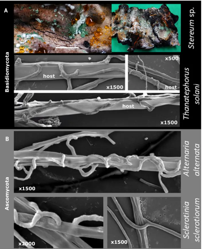
Fig 8. Mycoparasitism ofTrichoderma on Basidiomycota (A) and Ascomycota (B). Macrophotography for A shows T. simmonsi TUCIM 6527
onStereum sp. Bar indicates 1 cm. SEM images show hyphae of T. guizhouense NJAU 4742 on three hosts.
https://doi.org/10.1371/journal.pgen.1007322.g008
similar interactions betweenTrichoderma hyphae and Basidiomycota (Thanatephorus solani
[syn.Rhizoctonia solani, Cantharellales]) and Ascomycota hosts (Fig 8B), which include chas-ing, coilings, and penetration of the host hyphae.
We investigated interactions betweenT. reesei and the lignocellulolytic P. fici [24], which was several times identified as one of the putative LGT donors (Fig 6).P. fici was also selected
because it has been isolated from the same ecosystem whereT. reesei is common (the
phyllo-sphere ofShorea sp., Borneo) and it has comparable growth rates in vitro (Supporting
Informa-tionS1 Fig). In dual confrontation assays on agar plates,T. reesei overgrew a colony of P. fici,
but did not kill it (Fig 9A). Microscopic examination revealed a tight association between the hyphae, suggesting endoparasitism ofP. fici by T. reesei (Fig 9B). Confocal microscopy revealed that cords ofP. fici hyphae were penetrated and colonized by the thinner hyphae of T. reesei (Fig 9C). This experiment shows thatTrichoderma hyphae can grow inside hyphae of at
least some extant putative Ascomycota donors. Dual confrontation assays with a set of ran-domly selected Eurotiales fungi showed thatTrichoderma is capable of attacking these fungi as
well (Supporting InformationS3 Fig). However, endoparasitism was not observed, possibly because the hyphae of the tested Eurotiales fungi were comparable in size withTrichoderma
spp., making internal penetration difficult.
A surprising finding of this study was the detection of four cases of LGT of cellulolytic enzymes from other Hypocreales. Interestingly,Trichoderma is also capable of parasitizing
fungi belonging to its very close phylogenetic neighbors (adelphoparasitism [62]), including
Fusarium [55,63]. To investigate the range ofTrichoderma adelphoparasitism, we confronted
differentTrichoderma strains with fungi from the same genus, family, and order (Fig 10). The microscopic study revealed numerous cases of hyphal fusion that may be linked to self/non-self-recognition mechanisms inTrichoderma species and only in part to parasitism. Therefore,
evidence for adelphoparasitism was only accepted when one colony overgrew the other. Our results showed thatT. harzianum might attack its sister species, T. guizhouense (Fig 10A, see Fig 1for phylogenomics). Any of the nineTrichoderma species can parasitize E. weberi (Fig 10BforT. atroviride), while the latter fungus did not attack Trichoderma. The majority of Tri-choderma strains attacked and/or killed Fusarium spp. (Fig 10C) [55,63], although individual strains of the latter host fungus resistedTrichoderma infections. A similar interaction was
observed in a confrontation withEmericellopsis alkalina, which belongs to an Acremonium
species complex in Hypocreales (Fig 10D). Our results show that all species ofTrichoderma
studied are capable of adelphoparasitism in the strictest sense of this term (parasitism on organisms belonging to the same genus or family [62]), and this property extends to intera-ctions with other filamentous Ascomycota. Along with the unique ability to perform adel-phoparasitism,Trichoderma maintains its alloparasitic properties (Supporting Information
S3 Fig).
Discussion
LGT as an evolutionary shortcut to achieving nutritional versatility
In this work, we uncovered a possible evolutionary process that contributed to the develop-ment of the nutritional versatility ofTrichoderma. Phylogenomic analysis showed that the
genus shared a last common ancestor with entomoparasitic hypocrealean fungi (Cordycipita-ceae, Ophiocordycipita(Cordycipita-ceae, and Clavicipitaceae). Since then,Trichoderma evolution has been
directed towards mycotrophy. Although this path has also been taken by a number of other fungi of the family Hypocreaceae [20,55],Trichoderma is the most taxonomically diverse
mycoparasitic fungus, harboring at least 260 molecularly defined species [64] found world-wide (NCBI Taxonomy browser, Nov. 2016).Trichoderma can also interact with animals [65],
Fig 9. Mycoparasitism of GFP-labeledT. reesei TUCIM 4817 on Pestalotiopsis fici TUCIM 5788. A. Dual confrontation assay after 10 days of
incubation at 28˚C in darkness. B. Hyphal interactions observed using light microscopy (400x magnification). C. Confocal image showing endoparasitism ofT. reesei on hyphae of P. fici on a glass slide prepared as shown inS3 Fig. D. Hyphae ofP. fici TUCIM 5788 and a fluorescent
chlamydospore ofT. reesei. E. T. reesei TUCIM 4817 mycelium. Scale bar on C–E—40 μm.
https://doi.org/10.1371/journal.pgen.1007322.g009
Fig 10. Adelphoparasitism ofTrichoderma on members of the same genus (A), same family (B), and same order (C, D). Parasites were inoculated
on the right side of each plate, and hosts are on the left side. Images were taken after 10 days of incubation at 28˚C in the dark. Parasitism is assigned as a function of active overgrowth of the opponent colony. NCBI accession numbers for the DNA barcodes for fungi are given in Supporting Information
S6 Table. Note to A: In this experiment, the host fungusT. guizhouense NJAU 4742 did not produce conidia (see other images in Supporting
InformationS3 Fig).
although the evolutionary state and mechanisms are not understood (see also Supporting InformationS1 Text). It is known that the evolutionary history of some hypocrealean fungi involved the emergence of mycotrophy from a entomoparasitic/sarcophagic background. For example,Elaphocordyceps spp., deriving from the mainly entomoparasitic order
Cordycipita-ceae, are parasites of false truffles of the genusElaphomyces (Eurotiales) [66].
Nikoh and Fukatsu [20] invoked the host-habitat hypothesis for such a “jump” from feeding on cicada nymphs to parasitism on truffles.
Due to the chemical composition of animals and fungi, the host shift from feeding on arthropods to feeding on fungi does not appear to be a difficult metabolic transition. Instead, it would only require a fine-tuning of ecological adaptations for specific hosts in one or another kingdom (i.e., mechanisms for recognition, defense, and overcoming the host). In contrast, feeding on plant biomass is an evolutionary challenge for any fungus specialized for feeding on insects or fungi. Our comparative analysis of the pcwdCAZome of hypocrealean fungi revealed that members of entomoparasitic families have a relatively poor repertoire of genes required for degradation of plant biomass compared to those of the hypocrealean phytopara-sites. This paucity is also present in theEscovopsis weberi, a parasite of Agaricales and the
clos-est phylogenetic neighbor ofTrichoderma for which genome information is available [22]. The reduced number of pcwdCAZymes ofE. weberi contradicts the predictions of the
host-habitat hypothesis (see above) because the host-habitat of this fungus is directly linked to plant biomass, which is used by ants to cultivateE. weberi’s host fungus Leucoagaricus spp. The
pcwdCAZome of the nineTrichoderma spp. investigated here was found to be of intermediate
size between entomoparasitic and phytoparasitic Hypocreales fungi. We demonstrate that the abilities ofTrichoderma to feed on plant and fungal biomass are equally developed in the
studied species. Consequently, nutritional extension—not shifts or “jumps”—results in nutri-tional versatility and provides the basis for the general environmental opportunism of this genus [4].
Trichoderma gained pcwdCAZymes from filamentous Ascomycota hosts
Our data suggest that nearly half of the genes encoding pcwdCAZymes have been obtained by LGT from other fungi. Gene duplication, which has been described as a major source of gene innovation in fungi [67] and other organisms [68] apparently played only a minor role in the evolution of theTrichoderma pcwdCAZome. It has been reported that 0.1–3% of the genes in a
given Pezizomycotina genome were derived by LGT, usually indicating interdomain
exchanges [43,67]. When this estimation is applied to the 122 proteins of the pcwdCAZome of
Trichoderma, maximally five genes would be expected to have originated from LGT. This
sug-gests that the frequency of LGT in pcwdCAZome is an exceptional case. Surprisingly, we did not detect any transfer event from prokaryotes, and we also did not observe LGT events from Basidiomycota fungi. Marcet-Houben and Gabaldon [69] and Savoryet al. [59] reviewed LGT events between bacteria and fungi, and listedT. reesei as one of the fungi comprising the
high-est number of bacterial-derived proteins. However, the genes transferred encoded arsenite reductases, catalases, different racemases and enzymes of peptidoglycan metabolism, but no pcwdCAzymes. Because the transfer of bacterial glycoside hydrolase genes to ciliates [70] or rotifers [71] has been demonstrated to have shaped their adaptation to polysaccharide-rich environments, we expected to find such cases forTrichoderma. However, none of the 50 LGT
events detected in this study involved a bacterial donor.
The only example of non-fungal putative LGT toTrichoderma was that of the gene
encod-ing the auxiliary protein swollenin [72,73]. The plant expansins were described to have under-gone at least two LGT events to other organisms, including one event that gave rise to
amoebozoa expansins and fungal swollenins and another that gave rise to the bacterial expan-sins [73]. Our data are in accordance with these findings and further suggest thatTrichoderma
was among the first fungal genera to undergo LGT from plants (either directly or through other fungi).
Which features of
Trichoderma mycoparasitism may be linked to LGT?
Historically, LGT between eukaryotes containing cell walls has been considered to be rare and linked to phagotrophy [74]. However, nearly two decades ago, Wo¨stemeyeret al. [56] hypothe-sized that hyphal fusion mycoparasitism might offer nearly ideal conditions for interfungal DNA exchanges. They demonstrated the transfer of genesin vitro from the Mucoromycotina
mycoparasiteParasitella parasitica to its Mucoromycotina host, Absidia glauca [75]. Our dis-covery that a massive but taxonomically restricted putative LGT of pcwdCAZymes occurred in
Trichoderma from filamentous Ascomycota hosts correlates with the expansion of Tricho-derma mycoparasitic host range to Ascomycota. This has not occurred in other Hypocreaceae
(Escovopsis, Hypomyces, Sphaerostilbella, etc.) that feed on Basidiomycota. Chaverri and
Samuels [16] proposed that the ability to parasitize Ascomycota is likely a dominant force that has driven diversification inTrichoderma.
In nature, alloparasitism (parasitism of taxonomically remote hosts) is widespread, while adelphoparasitism is rare. This has mainly been described as social parasitism in Hymenoptera (Arthropoda, Animalia), while cases of cellular interactions are limited to the Rhodophyta (red algae)Gracilariopsis andersonii and its closely related endoparasite, Gracilariophila oryzoides
[76]. Interestingly, a case of adelphoparasitism has also been reported recently in Hypocreales for the clavicipitoid ergot parasiteTyrannicordyceps sclerotium, which attacks closely related
species [66]. Contrary to nutritional expansions inTrichoderma, T. sclerotium offers an
addi-tional example of the apparently common nutriaddi-tional shift, at least in Hypocreales. The mycoparasitism ofTrichoderma on Pezizomycotina has been intensively studied in vitro for its use in plant protection, and, therefore, these studies are biased towards plant
path-ogenic fungi that are not necessarily the natural hosts. In nature,Trichoderma has only rarely
been observed on sporophores of ascomycetes from Xylariales and Helotiales [77]. We have investigated the interactions betweenTrichoderma and extant fungi that may represent or be
descendants of ancient LGT donors. A particularly convenient model donor forTrichoderma
spp. isPestalotiopsis fici (Xylariales) because both fungi are ecophysiologically compatible in vitro. Interestingly, Gazis and Chaverri [78] foundPestalotiopsis and Trichoderma are the most
frequent endophytic fungi on the leaves and stems of rubber trees (Hevea brasiliensis), which
confirms their sympatric occurrence in nature and, thus, the possibility for LGT. Notably, in our experiments,P. fici was not killed by Trichoderma, although intrahyphal growth was
observed.
Another very interesting finding of our study is the absence of putative LGT events of pcwdCAZymes from Basidiomycota fungi, which are commonTrichoderma hosts or
sub-strates in nature [4,13] and on mushroom farms [61]. Our results and numerous previous observations show thatTrichoderma is capable of penetrating the cell wall of fungi, such as Thanatephorus solani and Athelia rolfsii (Basidiomycota). This indicates that neither fusion
mycoparasitism alone nor the host-habitat hypothesis predict interfungal DNA exchanges. The reason why LGT from Basidiomycota was not detected is not easy to explain. This find-ing seems to not be restricted to pcwdCAZymes because we also found no hints of LGT from basidiomycete donors in other gene families. A single case of a putative LGT from Basidiomy-cota toT. reesei has been suggested by Slot and Hibbett [77] for the nitrate-utilizing gene clus-ter. Their analysis suggestedUstilago maydis (Ustilaginales) to be a donor. Interestingly, the
Ustilaginales belong to the simple-septate Basidiomycota fungi, which do not have the com-plex dolipore septae found in Agaricomycotina mushrooms [79]. The dolipore may prevent the penetration of the host hyphae byTrichoderma. It could also be that LGT requires the
growth of the parasite inside the host because during the proliferation of both hyphae, the cytoplasm and nuclei of both organisms may come in contact during mitosis. This might facili-tate DNA exchange.
The likely physical difficulty to grow inside of hyphae of Basidiomycota with dolipores also suggests that mycoparasitic Hypocreaceae (Escovopsis, Hypomyces, Sphaerostilbella, etc.),
which feed exclusively on such Basidiomycota, will not obtain genes from them. Indeed, such a case has not been reported thus far. Therefore, we hypothesize that the ability ofTrichoderma
to parasitize similar fungi (Pezizomycotina), even in extreme cases of adelphoparasitism, has been a significant ecological adaptation of this genus that subsequently enabled the observed putative LGT. We show thatTrichoderma spp. can parasitize (overgrow and kill) some fungi
belonging to the same genus, family, or order. In this study, we observed thatE. weberi lacks
this ability because it was parasitized byTrichoderma in all assays or did not interact.
The nutritional expansion ofTrichoderma towards plant biomass through LGT is
theoreti-cally concordant with the “you are what you eat” concept in which the integration of a foreign DNA is a key mechanism. Yet,Trichoderma LGT resulted only from feeding on a limited
group of hosts.
Glycoside hydrolase requirements for feeding on plant biomass
We propose that the major putative LGT events that resulted in the nutritional expansion from more ancient mycoparasitism to phytophagy took place before the diversification of Tri-choderma into extant infrageneric groups (sections, clades and species). It is also evident that
the fungus maintained both nutritional strategies. Thus, the composition of the pcwdCAZome allows us to speculate about the requirements for efficient feeding on plant biomass. The GH families, for which genes have been entirely acquired by LGT and are absent from the phyloge-netic neighbors ofTrichoderma (i.e. GH6, GH51, GH62, GH74, and swollenin), reveal that
improvements in cellulose and hemicellulose degradation were a key trait for the phytophagy of this fungus. Specifically, the gain of CEL6A that proceeds from the nonreducing cellulose ends complements the presence of CEL7A that acts at the reducing end, and therefore allows a processive movement along cellulose and an increase the speed of its degradation [80]. The addition of swollenin, which disrupts the cellulose structure and generates additional free chain ends [81], provides an increased number of accessible points for the two cellobiohydro-lases. Interestingly,Trichoderma also obtained a large number of GH27 α-D-galactosidases,
GH28 pectinases, and GH10, GH11, and GH30 xylanases, suggesting their importance for the hydrolysis of both hemicelluloses and pectin.
In this regard, it is meaningful that several GH families, that are absent from the entomo-parasitic hypocrealean fungi, are present inTrichoderma and E. weberi (GH7A, GH5
ß-man-nanases, GH12, GH67, GH74, GH95). This suggests that a part of the pcwdCAZome
repertoire must have already been acquired before the split of the genera. It may also indicate thatE. weberi likely lost the nutritional versatility of its ancestor along with a specialization for
parasitizingLeucoagaricus spp. [22].
Conclusions
In this study, we propose that the parasitism ofTrichoderma on phylogenetically close hosts
(up to adelphoparasitism) enabled LGT to build its unique pcwdCAZome and nutritional versatility. In support of this,Trichoderma spp. are frequently detected as a members of
endophytic fungal communities [78] where they may either parasitize their putative cellulolytic hosts or feed on plant biomass or do both. Further studies of the evolutionary consequences of adelphoparasitism may explain other unique genomic features ofTrichoderma. In addition,
the description of the complete pcwdCAZome of nineTrichoderma spp. and, in the case of
LGT, the identification of their putative donors may be of considerable interest to researchers studying the cellulolytic activity of this fungus for industrial applications.
Materials and methods
Organisms used in this study
Trichoderma strains used for the whole genome sequencing are given in Supporting
Informa-tionS1 Table. All fungal strains and other organisms used in experiments and their respective accession numbers for DNA barcode sequences deposited in public databases and/or refer-ences are given in Supporting InformationS6 Table.
Assessment of the growth on plant and fungal biomass
For inoculum preparation, fungi were cultivated on potato dextrose agar (Sigma Aldrich, Steinheim, Germany) at 28˚C for 4 days. Spore suspensions (3x10⁶spores/ml) were prepared in 0.9% (w/v) NaCl with 0.025% (w/v) Tween 20 (Carl Roth, Austria). Growth tests were per-formed in CELLSTAR 24 Well Cell Culture plates (Greiner bio-one International). 1 ml of spore suspension of the fungus to be tested was inoculated on the following substrates: i) heat-treated (100˚C for 3 hours) dried fruiting bodies ofGanoderma lucidum (Polyporales,
Basidio-mycota) (0.3% w/v), ii) epiphyte-free dried leaves ofShorea johorensis (Malvales, Angiosperms,
Plantae) (0.3% w/v), iii) naturally degraded dead wood ofS. johorensis (0.3% w/v), iv)
commer-cial saw dust (local supplier, Vienna, Austria) (0.3% w/v), v) microcrystalline cellulose (0.05 mM research grade; AMS Biotechnology, Milton park, UK) in 0.5% (w/v) Agar-Agar Kobe I (Carl-Roth, Mannheim, Germany), vi) 2% (w/v) pre-treated (steam exploded) wheat straw 3% (w/v) in Agar-Agar Kobe I, vii) 0.3% pectin (w/v) in Agar-Agar Kobe I. Growth in 0.5% (w/v) Agar-Agar Kobe I was also tested as a control. All non-powdered substrates were finely ground and then sterilized at 120˚C for 20 min. Experiment was carried out in quadruples. The plates were photographed after incubation at 28˚C for seven days in darkness.
Interfungal interactions
Dual confrontation assays between fungi were done as described in Atanasovaet al. [15]. For these experiments, fungi were incubated for 10 days on PDA at 25˚C and 12 hours with cyclic illumination. When required, slower growing fungi (such asLentinula edodes and Leucoagari-cus gongylophorus) were inoculated 2–3 days prior to the inoculation with fast growing
Hypo-creales. The set ofPenicillium spp. and Pestalotiopsis fici TUCIM 5788 strains was randomly
selected from a pool of strains isolated from phylloplane ofShorea johorensis
(Dipterocarpa-ceae, Plantae) from Borneo whereTrichoderma spp. are common.
Confocal microscopy. To analyze the interfungal interaction by confocal microscopy,
spores ofT. reesei strain TUCIM 4817, carrying a gfp gene under the control of a histone 3A
promoter, andPestalotiopsis fici TUCIM 5788 were inoculated on two adjacent but separated
PDA agar blocks mounted between the glass slides and the cover glass using a modified Riddell slide method [114] (Supporting informationS3 Fig). The construct was incubated for 72 hours in a sterile wet chamber that hyphae of both fungi established the contacts. Live-cell imaging was performed using a Nikon C1 confocal laser scanning unit mounted on a Nikon Eclipse TE2000-E inverted microscope base (Nikon GmbH, Vienna, Austria). A Nikon Plan Apo VC
100×/1.4 with oil immersion objective lens was used. GFP was excited with an argon ion laser at 488 nm. The emitted fluorescence was separated by a Nikon MHX40500b/C100332 filter cube and detected with a photomultiplier tube within the range of 500–530 nm. Bright light images were captured simultaneously with a Nikon C1-TD transmitted light detector mounted behind the condenser turret.
Scanning electron microscopy. For scanning electron microscopy, a coverslip (1 cm2) was placed on the centre of an agar plate inoculated with partner fungi and incubated until contact between hyphae was established (in average for 72 hours). The hyphae were then fixed with 2.5% (v/v) glutaraldehyde in 0.5 M potassium phosphate buffer and used for examining by SEM (HITACHI S-3000N, Tokyo, Japan).
Genome analysis
The genomes of fiveTrichoderma species (T. longibrachiatum ATCC 18648, T. citrinoviride
TUCIM 6016,T. harzianum CBS 226.95, T. guizhouense NJAU 4742 and T. asperellum CBS
433.97) were sequenced for this work (Supporting informationS1 Table). Four of them(T. longibrachiatum, T. citrinoviride, T. harzianum, and T. asperellum) were sequenced using an
Illumina platform. To this end, Illumina fragments (270 bp insert size) and 4 kbp long mate-pair (LMP) libraries were combined. The fragment libraries were produced from 1μg of geno-mic DNA, sheared to 270 bp using an E210 Focused-ultrasonicator (Covaris) and size selection was carried out using SPRIselect (Beckman Coulter). The fragments were treated with end-repair, A-tailing, and ligation of Illumina adapters (Eurofins MWG Operon), using a NEBNext Ultra DNA Library Prep Kit (New England Biolabs Inc.).
Two types of LMP libraries were used, CLIP (Cre-Lox Inverse PCR) and LFPE (Ligation Free Paired-End), both of which used 15μg of genomic DNA sheared with HydroShear (Genomic Solutions) using a selection size of 4 kb. For CLIP, the size selected DNA was ligated to adaptors containing loxP and the Illumina specific primer sequence. This adaptor ligated DNA fragments were then circularized via recombination by a Cre (NEB) excision reaction. The circularized DNA templates were then digested with a cocktail of four base cutter restric-tion enzymes, i.e. NlaIII, MseI, HypCH4IV (NEB), followed by self-ligarestric-tion. The paired end library was then amplified via inverted PCR using an Illumina specific primer set. The size of the amplified paired end library was selected by running on a 1.8% (w/v) agarose gel followed by gel purification of the desired fragment (300–600 bp).
For LFPE library, the sheared DNA was treated with end repair adapters and ligated with biotinylated adapters. The adapter ligated DNA fragments were circularized by intra-molecular hybridization. The circularized DNA templates were digested by T7 Exonuclease and S1 Nucle-ase (Thermo Fisher Scientific). The digested fragments were treated with A-tailing Enzyme (NEB), followed by immobilization of mate-paired fragments on strepavidin beads (Thermo Fisher Scientific). Illumina compatible adapters (IDT, Inc) were ligated to the mate paired frag-ments and 12 cycles of PCR was used to enrich for the final library (KAPA Biosystems).
All prepared libraries were quantified using KAPA Biosystem’s next-generation sequencing library qPCR kit and run on a Roche LightCycler 480 real-time PCR instrument. The quanti-fied libraries were then prepared for sequencing on the Illumina HiSeq sequencing platform utilizing a TruSeq paired-end cluster kit v3 and Illumina’s cBot instrument to generate a clus-tered flowcell for sequencing. Sequencing of the flowcell was performed on the Illumina HiSeq2000 sequencer using a TruSeq SBS sequencing kit 200 cycles v3, following a 2 x 100 bp or 2 x 150 bp run recipe.
Illumina data were QC filtered for artifact/process contamination and subsequently assem-bled using Rnnotator [82] for transcriptomes, and AllPathsLG [83] for genomes. The Pacific
Biosciences library was prepared from 5μg of gDNA sheared using a Covaris LE220 focused-ultrasonicator with their Blue miniTUBES to generate sheared fragments of 3kb in length. The sheared DNA fragments were then prepared according to the Pacific Biosciences protocol and using their SMRTbell Template Preparation Kit, where the fragments were treated with DNA damage repair, had their ends repaired so that they were blunt-ended, and 5’ phosphorylated. Pacific Biosciences hairpin adapters were then ligated to the fragments to create the SMRTbell template for sequencing. The SMRTbell templates were then purified using exonuclease treat-ments and size-selected using AMPure PB beads. Sequencing primer was then annealed to the SMRTbell templates and Version C2 sequencing polymerase was bound to them. The prepared SMRTbell template libraries were then sequenced on a Pacific Biosciences RSII sequencer using Version C2 chemistry and 2x45min sequencing movie run times.
Genomes were annotated using the JGI Annotation pipeline and made available via JGI fungal genome portal MycoCosm (jgi.doe.gov/fungi[84]). They have also been deposited at DDBJ/EMBL/GenBank as specified in Supporting InformationS1 Table.
The genome ofT. guizhouense NJAU 4742 was shotgun sequenced using a Roche 454 GS
FLX system at the Chinese National Human Genome Center (Zhangjiang Hi-tech Park, Shanghai, China) with 28.4X coverage. The fragment libraries were produced from 5μg of genomic DNA, sheared to 300–500 bp using M220 Ultrasonicator (Covaris, America) and was purified with Agencourt Ampure beads (Beckman, America). The fragment libraries were con-structed with Purified DNA fragments by using DNA Library Preparation kit (Roche Applied Science, Switzerland) and fixed on magnetic beads with GS emPCR kit (Roche Applied Sci-ence). The 569 Mb raw data were achieved from 454 GS FLX system with 1,435,699 reads.
For sequence scaffolding, Solexa Mate Pair reads were used to establish the genome scaf-folds. 5μg of genomic DNA was sheared with a Hydroshear device (Gene Machine) to gener-ate 3–5 kb DNA fragments. The library was prepared by using TruSeq DNA Sample Prep Kit-SetA (illumina, America), and amplified using TruSeq PE Cluster Kit (illumina, America), and then sequenced in Solexa sequencing machine (illumina, America). Gene calls were generated using FGENESH [85], ExonHunter [86] and AUGUSTUS version 2.7 [87].
Composition of
Trichoderma pcwdCAZome
Annotation of the genes encoding carbohydrate active enzymes involved in plant cell wall deg-radation (pcwdCAZome) in the nineTrichoderma genomes was performed using the
Carbo-hydrate-Active Enzyme database (CAZy) nomenclature [88,89], by comparing each protein model from the genome by the sequence similarity search tool (blastp) to a collection of pro-tein modules corresponding to catalytic and carbohydrate-binding modules derived from CAZy. Individual hits were then compared by HMMer to models corresponding to each CAZy family to allow an assignment of each identified protein.
Accession numbers of genes composing pcwdCAZome and respective regulatory proteins inTrichoderma genomes are given in Supporting InformationS3 Table.
Principal component analysis and two-way cluster analysis of the Hypocreales pcwdCA-Zomes (Supporting InformationS3 Table) were made with the use ofhttps://biit.cs.ut.ee/ clustvis/[90]. Cluster analysis was made with Euclidian distance and complete linkage method.
Genomic location of individual genes encoding for pcwdCAZome
To analyse whether the genomic location of the identified pcwdCAZomes would be syntenic among the nineTrichoderma species, we used the manually annotated chromosomes of T. ree-sei [45] as a template. Orthologs for each individual gene from the otherTrichoderma spp.
were then located on their genomic scaffolds, and at least five genes flanking its 5’ and 3’ area were retrieved and identified by blastp. A synteny value of 100% was assigned betweenT. reesei
and anotherTrichoderma sp. when all investigated flanking genes were orthologues and their
order was conserved.
Phylogenetic analyses
Phylogenomic analysis ofTrichoderma and other Hypocreales using 100 neutrally
evolving genes. One-hundred genes were randomly selected from the genomes of the nine
Trichoderma spp. and 12 reference Hypocreales (Escovopsis weberi, Metarhizium acridium, M. robertsii, Calviceps purpurea, Ophiocordyceps sinensis, Beauveria bassiana, Cordyceps militaris, Fusarium graminearum, F. oxysporum f. sp. lycopersici, F. pseudograminearum, F. fujikuroi,
andNectria haematococca; see Supporting InformationS1 TableandS2 Table) based on two requirements: (a) they should display a syntenic position in all genomes, and (b) be true ortho-logues (no other gene encoding a protein with amino acid similarity >50% present).Neurospora crassa and Chaetomium globosum (Sordariales) were chosen as outgroups. For each gene the
alignments of nucleotide sequences consisting of coding regions were prepared using ClustalW [91] and analyzed for the neutral evolution [92] using DNASp V5.10.01 [93] based on Tajima’s D test [94]as described by Rozas [95] (Supporting InformationS2 Table). Multiple sequence alignments of each protein were done using ClustalW [91]. Resulting alignments were exam-ined in Genedoc [96] and then subjected to the phylogenetic analysis online in PhyML [97] based on best amino acid substitution model acquired using “smart model selection” option (http://www.atgc-montpellier.fr/phyml-sms/). Maximum likelihood trees assessed using 1000 bootstrap replicates were also constructed individually for each of the 100 protein sequences and the phylome is deposited athttp://itol.embl.de/shared/druzhininaetal. Accession numbers of all genes used in phylogenomic analysis are given in Supporting InformationS2 Table.
For a combined analysis, a concatenated set of 100 proteins for each of 23 species was sub-jected to the alignment algorithm using the stand alone MAFFT tool [98] with G-INS-i parame-ters. Selection of conserved blocks was done using “relaxed” conditions in Gblocks [99]. The final concatenated alignment contained 47 726 amino acids. The selection of best amino acid substitution model was done using ProtTest 3 [100] based on BIC criterion. The Bayesian analy-sis was performed using MrBayes v3.2.5 [101,102], 1 million generations and the Dayhoff I+G +F amino acid substitution model [103]. Two simultaneous, independent analyses starting from different random trees were run, each using three heated chains and one "cold" chain. Once the analyses were completed, 7500 trees were summarized after discarding the first 25% of the obtained 10,000 trees, resulting in a consensus tree. The parameters of phylogenetic analyses and accession numbers of individual genes are given in Supporting InformationS2 Table.
Multilocus phylogeny of Ascomycota. To reconstruct a phylogenetic tree that included
also all fungi for which putative pcwdCAZyme homologs toTrichoderma have been identified
(see below), the amino acid sequences from four nuclear genes that have previously been shown to be suitable phylogenetic markers for Ascomycota multilocus phylogeny (histone acetyltransfer-ase subunit of the RNA polymeracetyltransfer-ase II holoenzyme, FG533; NAD-dependent glutamate dehydro-genase, FG570; translation initiation factor eIF-5, FG832; and TSR1p, a protein required for processing of 20S pre-rRNA, MS277) were retrieved from FunyBase [104] (http://genome.jouy. inra.fr/funybase), GenBank (http://www.ncbi.nlm.nih.gov/genbank/), the Joint Genome Institute (http://genome.jgi-psf.org/programs/fungi/index.jsf?projectList), EnsemblFungi (http://fungi. ensembl.org/index.html) and Broad Institute (http://www.broadinstitute.org/) databases. Com-plete sets of amino acid sequences for 128 fungi (including the nineTrichoderma spp.) were
pre-pared. Concatenated alignments (provided in Supporting InformationS1 Data) and an MCMC Massive interfungal LGT of plant cell wall-degrading enzymes