HAL Id: inserm-01144516
https://www.hal.inserm.fr/inserm-01144516
Submitted on 21 Apr 2015HAL is a multi-disciplinary open access
archive for the deposit and dissemination of sci-entific research documents, whether they are pub-lished or not. The documents may come from teaching and research institutions in France or abroad, or from public or private research centers.
L’archive ouverte pluridisciplinaire HAL, est destinée au dépôt et à la diffusion de documents scientifiques de niveau recherche, publiés ou non, émanant des établissements d’enseignement et de recherche français ou étrangers, des laboratoires publics ou privés.
Distributed under a Creative Commons Attribution| 4.0 International License
Fourier-transform infrared imaging and clustering:
toward an automated histology of normal colon
Thi Nguyet Que Nguyen, Pierre Jeannesson, Audrey Groh, Dominique
Guenot, Cyril Gobinet
To cite this version:
Thi Nguyet Que Nguyen, Pierre Jeannesson, Audrey Groh, Dominique Guenot, Cyril Gobinet. Fourier-transform infrared imaging and clustering: toward an automated histology of normal colon. Journées RITS 2015, Mar 2015, Dourdan, France. pp.146-147. �inserm-01144516�
Actes des Journées Recherche en Imagerie et Technologies pour la Santé - RITS 2015 146
Fourier-transform infrared imaging and clustering: toward an automated
histology of normal colon
Thi Nguyet Que NGUYEN
1,2, Pierre JEANNESSON
1,2, Audrey GROH
3, Dominique GUENOT
3,
Cyril GOBINET
1,2∗1 Universit´e de Reims Champagne-Ardenne, Equipe Biophotonique et Technologies pour la Sant´e, UFR de Pharmacie, Reims, France. 2 CNRS UMR 7369, Matrice Extracellulaire et Dynamique Cellulaire (MEDyC), Reims, France.
3 Universit´e de Strasbourg (UdS), EA 3430 Progression tumorale et microenvironnement. Approches translationnelles et Epid´emiologie.
F´ed´eration de M´edecine Translationnelle de Strasbourg (FMTS), Strasbourg, France.
∗ Corresponding author (email: cyril.gobinet@univ-reims.fr).
Abstract - Fourier-transform infrared (FTIR) imag-ing is currently used as a non-destructive and label free method for analyzing biological specimens. Combined with unsupervised clustering method, this biophotonic approach allows to perform a spectral histopathology of human tissues. However, this method requires the sub-jective choice of the number of clusters. To overcome this problem, we developed a hierarchical double application of 9 cluster validity indices (CVIs) using K-Means clus-tering. Applying this approach to FTIR images of normal human colon tissue samples, PBM and SI reveal to be the most efficient indices in retreving the main structures of colon histology. These results suggest that the hierarchi-cal double CVI application is thus a promising method for an automated spectral histology.
Index Terms - Optical Imaging, Image Processing, Sig-nal Processing.
I. INTRODUCTION
In biophotonic, Fourier-transform infrared (FTIR) imaging is a non destructive and label-free technique for analyzing samples. This approach spatially records vibrational infor-mation which is directly linked to the chemical composi-tion of samples. Recent studies [1] have shown that the combination of this biophotonic approach with unsuper-vised clustering method, such as K-Means (KM), allows to perform a spectral histology of human tissues. However, this method requires the subjective choice of the number of clusters k. To automatically estimate the optimal k, cluster validity indices (CVIs) have been developed for clustering methods.
In this study, to automate the spectral histology, we de-veloped a hierarchical double CVI application for FTIR imaging. The performance of this approach was then eval-uated on 9 different CVIs, using KM partitions estimated on FTIR images acquired on healthy human colon tissue sections.
II. MATERIALS AND METHODS
II.1. FTIR imaging datasets
Five formalin-fixed paraffin-embedded tissue blocks of normal colon zones were obtained from colon cancer surgery of four patients. For each block, two adjacent 6µm thick slices were prepared. The first slice was mounted on a CaF2 window for FTIR imaging (Perkin Elmer). The
second slice was stained with Hematoxylin-Eosin (HE) for conventional histology, and used as a reference for com-parison with FTIR imaging.
Each FTIR image was recorded with 6.25µm spatial res-olution, on the mid-IR range of 900 to 1800 cm−1with 4
cm−1 spectral resolution. In order to avoid chemical de-paraffinization, an in-house Matlab code was applied for neutralizing the spectral interferences from paraffin. II.2. Hierarchical double CVI application
A CVI is a mathematical function that measures the quality of a partition. By performing a KM clustering for different values of k, 2 ≤ k ≤ kmax, this function calculates the
ratio between the compactness and separation of clusters for each KM partition. The optimal number of clusters is defined as the number of clusters giving the optimal CVI value. Patient sample 1 2 3 4 5 CVI PBM 10 9 8 9 10 SI 13 9 9 20 10 Dunn 326 316 180 257 278 OS 40 392 394 333 374 SV 228 378 22 58 21 DB 7 7 8 4 4 COP 5 4 4 7 4 SWC 4 5 4 4 4 XB 5 6 8 4 4
Table 1: koptestimated by hierarchical double CVI
appli-cation on the FTIR images. Bold values represent the op-timal partitions that retrieved at least the main histological structures of normal human colon.
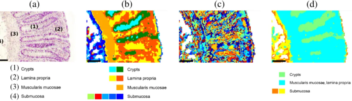
147 Actes des Journées Recherche en Imagerie et Technologies pour la Santé - RITS 2015 (a) (b) (c) (d) (1) (2) (3) (4)
Figure 1: Comparison between conventional histology and pseudo color-coded images reconstructed by double CVI applica-tions for patient sample 1. (a) HE stained image of tissue with 4 histological structures of colon. Optimal partition estimated by (b) PBM (kopt = 10), (c) Dunn (kopt= 316) and (d) SWC (kopt= 4). Scale bars indicate 100µm.
In this study, the nine following CVIs [2,3] were applied: Dunn, Davies-Bouldin (DB), Silhouette-Width-Criterion (SWC), Xie-Beni (XB), Pakhira-Bandyopadhyay-Maulik (PBM), Sym-Index (SI), Context-independent Optimal-ity and PartialOptimal-ity (COP), Separation-Variance (SV) and Overlap-Separation (OS).
To realize an objective and automated spectral histology, a hierarchical double application of CVIs to KM results with 2 ≤ k ≤ 20 is proposed. For each spectral image, this method is applied using the following steps: i) The CVI is first applied on the KM results of the spectral image. The optimal number of clusters estimated by the CVI is the number of main clusters kmaincomposing the dataset. ii)
Then, on each main cluster, KM is applied for 2 ≤ k ≤ 20, and a second application of the CVI on these KM results estimates the optimal number of sub-clusters. iii) The fi-nal optimal number of clusters koptis thus the sum of
esti-mated optimal numbers of sub-clusters. The corresponding optimal double CVI partition is obtained by assembling all the kmainestimated optimal sub-partitions together.
III. RESULTS
The koptestimated by this approach applied on the 5
spec-tral images are listed in Table 1. Data show that PBM and SI are the most effective indices since their koptvaried
be-tween 8 and 20 while exactly matching the main colon tis-sue structures (Figure 1(a) and (b)) for the 5 samples. Concerning Dunn, OS and SV, they often exhibited dra-matically high kopt (kopt ≥ 40) , thus complicating the
assignment of clusters to the corresponding histological structures (Figure 1(c)). By contrast, DB, COP, SWC and XB estimated low kopt inducing optimal partitions which
partially correspond to the histological structures. For ex-ample, the optimal SWC partition (Figure 1(d)), assigned the cyan cluster to both the muscularis mucosae and the lamina propria.
IV. DISCUSSION-CONCLUSION
In this study, the traditional single application of CVIs has been tested on the FTIR images (data not shown) and
mainly leads to an under-estimation of the number of clus-ters, thus preventing retrieval of the main structures of hu-man normal colon tissue. Theoretically, CVI works cor-rectly for dataset composed of compact and separated clus-ters, which is not a property fulfilled by FTIR spectral datasets acquired on normal human colon.
For this, the proposed hierarchical double application of CVI succeeds in retrieving the structures of colon tissue, permitting to realize an automatic spectral histology. How-ever, the number of layers of the hierarchical application of CVI is obviously dependent on the considered dataset. An objective criterion needs thus to be defined.
In conclusion, spectral histology is a new concept associat-ing FTIR imagassociat-ing and clusterassociat-ing. To overcome the subjec-tive choice of the number of clusters, a hierarchical double CVI application for KM partition was proposed. This pro-cedure achieves an automated spectral histology, since the main human normal colon tissue structures are detected for all the analyzed FTIR images.
ACKNOWLEDGMENTS
Authors thank Canc´eropˆole Grand-Est, Ligue contre le Cancer, the URCA technological platform of cellular and tissular imaging PICT-IBiSA, R´egion Champagne-Ardenne, R´egion Alsace and Minist`ere de l’Enseignement Sup´erieur et de la Recherche for financial support.
REFERENCES
[1] J. Trevisan, P.P. Angelov, P.L. Carmichael, et al. ”Extracting biological information with computational analysis of Fourier-transform infrared (FTIR) biospec-troscopy datasets: current practices to future perspec-tives”, The Analyst, 2012, Vol. 137, pp. 3202-3215. [2] O. Arbelaitz, I. Gurrutxaga, J. Muguerza, et al. ”An
extensive comparative study of cluster validity indices”, Pattern Recognition, 2013, Vol. 46, pp. 243-256. [3] W. Wang, Y. Zhang, J. Muguerza, et al. ”On fuzzy
clus-ter validity indices”, Fuzzy Sets and Systems, 2007, Vol. 158, pp. 2095-2117.