HAL Id: hal-02541681
https://hal.uvsq.fr/hal-02541681
Submitted on 14 Apr 2020HAL is a multi-disciplinary open access archive for the deposit and dissemination of sci-entific research documents, whether they are pub-lished or not. The documents may come from teaching and research institutions in France or abroad, or from public or private research centers.
L’archive ouverte pluridisciplinaire HAL, est destinée au dépôt et à la diffusion de documents scientifiques de niveau recherche, publiés ou non, émanant des établissements d’enseignement et de recherche français ou étrangers, des laboratoires publics ou privés.
Neuromotor Strategy of Gait Rehabilitation for
Lower-Limb Spasticity
Jinan Charafeddine, Sylvain Chevallier, Mohamad Khalil, Didier Pradon,
Samer Alfayad
To cite this version:
Jinan Charafeddine, Sylvain Chevallier, Mohamad Khalil, Didier Pradon, Samer Alfayad. Neuromotor Strategy of Gait Rehabilitation for Lower-Limb Spasticity. International Con-ference on Advances in Biomedical Engineering (ICABME), Oct 2019, Tripoli, Lebanon. �10.1109/ICABME47164.2019.8940263�. �hal-02541681�
icabme-5 INTERNATIONALXXXX-X/XX/$XX.00 ©2019 IEEE
Neuromotor Strategy of Gait Rehabilitation for
Lower-Limb Spasticity
Jinan Charafeddine1, Sylvain Chevallier2 , Mohamad Khalil3,
Didier Pradon1, Samer Alfayad2.
1- EndiCAP U1179 - APHP – UVSQ Hopital Raymond Poincare, Garches,
France
2- Laboratoire d’Ingenierie des Systemes de Versailles Universite de
Versailles Saint-Quentin, France 3- Faculty of engineering, lebanese
university, Tripoli, Lebanon
Abstract— Assisting users and restoring human locomotion for patients with lower limb spasticity is a challenging task. Studies focusing on patients with abnormal walking behavior are scarce because there is an important variability from one patient to another. Those patients could benefit the most from rehabilitation and assistive mechatronic devices, there is no generic controlling scheme or any dynamical gain indicator. This contribution introduces a bio-kinematic index which is called Neuro-motor index (NMI), based on electromyographic (EMG) and joint angles measurements. NMI is derived from the nonlinear regression with a combination of two co-contraction indices (CCI), which allows addressing the variability of walking situations. This new index is evaluated on patients with cerebral palsy and a stroke. Then, the estimation error was calculated in comparison with the other co-contraction indices. This estimation shows that this index has the highest signification of joint angles prediction. Thus it can be suitable in adaptive rehabilitation control for spasticity cases.
Keywords— Rehabilitation Exoskeleton, Spasticity, Bio-kinematic, co-contraction, Neuro-motor.
I. INTRODUCTION
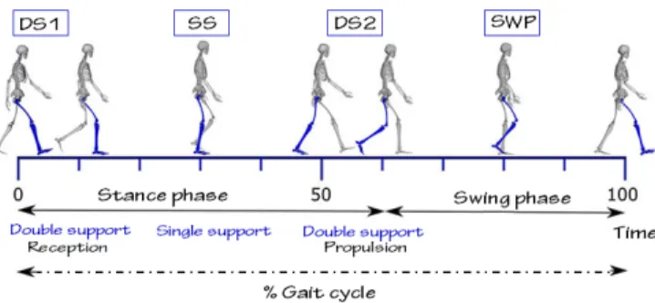
The neurological diseases, such as Cerebral Palsy (CP) and strokes (Cerebral Accident, CA), may lead to lower-limb spasticity which conducts to gait disorders. CP is a disorder that affects muscle tone and motor skills, inducing uncoordinated movements [1]. For CA, this disease causes death or spasticity due to low blood flow to brain cells [2]. Spasticity is an exaggerated contraction of the antagonist muscle, which generates an opposition couple or even an inversion of the desired movement [3]. This could affect all the walk phases which are first double support (DS1), single support (SS), second double support (DS2) and swing phase (SWP) that are detailed on Fig. 1. Mechatronic devices such as exoskeletons are one of the solutions for the rehabilitation process. However, they are difficult to control as each patient has specific motor capabilities and physical integrity should be ensured. Control strategies rely on the fundamental assumption that the system should intervene if the patient is moving along a trajectory considered as “correct”. When deviating from this reference trajectory, the exoskeleton should generate a compensation force, through an adjustment of the mechanical impedance in the controller [4]. This kind of correction includes adjusting the walking speed or step length. There are two known control strategies for these devices (i) Interaction force controllers using impedance or
admittance, which are generally predetermined and do not consider the user's physical condition [5],[6] (ii) Musculoskeletal model based on EMG signals which rely on the detection of muscle activation. But EMG signals are not effective while applying on patients having a muscular disorder[5],[6]. Consequently, the aforementioned strategies are inappropriate for patients with cerebral palsy (CP) or after stroke. The EMG signals not only contain data about patient intention but also give information about the muscle co-contraction around the joint [7]. Co-co-contraction is an operation between a pair of muscles (agonist/antagonist). The quadriceps (contains rectus femoral muscle (RF)) and hamstring (contains biceps femoral muscle (BF)) are bi-articular muscles for knee and hip joints. The angles of these joints result while movement due to the exchange of muscles roles between agonist and antagonist. Co-contraction is related to joint stability [8] and is an important factor which participates to the inefficiency of pathological movement [9] .
Fig. 1: Illustration of the walk phase: First double support (DS1), single support (SS), second double support (DS2) and swing phase (SWP).
The most three important approaches to estimate this co-contraction are proposed by Falconer and Winter [9], Hessee et al. [10] and Unihan et al. [11]. These approaches are efficient for offline analysis and diagnosis but they are not appropriate for designing a control strategy. Consequently, a new index is needed which depends on physiological signals (Eelectromyography EMG) to estimate joint angles of walking. The strategy is to use the EMG as a proxy for determining the patient’s intention, initiating assistance when the muscular contraction is characteristic of a predefined move and detected with sufficient intensity. This approach stimulates movements initiated by the patient, which is an essential part of motor rehabilitation. The main objective of this work is to propose an approach suitable for people with
cerebral palsy or strokes and can be used in the future for exoskeleton control. Where the patient is considered as an expert while moving. The contributions are the following:
•A complete and uniform description for the most important co-contraction indices existing in the literature.
•A novel index proposition to characterize the relation between electromyographic activity and kinematic parameters.
•An evaluation on bio-kinematic recordings from 20subjects (9 healthy, 6 strokes and 5 with cerebral palsy).
Section II presents the materials and methods. Section III details the results by evaluating the proposed index. Section IV concludes this paper.
II. MATERIALS AND METHODS
There were twenty subjects (9 healthy adults: 4 females and 5 males, aged 50 years±7; five kids with cerebral palsy:3 females and 2 males, aged 10 years±2; 6 adults with stroke: 3 females and 3 males, aged 50 years±7), were recorded in Gait Laboratory (Raymond Poincare Hospital, arches, France).
A. Experimental protocol
The normal gait for each subject was tested on the ground by performing 11 trials. The activity of quadriceps and hamstring muscles were recorded using a surface EMG system (MA311). Using a 3D optoelectronic system, the gait analysis was recorded at 100 Hz following the Helen Hayes model which is commonly used by the biomechanical community for gait analysis, a group of markers were placed on the lower part of the patient [12].The marker trajectories were then filtered using a Butterworth filter (4th order, 6 Hz cut-off frequency) [13].The peak of flexion and extension was the main kinematic parameters as appropriate for the hips and knees. For both lower limbs: a calculation was made for spatiotemporal parameters (velocity, cadence, step and stride length, step width) and for kinematic parameters on each sub-phase of the gait cycle.
B. Signal Processing
The processing of the raw EMG signals was divided into several steps: first were band-pass filtered (10–400 Hz),second were rectified, third were low-pass filtered using Butterworth filter (4th order) from 4 till 6 Hz
cut-off frequency depends on the subject cadence [14], and finally normalized by the detected maximal voluntary contraction value (MVC) of each muscle on gait trials [4]. For the kinematic data, the processing was begin by filtering using a Butterworth filter (4th order, 6 Hz cut-off frequency)
which is connected to the acquisition system, then these data is segmented in 1001 values for knee and hip joints to obtain matrices with fixed dimensions which are equal to those of recorded EMG matrices. After that, the estimation of their mean and variance was done.
C. Co-contractions Indices
In the case of a normal walk, there is a very large literature on the CCI, which depends on numerous parameters such as the walking speed, inertia, considered muscle groups, age and sex [11]. The CCIs are always computed as a ratio between agonist and antagonist muscles, and as a function of the phase in the walking cycle. Among the various methodologies to
compute CCI, three are more robust and accurate and will be detailed hereafter.
The CCI1(t) is introduced by [9] and is computed as:
CCI1(t)=
∫ #$%+-+, &'()*' (/) ∩ #$%&2+&'()*' (/) 3/
∫ #$%+-+, &'()*' (/) ∪ #$%&2+&'()*' (/)3/ x 100 (1)
Where ENVemg·(t) is the normalized envelop of EMG signals, as shown on Fig. 3. The period [t1, t2] denotes the
time span where the envelop of Rectus Femoral (RF) is lower than the envelop of Biceps Femoral (BF), whereas [t2, t3]
denote the period where the BF envelop is lower than the RF envelop. CCI1(t) is computed by finding the area of the total
antagonistic activity (area under RF + BF) divided by the integral of the sum of the EMG envelop (area under RF + BF). This method is commonly used in the literature, for quantifying the co-activation of muscle groups during multijoint movements, such as gait. However, it exhibits discontinuities in two points of the cycle, between the single support (SS) and the second double support (DS2) on the one hand and, on the other hand, between the
The CCI2(t) is proposed by [10] and is defined as:
CCI2 (t)=
7 ∫ #$%+-+, &'()*' (/) ∩ #$%&2+&'()*' (/) 3/
∫ #$%+-+, &'()*'(/) 8 #$%9:;9<=)*' (/) 3/ x 100 (2)
This method derived by the equation proposed by [15] and [9] The CCI2 (t)is evaluated by finding the overlap between
the agonist and antagonist muscle (area under RF ∩ BF) curves and divided by the integral of the sum of the EMG envelop (area under RF + BF). On could note thatCCI2(t)is continuous throughout a complete gait cycle.
CCI3 (t)=
7 ∫ #$%+-+, &2+&'()*' (/) 3/8 ∫ #$%+>+- &2+&'()*' (/) 3/
∫ #$%+>+, &'()*' (/)8 #$%&2+&'()*' (/) 3/ x 100 (3)
The last co-contraction index is a reformulation of the index proposed by [15] and [11] .CCI3(t) is evaluated by finding the
overlap between the agonist and antagonist (area under RF∩BF) curves and normalized by the area under RF∪BF.
Fig. 2 Examples of muscles study, biceps femoral (BF) and rectus femoral (RF), conducted on one subject. a) normalized EMGs of the agonist and antagonist muscles. b) normalized EMGs of the antagonist and the total
muscle activity used to calculate CCI1(t). c) normalized EMGs of the agonist and antagonist muscles, and the overlap between the two muscles used to calculate CCI2(t). d) normalized EMGs of the agonist union the antagonist muscles, and the overlap between the two muscles used to calculate CCI3(t).
As CCI2(t), CCI3(t) is also continuous during the gait cycle.
Nonetheless, CCI2(t) andCCI3(t) relies on the arbitrary
definition of the peak co-contractions, that is the precise value of t1, t2 and t3which could be set priori for normal walk but is
not suitable for abnormal walk without manual annotations of the data. They are thus not appropriate for the control of rehabilitation or assistive exoskeleton.
D. Proposed Index
Even with this ensured continuity for CCI2(t) and CCI3(t), the
gait angles and the CCI varies independently along the walk cycle. It is difficult to find a mapping from the joint angle variation to the CCI, thus to propose a mapping function for the control process. We address this problem by defining a regression from the CCI to the angle variation of the considered articulation, that preserve the necessary information and act as a smoothing function. We use a Hermit polynomial to combine the CCI that are estimated from the EMGs recorded from the quadriceps and the hamstrings muscles. The proposed neuro-motor index is built on the care-full detection of user-specific peak co-contraction activities which are then injected in non-linear combination of two co-contraction indices [10],[16]. The NMI allows to characterize the envelop peak of a muscle pair during each flexion/extension of a walk cycle, and to estimate the joint angular trajectory. The first step is to detect the peak of f(t) defined as:
NMI (t) = ?
7 {CCI1 (t ) + Rx (t) CCI2 (t ) } (4)
Where, Rx(t) is a nonlinear regression based on Hermitian function
Rx (t) = h1 (t) f0 + h2 (t) p0 + h3(t) f1 + h4 (t) p1
The peaks are identified with: argmin t | f’(t)|
f (t) = EMGantago (t) ∩ EMGago (t)
h1, h2, h3, h4∈ He (t); p0 and p1 are tangent to f0 and f1.
The NMI is determined based on the flexion/extension of a joint and does not depend on the definition of a standard gait cycle, a requirement for working with cerebral palsy and stroke patients. The non-linear regression ensures that this method could be applied for an abnormal walk, either for adult or children. Starting from a minimal calibration to reduce the patient fatigue, in order to provide comfortable, stable and secure rehabilitation for the patient, the NMI allows a direct prediction of the patient intention because it depends on his EMG, and since it is derived from the co-contraction index, its results rely on the residual capabilities of the patient.
III. RESULTATS
The evaluation is conducted on all 20 subjects. CCI1(t),
CCI2(t) ,CCI3(t) and NMI(t) are calibrated for one walking
cycle of each subject and evaluated on the 10 other cycles.
Each index allows to estimate the angular value of the knee, and an estimation error is computed using kinematics data as ground truth. To proceed this evaluation, we manually annotate the dataset to indicate the t1, t2 and t3 time values
corresponding to each phase of the walking cycle (first double support, single support, second double support, and swing phase). Healthy subjects that have a normal walking cycle do not present any change. However this provides a valuable information for assisting patients with an abnormal walk, as the CCIs are not appropriate for the latter case. Moreover, it should be mentioned that the Θ(t) angular value of the knee, which is to be predicted, is used only during training for NMI (calibration phase on 1 cycle) and is not provided to the algorithm during the rest of the test (evaluation phase on 11 cycles). Fig.3 shows the relation between the joint angles and the CCIs during the gait cycle. It shows that the CCI do not provide a reliable or proper function to predict the joint angle for the control of an actuator. However, the determination of these angles is possible using NMI, which is a surjective function that is valid for control. It could be seen that each input corresponds to a single output, that is crucial requirement for designing a control function. As it could be seen on Fig.4, the NMI consistently achieved the best prediction for the knee angle, based on the ground truth and only after a short calibration. The CCIs performed well for subjects with a normal walk but the main source of error was with patients suffering from cerebral palsy or stroke.
Fig.3. Knee joint angle variation Θ(t) computed for CCI1, CCI2, CCI3 and NMI as a function of walk cycle.
Fig.4 Comparison of error estimation for CCI1, CCI2, CCI3 and NMI of
stroke patient. Walking phases are DS1 (first double support), SS (single support), DS2 (second double support) and SWP (swing phase).
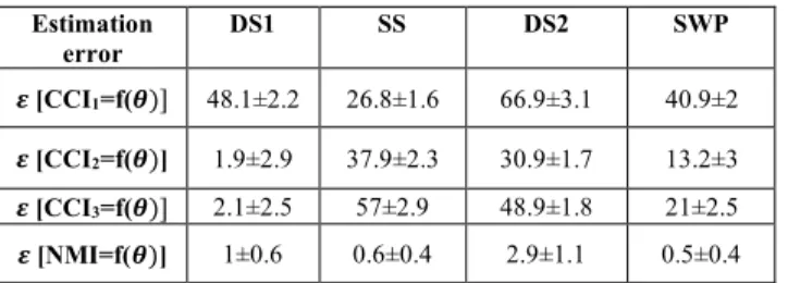
Table 1: Comparison results between CCIs and NMI. Estimation error DS1 SS DS2 SWP 𝜺 [CCI1=f(𝜽)] 48.1±2.2 26.8±1.6 66.9±3.1 40.9±2 𝜺 [CCI2=f(𝜽)] 1.9±2.9 37.9±2.3 30.9±1.7 13.2±3 𝜺 [CCI3=f(𝜽)] 2.1±2.5 57±2.9 48.9±1.8 21±2.5 𝜺 [NMI=f(𝜽)] 1±0.6 0.6±0.4 2.9±1.1 0.5±0.4
The estimation error, in Table 1, shows a big variability for CCIs in function of Knee angles for 11 gait cycle of a stroke subject, where the estimation error of CCI1 reaches (48. 1 ± 2.2 ) in DS1, for each CCI between (26.8±1.6 to 57±2.9) in SS, (30.9 ± 1.7 to 66.9 ± 3.1) in DS2, and (13.2±3 to 40.9 ±2) in SWP, however, this estimation reaches a maximum value ( 2.9±1.1 ) for NMI in all the phases of the gait cycle.
IV. CONCLUSION
The NMI is a new bio-kinematic index, focused on producing a reliable and generic estimation of the articulation flexion/extension using EMG to determine muscle co-contraction. The main contribution of this new index is to provide a model-free estimation of the walk cycle, without imposing a pre-defined trajectory on the patient. The patient is thus considered as an expert, providing a personal optimal walk cycle for calibration, and the NMI yields a robust index for designing an exoskeleton controller. This index evaluated on a dataset of 20 subjects, some with normal walk cycle and others with abnormal ones, using kinematic data as ground truth. The NMI largely outperforms existing CCIs. Following this work, the NMI will be integrated in the control scheme for a lower limb exoskeleton used as walking assistance for people suffering of spasticity.
REFERENCES
[1] J. K. Grether and K. B. Nelson, ‘Maternal infection and cerebral palsy in infants of normal birth weight’, Jama, vol. 278, no. 3, pp. 207–211, 1997.
[2] S. Haldal, J. Beary, P. Nattanmai, P. George, and C. Newey, ‘Acute ischemic stroke management review for the hospitalist’, 2018.
[3] B. Zagrodny, M. Ludwicki, W. Wojnicz, J. Mrozowski, and J. Awrejcewicz, ‘Cooperation of mono-and bi-articular muscles: human lower limb’, Journal of musculoskeletal & neuronal interactions, vol. 18, no. 2, p. 176, 2018.
[4] K. Anam and A. A. Al-Jumaily, ‘Active exoskeleton control systems: State of the art’, Procedia Engineering, vol. 41, pp. 988–994, 2012.
[5] T. Yan, M. Cempini, C. M. Oddo, and N. Vitiello, ‘Review of assistive strategies in powered lower-limb orthoses and exoskeletons’, Robotics and Autonomous Systems, vol. 64, pp. 120–136, 2015.
[6] G. Chen, C. K. Chan, Z. Guo, and H. Yu, ‘A review of lower extremity assistive robotic exoskeletons in rehabilitation therapy’, Critical ReviewsTM in Biomedical Engineering, vol.
41, no. 4–5, 2013.
[7] E. Kellis, F. Arabatzi, and C. Papadopoulos, ‘Muscle activation around the knee in drop jumping using the co-contraction index’, Journal of Electromyography and Kinesiology, vol. 13, no. 3, pp. 229–238, 2003.
[8] J. V. Basmajian, ‘Motor learning and control: a working hypothesis.’, Archives of physical medicine and rehabilitation, vol. 58, no. 1, pp. 38–41, 1977.
[9] K. Falconer and D. A. Winter, ‘Quantitative assessment of co-contraction at the ankle joint in walking.’, Electromyography and clinical neurophysiology, vol. 25, no. 2–3, pp. 135–149, 1985.
[10] S. Hesse, B. Brandl-Hesse, U. Seidel, B. Doll, and M. Gregoric, ‘Lower limb muscle activity in ambulatory children with cerebral palsy before and after the treatment with Botulinum toxin A’, Restorative neurology and neuroscience, vol. 17, no. 1, pp. 1–8, 2000.
[11] V. B. Unnithan, J. J. Dowling, G. Frost, B. A. Volpe, and O. Bar-Or, ‘Cocontraction and phasic activity during GAIT in children with cerebral palsy.’, Electromyography and clinical neurophysiology, vol. 36, no. 8, pp. 487–494, 1996. [12] M. P. Kadaba, H. K. Ramakrishnan, and M. E. Wootten,
‘Measurement of lower extremity kinematics during level walking’, Journal of orthopaedic research, vol. 8, no. 3, pp. 383–392, 1990.
[13] D. A. Winter, H. G. Sidwall, and D. A. Hobson, ‘Measurement and reduction of noise in kinematics of locomotion’, Journal of biomechanics, vol. 7, no. 2, pp. 157– 159, 1974.
[14] R. Shiavi, C. Frigo, and A. Pedotti, ‘Electromyographic signals during gait: criteria for envelope filtering and number of strides’, Medical and Biological Engineering and Computing, vol. 36, no. 2, pp. 171–178, 1998.
[15] D. J. Keefer et al., ‘Interrelationships among thigh muscle co-contraction, quadriceps muscle strength and the aerobic demand of walking in children with cerebral palsy.’, Electromyography and clinical neurophysiology, vol. 44, no. 2, pp. 103–110, 2004.
[16] G. Frost, J. Dowling, K. Dyson, and O. Bar-Or, ‘Cocontraction in three age groups of children during treadmill locomotion’, Journal of Electromyography and Kinesiology, vol. 7, no. 3, pp. 179–186, 1997.