HAL Id: hal-01581091
https://hal.archives-ouvertes.fr/hal-01581091
Submitted on 17 May 2019
HAL is a multi-disciplinary open access
archive for the deposit and dissemination of
sci-entific research documents, whether they are
pub-lished or not. The documents may come from
teaching and research institutions in France or
abroad, or from public or private research centers.
L’archive ouverte pluridisciplinaire HAL, est
destinée au dépôt et à la diffusion de documents
scientifiques de niveau recherche, publiés ou non,
émanant des établissements d’enseignement et de
recherche français ou étrangers, des laboratoires
publics ou privés.
Microtubules acquire resistance from mechanical
breakage through intralumenal acetylation
Zhenjie Xu, Laura Schaedel, Didier Portran, Andrea Aguilar, Jérémie
Gaillard, M. Peter Marinkovich, Manuel Théry, Maxence V. Nachury
To cite this version:
Zhenjie Xu, Laura Schaedel, Didier Portran, Andrea Aguilar, Jérémie Gaillard, et al.. Microtubules
acquire resistance from mechanical breakage through intralumenal acetylation. Science, American
Association for the Advancement of Science, 2017, 356 (6335), pp.328 - 332. �10.1126/science.aai8764�.
�hal-01581091�
REPORT
◥CYTOSKELETON
Microtubules acquire resistance from
mechanical breakage through
intralumenal acetylation
Zhenjie Xu,1,2*† Laura Schaedel,3
Didier Portran,1Andrea Aguilar,1Jérémie Gaillard,3 M. Peter Marinkovich,2,4Manuel Théry,3,5Maxence V. Nachury1†
Eukaryotic cells rely on long-lived microtubules for intracellular transport and as compression-bearing elements. We considered that long-lived microtubules are acetylated inside their lumen and that microtubule acetylation may modify microtubule mechanics. Here, we found that tubulin acetylation is required for the mechanical stabilization of long-lived microtubules in cells. Depletion of the tubulin acetyltransferase TAT1 led to a significant increase in the frequency of microtubule breakage. Nocodazole-resistant microtubules lost upon removal of acetylation were largely restored by either pharmacological or physical removal of compressive forces. In in vitro reconstitution experiments, acetylation was sufficient to protect microtubules from mechanical breakage. Thus, acetylation increases mechanical resilience to ensure the persistence of long-lived microtubules.
H
ow some cytoplasmic microtubules are sta-bilized and persist for several hours remains an open question (1). After stabilization, mi-crotubules are posttranslationally detyrosi-nated and acetylated on lysine 40 ofa-tubulin (aK40). Although detyrosination alters the bind-ing site for microtubule-associated proteins (MAPs), severing enzymes, and motors to create specialized microtubule tracks (2), the molecular consequences ofaK40 acetylation remain elusive. It is difficult to conceptualize how the modification of a resi-due inaccessible from outside the microtubule could alter MAP and motor binding (2, 3). We re-cently proposed that acetylation modifies micro-tubule mechanics by weakening interprotofilament interactions (4).TAT1 is responsible for nearly all acetylation onaK40 in every organism studied (2). Although TAT1 depletion from retinal pigment epithelial (RPE) cells did not measurably affect global mi-crotubule polymerization or organization (fig. S1),
tubulin detyrosination was significantly decreased at the bulk level (Fig. 1A and fig. S2, A and B) and reduced on microtubules of TAT1-depleted cells (Fig. 1B) andTat1–/–mouse embryonic fibroblasts (MEFs) (fig. S2C). Given that acetylation and dety-rosination sites are separated by the microtubule wall, it is unlikely that the two modifications are enzymatically coupled. Instead, long-lived micro-tubules, including detyrosinated micromicro-tubules, may be lost when acetylation is reduced. After treating cells with nocodazole, dynamic microtubules were depolymerized, and most remaining microtubules displayed the typical characteristics of long-lived microtubules with high levels of acetylation and detyrosination and long and curvy morphology (Fig. 1C and fig. S2D). Under the same conditions, very few microtubules remained in TAT1-depleted cells, and these were very short and dispersed throughout the cell (Fig. 1C). The number of micro-tubules that remained after nocodazole treatment was significantly decreased upon TAT1 depletion in RPE cells (Fig. 1D) or inTat1–/–MEFs (fig. S2, E and F). The effect of TAT1 removal was even more striking when the length of nocodazole-resistant microtubules was examined (Fig. 1E). After 60 min of nocodazole treatment, most microtubules were normally longer than 4mm but shorter than 2 mm in TAT1-depleted cells.
Because pharmacological reduction of tubu-lin detyrosination did not affect the length of nocodazole-resistant microtubules or the levels of acetylation (fig. S3), the effects of TAT1 depletion on nocodazole-resistant microtubules are unlikely to be caused by the observed reduction in detyrosi-nation. Because overexpression of TAT1—but not a catalytically dead mutant—significantly elevated the mass of nocodazole-resistant microtubules
(fig. S4) and because nocodazole-resistant micro-tubules are increased in MEFs that lack the tubu-lin deacetylase HDAC6 (5), it is aK40 acetylation rather than an acetyltransferase-independent ac-tivity of TAT1 (6–8) that is required for the main-tenance of long-lived cytoplasmic microtubules in mammalian cells. Long-lived microtubules are not lost from TAT1-depleted cells because of an increased susceptibility to severing enzymes, because acety-lation does not influence the activity of spastin in vitro (9) and did not affect the activity of spastin or katanin in vivo (fig. S5). Defective centrosomal microtubule anchoring in TAT1-depleted cells was ruled out by imaging microtubule regrowth after depolymerization (fig. S6).
Long-lived microtubules display frequent buck-ling because of compressive forces generated by microtubule-based motors and actomyosin contrac-tility (10–12). Because microtubules are very stiff polymers that rupture when subjected to flexural stresses (13), this highly bent morphology suggests the existence of protective mechanisms for long-lived microtubules. The repair of lattice defects has emerged as an intrinsic property of microtubules that are subjected to mechanical stress (14, 15), and acetylation protects microtubules from mechan-ical fatigue in vitro (4). A further suggestion that acetylation may confer mechanical protection to microtubules comes from the observation that re-moving TAT-1 from touch receptor neurons of nem-atodes results in profound microtubule lattice defects (6, 16) that can be rescued by paralyzing the animals (8). Finally, although detyrosination is evenly distributed along microtubules (fig. S7A) (17), the pattern of acetylation is discontinuous with a preference for highly curved areas of nocodazole-resistant microtubules (Fig. 1, F and G, and fig. S7B), suggesting that TAT1 may preferentially acet-ylate segments experiencing stress. Alternatively, it is conceivable that only bends at regions that are acetylated were preserved after fixation (see the sup-plementary text). Localized acetylation is thus a prime candidate for the mechanical adaptation of microtubules to mechanical stresses.
We sought to test the hypothesis that long-lived microtubules disappear in the absence of TAT1 be-cause of an increased rate of breakage under me-chanical stress. In the past, imaging of microtubule breakage has been limited to very thin areas of the cell, such as lamellipodia, where single microtubules can be readily resolved (11, 18, 19). However, most of the microtubules in lamellipodia are dynamic and thus not acetylated (20). To specifically image the breakage of long-lived microtubules, we followed microtubules in real time in the presence of no-codazole using a triple green fluorescent protein (GFP) fusion with the microtubule-binding domain of ensconsin (EMTB), a MAP that does not affect microtubule dynamics when expressed at low lev-els (21). After 15 min in nocodazole, most remain-ing microtubules were highly bent, acetylated, and detyrosinated (fig. S7A), and microtubule number was similar in control and TAT1-depleted RPE-[EMTB-GFP3] cells (fig. S8, A and B). Over the next
30 min of live cell imaging, microtubules rapidly disappeared in TAT1-depleted cells, whereas most microtubules persisted in control cells (fig. S8, A
1Department of Molecular and Cellular Physiology, Stanford
University School of Medicine, Stanford, CA 94305-5345, USA.2Program in Epithelial Biology, Stanford University
School of Medicine, Stanford, CA 94305-5168, USA.
3CytoMorpho Laboratory, Laboratory of Cell and Plant
Physiology (LPCV), UMR 5168, Biosciences and Biotechnology Institute of Grenoble, CEA/INRA/CNRS/ Université Grenoble-Alpes, 17 rue des Martyrs, 38054 Grenoble, France.4Division of Dermatology, Palo Alto
Veterans Affairs Medical Center, Palo Alto, CA 94305, USA.
5CytoMorpho Laboratory, A2T, UMRS 1160, Institut
Universitaire d’Hématologie, Hôpital Saint Louis, INSERM/ AP-HP/Université Paris Diderot, 1 Avenue Claude Vellefaux, 75010 Paris, France.
*Present address: Department of Anatomy, University of California, San Francisco, CA 94143-0452, USA.†Corresponding author. Email: nachury@gmail.com (M.V.N.); zhenjie.xu@ gmail.com (Z.X.)
on May 17, 2019
http://science.sciencemag.org/
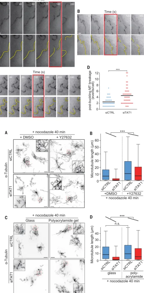
and C) and the mean microtubule length was sig-nificantly decreased in TAT1-depleted cells after 50 min in nocodazole (fig. S8D). Consistent with prior observations of microtubule breakage in fi-broblasts (18, 19), typical rupture events (Fig. 2, A to C; fig. S9, A and B; and movies S5 to S9) were preceded by local microtubule buckling, with the breakage site coinciding with the region of highest curvature. Tracking individual microtu-bules demonstrated that the frequency of micro-tubule breakage events preceded by buckling was increased twofold in TAT1-depleted cells compared with control cells (Fig. 2D). The frequency of mi-crotubule breakage events that were not preceded by buckling (shown in fig. S9C and movie S10) showed no significant difference between control and TAT1-depleted cells (fig. S9D). These findings suggest that tubulin acetylation protects micro-tubules from breakage resulting from compres-sive forces.
The two known types of forces responsible for buckling and breakage of cytoplasmic microtubules are microtubule motors pushing onto anchored mi-crotubules (11) and actomyosin contractility trans-mitted through actin-microtubule linker proteins (19). Contractility is likely to represent the major factor responsible for microtubule compression in nocodazole-treated cells because microtubule de-polymerization leads to activation of Rho and Rho-associated kinase (ROCK), thereby increasing myo-sin activity and stress fiber assembly (22). To test the hypothesis that long-lived microtubules in TAT1-depleted cells break under actomyosin-mediated compression, we treated cells with the ROCK inhib-itor Y27632 or the myosin inhibinhib-itor blebbistatin and then removed dynamic microtubules using no-codazole (Fig. 3A, and figs. S10 and S11). Pharma-cological release of tension increased the mean length of nocodazole-resistant microtubules 1.5-fold in control cells (Fig. 3B). The effect of Y27632 on TAT1-depleted cells was much more dramatic, with the mean length of nocodazole-resistant mi-crotubules increasing fourfold (Fig. 3B) and the length distribution of nocodazole-resistant micro-tubules approaching that of control cells in the ab-sence of Y27632 (Fig. 3B). A statistical test for the rescue of microtubule length in TAT1-depleted cells was highly significant with Y27632 (Fig. 3B) and significant with blebbistatin (fig. S11). ROCK inhi-bition did not restore tubulin acetylation in TAT1-depleted cells (fig. S10, B and C). Thus, inhibition of the major Rho effectors largely restores the nocodazole-resistant microtubules lost from TAT1-depleted cells.
Because ROCK inhibition may stabilize mi-crotubules in TAT1-depleted cells through other mechanisms than the release of compressive forces [e.g., inhibitory phosphorylation of MAPs (23)], we sought to release the compressive forces exerted onto microtubules in a more direct and specific manner. When cells are plated onto soft substrates made of fibronectin-coated polyacrylamide (24, 25) (Fig. 3C and fig. S12), the force-dependent matu-ration of focal adhesions is stunted, stress fiber assembly is limited, and contractility is low (25). Plating cells on polyacrylamide largely rescued the length of nocodazole-resistant microtubules
Fig. 1. Long-lived microtubules are lost in the absence ofa-tubulin K40 acetylation. (A)a-Tubulin K40 acetylation and detyrosination levels were measured by immunoblotting lysates of RPE cells treated with two different small interfering RNAs (siRNAs) against TAT1 (siTAT1 #2 and siTAT1 #3) or control siRNAs (siControl). (B) Immunofluorescence (IF) images of siRNA-treated RPE cells stained for acetylateda-tubulin K40 (red), detyrosinated tubulin (green), and DNA (blue). Scale bar, 10mm. Insets are 7 by 7mm. (C) IF images of siRNA-treated RPE cells treated with 2mM nocodazole and stained fora-tubulin (white), acetylateda-tubulin (red), and DNA (blue). (Bottom) Thea-tubulin channel alone. (Insets) The highly curved microtubules present in control cells and the very short microtubules in TAT1-depleted cells. Scale bar, 10mm (main panels). Insets are 10 by 10mm. The number (D) and length (E) of microtubules remaining after nocodazole treatment were measured in siRNA-treated RPE cells. (D)N (30 min) = 153 (siCTRL), 157 (siTAT1 #3), and 155 (siTAT1 #2) cells, four independent experiments; N (60 min) = 236 (siCTRL), 302 (siTAT1 #3) and 206 (siTAT #2) cells, three independent experiments. Error bars indicate SD. Asterisks indicatet test significance values; ***P < 10−4. (E) The box is bound by the 25th to 75th percentile, whiskers span 5th to 95th percentile, and the bar in the middle is the median. N (40 min) = 3058 (siControl), 4659 (siTAT1#3) microtubules from at least 500 cells, six independent experiments;N (60 min) = 880 (siControl), 1783 (siTAT1#3) and 1323 (siTAT1#2) microtubules from at least 180 cells, three independent experiments. Asterisks indicate Mann-Whitney U test significance values; ***P < 10−4. (F) IF images of RPE cells treated with nocodazole for 45 min and stained for acetylateda-tubulin anda-tubulin. Scale bar, 10mm. (G) The level ofa-tubulin K40 acetylation and the curvature were measured along microtubules in IF images of cells treated with nocodazole for 45 min. The whiskers indicate 1.5 times the range.N = 1904 data points from 23 microtubules. Asterisks indicate Mann-WhitneyU test significance values; **P < 10−3, ***P < 10−4.
RESEARCH | REPORT
on May 17, 2019
http://science.sciencemag.org/
Fig. 2. TAT1 depletion sensitizes nocodazole-resistant microtubules to mechanical breakage. (A to C) Microtubules were imaged in real-time in siRNA-treated RPE-[EMTB-GFP3] cells after at least
15 min in the presence of 2mM nocodazole. Projec-tion images were generated to capture microtubules across the entire cell thickness and to avoid missing microtubule segments because they left the focal plane. The yellow lines highlight microtubule be-havior, and the red box indicates the first frame where rupture is clearly detected. Scale bar, 1mm. The time series are extracted from movies S5 and fig. S7. (D) Microtubule breakage events preceded by buckling were counted in control and TAT1-depleted RPE-[EMTB-GFP3] cells during the 15- to 80-min period of nocodazole treatment.N = 46 (siControl) and 52 (siTAT1) cells, six independent experiments. The bar marks the mean. Asterisks indicatet test significance values; ***P < 10−4.
Fig. 3. Release of cell tension restores the length of nocodazole-resistant microtubules in TAT1-depleted cells. (A) Control and TAT1-depleted cells were treated with Y27632 or vehicle for 1 hour, then nocodazole was added for 40 min, and cells were fixed and stained for
a-tubulin. (Insets) The detailed morphology of nocodazole-resistant microtubules. Scale bar, 10mm. Insets are 10 by 10mm. Cells before nocodazole treatment are shown in fig. S10B. Although nocodazole-resistant microtubules are few and short in the absence of TAT1, the addition of Y27632 leads to the presence of numerous long nocodazole-resistant microtubules in TAT1-depleted cells. (B) Measure-ment of individual microtubule length. The box plots follow the same conventions as Fig. 1E.N = 128 cells (2371 micro-tubules) siControl stained with dimethyl sulfoxide (DMSO), 369 cells (2879 microtubules) siTAT1 with DMSO, 152 cells (2462 microtubules) siControl/Y27632 and 359 cells (2446 microtubules) siTAT1/Y27632, three independent ex-periments. Asterisks indicate multiple regression test significance values. ***P < 10−4. (C) Control and TAT1-depleted cells plated on glass coverslips (elastic modulus 50 GPa) or polyacrymide gel (PA)–coated coverslips (elastic modulus 7 kPa) were treated with nocodazole for 40 min, fixed with paraformaldehyde and stained fora-tubulin. (Insets) The detailed morphology of nocodazole-resistant microtubules. Scale bar, 10mm (main panels). Insets are 10 by 10mm. Cells before nocodazole treatment are shown in fig. S12. Nocodazole-resistant microtubules in TAT1-depleted cells are nearly absent when cells are plated onto glass but largely intact when cells are plated onto soft sub-strates. (D) Measurement of individual microtubule length. The box plots follow the same conventions as Fig. 1E.N = 1489 microtubules (siControl/glass), 2201 (siTAT1/glass), 1739 (siControl/PA), and 2138 (siTAT1/PA), three indepen-dent experiments. Asterisks indicate multiple regression test significance values. ***P < 10−4. n.s. indicates Mann-WhitneyU test significance value P > 0.01.
on May 17, 2019
http://science.sciencemag.org/
in TAT1-depleted cells (Fig. 3D). Meanwhile, the length of nocodazole-resistant microtubules was not significantly changed by plating control cells
on polyacrylamide gel, and this indicates that ad-hesion signaling does not affect long-lived micro-tubules under these experimental conditions (Fig.
3D and fig. S12D). Together, the partial rescue of microtubule length by pharmacological and physical treatments strongly suggests that acetylation protects long-lived microtubules from breakage re-sulting from the compressive forces generated by the actomyosin cytoskeleton and makes it unlike-ly that acetylation prevents nocodazole-induced depolymerization. The observation that microtu-bule length is not fully rescued is consistent with residual forces (e.g., microtubule motors) apply-ing stress on microtubules after these treatments. Considering that most tissues have the stiff-ness of polyacrylamide gels, the observation that nocodazole-resistant microtubules are largely im-mune to TAT1 depletion when cells are plated on polyacrylamide gels provides a cogent explanation for the very mild phenotypes ofTat1–/–mice. The marked effects of TAT1 depletion when cells are plated on glass suggest that TAT1 will be required in specialized cell types where the stiffness is con-siderably higher than a few kilopascals (e.g., bone) or where microtubules are subjected to repeated mechanical stresses. Congruent with the latter hy-pothesis, microtubules appear damaged in touch receptor neurons of nematodes that lack TAT-1 (16, 26) and tubulin acetylation sets the optimal cell stiffness for touch sensation in mammalian mechanosensory neurons (27). Further work exam-ining microtubule breakage in these specialized settings is needed to establish the role of tubulin acetylation in physiological contexts.
Because some MAPs can change the mechanical properties of microtubules (28, 29), acetylation could confer mechanical resistance to microtubules by altering the recruitment of specific MAPs. Alter-natively, acetylation may change interactions within the lattice to directly alter microtubule mechanics (4). To determine whether acetylation directly pro-tects microtubules from physical rupture, we gen-erated pure preparations of enzymatically acetylated and deacetylated microtubules (4) and reconstituted microtubule breakage in vitro using a modification of our microfluidics- and micropatterning-based microtubule-bending system (14) (Fig. 4A). First, we confirmed that flexural rigidity is decreased by acetylation (Fig. 4, B and C) (4). By including large stationary beads in the path of the bending micro-tubules, the microtubule sharply kinked and fre-quently broke at the site of maximal curvature (Fig. 4D). Although nearly one-third of the deacetylated microtubules (Ac1) ruptured under the mechanical stress, only 2% of the highly acetylated microtu-bules (Ac97) broke under the same conditions (Fig. 4E). Thus, acetylation directly protects microtu-bules from rupture. We propose that, by weakening interprotofilament interactions (4), acetylation in-creases lattice plasticity and limits the spread of preexisting lattice damage under repeated mechan-ical stress and thus protects microtubules from material fatigue (4) or mechanical breakage (Fig. 4E). Because acetylation is enriched in regions of high curvature (Fig. 1G), microtubule mechanics are likely to be modified locally. Furthermore, cy-clic stretch of cells increases acetylation (30), and lattice openings are found in bent segments of microtubules (31). Stress-induced bending may thus produce transient openings that let TAT1
Fig. 4. Acetylation pro-tects microtubules from mechanical breakage. (A) The microfluidic device used to reconsti-tute microtubule bending and breaking comprised two inlets and two outlets to control fluid flow along two orthogonal axes. By flowing them along the long axis, microtubule seeds (red) were grafted normally to the micropat-terned lines, which forced microtubules to elongate parallel to the long axis. For the breakage assay, large beads (pink) that nonspecifically adhere to the surface were included to serve as fixed obstacles. A controlled fluid flow was applied along the short axis to subject microtubules to a normal bending force (right). The solution applied during the bending step contained free tubu-lin to keep microtubules dynamic and small beads (red) were added to the flowed solution to mea-sure the flow in situ. (B) Time series showing the progressive bending of a microtubule (green) upon application of fluid flow. Scale bar, 5mm. The pseudocolored image shows the overlay of suc-cessive time points. (C) Quantification of the persistence length of microtubules made from enzymatically acetylated
and deacetylated tubulin. The box plot follows the conventions of Fig. 1G. The levels of aK40 acetylation were 97.2% (Ac97) or 0.8% (Ac1).N = 29 (Ac1) and 25 (Ac97) microtubules, three
inde-pendent experiments. Asterisks indicate Mann-WhitneyU test significance values, ***P < 10−4. (D) Time series showing the breaking of a microtubule (green) upon application of fluid flow. Large beads nonspecifically adhering to the surface (arrowhead) were used as fixed obstacles to enhance mi-crotubule bending upon flow thus resulting in mimi-crotubule rupture at the site of maximal bending. Scale bar, 10mm. (E) Time taken for microtubules to break after application of flow. The shortest experimental application of flow was 9.55 s, and all microtubules not broken at 9.55 s are displayed as dots.N = 46 (Ac1) and 42 (Ac97) microtubules, two independent experiments. The frequency of breakage is 28% for
Ac1microtubules and 2% for Ac97microtubules. A Mann-WhitneyU test was conducted on the entire
data set and asterisks indicate significance values. **P < 0.005. (F) Model for regulation of microtubules mechanics by TAT1-mediated acetylation. We propose a two-step adaptive model for the mechanical stabilization of microtubules where bending results in sidewall breathing and allows TAT1 to enter the lumen. Subsequent acetylation locally modifies the mechanical properties of the microtubule to protect it against flexural breakage.
RESEARCH | REPORT
on May 17, 2019
http://science.sciencemag.org/
access the microtubule lumen in areas experienc-ing the highest stress (32) and result in an adapt-ive and local increase in mechanical resilience (Fig. 4F).
R E FE R E N C ES A N D N OT ES
1. R. Li, G. G. Gundersen, Nat. Rev. Mol. Cell Biol. 9, 860–873 (2008). 2. Y. Song, S. T. Brady, Trends Cell Biol. 25, 125–136 (2015). 3. C. Janke, J. C. Bulinski, Nat. Rev. Mol. Cell Biol. 12, 773–786 (2011). 4. D. Portran, L. Schaedel, Z. Xu, M. Théry, M. V. Nachury,
Nat. Cell Biol. 19, 391–398 (2017).
5. A. D.-A. Tran et al., J. Cell Sci. 120, 1469–1479 (2007). 6. I. Topalidou et al., Curr. Biol. 22, 1057–1065 (2012). 7. N. Kalebic et al., Mol. Cell. Biol. 33, 1114–1123 (2013). 8. B. Neumann, M. A. Hilliard, Cell Rep. 6, 93–103 (2014). 9. M. L. Valenstein, A. Roll-Mecak, Cell 164, 911–921 (2016). 10. C. P. Brangwynne et al., J. Cell Biol. 173, 733–741 (2006). 11. A. D. Bicek et al., Mol. Biol. Cell 20, 2943–2953 (2009). 12. P. Robison et al., Science 352, aaf0659 (2016).
13. T. Hawkins, M. Mirigian, M. S. Yasar, J. L. Ross, J. Biomech. 43, 23–30 (2010).
14. L. Schaedel et al., Nat. Mater. 14, 1156–1163 (2015). 15. C. Aumeier et al., Nat. Cell Biol. 18, 1054–1064 (2016). 16. J. G. Cueva, J. Hsin, K. C. Huang, M. B. Goodman, Curr. Biol.
22, 1066–1074 (2012).
17. G. Geuens et al., J. Cell Biol. 103, 1883–1893 (1986). 18. D. J. Odde, L. Ma, A. H. Briggs, A. DeMarco, M. W. Kirschner,
J. Cell Sci. 112, 3283–3288 (1999).
19. S. L. Gupton, W. C. Salmon, C. M. Waterman-Storer, Curr. Biol. 12, 1891–1899 (2002).
20. E. J. Ezratty, M. A. Partridge, G. G. Gundersen, Nat. Cell Biol. 7, 581–590 (2005).
21. K. Faire et al., J. Cell Sci. 112, 4243–4255 (1999). 22. Y.-C. Chang, P. Nalbant, J. Birkenfeld, Z.-F. Chang,
G. M. Bokoch, Mol. Biol. Cell 19, 2147–2153 (2008).
23. M. Amano, M. Nakayama, K. Kaibuchi, Cytoskeleton 67, 545–554 (2010).
24. R. J. Pelham Jr., Y. Wang, Proc. Natl. Acad. Sci. U.S.A. 94, 13661–13665 (1997).
25. J. Solon, I. Levental, K. Sengupta, P. C. Georges, P. A. Janmey, Biophys. J. 93, 4453–4461 (2007).
26. I. Topalidou, M. Chalfie, Proc. Natl. Acad. Sci. U.S.A. 108, 19258–19263 (2011).
27. S. J. Morley et al., eLife 5, 618 (2016). 28. H. Felgner et al., J. Cell Biol. 138, 1067–1075
(1997).
29. D. Portran et al., Mol. Biol. Cell 24, 1964–1973 (2013).
30. D. A. Hoey, M. E. Downs, C. R. Jacobs, J. Biomech. 45, 17–26 (2012).
31. I. A. T. Schaap, C. Carrasco, P. J. de Pablo, F. C. MacKintosh, C. F. Schmidt, Biophys. J. 91, 1521–1531 (2006). 32. C. Coombes et al., Proc. Natl. Acad. Sci. U.S.A. 113,
E7176–E7184 (2016).
AC K N OW L E D G M E N TS
We are grateful to Z. Werb for hosting experiments in her laboratory, S. Triclin and L. Kurzawa for investigating microtubule lifetime in TAT1-depleted cells, D. Nager and F. Ye for assistance with statistical analysis, T. Vignaud and Q. Tseng for preparing
polyacrylamide-coated coverslips, to G. G. Gundersen for the detyrosinated tubulin antibody, to C. Bulinski for p3xGFP-EMTB, to C. Janke for the spastin cDNA, and to F. McNally for the katanin p60 construct. This work was funded by Stanford School of Medicine (Deans’ Fellowships to Z.X. and A.A.); U.S. Department of Defense (BC103963, Z.X.); National Cancer Institute, NIH (CA108462, Z.X., and CA057621, Z. Werb); NIH funding (GM089933) to M.V.N.; the Human Frontier Science Program (RGY0088, M.V.N. and M.T.); the French National Research Agency (ANR) (14-CE09-0014-02, M.T.); and the Palo Alto Veterans Administration (M.P.M.). M.V.N., M.T., and Z.X. conceived and coordinated the project with help from M.P.M; M.V.N. and Z.X. wrote the paper with contributions from all authors; L.S., M.T., and J.G. developed the microfluidics system and performed the bending and breakage experiments; D.P. prepared tubulin with defined acetylation level; A.A. conducted experiments with MEFs, and Z.X. conducted all other experiments. Data described can be found in the main figures and supplementary materials. The authors declare no conflict of interest.
SUPPLEMENTARY MATERIALS
www.sciencemag.org/content/356/6335/328/suppl/DC1 Materials and Methods
Supplementary Text Figs. S1 to S12 References (33–50) Movies S1 to S13
25 August 2016; accepted 24 March 2017 10.1126/science.aai8764
on May 17, 2019
http://science.sciencemag.org/
Microtubules acquire resistance from mechanical breakage through intralumenal acetylation
Maxence V. NachuryZhenjie Xu, Laura Schaedel, Didier Portran, Andrea Aguilar, Jérémie Gaillard, M. Peter Marinkovich, Manuel Théry and
DOI: 10.1126/science.aai8764 (6335), 328-332. 356 Science , this issue p. 328 Science
and makes the lattice more plastic.
rupture after buckling. Acetylation makes microtubules more mechanically stable, facilitates sliding between filaments, found that if they were not acetylated, long-lived microtubules underwent frequent
et al.
breakage in live fibroblasts, Xu
Cells need microtubules for intracellular transport and to avoid being crushed. On investigating microtubule
Acetylation keeps microtubules strong
ARTICLE TOOLS http://science.sciencemag.org/content/356/6335/328 MATERIALS SUPPLEMENTARY http://science.sciencemag.org/content/suppl/2017/04/19/356.6335.328.DC1 CONTENT RELATED http://stke.sciencemag.org/content/sigtrans/10/506/eaan5748.full REFERENCES http://science.sciencemag.org/content/356/6335/328#BIBL
This article cites 50 articles, 23 of which you can access for free
PERMISSIONS http://www.sciencemag.org/help/reprints-and-permissions
Terms of Service
Use of this article is subject to the
is a registered trademark of AAAS.
Science
licensee American Association for the Advancement of Science. No claim to original U.S. Government Works. The title Science, 1200 New York Avenue NW, Washington, DC 20005. 2017 © The Authors, some rights reserved; exclusive
(print ISSN 0036-8075; online ISSN 1095-9203) is published by the American Association for the Advancement of
Science
on May 17, 2019
http://science.sciencemag.org/