University of Dundee
CariesCare practice guide
Martignon, Stefania; Pitts, Nigel B.; Goffin, Guy; Mazevet, Marco; Douglas, Gail V. A.;
Newton, J. Tim
Published in:
British Dental Journal
DOI:
10.1038/s41415-019-0678-8
Publication date:
2019
Document Version
Peer reviewed version
Link to publication in Discovery Research Portal
Citation for published version (APA):
Martignon, S., Pitts, N. B., Goffin, G., Mazevet, M., Douglas, G. V. A., Newton, J. T., Twetman, S., Deery, C.,
Doméjean, S., Jablonski-Momeni, A., Banerjee, A., Kolker, J., Ricketts, D., & Santamaria, R. M. (2019).
CariesCare practice guide: consensus on evidence into practice. British Dental Journal, 227(5), 353-362.
https://doi.org/10.1038/s41415-019-0678-8
General rights
Copyright and moral rights for the publications made accessible in Discovery Research Portal are retained by the authors and/or other copyright owners and it is a condition of accessing publications that users recognise and abide by the legal requirements associated with these rights.
• Users may download and print one copy of any publication from Discovery Research Portal for the purpose of private study or research. • You may not further distribute the material or use it for any profit-making activity or commercial gain.
• You may freely distribute the URL identifying the publication in the public portal.
Take down policy
If you believe that this document breaches copyright please contact us providing details, and we will remove access to the work immediately and investigate your claim.
CariesCare Practice Guide: Consensus on Evidence into Practice
Stefania Martignon
1,2, Nigel B. Pitts
1, Guy Goffin
1, Marco Mazevet
1,
Gail V. A. Douglas
3, J. Tim Newton
1Co-author Chairs and Co-Chairs for each D element:
Svante Twetman
4and Christopher Deery
5(1
stD); Sophie Doméjean
6and Anahita Jablonski-Momeni
7(2
ndD); Avijit
Banerjee
1and Justine Kolker
8(3
rdD); David Ricketts
9and Ruth M Santamaria
10(4
thD).
1Faculty of Dentistry, Oral & Craniofacial Sciences, King’s College London Dental Institute, UK; 2UNICA Caries Research Unit, Universidad El Bosque, Bogotá, Colombia; 3School of Dentistry, University of Leeds, UK; 4University of Copenhagen, Denmark; 5University of Sheffield, UK; 6Université Clermont Auvergne, Clermont-Ferrand,
France; 7Dental School, Philipps University Marburg, Germany; 8University of Iowa, USA; 9University of Dundee, UK; 10Greifswald University, Germany.
Acknowledgement as Consensus Contributors:
Ninoska Abreu-Placeres, Universidad Iberoamericana, Dominican Republic; University of Copenhagen, Denmark; Ben Amaechi, University of Texas Health Science Center at San Antonio, USA; Matteo Basso, University of Torino, Italy; Mariana Braga, University of São Paulo, Brazil; Jeroen Van den Bulcke, Ghent University, Belgium; Iain L. C. Chapple, University of Birmingham, UK; Andrea Cortes, Universidad El Bosque, Colombia; Bhupinder Dawett, Hafren House Dental Practice at Derbyshire, UK; Bernadette K. Drummond, University of Leeds, UK; Kim Ekstrand, University of Copenhagen, Denmark; Margherita Fontana, University of Michigan, USA; Thomas Lamont, University of Dundee, UK; Adrian Lussi, University of Bern, Switzerland; David Manton, University of Melbourne, Australia; Paulo Melo, University of Oporto, Portugal; Michelle Muller-Bolla, Université Côte d’ Azur, France; Mike McGrady, NHS Greater Glasgow and Clyde, UK; Marcelle Nascimento, University of Florida, USA; Hien Ngo, Kuwait University, Kuwait; Francisco Ramos-Gomez, UCLA School of Dentistry, USA; Eric Rooney, NHS Central Lancashire, UK; Susie Sanderson, British Dental Association (BDA), UK; Falk Schwendicke, Charité - Universitätsmedizin Berlin, Germany; Woosung Sohn, University of Sydney School of Dentistry, Australia; Christian Splieth, Greifswald University, Germany; Seiichi Sugiyama, Sugiyama Dental Clinic Chiba-ken, Japan; Angus Walls, University of Edinburgh, UK; David Wiliams, Bart’s and The London School of Medicine and Dentistry, UK; Alix Young, University of Oslo, Norway; Andrea Zandona, Tufts University, USA; Olga Lucía Zarta, Universidad El Bosque, Colombia; Dom Zero, Indiana University, USA.
Correspondence: Stefania Martignon. Dental Innovation and Translation Hub, Centre for Oral, Clinical and Translational Sciences, Faculty of Dentistry, Oral & Craniofacial Sciences, King’s College London Dental Institute; Floor 17, Tower Wing; Guy’s Hospital, London SE1 9RT, UK. [email protected]
What is CariesCare International?
CariesCare International is a charity promoting a patient-centred, risk-based approach to caries management designed for dental
practice. This comprises a health outcomes focused system that aims to maintain oral health and preserve tooth structure in the long
term.
Why is this useful for dentists?
This practice guide provides a structured update for dentists to help them deliver optimal caries
care and outcomes for their patients. This 4D cycle is a practice-building format which both
prevents and controls caries and can engage patients as long-term health partners with their
practice.
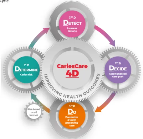
Figure 1. CariesCare 4D Cycle.
Author Accepted Manuscript version of Martignon, Stefania et al. "CariesCare practice guide: consensus on evidence into practice". British
Dental Journal. 2019, 227(5). 353-362. https://doi.org/10.1038/s41415-019-0678-8
What does CariesCare International aim to do?
It shares the same goals as ICCMS™ (International Caries Classification and Management System)1-4, which are:
- To prevent new caries lesions from appearing
- To prevent existing caries lesions from advancing further
- To preserve tooth structure
- with non-operative care at more initial stages and
- conservative operative care at more extensive caries stages - To manage caries risk factors
- To be alert to changes at both the tooth & patient levels through with periodic monitoring and review - To lead to improved health outcomes for patients.
Where does the CariesCare Practice Guide come from?
The CariesCare Practice Guide is derived from ICCMS™, which was developed through consensus with international experts by the ICDAS Foundation charity. This journey was started in 2002 with the founding of ICDAS; an international group of cariologists who, working with a wide range of other parties along the way, have systematically involved a large number of international experts in a number of high-quality, international, peer-reviewed activities1-10.
The CariesCare Practice Guide is based upon best available evidence and expert opinion and has been specifically tailored for use in dental practice. It has been developed through current consensus from experts as a simpler and shorter version of the full ICCMS™ Guide2, which was itself developed using consensus agreement based on quality ranked evidence. Methods of defining consensus
vary11; in these cases, large numbers of acknowledged experts in the field from many different countries have together considered
systematic reviews and research evidence and agreed the final text which they viewed as best practice for the clinical care of patients in a primary care setting.
Who is the CariesCare Practice Guide for?
It is designed to help dentists and healthcare teams help patients of all ages control the caries process and maintain health.
How can the CariesCare Practice Guide be used?
It guides the dental team through a 4-step structured process, leading to personalised interventions specific for each individual patient’s risks and needs. The 4 interlinked steps in the cycle (see Figure 1) all start with ‘D’: Determine Caries-risk; Detect lesions, stage their severity and assess their activity status; Decide on the most appropriate Care Plan for the specific patient at that time; and then Do the preventive and tooth preserving care which is needed (including risk-appropriate preventive care; control of initial non-cavitated lesions; and conservative restorative treatment of deep dentinal and cavitated caries lesions). These are referred to in CariesCare International as the 4D’s.
Why this Guide and why this approach?
Multiple sources for evidence-based dentistry exist, however, it can be confusing for clinicians to collect, critically analyse and implement all the relevant information into daily practice. CariesCare International has designed this practice-friendly consensus guide to summarise best practice as informed by the best available evidence. It has the objective of putting the patient’s health at the center of a risk based personalised care plan. It will also help with international trends in Practice that “Puts the mouth back in
the body” and links oral health to general health. For example, routinely determining and then addressing excessive sugar
consumption in dental practice may not only impact on oral health positively but also on disorders with the same risk factors such as obesity and diabetes. Following the guide should also increase patient satisfaction, involvement, wellbeing and value by being less invasive and health focused. For the dentist it should also provide benefits at the professional and practice levels including improved medico-legal protection.
Further references can be found at the end of this document for readers that may wish to consult the bibliography selected by the CariesCare international experts.
Core References:
1. Pitts N B, Ekstrand K R; ICDAS Foundation International Caries Detection and Assessment System (ICDAS) and its International Caries Classification and Management System (ICCMS) - methods for staging of the caries process and enabling dentists to manage caries. Community Dent Oral Epidemiol 2013; 41:e41-52.
2. Pitts N B, Ismail A I, Martignon S, Ekstrand K, Douglas G V A, Longbottom C. ICCMS™ Guide for Practitioners and Educators. Zenodo. 2014. Online information available at https://doi.org/10.5281/zenodo.853106 (accessed February 2018).
3. Ismail A I, Pitts N B, Tellez M; Authors of International Caries Classification and Management System (ICCMS), Banerjee A, Deery C, Douglas G, Eggertsson H, Ekstrand K, Ellwood R, Gomez J, Jablonski-Momeni A, Kolker J, Longbottom C, Manton D, Martignon S, McGrady M, Rechmann P, Ricketts D, Sohn W, Thompson V, Twetman S, Weyant R, Wolff M, Zandona A. The International Caries Classification and Management System (ICCMS™) An Example of a Caries Management Pathway. BMC Oral
Health 2015;15 Suppl 1:S9.
4. International Caries Classification and Management system (ICCMS™). 2019. Online information available at https://www.iccms-web.com/ (accessed February 2018). 5. Pitts N. “ICDAS” – an international system for caries detection and assessment being developed to facilitate caries epidemiology, research and appropriate clinical
management. Community Dental Health 2004; 21:193-198.
6. Ismail A I, Sohn W, Tellez M, Amaya A, Sen A, Hasson H, Pitts N B. The International Caries Detection and Assessment System (ICDAS): an integrated system for measuring dental caries. Community Dent Oral Epidemiol 2007; 35:170-178.
7. Selwitz R H, Ismail A I, Pitts N B. Dental caries. Lancet 2007; 369(9555):51-59.
8. Pitts N B, Zero D T, Marsh P D, Ekstrand K, Weintraub J A, Ramos-Gomez F, Tagami J, Twetman S, Tsakos G, Ismail A. Dental caries. Nat Rev Dis Primers 2017; 3:17030. 9. Dental Policy Lab 1 – June 2017, King’s College London Dental and Policy Institutes and the Alliance for a Cavity Free Future. Towards a Cavity Free Future – How do we accelerate a policy shift towards increased resource allocation for caries prevention and control? 2017. Online information available at https://www.kcl.ac.uk/dentistry/newsevents/news/newsrecords/2017/November/Towards-a-cavity-free-future.aspx (accessed February 2018).
10. Dental Policy Lab 2 – July 2018, King’s College London Dental and Policy Institutes and the Alliance for a Cavity Free Future. Paying for Health in Dentistry – How do we create and implement acceptable prevention based dental payment systems to achieve and maintain health outcomes? 2019. Online information available at https://www.acffglobal.org/wp-content/uploads/2019/02/Towards-paying-for-health-in-Dentistry-Policy-Lab-Report.pdf (accessed January 2019).
1
stD: DETERMINE Caries Risk
1-8, 12-21The Patient Perspective: Understanding their personal level of risk of disease is a key determinant of a
patient’s motivation to engage with healthcare and modify their own behavior to enhance their oral health.
Patients who perceive that they are susceptible to a disease are more likely to take action to ameliorate the
impact of that susceptibility.
What it is:
Caries risk assessment is the first essential step in the 4D-cycle for effective and personalised care. The aim of this step
is to assess the probability of whether a patient will develop carious lesions in the near future, and the likelihood that
there will be a progression of lesions if already present. Caries risk assessment also helps the dental team understand
why the patient has disease activity and consequently informs on adjustments that might be made to improve their risk
status. Knowing a patient’s caries risk will aid clinical decision-making and enable an personalised caries management
plan to be developed.
How to assess the patient’s caries risk:
Lots of tools already exist which help clinicians to systematically assess caries risk. Common tools are Cariogram, ADA,
CAMBRA and ICCMS™; many of these use 3 or more categories of caries risk. However, in practice, it is probably quicker,
easier and sufficient to focus on correctly identifying patients at the extremes of the spectrum of risk because those at
‘low risk’ of caries and those at ‘high risk’ of caries have clear management needs. Therefore, the CariesCare Practice
Guide uses just two risk categories, “at lower risk” and “at higher risk”, when choosing between caries management
options.
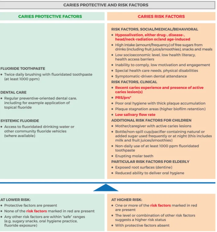
Risk factors and protective factors:
A patient’s risk level is derived from social, medical, behavioural (oral hygiene, diet, etc.) and past dental histories,
together with an oral examination. The clinician must weigh-up the patient’s risk and protective factors against each
other in order to assess the likely risk of future caries. Low risk is easy to identify as the absence of both caries risk
factors and active lesions. The most important information to consider is set out in Figure 2.
Good practice points:
• Patients’ caries risk must be assessed regularly since their risk category may change over time and should be
documented in their health record.
• Risk assessment should inform the frequency of patient recall. Patients with higher caries risk should have shorter
recall period than patients at lower risk patients, for monitoring, re-evaluation, and provision of preventive
interventions.
• The risk level should be clearly communicated to the patient and influence clinical decision-making regarding
treatment needs and alternatives, and the provision of other services.
• Whichever of the many risk assessment tools available is used, it should be integrated into the oral health record
and if possible, into a digital record system.
• Sugar is an important risk factor for caries initiation and progression but it is also a common risk factor for obesity,
diabetes and cardiovascular disease. Reducing sugar consumption is therefore important for both oral and general
health.
Figure 2. Caries Protective and Risk Factors
.
Guidance References:
12. Bratthall D, Hänsel Petersson G. Cariogram - a multifactorial risk assessment model for a multifactorial disease. Community Dent Oral Epidemiol 2005; 33:256-264. 13. Twetman S, Fontana M. Patient caries risk assessment. Monogr Oral Sci 2009; 21:91-101.
14. Twetman S, Fontana M, Featherstone J D. Risk assessment - can we achieve consensus? Community Dent Oral Epidemiol 2013; 41:e64-70.
15. Tellez M, Gomez J, Pretty I, Ellwood R, Ismail A I. Evidence on existing caries risk assessment systems: are they predictive of future caries? Community Dent Oral
Epidemiol 2013; 41:67-78.
16. Baginska J, Stowska W. Pulpal Involvement-Roots-Sepsis Index: A New Method for Describing the Clinical Consequences of Untreated Dental Caries. Med Princ Pract 2013; 22:555-560.
17. Moynihan P J, Kelly S A. Effect on caries of restricting sugars intake: systematic review to inform WHO guidelines. J Dent Res 2014; 93:8-18.
18. Cagetti M G, Bontà G, Cocco F, Lingstrom P, Strohmenger L, Campus G. Are standardized caries risk assessment models effective in assessing actual caries status and future caries increment? A systematic review. BMC Oral Health 2018; 18:123.
19. Featherstone J D B, Alston P, Chaffee B W, Rechmann P. Caries Management by Risk Assessment (CAMBRA): An Update for Use in Clinical Practice for Patients Aged 6 Through Adult. CDA CAMBRA: A Comprehensive Caries Management Guide for Dental Professionals. 2019. Online information available at https://www.cdafoundation.org/Portals/0/pdfs/cambra_handbook.pdf (accessed February 2019).
20. Featherstone JDB, Crystal YO, Chaffee BW, Zhan L, Ramos-Gomez F. An Updated CAMBRA Caries Risk Assessment Tool for Ages 0 to 5 Years. CDA CAMBRA: A Comprehensive Caries Management Guide for Dental Professionals. 2019. Online information available at CDA https://www.cdafoundation.org/Portals/0/pdfs/cambra_handbook.pdf (accessed February 2019).
2
ndD: DETECT &
ASSESS- Caries staging and activity
1-10, 22-36The Patient Perspective:
Assessment is the foundation of all care planning. Practitioner and patient work together to
create a shared understanding of the patients’ current health status and their priorities. Conceiving of caries severity
as a series of stages helps to identify the importance of both patient and practitioner behavior in modifying the
disease process.
What it is:
Caries staging and activity assessment is the second essential step in the 4D-cycle for effective and personalised care. It
builds on the knowledge acquired from the 1
stD. The aim is to examine the patient carefully for caries lesions, combining
this clinical assessment with information from radiographs when available. This step will involve differentiating caries
lesions from other pathologies/conditions such as erosive tooth wear, developmental defects, etc., as well as noting the
stage of any caries present (initial, moderate or extensive) and the activity of lesions (likely active or likely inactive).
Additionally, this step considers the patient’s past caries experience (including number of restorations, state of previous
restorative work, teeth extracted due to caries, and dental sepsis). Caries staging and activity assessment also helps
clinical decision-making and enables the development of an individualised caries management plan.
How to conduct the caries staging and activity assessment:
The caries assessment is based on visual examination of clean teeth in combination with, where possible, a radiographic
examination of posterior teeth (bite-wing x-rays). It is worth remembering that detecting smaller initial stage caries
lesions may be more difficult as they develop in areas of plaque stagnation, thus removing plaque is essential.
• Stage the severity of caries lesions (Table 1: coronal caries; Table 4: root caries). These categories based upon surface
characteristics of the lesion seen clinically are linked to the histological depth of the lesion.
• Where there are radiographs, the radiographic depth of a lesion is combined with its clinical appearance (as shown in
Table 2 for coronal caries) to determine the stage of caries.
• Once the severity stage of a caries lesion has been determined, its activity is assessed (Table 3: coronal caries).
Coronal caries
Severity staging:
3 key visual caries stages can be discriminated to help inform non-operative/operative care decisions: Table 1. ICDAS-merged visual coronal caries stages and related characteristics.ICDAS-merged visual coronal caries stages and related characteristics
Sound
(ICDAS 0)
No evidence of change in enamel translucency due to caries after plaque removal and air-drying.
Initial caries lesions
(ICDAS 1-2)Changes in enamel seen as a carious opacity or visible discolouration (white/brown spot) not consistent with clinical appearance of sound enamel with no evidence of surface breakdown, no underlying dentine shadowing or cavitation.
Moderate
caries
lesions
(ICDAS 3-4) Moderate Enamel breakdown (3)White/brown spot lesion with localized micro-cavity/ discontinuity, without visible dentine exposure. Best seen after air-drying.
Note: this category represents the highest challenge to inform non-operative/ operative care, which will depend on further information: radiographic lesion’s depth (deeper than external dentine third), together with patient´s caries risk.
Moderate Underlying dentinal shadow(4)
Obviously discoloured dentine visible through apparently intact or microcavitated enamel surface, which originated on the surface being evaluated. Often seen easiest with the tooth surface wet.
Extensive caries lesions
(ICDAS 5-6) Obvious visible dentine cavity in opaque/discoloured enamel. A WHO/CPI/PSR probe can gently confirm the cavity extends into dentine.
Note: Non-carious surfaces with developmental defects of enamel (including fluorosis), erosive tooth wear, and extrinsic/intrinsic stains are considered as sound for caries.
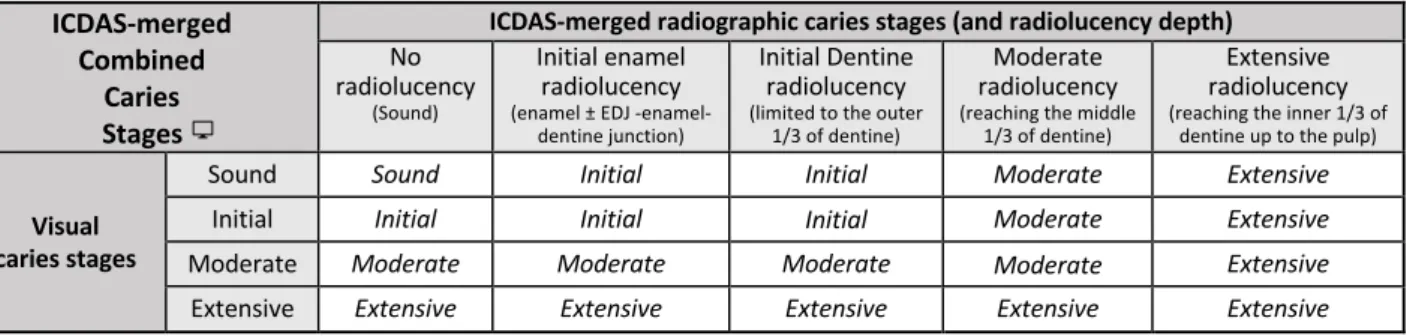
Visual combined with radiographic:
Radiographically,
ICDAS classifies coronal caries into three key caries stages (initial, moderate and extensive) that, in combination with the visual staging help inform non-operative/operative care decisions.Table 2. ICDAS-merged radiographic and visual combined caries stages.
ICDAS-merged
Combined
Caries
Stages
ICDAS-merged radiographic caries stages (and radiolucency depth)
No radiolucency
(Sound)
Initial enamel radiolucency
(enamel ± EDJ -enamel-dentine junction)
Initial Dentine radiolucency
(limited to the outer 1/3 of dentine)
Moderate radiolucency
(reaching the middle 1/3 of dentine)
Extensive radiolucency
(reaching the inner 1/3 of dentine up to the pulp)
Visual caries stages
Sound Sound Initial Initial Moderate Extensive
Initial Initial Initial Initial Moderate Extensive
Moderate Moderate Moderate Moderate Moderate Extensive
Extensive Extensive Extensive Extensive Extensive Extensive
Note for edition: (ICON to link to ICCMS webpage, here and elsewhere)
CARS (Caries associated to restoration or sealant):
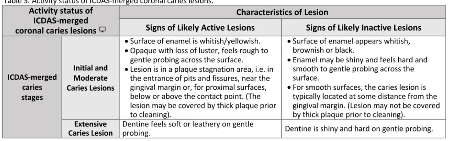
Same stages as coronal primary caries apply, but the caries lesion is located in association to a restoration or a sealant. Differentiation should be made from the status of the restoration or sealant: Good margin; Defective (plaque-retentive - can be adapted); Defective (needs replacement).Activity status:
For each coronal caries lesion assess the activity status using clinical parameters to inform either Likely active orLikely inactive:
Activity status of
ICDAS-merged
coronal caries lesions
Characteristics of Lesion
Signs of Likely Active Lesions
Signs of Likely Inactive Lesions
ICDAS-merged caries stages Initial and Moderate Caries Lesions
• Surface of enamel is whitish/yellowish. • Opaque with loss of luster, feels rough to
gentle probing across the surface. • Lesion is in a plaque stagnation area, i.e. in
the entrance of pits and fissures, near the gingival margin or, for proximal surfaces, below or above the contact point. (The lesion may be covered by thick plaque prior to cleaning).
• Surface of enamel appears whitish, brownish or black.
• Enamel may be shiny and feels hard and smooth to gentle probing across the surface.
• For smooth surfaces, the caries lesion is typically located at some distance from the gingival margin. (Lesion may not be covered by thick plaque prior to cleaning).
Extensive
Caries Lesion Dentine feels soft or leathery on gentle probing. Dentine is shiny and hard on gentle probing.
Root caries:
Severity staging: Characterised by colour change (light/dark brown or black). Three key root caries stages can be
discriminated that will help inform non-operative/operative care decision:
Table 4. ICDAS-merged root caries stages and related characteristics.
ICDAS-merged root caries stages and related characteristics
Sound No evidence of change in colour.
Root Initial caries lesion Loss of anatomic contour continuity <0.5 mm (without a frank caries cavity).
Root Moderate caries lesion Depth/Width: 0.5mm - 2mm.
Root Extensive caries lesion Depth/Width: > 2mm.
Activity status:
For each root caries lesion assess the activity status using clinical parameters to inform either Likely Active or Likely Inactive. If the lesion is located ≥1 mm from the gingival margin, hard to gentle probing, no cavitation or the surroundings of thecavity, smooth to probing, and dark brown/black it represents a Likely Inactive Root Caries Lesion. Conversely, if the lesion is located ≤1 mm from the gingival margin, leathery/soft to gentle probing, cavitation, and light brown/yellowish it represents a Likely Active Root Caries Lesion.
Together, the caries severity stage, along with the activity likelihood of each lesion and the patient’s caries risk status
directs care.
Good practice points:
• Clinical caries severity staging is fast and easy after training which is available through the ICDAS/ICCMS™ webpage . • Sharp probing does not improve detection and it causes further damage to caries lesions.
• Clinical caries severity staging does not require any specific device.
•
Remember that the radiographic images on bite-wing projections show a range of sizes of approximal lesions but are not able to reveal many occlusal lesions until they are quite extensive.•
The caries staging and activity assessment should be integrated into the oral health record and if possible into a digital record system.Guidance References:
22. Ekstrand K R, Ricketts D N, Kidd E A. Occlusal caries: pathology, diagnosis and logical management. Dent Update 2001;28: 380-387.
23. Ekstrand K R, Martignon S, Ricketts D J, Qvist V. Detection and activity assessment of primary coronal caries lesions: a methodologic study. Oper Dent 2007; 32:225-235.
24. Braga M M, Martignon S, Ekstrand K R, Ricketts D N, Imparato J C, Mendes F M. Parameters associated with active caries lesions assessed by two different visual scoring systems on occlusal surfaces of primary molars - a multilevel approach. Community Dent Oral Epidemiol 2010; 38:549-558.
25. Braga M M, Ekstrand K R, Martignon S, Imparato J C, Ricketts D N, Mendes F M. Clinical performance of two visual scoring systems in detecting and assessing activity status of occlusal caries in primary teeth. Caries Res 2010; 44:300-308.
26. Ekstrand K R, Luna L E, Promisiero L, Cortes A, Cuevas S, Reyes J F, Torres C E, Martignon S. The reliability and accuracy of two methods for proximal caries detection and depth on directly visible proximal surfaces: an in vitro study. Caries Res 2011; 45:93-99.
27. Brocklehurst P, Ashley J, Walsh T, Tickle M. Relative performance of different dental professional groups in screening for occlusal caries. Community Dent Oral Epidemiol 2012; 40:239-246.
28. Ekstrand K R, Poulsen J E, Hede B, Twetman S, Qvist V, Ellwood R P. A randomized clinical trial of the anti-caries efficacy of 5,000 compared to 1,450 ppm fluoridated toothpaste on root caries lesions in elderly disabled nursing home residents. Caries Res 2013; 47:391-398.
29. Banerjee A, Watson T F. Pickard's Guide to Minimally Invasive Operative Dentistry. 10th ed. Oxford: Oxford University Press, 2015.
30. Gimenez T, Piovesan C, Braga M M, Raggio D P, Deery C, Ricketts D N, Ekstrand K R, Mendes F M. Visual inspection for caries detection: A systematic review and meta-analysis. J Dent Res 2015; 94:895-904.
31. Pretty I A, Ekstrand K R. Detection and monitoring of early caries lesions: A review. Eur Arch Paediatr Dent 2016; 17:13-25.
32. Mattos-Silveira J, Oliveira M M, Matos R, Moura-Netto C, Mendes F M, Braga M M. Do the ball-ended probe cause less damage than sharp explorers? An ultrastructural analysis. BMC Oral Health 2016; 16:39.
33. Cortes A, Ekstrand K R, Martignon S. Visual and radiographic merged-ICDAS caries progression pattern in 2-6 years old Colombian children: Two-year follow-up. Int J
Paediatr Dent 2018. doi: 10.1111/ipd.12448. PMID: 30431189.
34. Ekstrand K R, Gimenez T, Ferreira F R, Mendes F M, Braga M M. The International Caries Detection and Assessment System - ICDAS: A Systematic Review. Caries Res 2018; 52:406-419.
35. Martignon S, Cortes A, Gómez S I, Castiblanco G A, Baquero X, Franco-Triviño A M, Palacio-Benavides J C, Gamboa L F, Villena R S. How long does it take to examine young children with the caries ICDAS system and how do they Respond? Braz Dent J 2018; 29:374-380.
36. Drancourt N, Roger-Leroi V, Martignon S, Jablonski-Momeni A, Pitts N, Doméjean S. Carious lesion activity assessment in clinical practice: a systematic review. Clin Oral
Investig 2019; 23:1513-1524.
3
rdD:
DECIDE - Personalised care plan: Patient and tooth levels
1-45The Patient Perspective:
A core component of Patient Centred Care is the discussion of a shared personalised plan
of care. The DECIDE stage focuses on identifying that plan and making it explicit. The co-creation of a care plan with
the patient enhances the patients’ understanding and commitment to the plan.
What it is:
DECIDE: The Personalised care plan, at the patient and the tooth levels, is the third essential step in the 4D-cycle for
effective and personalised care. The aim of this step is to synthesise all of the information gathered about the patient’s
caries risk (1
stD) and any caries lesions (severity and activity) (2
ndD) to develop an informed, risk-based, tooth preserving
care plan. This step is very important because:
• It determines at the tooth level what type of treatment to provide: preventive vs. surgical. • It helps to maintain good oral health and avoid unnecessary removal of tooth tissue. • It helps allocate resources appropriately based on risk.
• It involves the patient’s active engagement on the importance of oral health, avoiding future caries and operative treatment. • It helps clinicians to determine the recall interval for the patient.
How to develop the personalised care plan:
This step involves discussion with the patient as well as synthesis and consolidation of all the information gathered
about the patient’s history and clinical findings as described below. There are practical synthesis guides in relation to
caries lesions’ stage and activity likelihood, and patient’s risk classification (e.g. ICCMS™ Guideline and webpage).
Decision trees will help determine the Personalised care plan, both at the patient and at the tooth levels.
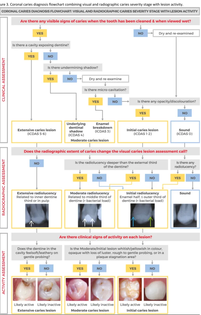
Arrive at a diagnosis for each Caries lesion:
Caries diagnosis is the result of combining the caries severity stage, as determined from visual and radiographic
examination (initial, moderate or extensive), with an accompanying lesion activity assessment into the categories
below:
o Initial active or inactive o Moderate active or inactive o Extensive active or inactive
Note that as with patient’s caries risk, lesion activity can change over time, and therefore so can a lesion’s diagnosis.
Taking into account the information gathered from the 2
ndD (described in Tables 1-4), Figure 3 shows a flowchart that
outlines how the process flows from clinical assessment through radiographic assessment and lesion activity assessment
to categorise coronal caries lesions as an example.
Arrive at a diagnosis of Patient’s caries risk:
Patient’s caries risk will have been determined after analysing history (1
stD) and intraoral risk factors, including the
presence of active caries lesions (2
ndD) (Figure 2). The influence of caries risk on deciding the appropriate care plan lies
mostly at the two extremes of high and low risk. Correctly identifying patients who are at particularly at lower risk and
those who are at higher risk guides risk-informed appropriate care.
Figure 4 shows the Caries risk level classification flowchart outlining the process flows to establish the level of caries
risk.
Decide upon the Personalised care plan - Patient and Tooth levels:
Patient’s risk management plan: This is tailored to the individual patient and will involve actions to protect sound tooth
surfaces from developing new caries lesions, arresting currently active lesions, and maintaining inactive lesions from
progressing. In addition, it aims to lower the risk status of the patient if not already low, and to maintain low risk status.
A preventive plan should address both homecare and clinical interventions/approaches informed by the caries risk
status of the patient.
Tooth-level management plan: Information on each caries lesion will be synthesised in terms of, whether or not, they
are likely active and if they are of Initial, Moderate or Extensive severity:
o Initial likely active / Initial likely inactive
o Moderate likely active / Moderate likely inactive o Extensive likely active / Extensive likely inactive.
Figure 5 shows the Patient’s Care Plan flowchart outlining the logical flow of integrating the management of individual
lesions assessed for activity and management of risk at the patient level. The three management options for surfaces
at the end of this flow are keeping sound surfaces sound, controlling lesions with non-operative care and providing
tooth-preserving operative care for only those lesions that need it. The management options to control caries risk at
the patient level are also outlined.
Figure 4. Patient’s caries risk level classification flowchart.
Figure 5. Patient’s Care Plan Decision Flowchart.
Guidance References:
37. Hänsel Petersson G, Åkerman S, Isberg PE, Ericson D. Comparison of risk assessment based on clinical judgement and Cariogram in addition to patient perceived treatment need. BMC Oral Health 2016; 17:13.
38. Schwendicke F, Frencken J E, Bjørndal L, Maltz M, Manton D J, Ricketts D, Van Landuyt K, Banerjee A, Campus G, Doméjean S, Fontana M, Leal S, Lo E, Machiulskiene V, Schulte A, Splieth C, Zandona A F, Innes N P. Managing Carious Lesions: Consensus Recommendations on Carious Tissue Removal. Adv Dent Res 2016; 28:58-67. 39. Kühnisch J, Ekstrand K R, Pretty I, Twetman S, van Loveren C, Gizani S, Spyridonos Loizidou M. Best clinical practice guidance for management of early caries lesions
in children and young adults: an EAPD policy document. Eur Arch Paediatr Dent 2016; 17:3-12.
40. Tonetti M S, Bottenberg P, Conrads G, Eickholz P, Heasman P, Huysmans M C, López R, Madianos P, Müller F, Needleman I, Nyvad B, Preshaw P M, Pretty I, Renvert S, Schwendicke F, Trombelli L, van der Putten G J, Vanobbergen J, West N, Young A, Paris S. Dental caries and periodontal diseases in the ageing population: call to action to protect and enhance oral health and well-being as an essential component of healthy ageing - Consensus report of group 4 of the joint EFP/ORCA workshop on the boundaries between caries and periodontal diseases. J Clin Periodontol 2017; 44 Suppl 18:S135-S144.
41. Slayton RL, Urquhart O, Araujo MWB, Fontana M, Guzmán-Armstrong S, Nascimento MM, Nový BB, Tinanoff N, Weyant RJ, Wolff MS, Young DA, Zero DT, Tampi MP, Pilcher L, Banfield L, Carrasco-Labra A. Evidence-based clinical practice guideline on nonrestorative treatments for carious lesions. A report from the American Dental Association. JADA 2018; 149:837-849.
42. Ricketts D, Innes N, Schwendicke F. Selective Removal of Carious Tissue. Monogr Oral Sci 2018; 27:82-91.
43. Fontana M, Pilcher L, Tampi M P, Urquhart O, Slayton R L, Araujo M W B, Carrasco-Labra A. Caries management for the modern age: Improving practice one guideline at a time. J Am Dent Assoc 2018; 149:935-937.
44. Rechmann P, Chaffee B W, Rechmann B M T, Featherstone J D B. Caries Management by Risk Assessment: Results from a Practice-Based Research Network Study. J
Calif Dent Assoc 2019; 47:15-24.
45. Urquhart O, Tampi M P, Pilcher L, Slayton R L, Araujo M W B, Fontana M, Guzmán-Armstrong S, Nascimento M M, Nový B B, Tinanoff N, Weyant R J, Wolff M S, Young D A, Zero D T, Brignardello-Petersen R, Banfield L, Parikh A, Joshi G, Carrasco-Labra A. Nonrestorative Treatments for Caries: Systematic Review and Network Meta-analysis. J Dent Res 2019; 98:14–26.
4
thD: DO - Appropriate Tooth-preserving & patient-level prevention & control
1-10, 37-54The Patient Perspective:
Having finalized a mutually agreed care plan in the DECIDE phase, the DO phase involves
both the planning and implementation of that care plan. Planning is good for both the practitioners and patients -
making an explicit plan has repeatedly been shown to ensure adherence to healthcare recommendations. The DO
stage is not only about professional treatment but working with the patient to ensure that they have a clear plan of
action to support their own oral health.
What it is:
DO: Appropriate Tooth-preserving & patient-level prevention & control is the fourth essential step in the 4D-cycle that
delivers the Personalised Comprehensive Caries Care Plan built on the outcomes of the first three Ds. This 4
thD consists
of two elements:
• Managing patient’s caries risk, tailored at the individual level with actions to improve the risk status where possible. • Managing individual caries lesions according to their severity and activity. Caries care options may differ between the
primary and permanent dentition.
How to conduct appropriate tooth-preserving & patient level prevention and control:
Managing the patient’s caries risk
• The caries risk factor management plan can involve two levels:
o Homecare approach: Activities to be conducted at home by the patient or their parent/guardian/carer as instructed by a member of the dental team, which takes into account the patient’s needs, opportunities and preferences. Activities include fluoridated toothpaste use, fluoride rinse/gel, toothbrushing, interproximal cleaning and behaviours related to oral health including diet and other oral hygiene advice.
o Clinical interventions: Activities conducted at the practice, including discussing personalised ways of improving oral-health related behaviour, topical fluoride application at a frequency appropriate to the patient’s risk classification, sealant application, one-to-one dietary advice (with emphasis on sugars), and if required, managing hyposalivation or other specific risk factors.
• There is strong evidence for the use of topical fluoride both professionally applied and for home use in the prevention of dental caries.
• Based on the available evidence – concentrate on delivering advice on brushing twice-a-day with a fluoridated toothpaste that is appropriate to the age of the patient and their risk factors.
• Advice should include basic details of when brushing is most effective and how to maintain the fluoridated toothpaste in contact with the teeth (spit, don’t rinse).
• Emphasis should be placed on improving oral hygiene and delivery of topical fluoride in plaque stagnation areas where caries commonly occurs.
• Given the understanding of the disease process dietary advice should be directed at identifying sugars in the diet (including hidden sugars), reducing the amount/frequency of sugar intake and suggesting safe alternatives.
• Organise and agree with the patient or their carer a risk-based recall (re-care) interval depending on risk classification.
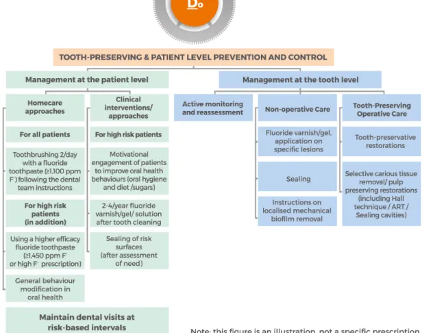
Managing tooth-level caries lesions
• Care Options for caries lesions include:
o Non-Operative care (NOC) – this is non-surgical preventive care to control caries
o Tooth-preserving Operative care (TPOC) – this is minimally interventive surgical treatment
• The severity status of the caries lesion will inform and dictate preventive (non-operative) or operative management, but lesion activity should also be considered:
o Initial caries lesions should be managed with Non-Operative care when Active, and when Inactive should be reviewed at recall appointments for any change in status
o Moderate caries lesions (ICDAS 3 and 4) management depends on a number of factors including patient-level risk status, radiographic appearance, lesion activity, and whether or not there is surface breakdown: if the lesion radiographically extend up to the outer dentine third (mainly in ICDAS 3 or microcavity) it is feasible to manage with Non-Operative Care if inactive, and in some cases in the absence of other risk factors and if the patient is compliant.
o Extensive caries lesions (ICDAS 5 and 6) should generally be managed with Tooth-preserving Operative care according to lesion severity and pulp involvement.
• Take into account patient level strategies for therapeutic control of initial lesions and:
o In children there is strong evidence for the use of fissure sealants for the caries management of pit and fissure caries, but there is a trend for strict indications for preventive sealants on sound teeth in high caries risk children and an increased focus on therapeutic sealants for initial caries lesions which cannot be controlled by non-operative caries measures. o Where operative intervention is required tooth-preserving operative care should be provided according to the patient’s
needs (age, setting, environment). There is evidence that more conservative caries removal techniques are effective in preserving tooth tissue and avoiding pulpal complications.
o In restored teeth consideration should be given to repair of a restoration rather than re-restoration where possible to avoid further loss of tooth tissue by unnecessary removal of sound sections of the old restoration.
Figure 6. Tooth-preserving and Patient Level Prevention and Control Flowchart.
Good practice points:
• Wherever possible caries should be managed with prevention (non-operative interventions) to avoid unnecessary surgical intervention.
• Where surgical intervention is required tooth-preserving operative care should be considered.
• Management options are dependent on patient- and tooth-level assessment (risk, caries lesions, restorative status and patient compliance).
• In some cases moderate or extensive inactive caries lesions may require TPOC due to local factors such as the presence of a removable prosthesis or a clasp contacting the lesion.
• Recall interval should be based upon a combination of risk assessment and management and clinical procedures carried out. • Erosive tooth wear, developmental defects of enamel, and periodontal status should be considered for comprehensive care. • Most evidence is based on children, adolescents and young adults but good clinical practice would suggest this is applicable for
older adults.
• Note: Local adaptations may be required, for example according to varying levels of systemic fluoride concentration.
• The intensity of the risk-based intervention is cumulative, so for patients with higher risk all preventive interventions prescribed for patients with lower caries risk should also be considered.
• Clinicians and their teams should be familiar with evidence-based prevention guidance applicable locally to them.
• Dentists should keep up to date with both changes in cavity preparation philosophy and the requirements and opportunities given by new developments in dental materials.
• Successful use of direct adhesive restorative techniques and materials require effective moisture control and rubber dam isolation should be considered over relative isolation with cotton rolls.
• As the Minamata Treaty is implemented internationally there is both an opportunity for prevention and a need for caution when dental amalgam is replaced by more technique-sensitive materials.
Guidance References:
46. Clinical Guideline 19. Dental recall: recall interval between routine dental examinations National Institute for Clinical Excellence (NICE), Department of Health, London. 2004. Online information available at www.nice.org.uk/CG019NICEguideline (accessed November 2018).
47. Splieth C H, Ekstrand K R, Alkilzy M, Clarkson J, Meyer-Lueckel H, Martignon S, Paris S, Pitts N B, Ricketts D N, van Loveren C. Sealants in dentistry: outcomes of the ORCA Saturday Afternoon Symposium 2007. Caries Res 2010; 44:3-13.
48. Ricketts D, Lamont T, Innes N P, Kidd E, Clarkson J E. Operative caries management in adults and children. Cochrane Database Syst Rev 2013; 3:CD003808. 49. Tellez M, Gomez J, Kaur S, Pretty I A, Ellwood R, Ismail A I. Non-surgical management methods of noncavitated carious lesions. Community Dent Oral Epidemiol 2013;
41:79-96.
50. Marinho V C, Worthington H V, Walsh T, Clarkson J E. Fluoride varnishes for preventing dental caries in children and adolescents. Cochrane Database Syst Rev 2013; 7:CD002279.
51. Marinho V C, Chong L Y, Worthington H V, Walsh T. Fluoride mouthrinses for preventing dental caries in children and adolescents. Cochrane Database Syst Rev 2016; 7:CD002284.
52. Innes N P, Frencken J E, Bjørndal L, Maltz M, Manton D J, Ricketts D, Van Landuyt K, Banerjee A, Campus G, Doméjean S, Fontana M, Leal S, Lo E, Machiulskiene V, Schulte A, Splieth C, Zandona A, Schwendicke F. Managing Carious Lesions: Consensus Recommendations on Terminology. Adv Dent Res 2016; 28:49-57.
53. Ahovuo-Saloranta A, Forss H, Walsh T, Nordblad A, Mäkelä M, Worthington H V. Pit and fissure sealants for preventing dental decay in permanent teeth. Cochrane
Database Syst Rev 2017; 31;7:CD001830.
54. Walsh T, Worthington H V, Glenny A M, Marinho V C, Jeroncic A. Fluoride toothpastes of different concentrations for preventing dental caries. Cochrane Database Syst
Concluding key points and Guidance for implementation
• CariesCare International is designed to support dentists and healthcare teams help patients of all ages control the caries process and maintain health over the life course.
• The systematic approach ensures that all the important steps involved in assessing and managing patients’ caries status are completed and recorded routinely.
• It is important to deliver all 4 Ds, they all contribute to optimal care and are required for the continuation of the cycle of care.
• It focuses attention on keeping sound surfaces sound and seeks the arrest/remineralisation of early caries lesions and encourages the use of minimally invasive techniques when caries removal is necessary.
•
It helps to clearly delineate for clinicians when preventive or surgical management of a caries lesion is most appropriate for each patient (taking into account attitudes and attendance behaviour).• Risk-based personalised recall and review is a key to deciding how rapidly the 4D cycle repeats.
• The 4D approach may help stimulate discussion with patients about their risk factors and involve them in determining what can be done to reduce them. For some risk factors this may not only reduce caries risk but may also reduce the risk of other disorders like obesity and diabetes.
• Health outcomes matter and are the purpose of this approach to care.
• 4D caries management is suitable for all ages throughout the lifecourse – but needs some change of emphasis at particular life stages.
• Dentists and the Dental and Health care teams can derive improved professional satisfaction using this patient-centred and preventive approach to caries management.
• Patients value a health focussed and personalised approach to care.
• The CariesCare aspects should integrate well with the rest of routine oral health care, including in particular erosive tooth wear, and periodontal disease assessment and management.
• CariesCare aims to integrate with the rest of health and well-being in a holistic way.
Implementation Points:
• “Glocal” is the watchword for successful implementation. This concept has been used successfully by the Alliance for a Cavity Free Future and takes key evidence from Global Evidence and Consensus and adapts it to the Local realities and cultures in specific Countries, areas and Practice settings.
• Modifications to reflect Local needs are acceptable, but care needs to be taken not to destroy the fundamentals of the CariesCare 4D System.
• An Educational online course (a “MOOC”) will be made available in the near future and there are tools already available, like training in visual caries criteria e-learnings and the CariesCare clinical case which follows.
• Collecting the required information efficiently on paper records can and is being done.
• However, moving forward, software development will ultimately help further integration into practice and help with integrated longitudinal health assessments.
• The shift in resource allocation towards prevention and “Paying for Health in Dentistry” are important in supporting dental teams in delivering the CariesCare International approach to caries prevention, control and management (Dental Policy Labs 1 & 2)9,10.
• In due course CariesCare International plans to develop as a community to support implementation and development - current examples include: 1) a consensus group of stakeholders co-creating a core Colombian oral health record, 2) a National French experiment looking at supporting the introduction of 4D caries management in general practice. CariesCare International (Figure 7) is working with the Alliance for a Cavity Free Future and King’s College London under the umbrella of the Global Collaboratory for Caries Management to help further implementation of this Guide.
Acknowledgements
The Authors acknowledge the research conducted by many contributors that underpinned this CariesCare International Guide and are indebted to the contributions made by all of the internationally mixed groups who attended the launch meeting of the Global Collaboratory for Caries Management at Kings College London in 2013 and the many who have helped since at meetings in Liverpool, Seattle, Philadelphia, London, Capetown, Greifswald, Dubai, Delhi and Tokyo to drive the ICCMSä initiative forward. We are also exceedingly grateful to all the individuals who have helped shape the CariesCare International initiative at meetings in Athens, Oslo, Copenhagen, Buenos Aires and London as well as to the numerous Organisations and Companies who have helped support this work and enabled progress to date.