HAL Id: hal-01308328
https://hal.sorbonne-universite.fr/hal-01308328
Submitted on 27 Apr 2016
HAL is a multi-disciplinary open access
archive for the deposit and dissemination of
sci-entific research documents, whether they are
pub-lished or not. The documents may come from
teaching and research institutions in France or
abroad, or from public or private research centers.
L’archive ouverte pluridisciplinaire HAL, est
destinée au dépôt et à la diffusion de documents
scientifiques de niveau recherche, publiés ou non,
émanant des établissements d’enseignement et de
recherche français ou étrangers, des laboratoires
publics ou privés.
Distributed under a Creative Commons Attribution| 4.0 International License
IDH1R132H Mutation Increases U87 Glioma Cell
Sensitivity to Radiation Therapy in Hypoxia
Xiao-Wei Wang, Marianne Labussière, Samuel Valable, Elodie A. Pérès,
Jean-Sébastien Guillamo, Myriam Bernaudin, Marc Sanson
To cite this version:
Xiao-Wei Wang, Marianne Labussière, Samuel Valable, Elodie A. Pérès, Jean-Sébastien Guillamo,
et al.. IDH1R132H Mutation Increases U87 Glioma Cell Sensitivity to Radiation Therapy in
Hy-poxia. BioMed Research International , Hindawi Publishing Corporation, 2014, 2014, pp.198697.
�10.1155/2014/198697�. �hal-01308328�
Research Article
IDH1
R132H
Mutation Increases U87 Glioma Cell
Sensitivity to Radiation Therapy in Hypoxia
Xiao-Wei Wang,
1,2,3Marianne Labussière,
1,2,3Samuel Valable,
4,5,6Elodie A. Pérès,
4,5,6Jean-Sébastien Guillamo,
4,5,6,7Myriam Bernaudin,
4,5,6and Marc Sanson
1,2,3,8,91Universit´e Pierre et Marie Curie-Paris 6, Centre de Recherche de l’Institut du Cerveau et de la Mo¨elle ´epini`ere (CRICM),
UMR-S975, 75013 Paris, France
2INSERM, U 975, 75013 Paris, France
3CNRS, UMR 7225, 75013 Paris, France
4CNRS, UMR 6301 ISTCT, CERVOxy group, GIP CYCERON, Boulevard Henri Becquerel, BP 5229, 14074 Caen cedex, France
5Universit´e de Caen Basse-Normandie, UMR 6301 ISTCT, 14000 Caen, France
6CEA, DSV/I2BM, UMR 6301 ISTCT, 14000 Caen, France
7CHU de Caen, Service de Neurologie, Boulevard Cˆote de Nacre, 14000 Caen, France
8AP-HP, Groupe Hospitalier Piti´e-Salpˆetri`ere, Service de Neurologie 2, 75013 Paris, France
9Service de Neurologie 2, Groupe Hospitalier Piti´e-Salpˆetri`ere, 75651 Paris Cedex 13, France
Correspondence should be addressed to Marc Sanson; [email protected] Received 12 February 2014; Accepted 6 April 2014; Published 7 May 2014 Academic Editor: Andrea Pace
Copyright © 2014 Xiao-Wei Wang et al. This is an open access article distributed under the Creative Commons Attribution License, which permits unrestricted use, distribution, and reproduction in any medium, provided the original work is properly cited. Objective. IDH1 codon 132 mutation (mostly Arg132His) is frequently found in gliomas and is associated with longer survival. However, it is still unclear whether IDH1 mutation renders the cell more vulnerable to current treatment, radio- and chemotherapy.
Materials and Methods. We transduced U87 with wild type IDH1 or 𝐼𝐷𝐻1𝑅132𝐻 expressing lentivirus and analyzed the
radiosensitivity (dose ranging 0 to 10 Gy) under normoxia (20% O2) and moderate hypoxia (1% O2). Results. We observed that
𝐼𝐷𝐻1𝑅132𝐻U87 cells grow faster in hypoxia and were more sensitive to radiotherapy (in terms of cell mortality and colony formation
assay) compared to nontransduced U87 and IDH1𝑤𝑡cells. This effect was not observed in normoxia. Conclusion. These data suggest
that𝐼𝐷𝐻1𝑅132𝐻mutation increases radiosensitivity in mild hypoxic conditions.
1. Introduction
The IDH1 gene encoding the cytoplasmic NADP+-dependent isocitrate dehydrogenase—and more rarely IDH2, encod-ing the mitochondrial isoform—are frequently mutated in gliomas, especially low grade gliomas and secondary glioblas-tomas [1]. IDH1/IDH2 mutation is associated with better clinical outcome, whatever the grade, but it is still not clear whether it is merely a prognostic marker or a predictor of the response to radiotherapy or chemotherapy [2–6]. Recent data IDH1/IDH2 mutation results in a new enzyme function catalyzing the NADPH-dependent reduction of
alpha-ketoglutarate to D-2-hydroxyglutarate (D-2HG) [7].
IDH1/IDH2 mutations result in D-2HG accumulation and
lowering NADPH levels. On one hand D-2HG inhibits various alpha-ketoglutarate dependant reactions, including histone and DNA demethylation, and is likely to promote— rather than inhibit—HIF1𝛼 degradation [8–11]. On the other hand, low NADPH levels might sensitize tumors to oxida-tive stress, potentiating response to radiotherapy, and may account for the prolonged survival of patients harboring the mutations.
Since the majority of gliomas are poorly responsive to current treatment regimens, the ability to enhance cell
Volume 2014, Article ID 198697, 5 pages http://dx.doi.org/10.1155/2014/198697
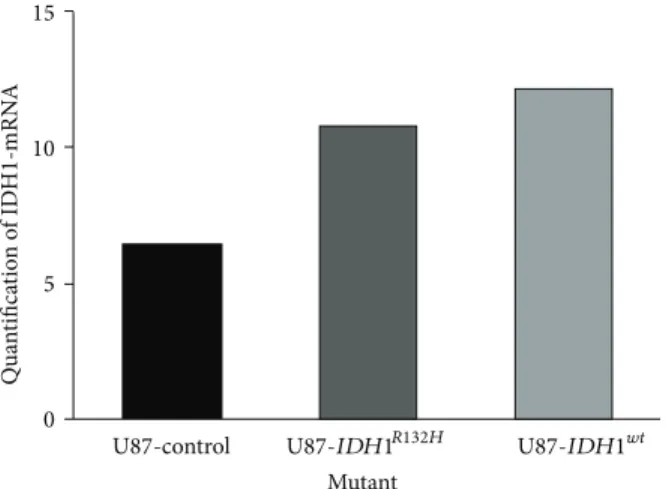
2 BioMed Research International 15 10 5 0 U87-control Mutant Qua n tifica tio n o f ID H 1-mRN A U87-IDH1R132H U87-IDH1wt
Figure 1: Real time PCR quantified the expression of the IDH1wtand IDH1R132Htransduced genes.
0 1 2 3 4 5 6 0 2 4 6 8 10 0 2 4 6 8 10
Days after seeding Days after seeding
0 1 2 3 4 5 6 U87 V ia b le cells (A U) V ia b le cells (A U) ∗ U87-IDH1wt U87 U87-IDH1wt U87-IDH1mutation U87-IDH1mutation
Figure 2: Effect of IDH1R132Hon U87 cell proliferation. U87, IDH1wt-U87, and IDH1R132H-U87 cells were incubated in normoxia 20% (left) or
hypoxia 1% (right) and cells were counted after 1, 3, and 7 days.
radio-chemosensitivity would be of clinical benefit. In this study, we characterized the impact of IDH1 mutation on U87 glioma cell growth and radiosensitivity.
2. Methods and Materials
2.1. Cell Culture and Hypoxia Treatment. The human
glioblastoma cell line U87 MG (HTB14) was obtained from the American Type Culture Collection (ATCC, Rockville, MD) and maintained in Dulbecco’s modified Eagle’s medium (DMEM), supplemented with 10% fetal bovine serum (FBS) and 1% penicillin/streptomycin. Normoxic cells (21% O2) were grown in a humidified-air atmosphere incubator containing 95% air/5% CO2 at 37∘C. Hypoxia experiments were performed in a controlled atmosphere chamber (INVIVO2 1000, Ruskinn, Awel, France) set at 1% O2, 94% N2, and 5% CO2at 37∘C.
2.2. Production of Recombinant Expression Lentiviruses.
A recombinant pLenti7.3/V5-TOPO expression vector
(Invitrogen’s ViraPowerTM HiPerformTM Lentiviral Expression Systems; catalog number K5320-00) containing the human IDH1 wild type and IDH1R132H cDNA was generated. The expression clones and the ViraPower Packaging Mix were cotransfected into the 293FT Cell line to produce lentiviral stocks, which were used to transduce the mammalian U87 cell line. U87-IDH1wtand U87-IDH1R132 stable cell lines were acquired using EmGFP selection by flow cytometry. The constructs was verified by DNA sequencing and RT-qPCR analysis.
2.3. Cell Proliferation Assay in Normoxia and in Hypoxia. To
evaluate the impact of IDH1 mutation on cell growth in nor-moxia and in hypoxia by trypan blue dye exclusion method, U87, U87-IDH1wt, and U87-IDH1R132Hcells (4000/well) plated in 24-well plates (6 plates in total) were incubated at 37∘C for six hours in normoxia to adhere; then 3 plates were removed at 37∘C in the controlled atmosphere chamber overnight. At 1, 3, and 7 days after exposure to normoxia and hypoxia, the cells were trypsinized, and the number of viable cells
0 2 4 6 8 10 0.1
1
0.1 1
Radiotherapy dose (Gy)
0 2 4 6 8 10
Radiotherapy dose (Gy)
Su rv iv in g f rac tio n Su rv iv in g f rac tio n U87 ∗ U87-IDH1wt U87-IDH1mutation U87 U87-IDH1wt U87-IDH1mutation
Figure 3: Effect of IDH1R132Hon U87 cell viability after irradiation. Transduced cells were plated and then irradiated with doses ranging from
0 to 10 Gy, in normoxia (20%) (left) and in hypoxia (1%) (right). Cells were counted 5 days later.
0 20 40 60 80 100 Normoxia Hypoxia R T ind uced cell de at h ( % ) U 87 U87 U 87 -ID H 1 wt U 87 -I DH1 wt U 87 -ID H1 mu ta ti o n U 87 -ID H1 mu ta ti o n
Figure 4: Cell viability after 8 Gy irradiation. Cells were counted
before 8 Gy irradiation and 5 days after, in normoxia (20% O2) (left)
and in hypoxia (1% O2) (right).
per well was determined by counting with trypan blue. The experiment was performed three times in triplicate each.
2.4. Comparative Cell Viability Assay after Irradiation, in Normoxia and in Hypoxia. To evaluate the effect of IDH1R132H
in the response to radiotherapy, U87 cells, IDH1wt-U87, and
IDH1R132H-U87 cells were plated (4.103 per well) in 24-well
plates. Six hours later at 37∘C in normoxia, plates were either kept in normoxia or incubated in the controlled atmosphere chamber 1% O2 overnight. The next day, cells were irradiated with doses ranging from 0 to 10 Gy in order to determine the most discriminating dose. Cells were fixed in paraformaldehyde (PFA) 4%, then stained with Hoechst 33342 (10𝜇g/mL in PBS, Sigma-Aldrich, France) and photographed in a blinded fashion under fluorescence (4 wells per condition; 4 photographs per well) at 24 h, 48 h, and
120 h, respectively. Cells were counted with ImageJ software (Rasband, WS, ImageJ, US NIH).
2.5. Colony-Formation Assay in Normoxia and in Hypoxia.
U87, IDH1wt-U87, and IDH1R132H-U87 cells were plated in 6-well containing 0.3% base agar layer. Six hours later, cells were either incubated in the hypoxic or normoxic chamber overnight. The next day, the cells were treated by radiotherapy at the Radiotherapy Department of the Centre de Lutte Contre le Cancer (CLCC) Franc¸ois Baclesse (Caen, France) using an X-ray generator with doses ranging 0–8 Gy (Therac 15-Saturne with a dose rate of 2 Gy/min) and then incubated again for colony formation. One month later, the colonies were fixed in 20% ethanol and stained with 0.05% crystal violet. Colonies that contained more than 50 cells were counted. Survival was calculated as the average number of colonies counted divided by the number of cells plated multiplicated by plating efficiency (PE), where PE is the fraction of colonies counted divided by cells plated without radiation. The clonogenic survival data were generated using JMP software. The experiment was performed five times in triplicate each.
2.6. Statistical Analysis. Results obtained in vitro were
expressed as mean± SEM Image analysis was performed with in-house macros under the ImageJ Software (Rasband, WS, ImageJ, US NIH). All statistical analyses were determined using post hoc tests after significant ANOVA. Values of𝑃 < 0.05 were considered statistically significant.
3. Results
3.1. Transduced Cells Express High Quantities of IDH1wt and
IDH1R132H. The presence of IDH1R132Htransduced gene was
confirmed by DNA sequencing. Real time PCR showed a high expression of gene IDH1wtand IDH1R132Hin transduced U87 cells compared to nontransduced cells (Figure 1).
4 BioMed Research International 0 2 4 6 8 0.01 0.1 1 Su rv iv in g f rac tio n 0.01 0.1 1 Su rv iv in g f rac tio n
Radiotherapy dose (Gy)
0 2 4 6 8
Radiotherapy dose (Gy)
U87 U87-IDH1wt U87-IDH1mutation U87 U87-IDH1wt U87-IDH1mutation ∗ ∗
Figure 5: IDH1R132H-U87 cells have a reduced colony forming cell ability after irradiation in hypoxia. U87, IDH1wt-U87, and IDH1R132H-U87
cells were plated 24 h before irradiation (0-2-4-6-8 Gy) in agar and incubated for one month in normoxia (20% O2) and in hypoxia (1%
O2). The colonies were fixed in ethanol, stained with 0.05% crystal violet, and counted. Survival rate was estimated by the ratio between the
colonies count and the number of cells plated, multiplicated by the plating efficiency.
3.2. IDH1R132H Expressing U87 Glioma Cells Grow Faster in Hypoxia. We determined whether IDH1R132H expression directly influences cell growth in normoxia and in hypoxia. The viable cell number per well was determined by counting with trypan blue at 1, 3, and 7 days after incubation in normoxia and in hypoxia. Proliferation rate of U87-IDH1R132H cells was significantly higher in normoxia than in hypoxia for all the three cell lines. In normoxia, U87, U87-IDH1wt, and U87-IDH1R132Hcells grew at the same rate, whereas
U87-IDH1R132Hgrew faster than U87 and U87-IDH1wtin hypoxia
(Figure 2).
3.3. Effect of Transduced IDH1R132H on Cell Viability upon Exposure to Doses Ranging 0 to 10 Gy in Normoxia and in Hypoxia. To evaluate the role of IDH1R132H in the response to radiotherapy, U87, U87-IDH1wt, and U87-IDH1R132H were exposed to different doses (range: 0–10 Gy): in normoxia the three cell lines showed the same radiosensitivity profile, whereas in hypoxia, the viability of U87-IDH1R132Hcells was significantly lower after 5 days compared to control cells and
IDH1wt cells (Figure 3) (13% versus 23% and 22% for a dose of 10 Gy,𝑃 < 0.001), respectively. This result suggests that
IDH1R132Hmakes the cells more radiosensitive in hypoxic, but
not in normoxic conditions.
3.4. Effect of Transduced IDH1R132Hon Cell Mortality over Time following 8 Gy Irradiation in Normoxia and in Hypoxia. We
quantified then cell death at 24 h, 48 h, and 120 h after 8 Gy irradiation. There was no substantial cell death after 24 h. The effect appeared at 48 h in both normoxia and in hypoxia (data not shown) and was maximal after 5 days. Cell death was significantly higher for IDH1R132Htransduced cells in hypoxia but not in normoxia (Figure 4).
3.5. Radiosensitivity of U87- IDH1R132H in Hypoxia Is Con-firmed by Colony-Formation Assay. A colony-formation
assay was used to confirm the effect of IDH1R132H on the response to radiotherapy. Cells were treated with graded doses of radiation (0, 2, 4, 6, and 8 Gy). Colony-forming efficiency was determined 1 month later and surviving fractions were calculated. In normoxia, U87, U87-IDH1wt, and U87-IDH1R132Hhad the same colony-formation capacity after radiotherapy. In hypoxia, the colony number of
U87-IDH1R132H after radiotherapy was significantly lower than
U87 and U87-IDH1wt(Figure 5). Thus, U87-IDH1R132H signif-icantly sensitized U87 glioma cells to radiation.
4. Discussion
We observed here that IDH1 mutated U87 grew faster in moderate hypoxic conditions (1% O2) than in normoxia (21% O2). This contrast with data obtained in normoxia,
IDH1R132H overexpression in established glioma cell lines in
vitro, resulted in a marked decrease in proliferation and mice
injected with IDH1R132H-U87 cells had prolonged survival compared to mice injected with IDH1wt-U87 cells [12].
We found then that IDH1R132H-U87 were more sensitive to radiotherapy in hypoxic condition. Indeed a high rate of cell proliferation is per se a sensitive factor of the radiation therapy response. But on the other hand, IDH1/IDH2 mutated cells may be more sensitive to oxidative stress. The role of isocitrate dehydrogenase in cellular defense against oxidative stress has been suggested [13]. Indeed, IDH1/IDH2 serves as a major source of cytosolic and mitochondrial NADPH pro-duction necessary to regenerate reduced glutathione (GSH) by glutathione reductase and for the activity of NADPH-dependent thioredoxin system, both are important in the protection of cells from oxidative damage [14,15]. Thus, the
decrease of NADPH in IDH1/IDH2 mutated cells might result in an increase of ROS that can damage DNA. Partially in line with our results, U87 cells transduced with IDH1R132Hor
IDH2R172Kdemonstrated increased sensitivity to radiation but
the effect observed in normoxia and hypoxic conditions was not investigated [16].
Despite hypoxia being considered as a factor of radiore-sistance, we observed here a radiosensitizing effect of
IDH1R132H in glioblastoma cell line in hypoxic but not in
normoxic condition. Until recently, IDH1/2 mutations were believed to result in the stabilization of HIF1𝛼 [10, 17]. Interestingly Koivunen et al. [11] showed that D-2HG (but not L-2HG) instead of being an inhibitor of EGLN (HIF prolyl 4-hydroxylases) activity acts as a partial agonist of EGLN and promotes the degradation of HIF1𝛼. Because HIF protects cells from irradiation therapy under hypoxic condition, we may hypothesize that IDH mutation, by inducing an inappropriate degradation of HIF, could make the mutated cell more vulnerable to RT.
In conclusion, this study suggests a radiosensitizing effect of IDH1R132Hin glioblastoma cell lines U87 grown under mild hypoxic conditions, which are close to in vivo conditions. We need to confirm this finding on clinical setting: the 1p19q codeletion is a known marker of chemosensitivity. Whether the IDH1/2 mutation is a marker of radiosensitivity should be determined. The ongoing EORTC trial on low grade gliomas, which randomizes radiotherapy versus chemotherapy in low grade gliomas at progression and includes also a prospective observational cohort, will be pivotal to answer this question.
Conflict of Interests
The authors declare that there is no conflict of interests regarding the publication of this paper.
Acknowledgments
This work was supported by Grants from the Institut National du Cancer (INCA; PL 046), the Association pour la Recherche sur le Cancer, the Centre National de la Recherche Scientifique (CNRS), the University of Caen-Basse Normandie, the Conseil R´egional de Basse-Normandie. The authors wish to thank the Radiotherapy Department of the CLCC Franc¸ois Baclesse (Caen, France), especially A. Batalla and P. Chevallier for giving them an access to the irradiator.
References
[1] D. W. Parsons, S. Jones, X. Zhang et al., “An integrated genomic analysis of human glioblastoma multiforme,” Science, vol. 321, no. 5897, pp. 1807–1812, 2008.
[2] M. Sanson, Y. Marie, S. Paris et al., “Isocitrate dehydrogenase 1 codon 132 mutation is an important prognostic biomarker in gliomas,” Journal of Clinical Oncology, vol. 27, no. 25, pp. 4150– 4154, 2009.
[3] M. J. Van Den Bent, H. J. Dubbink, Y. Marie et al., “IDH1 and IDH2 mutations are prognostic but not predictive for outcome in anaplastic oligodendroglial tumors: a report of the European Organization for Research and Treatment of Cancer
Brain Tumor Group,” Clinical Cancer Research, vol. 16, no. 5, pp. 1597–1604, 2010.
[4] C. Houillier, X. Wang, G. Kaloshi et al., “IDH1 or IDH2 muta-tions predict longer survival and response to temozolomide in low-grade gliomas,” Neurology, vol. 75, no. 17, pp. 1560–1566, 2010.
[5] M. J. van den Bent, A. A. Brandes, M. J. B. Taphoorn et al., “Adjuvant PCV chemotherapy in newly diagnosed anaplastic oligodendroglioma—long term follow-up of EORTC Brain Tumor Group study 26951,” Journal of Clinical Oncology. In press.
[6] J. G. Cairncross, M. Wang, R. B. Jenkins et al., “Benefit from procarbazine, lomustine, and vincristine in oligodendroglial tumors is associated with mutation of IDH,” Journal of Clinical Oncology, vol. 32, no. 8, pp. 783–790, 2014.
[7] L. Dang, D. W. White, S. Gross et al., “Cancer-associated IDH1 mutations produce 2-hydroxyglutarate,” Nature, vol. 462, no. 7274, pp. 739–744, 2009.
[8] C. Lu, P. S. Ward, G. S. Kapoor et al., “IDH mutation impairs histone demethylation and results in a block to cell differentia-tion,” Nature, vol. 483, no. 7390, pp. 474–478, 2012.
[9] S. Turcan, D. Rohle, A. Goenka et al., “IDH1 mutation is sufficient to establish the glioma hypermethylator phenotype,” Nature, vol. 483, no. 7390, pp. 479–483, 2012.
[10] W. Xu, H. Yang, Y. Liu et al., “Oncometabolite
2-hydroxyglutarate is a competitive inhibitor of
𝛼-ketoglutarate-dependent dioxygenases,” Cancer Cell, vol. 19, no. 1, pp. 17–30, 2011.
[11] P. Koivunen, S. Lee, C. G. Duncan et al., “Transformation by the (R)-enantiomer of 2-hydroxyglutarate linked to EGLN activation,” Nature, vol. 483, no. 7390, pp. 484–488, 2012. [12] L. B. C. Bralten, N. K. Kloosterhof, R. Balvers et al., “IDH1
R132H decreases proliferation of glioma cell lines in vitro and in vivo,” Annals of Neurology, vol. 69, no. 3, pp. 455–463, 2011. [13] H. L. Jin, Y. K. Sung, S. K. In, and J.-W. Park,
“Regula-tion of ionizing radia“Regula-tion-induced apoptosis by mitochondrial NADP+-dependent isocitrate dehydrogenase,” Journal of Bio-logical Chemistry, vol. 282, no. 18, pp. 13385–13394, 2007. [14] J. Shi, H. Zuo, L. Ni et al., “An IDH1 mutation inhibits
growth of glioma cells via GSH depletion and ROS generation,” Neurological Sciences. In press.
[15] I. V. Mohrenz, P. Antonietti, S. Pusch et al., “Isocitrate dehy-drogenase 1 mutant R132H sensitizes glioma cells to BCNU-induced oxidative stress and cell death,” Apoptosis, vol. 18, pp. 1416–1425, 2013.
[16] S. Li, A. P. Chou, W. Chen et al., “Overexpression of isocitrate dehydrogenase mutant proteins renders glioma cells more sensitive to radiation,” Neuro-Oncology, vol. 15, pp. 57–68, 2013. [17] S. Zhao, Y. Lin, W. Xu et al., “Glioma-derived mutations in IDH1 dominantly inhibit IDH1 catalytic activity and induce HIF-1𝛼,” Science, vol. 324, no. 5924, pp. 261–265, 2009.
Submit your manuscripts at
http://www.hindawi.com
Neurology
Research International
Hindawi Publishing Corporation
http://www.hindawi.com Volume 2014
Alzheimer’s Disease
Hindawi Publishing Corporation
http://www.hindawi.com Volume 2014
International Journal of
Scientifica
Hindawi Publishing Corporationhttp://www.hindawi.com Volume 2014
Hindawi Publishing Corporation
http://www.hindawi.com Volume 2014 BioMed
Research International
Hindawi Publishing Corporation
http://www.hindawi.com Volume 2014 Research and Treatment
Schizophrenia
The Scientific
World Journal
Hindawi Publishing Corporationhttp://www.hindawi.com Volume 2014
Hindawi Publishing Corporation
http://www.hindawi.com Volume 2014
Neural Plasticity
Hindawi Publishing Corporation
http://www.hindawi.com Volume 2014
Parkinson’s
Disease
Hindawi Publishing Corporationhttp://www.hindawi.com Volume 2014
Research and Treatment
Autism
Sleep Disorders
Hindawi Publishing Corporation
http://www.hindawi.com Volume 2014
Hindawi Publishing Corporation
http://www.hindawi.com Volume 2014
Neuroscience
Journal
Epilepsy Research and Treatment Hindawi Publishing Corporation
http://www.hindawi.com Volume 2014
Hindawi Publishing Corporation
http://www.hindawi.com Volume 2014
Psychiatry
Journal
Hindawi Publishing Corporation
http://www.hindawi.com Volume 2014 Computational and Mathematical Methods in Medicine Depression Research and Treatment
Hindawi Publishing Corporation
http://www.hindawi.com Volume 2014
Hindawi Publishing Corporation
http://www.hindawi.com Volume 2014
Brain Science
International Journal ofStroke
Research and TreatmentHindawi Publishing Corporation
http://www.hindawi.com Volume 2014
Neurodegenerative
Diseases
Hindawi Publishing Corporation
http://www.hindawi.com Volume 2014
Journal of
Cardiovascular Psychiatry and Neurology Hindawi Publishing Corporation