HAL Id: hal-02288112
https://hal.archives-ouvertes.fr/hal-02288112
Submitted on 19 Nov 2020
HAL is a multi-disciplinary open access
archive for the deposit and dissemination of
sci-entific research documents, whether they are
pub-lished or not. The documents may come from
teaching and research institutions in France or
abroad, or from public or private research centers.
L’archive ouverte pluridisciplinaire HAL, est
destinée au dépôt et à la diffusion de documents
scientifiques de niveau recherche, publiés ou non,
émanant des établissements d’enseignement et de
recherche français ou étrangers, des laboratoires
publics ou privés.
Benoît Masquida
To cite this version:
Mélanie Meyer, Hélène Walbott, Vincent Oliéric, Jiro Kondo, Maria Costa, et al.. Conformational
adaptation of UNCG loops upon crowding. RNA, Cold Spring Harbor Laboratory Press, 2019, 25
(11), pp.1522-1531/rna.072694.119. �10.1261/rna.072694.119�. �hal-02288112�
Conformational adaptation of UNCG loops upon crowding
MÉLANIE MEYER,1HÉLÈNE WALBOTT,2VINCENT OLIÉRIC,3JIRO KONDO,4MARIA COSTA,2
and BENOÎT MASQUIDA5 1PPRS, 68000 Colmar, France
2Institute for Integrative Biology of the Cell (I2BC), CEA, CNRS, Université Paris‐Sud, Université Paris‐Saclay, 91198, Gif‐sur‐Yvette cedex, France 3Paul Scherrer Institute, Swiss Light Source, 5232 Villigen PSI, Switzerland
4Department of Materials and Life Sciences, Sophia University, 7-1 Kioi-cho, Chiyoda-ku, 102-8554 Tokyo, Japan 5UMR7156 GMGM Université de Strasbourg– CNRS, 67084 Strasbourg, France
ABSTRACT
If the A-form helix is the major structural motif found in RNA, the loops that cap them constitute the second most important family of motifs. Among those, two are overrepresented, GNRA and UNCG tetraloops. Recent surveys of RNA structures deposited in the PDB show that GNRA and UNCG tetraloops can adopt tertiary folds that are very different from their ca-nonical conformations, characterized by the presence of a U-turn of a Z-turn, respectively. Crystallographic data from both a lariat-capping (LC) ribozyme and a group II intron ribozyme reveal that a given UUCG tetraloop can adopt a distinct fold depending on its structural environment. Specifically, when the crystal packing applies relaxed constraints on the loop, the canonical Z-turn conformation is observed. In contrast, a highly packed environment induces“squashing” of the tetraloop by distorting its sugar-phosphate backbone in a specific way that expels the first and fourth nucleobases out of the loop, and falls in van der Waals distance of the last base pair of the helix, taking the place of the pair formed between the first and fourth residues in Z-turn loops. The biological relevance of our observations is supported by the presence of similarly de-formed loops in the highly packed environment of the ribosome and in a complex between a dsRNA and a RNase III. The finding that Z-turn loops change conformation under higher molecular packing suggests that, in addition to their demon-strated role in stabilizing RNA folding, they may contribute to the three-dimensional structure of RNA by mediating tertiary interactions with distal residues.
Keywords: UNCG tetraloop; Z-turn loop; lariat-capping ribozyme; group II intron
INTRODUCTION
To fulfill their biological roles, RNA molecules adopt spe-cific structures able to interact with their molecular partners, change conformations and carry out catalytic re-actions. Their structures result from folding, a process of condensation of the RNA chain in which concatenation of the Watson–Crick base pairs builds up A-form anti-par-allel helices interspersed by single-stranded regions. Nevertheless, the experimentally determined RNA struc-tures stored in the PDB indicate that single-stranded re-gions also adopt specific structures, which allow the RNA to acquire diverse and complex three-dimensional archi-tectures (Masquida et al. 2010; Westhof et al. 2011; Koculi et al. 2012). One of the most abundant types of mo-tifs found in the RNA structural repertoire is represented by apical loops, which cap helices and allow the outgoing RNA strand to be anti-parallel to the ingoing one.
Among the loop motifs, tetraloops of the GNRA and UNCG families are overrepresented.
GNRA tetraloops have long been known to weave long-range tertiary contacts (Michel and Westhof 1990; Pley et al. 1994; Cate et al. 1996). The orientation of the adeno-sine nucleobases in the loop favor interactions involving their sugar or Watson–Crick edges, called A-minor interac-tions (Doherty et al. 2001; Nissen et al. 2001). The receptors of these loops have been the subject of many studies (for review, see Fiore and Nesbitt 2013), including by in vitro se-lection methods (Costa and Michel 1997; Robertson et al. 1999; Barrick et al. 2001; Geary et al. 2008). However, in sharp contrast with GNRA loops, UNCG tetraloops have been mostly considered as nucleating RNA folding due to their exceptional thermodynamic stability (Varani et al.
Corresponding author: b.masquida@unistra.fr
Article is online at http://www.rnajournal.org/cgi/doi/10.1261/rna. 072694.119.
© 2019 Meyer et al. This article is distributed exclusively by the RNA Society for the first 12 months after the full-issue publication date (see http://rnajournal.cshlp.org/site/misc/terms.xhtml). After 12 months, it is available under a Creative Commons License (Attri-bution-NonCommercial 4.0 International), as described at http:// creativecommons.org/licenses/by-nc/4.0/.
1991; Antao and Tinoco 1992). Interestingly, the first at-tempts to crystallize tetraloops embedded in 4 bp hairpins led to solve structures of extended double helix dodeca-mers incorporating a set of four central mismatches (for re-view, see Masquida and Westhof 1999). This situation resulted from oligonucleotide dimerization at the high centration required for crystal growth. It is only in the con-text of more complex RNA structures, like in three-way junctions from ribozymes, riboswitches, and ribosomes that the actual tetraloop structures could be finally cap-tured by crystallography (Pley et al. 1994; Ban et al. 2000; Ennifar et al. 2000; Wimberly et al. 2000). These data are consistent with the notion that the collapse of the RNA chain into an organized architecture is dominated by the propagation of double-stranded helices and by the forma-tion of tertiary interacforma-tions, and not by loop conformaforma-tions (Woodson 2010).
The mention of the GNRA or UNCG consensus se-quence usually points to a characteristic structure thought to be canonical. However, evidence from recent tetraloop surveys indicates that a given sequence can adopt more than one conformation and, conversely, that sequences departing from GNRA or UNCG consensus can also adopt GNRA or UNCG-like loop structures (Bottaro and Lindorff-Larsen 2017; D’Ascenzo et al. 2017). Thus, naming a loop structure after its consensus sequence could be mislead-ing in some cases. To rule out this situation, D’Ascenzo et al. (2017) suggest to name tetraloops after their charac-teristic turn, that is, U-turn or Z-turn loops, since those turns are characteristic of GNRA or UNCG tetraloops, re-spectively. The U-turn intervenes between the first and second residue of the tetraloop, while the Z-turn takes place between the third and fourth nucleotide. U-turn-based loops are generally locked by a base pair between the sugar and Hoogsteen edges of the first and fourth res-idues, respectively. Z-turn loops present a head-to-tail ori-entation of ribose rings from the third and fourth residues. The fourth residue generally presents a syn conformation reminiscent of Z-RNA (Hall et al. 1984; Davis et al. 1986), and donates its Watson–Crick edge to the sugar edge of the first residue. Few exceptions with an anti conformation of the fourth residue have been observed. It is only recent-ly that rare but specific Z-turn loop receptors (onrecent-ly three examples up to now) have been identified by another sur-vey of RNA structures (D’Ascenzo et al. 2018). This survey also points to a rare conformational change of a GNRA loop, which binds a Z-turn receptor. These results lead to the update of tetraloop semantics, which now breaks into U-turn and Z-turn loops with their own set of receptors.
UNCG tetraloops adopt the Z-turn conformation far more frequently than any other conformation, as it has been concluded from NMR studies (Allain and Varani 1995; Nozinovic et al. 2010). However in this study, we re-port rare cases where the conformation of Z-turn
tetra-loops (D’Ascenzo et al. 2017) is altered due to the steric hindrance applied by crystal packing, which forces the backbone of the second and third residues to take the place of the U1-G4 pair, expelling those residues out of the helix. This arrangement can be seen as“squashing” of Z-turn loops following local densification of macromole-cules. The first observation arose from the different confor-mations adopted by the DP2 loop in two related crystal structures of the Didymium iridis lariat-capping ribozyme (LC) (Johansen and Vogt 1994; Meyer et al. 2014) differing in the length of the DP2 helix (Fig. 1). The second observa-tion arose from the close inspecobserva-tion of two crystal struc-tures of a chimeric group II ribozyme derived from the Oceanobacillus iheyensis intron. In this case, the intron ri-bozyme was crystallized both in the presence and in the absence of its 5′exon substrate, which led to different crys-tal packing interactions (Costa et al. 2016). In support to our findings based on crystal packing variability, an inspec-tion of RNA structures in the PDB also permitted to find various examples in ribosome structures of loops adopting a squashed conformation. Importantly, the observations made on ribosome structures indicate that the squashed conformations do not result from packing, but from bio-logically relevant highly packed environments such as those provided by the ribosomal subunits. Interestingly, RNA and/or protein contribute to interactions with the squashed loops. A squashed conformation is also ob-served in the tetraloop of the double-stranded RNA sub-strate from a yeast RNase III (Rnt1p) (Song et al. 2017). Altogether, our findings add a property to those of Z-turn loops, the conformation of which can adapt accord-ing to the degree of compaction of their immediate sur-roundings. In this conformational adaptation resulting from the increase of the local molecular density, the sug-ar-phosphate backbone expels nucleobases out of the loop, which become free to make stacking and H bond in-teractions with distal residues.
RESULTS
The circularly permutated (CP) form of the LC ribozyme presents a distorted UUCG loop
In vivo, the LC ribozyme (Fig. 1) catalyzes two reactions. The branching reaction results in the formation of a 3 nt lariat upon the nucleophilic attack of the phosphate group of C230 by the 2′hydroxyl of U232. Due to the chemical re-versibility of the transesterification reaction, the ligation is also observed, although the equilibrium is in favor of the branching reaction. In addition, in vitro only, hydrolysis at C230 is also noticeable (Nielsen et al. 2005). In vitro, the coexistence of three different reactions generates a va-riety of RNA products, which do not favor crystallization. In order to select a unique conformation and improve crys-tallization of the LC ribozyme, we engineered a circular
permutation so that the natural 5′and 3′ends are tethered by the UUCG loop, and the nucleotides at the catalytic cleavage site, C230 and G229, become the new 5′and 3′ ends, respectively. An optimized hammerhead construct was added upstream of the unfavorable transcriptional se-quence 5′ CAU 3′ corresponding to the lariat sequence (Meyer and Masquida 2014). No interference of the engi-neered UUCG loop with the overall architecture was expected.
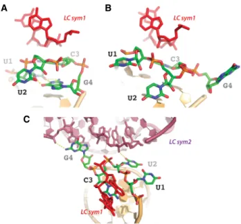
The crystal structure of the CP LC ribozyme (Meyer et al. 2014) shows that the DP2 UUCG loop adopts a squashed conformation where the backbone of residues 2 and 3 takes the place of the U–G pair formed between residues 1 and 4 characteristics of Z-turn loops, expelling nucleotides 1 and 4 into the solvent (Fig. 2A). Close in-spection of the crystal packing indicates that the DP2 loop contacts two symmetry-related LC ribozyme mole-cules. One symmetry-related molecule (sym1) makes van der Waals contacts with residues 2 and 3 and the second symmetry-related molecule (sym2) provides stack-ing and H-bondstack-ing interactions to the expelled G4 (Fig. 3A,C).
Removal of one base pair from the DP2 stem restores the canonical UNCG loop structure
Following this observation, we engineered a CP LC ribo-zyme construct (CP LC DP2ΔUA) in which one base pair was removed from the DP2 stem (Fig. 1). This RNA crystal-lized under the same conditions and in identical space group P212121 with each parameter expanded by <4% with respect to the parent CP LC ribozyme construct
(Table 1; PDB: 6gyv). The crystal struc-ture of the CP LC DP2ΔUA construct reveals that the canonical structure of the UUCG loop capping the DP2 stem has been restored (Fig. 2B) and displays the typical Z-turn loop struc-tural characteristics reported in D’Ascenzo et al. (2017). It appears that the removal of one base pair from the DP2 stem restores enough space so that residues U1 and G4 from the loop can stack within the helix and form the trans base pair between their Watson–Crick and sugar edges (Leontis and Westhof 2001), respec-tively. Although the crystal packing re-mains unchanged, the conformational change of the loop results in the loss of packing interactions due to the differ-ent spatial oridiffer-entation of the fourth residue of the loop. While sym1 still provides van der Waals distance con-tacts, the increased distance between G4 and sym2 does not allow anymore any type of interac-tions (Fig. 3C).
Crystal packing induces a conformational change on a UUCG loop engineered at a peripheral section of a group II intron RNA
Group II introns are large self-splicing RNAs and mobile genetic elements of bacterial origin. They are usually re-garded as the ancestors of spliceosomal introns and the spliceosome in eukaryotes. Similarly to nuclear pre-mRNA splicing, group II self-splicing proceeds through two consecutive transesterification reactions and the in-tron is released in a typical branched conformation called the“lariat.” The reversibility of the transesterification reac-tions allows the excised intron lariat to insert back into RNA or DNA targets through the reverse-splicing pathway. Reverse splicing into DNA is at the basis of the genomic mobility of group II introns. Recently, an engineered group II ribozyme derived from the O. iheyensis intron was used to obtain two crystal structures of a group II intron lariat primed for reverse splicing (Costa et al. 2016). The lariat form of this chimeric O. iheyensis intron was crystallized ei-ther alone or in the presence of a nonreactive 5′-exon an-alog RNA. Interestingly, each condition gave rise to a different crystal form having a distinct set of packing con-tacts. In the lariat-alone crystal structure, one of the pack-ing interactions involves a UUCG tetraloop engineered at the tip of intron domain III. Remarkably, this crystal contact forces the UUCG loop to adopt a squashed conformation similar to the one observed in the DP2 UUCG loop from the CP LC ribozyme (Fig. 4A). Conversely, however, in
B A
FIGURE 1. Secondary structure schemes of the circularly permutated (CP) LC ribozyme and CP DP2ΔUA derived from the D. iridis ribozyme. (A) The wild-type form of the LC ribozyme pre-sents both the 5′and 3′ends in the DP2 stem. The 2′hydroxyl group of the catalytic nucleotide, U232 cleaves the phosphate bond between the last nucleotide of the ribozyme G229 (cyan) and the downstream C230 (purple) leading to the formation of a 3 nt lariat. (B) In the CP form, the 5′and 3′ends reside at the scissile bond while DP2 has been circularized using a UUCG tetraloop. The U–A pair depleted to allow the loop to change conformation is shown in red.
the context of the 5′-exon-bound lariat structure, the UUCG loop of domain III points toward the solvent and is seen to adopt the canonical conformation expected for the Z-turn tetraloop family (Fig. 4B). Noteworthy, the crystal packing variations do not influence the canonical conformation of the other UUCG loop engineered in intron domain IV.
Occurrences of“squashed” loop conformations within other RNAs
To find biologically relevant examples of our observations, we then investigated whether other squashed tetraloops of any sequence could be found in the PDB. We took advan-tage of the complete and recent data set gathered by Bottaro and Lindorff-Larsen (2017). In this study, a cluster-ing approach identifies all the structurally distinct RNA tet-raloops. Among the 44 clusters reported, four gather loops with structures based on Z-turns typical of UNCG loops (clusters 2, 5, 37, and 44). Three clusters gather
conforma-tions close to the squashed loop observed in the LC ribozyme crys-tal structure (clusters 16, 19, and 41). The contents of the clusters encom-passing the Z-turn and the squashed loops are biased at two levels. First, re-dundancy of crystal or cryo-EM struc-tures of ribosomes contributes most of the yet independent observations. Second, NMR structures usually bring >10 models, which cannot be con-sidered as independent since they re-sult from calculations obtained from a given set of restraints. Consequent-ly, after pruning NMR structures, the final data set contains 1504 Z-turn and 380 squashed loops, respectively. Squashed loops represent ∼25% of the Z-turn loops, pointing to the signif-icance of this RNA motif.
Considering squashed loops, the first example extracted from cluster 41 with a UACG loop sequence obeys the UNCG consensus (Fig. 5 in green). This loop belongs to the 1450 region of the Thermus thermophilus 16S rRNA, located at the periphery of the 70S ribosome (Maehigashi et al. 2014; Rozov et al. 2015, 2016a,b). The main structural features of this loop are due to its first (U1450) and last (G1453) residues, which bulge out. G1453 interacts with ribosomal protein RPS20. Despite its peripheral location, this loop is not involved in packing. Strikingly, it adopts a canonical UNCG structure in the original crystal structure of the 30S particle alone (1fjg, Wimberly et al. 2000), providing an additional poten-tial biological role for the switch between canonical and squashed conformations. Interestingly, a 3 nt bulge is lo-cated exactly 3 bp away from the 1450 loop. This bulge bridges the 23S rRNA (2850 region) and also interacts with RPL19. Inspection of PDB files point to a squashed loop structure during elongation, while a Z-turn loop is preferred during other phases of translation, including ini-tiation, termination, and stress-dependent ribosome stall-ing. These observations are nonetheless restricted to the T. thermophilus ribosome, and require additional data to be confirmed.
The NGNN tetraloop from the double-stranded RNA substrate of the yeast Rnt1p (RNase III family homologous to human drosha and dicer) also adopts the squashed con-formation from cluster 41 in the crystal structure of the complex (Liang et al. 2014; Song et al. 2017). This tetra-loop (AGUC in pdb file 1k6g) adopts a regular
A
B
FIGURE 2. Secondary structures and conformations of the DP2 stem–loops. (A) The UUCG tet-raloop of the CP LC ribozyme presents a squashed conformation (PDB: 6gyv). Squashing re-sults from the proximity between the backbone of residues U2 and C3 and the last base pair of the stem (nucleotides with filled rings), which expels the residues from the loop toward the solvent. The helical conformation is interrupted at the level of U1 and restored only after G4. (B) The loop from the CP DP2ΔUA ribozyme adopts the canonical UUCG conformation with a maintained helical continuity at U1 and G4 (PDB: 6g7z). The Z-turn allows reorientation of the RNA chain toward the second strand of the stem. On each panel, the tetraloop is sur-rounded by a maximum likelihood map 2mFobs− DFcalccontoured at 1.5σ.
conformation in the free form of the RNA (Lebars et al. 2001), in the sense that the first and fourth residues from the loop form a base pair, which stacks upon the last base pair of the stem. Nevertheless, the kink in the back-bone is mediated at the phosphate group between resi-dues 2 and 3, a situation neither typical of U-turn nor Z-turn loops. Mapping the NMR distance constraints from Lebars et al. (2001) onto a Z-turn loop points to only two which cannot be accommodated between the 2′ and 3′ hydrogen atoms from residue L4 and the H6 atom from L3. Since the L4 residue is a purine in the Z-turn loop and not a pyrimidine as in the 1k6g NMR struc-ture, these two constraints may be specific from this se-quence. The torsional angles around the phosphate group (α and ζ) remained unconstrained, indicating that most of the torsion angles result from the distance con-straints. Moreover, this loop conformation is not represent-ed by any cluster from Bottaro and Lindorff-Larsen (2017), indicating that it does not represent a characteristic fold.
Loops from clusters 16 and 19 also adopt squashed con-formations, even though their sequences depart from the UNCG consensus (Table 2; Fig. 5 in cyan and purple, re-spectively). Cluster 16 characterizes GGAU loops in the 23S rRNA from T. thermophilus, and cluster 19, CGAA, UGAG and UUAG loops in the same region of the 23S
rRNA from three different organisms, Haloarcula Marismortui (Ban et al. 2000), Deinococcus radiodurans (Schlüenzen et al. 2001), and T. thermophilus (Maehigashi et al. 2014; Rozov et al. 2015). In the case of cluster 16, the loop is situated at the solvent interface of the rRNA, close to ribosomal protein L28. The expelled first residue (G2210) interacts by stacking with U1493 (T. thermophilus numbering, pdb 1vvj) and hydrogen-bonds with a non-bridging atom of the phosphate group from G1492. The fourth residue of the loop, also ejected into the solvent, in-teracts with an arginine residue (R52) from L28. No crystal packing interaction is observed, but the RNA is not fully modeled in this region opening the possibility that the con-formation of the loop may be restrained by unobserved contacts.
In contrast, the loops from cluster 19 are well buried in the 50S subunit and interact in the same way in the three considered organisms. The first and fourth residues of the UUAG loop stack on A2430 (T. thermophilus number-ing) and G2448, respectively. In addition, the WC edge from the fourth residue interacts with the sugar edge of G2445. The second and third residues make shallow groove contacts with nucleotides 2246 to 2248. The very same arrangement is observed for the other loops CGAA and UGAG. Only RNA contacts are observed in this case, reminding of the different situations observed in the crystal packing that drove our study.
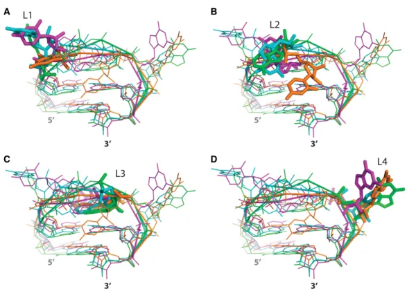
The structural characteristics of these loops can thus be summarized as follows. L1 and L4 are bulged out with a strong clustering for the latter. L2 and L3 are mostly locat-ed on the shallow groove and deep groove sides, respec-tively. The conformations for L3 residues are more
B A
C
FIGURE 3. Contacts between the two conformations of the DP2 stem–loops of CP LC ribozyme and the symmetry-related molecules. (A) When the loop adopts a canonical conformation, the backbone of U2 and C3 is in close contact with the sugar edge of two residues em-bedded in an A-form helix of a symmetry-related ribozyme (LC sym1). (B) Squashing of the loop results in spreading its residues that can con-tact residues at a much higher distance. LC sym1 remains concon-tacted by residues U2 and C3. (C) However, G4 now stacks and H-bonds (N2 group of G4) with an adenine (Phosphate group of A115) from a second symmetry-related molecule (LC sym2) already involved in A-minor interactions (Doherty et al. 2001).
TABLE 1. Crystallographic data for CP LC ribozyme DP2ΔUA
PDB Id 6g7z Resolution range 41.17− 3.337 (3.456 − 3.337) Space group P 212121 Unit cell 59.945 88.791 110.038 90 90 90 Unique reflections 8338 (299) Completeness (%) 92.64 (33.98) Wilson B-factor 88.10
Reflections used in refinement 8336 (299) Reflections used for R-free 417 (15)
R-work 0.2351 (0.3748) R-free 0.2933 (0.5942) Number of nonhydrogen atoms 3995 Macromolecules 3970 Ligands 25 RMS (bonds) 0.002 RMS (angles) 0.55 Clashscore 8.91
constrained than for L2 residues. L4 residues are often en-gaged in tertiary interactions with neighboring residues, which defines a set of clustered conformations.
DISCUSSION
It is usually accepted that UNCG tetraloops adopt a unique fold with characteristics accurately described elsewhere (D’Ascenzo et al. 2017), in brief, a Z-turn favoring the for-mation of a trans sugar edge-Watson–Crick pair between residues U1 and G4 of the tetraloop, provided that the G residue adopts a syn conformation. This conformation of the loop also presents a head-to-tail orientation of the third and fourth ribosomes reminiscent of Z-RNA (Hall et al. 1984; Davis et al. 1986). In another study, Bottaro and Lindorff-Larsen (2017) have shown that tetraloops with sequences distinct from UNCG could also adopt the canonical UNCG loop geometry and that, on rare occa-sions, UNCG sequences could acquire other conforma-tions. A UUAG loop sequence compatible with the
presence of a Z-turn between residues 3 and 4 has also been reported (D’Ascenzo et al. 2018), indicating that un-der relaxed constraints, loops UGAG and UUAG from clus-ter 19 may adopt a Z-turn-based conformation.
In the present study, we report observations from crystal structures of two different ribozymes, where a given UUCG loop adopts two distinct conformations according to the crystal packing variations resulting from slight structural changes of the RNA in the asymmetric unit. In each case, the UUCG loops were engineered in order to stabilize the underlying stem and thus, facilitate crystallization of the RNA molecule. Although the aim of these strategies was not to promote inter-molecular interactions, the engi-neered UUCG loops become involved in crystal packing contacts, which resulted in squashing these loops. The conformations induced by these interactions are very sim-ilar, and reveal that the squashed conformation is specifi-cally obtained in response to the backbone–backbone interaction through contacts between phosphate and 2′hydroxyl groups. In the two occurrences of this interac-tion observed in our crystal structures, a helix of a symme-try-related molecule contacts the tip of the loop formed by the second and third residues (Fig. 3). However, in the crys-tal structure of the group II ribozyme, the guanine (L4) of the squashed loop does not interact with a symmetry-relat-ed RNA like it does in the CP LC ribozyme crystal structure. This indicates that G-mediated tertiary interaction is not a prerequisite to the folding of the squashed conformation. The relevance of the distorted UUCG loop conformation we observe is supported by the behavior of specific loops in ribosomal RNAs. Interestingly, the UACG loop from cluster 41 in the study of Bottaro and Lindorff-Larsen (2017) adopts the squashed conformation only in the con-text of the 70S ribosome since the same loop adopts a ca-nonical conformation in the original structure of the 30S ribosomal subunit (Wimberly et al. 2000). Subunit associa-tion is well known to induce RNA conformaassocia-tional changes, suggesting that the one we describe in the context of the ribosome may have a biological significance. Inspection of the other clusters reveals that loops with other sequences can indeed adopt conformations very much related to the squashed loop from the LC ribozyme. The L2 nucleotide presents the most variable position. Residues at positions 3 and 4 occupy very identical positions in spite of belong-ing to different clusters (Table 3). It appears that the cen-troid approach developed by Bottaro and Lindorff-Larsen may be too sensitive, and that some clusters could actually be merged under more general structural features.
The resolution of the structures can also be questioned. Usually, structural data mining is performed on structures with resolution better than 2.5−3 Å. In this case, we also took into account structures with resolution worse than 3 Å. Although the positions of loop residues may result from the geometric restraints applied on the models dur-ing refinement, the generally crowded surrounddur-ings of
A
B
FIGURE 4. Structural flexibility of a UUCG tetraloop present in an en-gineered group II intron ribozyme. (A) The UUCG tetraloop at the tip of intron domain III is involved in a crystal packing contact and adopts a squashed conformation in which residues U1 and G4 are ejected into the solvent (PDB entry: 5j01). (B) The same UUCG tetraloop ac-quires the canonical conformation in a different crystal form (corre-sponding to the group II intron ribozyme bound to its 5′-exon) in which the loop is not involved in any interaction (PDB entry: 5j02). On each panel, the tetraloop is surrounded by a maximum likelihood map 2mFobs− DFcalccontoured at 1.5σ.
B A
D C
FIGURE 5. Comparison between the conformations of the squashed loops from clusters 41 (green, PDB: 4v6f), 16 (cyan, PDB: 1vvj), and 19 (pur-ple, PDB: 1j5a) from the Bottaro and Lindorff-Larsen study (Bottaro and Lindorff-Larsen 2017) and the squashed loop from the LC ribozyme (or-ange, PDB: 6gyv). Superimposition of the different types of loops seen from the shallow groove side shows that nucleotides cluster individually in spite of the large differences observed for the torsional angles. Emphasis is given for nucleotides from the loops at first (A, L1), second (B, L2), third (C, L3), and fourth position (D, L4).
TABLE 2. Contacts between squashed loops in ribosome structures
PDB Location RNA contacts Protein contacts
C41: UACGa 4v8b UACG
Loop 1448 (16S) L2 Hbond with 16S-G147GA149 RPS20. No packing interaction
C16: GGAU (23S)b 1vvj
GGAU
G2210GAU2214 G1 stack with U1493 Shallow groove contact W70
of RPL2 loop close to RPL28 C19: CGAA, UGAG, UUAG (23S)
1ffk CGAA
C920GAA923 (well buried into the structure)
L2–L3 interact with RNA backbone (2279–2280) L1 stack with A2467
Adenine L4 WC edge with S edge of G2480
None
1j5a UGAG
U840GAG843 (23S) (well buried into the structure) Same position regardless of nucleotide numbering
L2–L3 interact with RNA backbone (2225–2227) L1 stacks with A2409
Guanine L4 wc edge with s edge of G2424 (opposite strand from Adenine L4 from CGAA loop)
None
1vvj UUAG
U827UAG830 (23S) Same location as CGAA and UGAG (Same file as for C16)
L2–L3 interact with RNA backbone (2246–2248) L1 stacks on A2430
L4 stacks on A2448
Guanine L4 wc edge with s edge of G2445 (same strand as for CGAA)
None
aNo deformation of the loop in the original T. thermophilus crystal structure of 30S subunit (4kvb). bNo crystal packing interactions.
the squashed loops indicate that these conformations more likely result from molecular adaptation. Conse-quently, squashed loops gather similar structural features, which are (i) residues L1 and L4 bulging out, (ii) the back-bone from residues L2 and L3 oriented right above the last helical base pairs, and (iii) the base from residue L3 pointing toward the deep groove side. These features al-low residues 1 and 4 to interact with neighboring mole-cules, either RNAs or proteins, and the backbone of residues 2 and 3 to interact with the shallow groove of helices in the vicinity. Most importantly, the squashed conformation is reversible at least in four occurrences, in-cluding one in the ribosome, and one in the dsRNA sub-strate from Rnt1p.
In summary, our study presents for the first time evi-dence that loops belonging to the UNCG family can adopt more than one conformation according to the variations of their structural context. Considering that RNA molecules are highly dynamic and that they fulfill their biological functions through complex folding pathways and structural rearrangements, conformational UNCG loop flexibility may play an important role in RNA biology by allowing them to fulfill particular functional needs through their en-gagement in specific tertiary interactions with neighboring residues. The fact that the loops from cluster 19 are em-bedded in the same kind of structural environment than the crystal packing from the LC and group II ribozymes cor-roborates our conclusions.
Interestingly, a parallel can also be drawn between UNCG tetraloops and those belonging to the GANC con-sensus, which specifically caps the catalytic domain V hairpin in subgroup IIC self-splicing introns. Comparison of the crystal structures of the chimeric O. iheyensis intron revealed that the GANC tetraloops are flexible and adopt two different conformations according to the presence or absence of the 5′-exon substrate (Costa et al. 2016). In the presence of the 5′-exon, the GAAC loop of domain V acquires an alternate conformation that allows it to con-tact a specific intron receptor sequence through a single base stack interaction. Thus, in this case, the flexibility of the GAAC loop plays a crucial biological role by partici-pating in the proper folding of the catalytic center of
the intron. It is also interesting to note that this GAAC-re-ceptor interaction is for the moment restricted to group IIC introns since it is specifically adapted to function in the particular structural context of domain V found in these introns.
Finally, the recent description of Z-turn specific recep-tors (D’Ascenzo et al. 2018) also supports the idea that the different conformations of tetraloops of a given se-quence may have distinct receptors. The squashed tetra-loops can still interact with macromolecules in the vicinity, especially with the shallow groove of RNA helices, while expelled nucleobases 1 and 4 can stack or H bond with residues in the vicinity.
The present work can thus be seen as the description of a new kind of adaptive tetraloop interaction for which more occurrences may be found in RNA structures that will be solved in the future.
MATERIALS AND METHODS
In vitro transcription, crystallization, and structure resolution of the CP LC DP2ΔUA construct
The CP LC DP2ΔUA was cloned, transcribed and purified as de-scribed in Beckert and Masquida (2011) and Meyer and Masquida (2014, 2016). The purified RNA was crystallized under conditions obtained from the crystallization of the CP LC ribo-zyme (Meyer et al. 2014). One volume of 100 µM of RNA was mixed to one volume of crystallization solution containing 200 mM NaCl, 100 mM HEPES pH 7.5 and 5%–25% PEG 3350. The structure was solved by the molecular replacement method and refined using Refmac (Murshudov et al. 2011) and Phenix (Table 1; Adams et al. 2010).
Data analysis
Representative loops from the various clusters presented in the Bottaro and Lindorff-Larsen study (Bottaro and Lindorff-Larsen 2017) were superimposed to the squashed loop of the LC ribo-zyme to pick up clusters with similar folds. Torsion angles were de-termined with x3DNA-DSSR (Lu and Olson 2003). Figures, superimpositions and RMSD calculations were made in PyMol (Schrodinger 2010).
ACKNOWLEDGMENTS
B.M. is a USIAS recipient for the exchange program between University of Strasbourg (France) and Sophia University (Tokyo, Japan). This research is supported by the Centre National de la Recherche Scientifique (LABEX ANR-11-LABX-0057_MITOCROSS), the University of Strasbourg. M.C. acknowl-edges the French agency ANR (grant ANR-10-BLAN-1502) and the BIG Lidex program for funding.
Received July 22, 2019; accepted August 1, 2019. TABLE 3. Root-mean-square deviations (Å) between individual
residues in squashed loops using the squashed loop from the LC ribozyme as a reference L1 L2 L3 L4 4v8b UACG 10.06 7.51 2.25 5.01 1vvj UUAG 6.30 6.35 2.72 3.68 1j5a UGAG 6.44 10.24 4.67 3.61 5j01 Group II UUCG 12.27 6.32 4.29 10.67 Average 8.77 7.60 3.48 5.74
REFERENCES
Adams PD, Afonine PV, Bunkoczi G, Chen VB, Davis IW, Echols N, Headd JJ, Hung LW, Kapral GJ, Grosse-Kunstleve RW, et al. 2010. PHENIX: a comprehensive Python-based system for macro-molecular structure solution. Acta Crystallogr D Biol Crystallogr 66: 213–221. doi:10.1107/S0907444909052925
Allain FH, Varani G. 1995. Structure of the P1 helix from group I self-splicing introns. J Mol Biol 250: 333–353. doi:10.1006/jmbi.1995 .0381
Antao VP, Tinoco IJ. 1992. Thermodynamic parameters for loop for-mation in RNA and DNA hairpin tetraloops. Nucleic Acids Res 20: 819–824. doi:10.1093/nar/20.4.819
Ban N, Nissen P, Hansen J, Moore PB, Steitz TA. 2000. The complete atomic structure of the large ribosomal subunit at 2.4 Å resolution. Science 289: 905–920. doi:10.1126/science.289.5481.905 Barrick JE, Takahashi TT, Ren J, Xia T, Roberts RW. 2001. Large
librar-ies reveal diverse solutions to an RNA recognition problem. Proc Natl Acad Sci 98: 12374–12378. doi:10.1073/pnas.221467798 Beckert B, Masquida B. 2011. Synthesis of RNA by in vitro
transcrip-tion. Methods Mol Biol 703: 29–41. doi:10.1007/978-1-59745-248-9_3
Bottaro S, Lindorff-Larsen K. 2017. Mapping the universe of RNA tet-raloop folds. Biophys J 113: 257–267. doi:10.1016/j.bpj.2017.06 .011
CateJH, Gooding AR, Podell E, Zhou K, Golden BL, Kundrot CE, Cech TR, Doudna JA. 1996. Crystal structure of a group I ribozyme domain: principles of RNA packing. Science 273: 1678–1685. doi:10.1126/science.273.5282.1678
Costa M, Michel F. 1997. Rules for RNA recognition of GNRA tetra-loops deduced by in vitro selection: comparison with in vivo evo-lution. EMBO J 16: 3289–3302. doi:10.1093/emboj/16.11.3289 Costa M, Walbott H, Monachello D, Westhof E, Michel F. 2016. Crystal
structures of a group II intron lariat primed for reverse splicing. Science 354: aaf9258. doi:10.1126/science.aaf9258
D’Ascenzo L, Leonarski F, Vicens Q, Auffinger P. 2017. Revisiting GNRA and UNCG folds: U-turns versus Z-turns in RNA hairpin loops. RNA 23: 259–269. doi:10.1261/rna.059097.116
D’Ascenzo L, Vicens Q, Auffinger P. 2018. Identification of receptors for UNCG and GNRA Z-turns and their occurrence in rRNA. Nucleic Acids Res 46: 7989–7997. doi:10.1093/nar/gky578 Davis PW, Hall K, Cruz P, Tinoco I Jr., Neilson T. 1986. The
tetraribo-nucleotide rCpGpCpG forms a left-handed Z-RNA double-helix. Nucleic Acids Res 14: 1279–1291. doi:10.1093/nar/14.3.1279 Doherty EA, Batey RT, Masquida B, Doudna JA. 2001. A universal
mode of helix packing in RNA. Nat Struct Biol 8: 339–343. doi:10.1038/86221
Ennifar E, Nikulin A, Tishchenko S, Serganov A, Nevskaya N, Garber M, Ehresmann B, Ehresmann C, Nikonov S, Dumas P. 2000. The crystal structure of UUCG tetraloop. J Mol Biol 304: 35–42. doi:10.1006/jmbi.2000.4204
Fiore JL, Nesbitt DJ. 2013. An RNA folding motif: GNRA tetraloop-re-ceptor interactions. Q Rev Biophys 46: 223–264. doi:10.1017/ S0033583513000048
Geary C, Baudrey S, Jaeger L. 2008. Comprehensive features of nat-ural and in vitro selected GNRA tetraloop-binding receptors. Nucleic Acids Res 36: 1138–1152. doi:10.1093/nar/gkm1048 Hall K, Cruz P, Tinoco I Jr., Jovin TM, van de Sande JH. 1984.
‘Z-RNA’—a left-handed RNA double helix. Nature 311: 584–586. doi:10.1038/311584a0
Johansen S, Vogt VM. 1994. An intron in the nuclear ribosomal DNA of Didymium iridis codes for a group I ribozyme and a novel ribo-zyme that cooperate in self-splicing. Cell 76: 725–734. doi:10 .1016/0092-8674(94)90511-8
Koculi E, Cho SS, Desai R, Thirumalai D, Woodson SA. 2012. Folding path of P5abc RNA involves direct coupling of secondary and
ter-tiary structures. Nucleic Acids Res 40: 8011–8020. doi:10.1093/ nar/gks468
Lebars I, Lamontagne B, Yoshizawa S, Aboul-Elela S, Fourmy D. 2001. Solution structure of conserved AGNN tetraloops: insights into Rnt1p RNA processing. EMBO J 20: 7250–7258. doi:10.1093/ emboj/20.24.7250
Leontis NB, Westhof E. 2001. Geometric nomenclature and classifica-tion of RNA base pairs. RNA 7: 499–512. doi:10.1017/ S1355838201002515
Liang YH, Lavoie M, Comeau MA, Abou Elela S, Ji X. 2014. Structure of a eukaryotic RNase III postcleavage complex reveals a double-ruler mechanism for substrate selection. Mol Cell 54: 431–444. doi:10.1016/j.molcel.2014.03.006
Lu XJ, Olson WK. 2003. 3DNA: a software package for the analysis, rebuilding and visualization of three-dimensional nucleic acid structures. Nucleic Acids Res 31: 5108–5121. doi:10.1093/nar/ gkg680
Maehigashi T, Dunkle JA, Miles SJ, Dunham CM. 2014. Structural in-sights into +1 frameshifting promoted by expanded or modifica-tion-deficient anticodon stem loops. Proc Natl Acad Sci 111: 12740–12745. doi:10.1073/pnas.1409436111
Masquida B, Westhof E. 1999. Crystallographic structures of RNA ol-igonucleotides and ribozymes. In Oxford handbook of nucleic acid structures (ed. Neidle S), pp. 533–565. Oxford University Press, Oxford, UK.
Masquida B, Beckert B, Jossinet F. 2010. Exploring RNA structure by integrative molecular modelling. N Biotechnol 27: 170–183. doi:10.1016/j.nbt.2010.02.022
Meyer M, Masquida B. 2014. Cis-Acting 5′hammerhead ribozyme op-timization for in vitro transcription of highly structured RNAs. Methods Mol Biol 1086: 21–40. doi:10.1007/978-1-62703-667-2_2
Meyer M, Masquida B. 2016. Polyacrylamide gel electrophoresis for purification of large amounts of RNA. Methods Mol Biol 1320: 59–65. doi:10.1007/978-1-4939-2763-0_5
Meyer M, Nielsen H, Olieric V, Roblin P, Johansen SD, Westhof E, Masquida B. 2014. Speciation of a group I intron into a lariat cap-ping ribozyme. Proc Natl Acad Sci 111: 7659–7664. doi:10.1073/ pnas.1322248111
Michel F, Westhof E. 1990. Modelling of the three-dimensional archi-tecture of group-I catalytic introns based on comparative se-quence analysis. J Mol Biol 216: 585–610. doi:10.1016/0022-2836(90)90386-Z
Murshudov GN, Skubák P, Lebedev AA, Pannu NS, Steiner RA, Nicholls RA, Winn MD, Long F, Vagin AA. 2011. REFMAC5 for the refinement of macromolecular crystal structures. Acta Crystallogr D Biol Crystallogr 67: 355–367. doi:10.1107/ S0907444911001314
Nielsen H, Westhof E, Johansen S. 2005. An mRNA is capped by a 2′, 5′lariat catalyzed by a group I-like ribozyme. Science 309: 1584– 1587. doi:10.1126/science.1113645
Nissen P, Ippolito JA, Ban N, Moore PB, Steitz TA. 2001. RNA tertiary interactions in the large ribosomal subunit: the A-minor motif. Proc Natl Acad Sci 98: 4899–4903. doi:10.1073/pnas.081082398 Nozinovic S, Fürtig B, Jonker HR, Richter C, Schwalbe H. 2010.
High-resolution NMR structure of an RNA model system: the 14-mer cUUCGg tetraloop hairpin RNA. Nucleic Acids Res 38: 683–694. doi:10.1093/nar/gkp956
Pley HW, Flaherty KM, McKay DB. 1994. Model for an RNA tertiary in-teraction from the structure of an intermolecular complex between a GAAA tetraloop and an RNA helix. Nature 372: 111–113. doi:10 .1038/372111a0
Robertson ME, Seamons RA, Belsham GJ. 1999. A selection system for functional internal ribosome entry site (IRES) elements: analysis of the requirement for a conserved GNRA tetraloop in the
encephalomyocarditis virus IRES. RNA 5: 1167–1179. doi:10 .1017/S1355838299990301
Rozov A, Demeshkina N, Westhof E, Yusupov M, Yusupova G. 2015. Structural insights into the translational infidelity mechanism. Nat Commun 6: 7251. doi:10.1038/ncomms8251
Rozov A, Demeshkina N, Khusainov I, Westhof E, Yusupov M, Yusupova G. 2016a. Novel base-pairing interactions at the tRNA wobble position crucial for accurate reading of the genetic code. Nat Commun 7: 10457. doi:10.1038/ncomms 10457
Rozov A, Westhof E, Yusupov M, Yusupova G. 2016b. The ribosome prohibits the G∗U wobble geometry at the first position of the co-don–anticodon helix. Nucleic Acids Res 44: 6434–6441. doi:10 .1093/nar/gkw431
Schlüenzen F, Zarivach R, Harms J, Bashan A, Tocilj A, Albrecht R, Yonath A, Franceschi F. 2001. Structural basis for the interaction of antibiotics with the peptidyl transferase centre in eubacteria. Nature 413: 814–821. doi:10.1038/35101544
Schrodinger LLC. 2010. The PyMOL Molecular Graphics System, Version 1.3r1.
Song H, Fang X, Jin L, Shaw GX, Wang YX, Ji X. 2017. The functional cycle of Rnt1p: five consecutive steps of double-stranded RNA processing by a eukaryotic RNase III. Structure 25: 353–363. doi:10.1016/j.str.2016.12.013
Varani G, Cheong C, Tinoco I Jr. 1991. Structure of an unusually stable RNA hairpin. Biochemistry 30: 3280–3289. doi:10.1021/ bi00227a016
Westhof E, Masquida B, Jossinet F. 2011. Predicting and modeling RNA architecture. Cold Spring Harb Perspect Biol 3: a003632. doi:10.1101/cshperspect.a003632
Wimberly BT, Brodersen DE, Clemons WM Jr., Morgan-Warren RJ, Carter AP, Vonrhein C, Hartsch T, Ramakrishnan V. 2000. Structure of the 30S ribosomal subunit. Nature 407: 327–339. doi:10.1038/35030006
Woodson SA. 2010. Compact intermediates in RNA folding. Annu Rev Biophys 39: 61–77. doi:10.1146/annurev.biophys.093008.131334
10.1261/rna.072694.119
Access the most recent version at doi:
2019 25: 1522-1531 originally published online August 19, 2019 RNA
Mélanie Meyer, Hélène Walbott, Vincent Oliéric, et al.
Conformational adaptation of UNCG loops upon crowding
References
http://rnajournal.cshlp.org/content/25/11/1522.full.html#ref-list-1
This article cites 45 articles, 14 of which can be accessed free at:
License
Commons
Creative
. http://creativecommons.org/licenses/by-nc/4.0/ 4.0 International), as described atmonths, it is available under a Creative Commons License (Attribution-NonCommercial ). After 12
http://rnajournal.cshlp.org/site/misc/terms.xhtml
full-issue publication date (see
This article is distributed exclusively by the RNA Society for the first 12 months after the
Service
Email Alerting
click here.
top right corner of the article or
Receive free email alerts when new articles cite this article - sign up in the box at the
http://rnajournal.cshlp.org/subscriptions
go to:
RNA