HAL Id: inserm-01139861
https://www.hal.inserm.fr/inserm-01139861
Submitted on 7 Apr 2015
HAL is a multi-disciplinary open access
archive for the deposit and dissemination of
sci-entific research documents, whether they are
pub-lished or not. The documents may come from
teaching and research institutions in France or
abroad, or from public or private research centers.
L’archive ouverte pluridisciplinaire HAL, est
destinée au dépôt et à la diffusion de documents
scientifiques de niveau recherche, publiés ou non,
émanant des établissements d’enseignement et de
recherche français ou étrangers, des laboratoires
publics ou privés.
In vivo and in vitro sensitivity of blastic plasmacytoid
dendritic cell neoplasm to SL-401, an interleukin-3
receptor targeted biologic agent.
Fanny Angelot-Delettre, Anne Roggy, Arthur Frankel, Baptiste Lamarthee,
Estelle Seilles, Sabeha Biichle, Bernard Royer, Eric Deconinck, Eric Rowinsky,
Christopher Brooks, et al.
To cite this version:
Fanny Angelot-Delettre, Anne Roggy, Arthur Frankel, Baptiste Lamarthee, Estelle Seilles, et al.. In
vivo and in vitro sensitivity of blastic plasmacytoid dendritic cell neoplasm to SL-401, an interleukin-3
receptor targeted biologic agent.. Haematologica, Ferrata Storti Foundation, 2015, 100 (2), pp.223-30.
�10.3324/haematol.2014.111740�. �inserm-01139861�
Introduction
Blastic plasmacytoid dendritic cell neoplasm (BPDCN) is an aggressive neoplasm derived from plasmacytoid dendritic cells.1 In 2008, BPDCN was classified by the World Health
Organization (WHO) as a distinct entity in the group of “acute myeloid leukemia (AML) and related precursor neo-plasms”.2Although elderly subjects are principally affected,
BPDCN can also arise in young adults and children.3,4
Approximately 90% of patients exhibit cutaneous lesions at diagnosis, which upon microscopic analysis appear as a der-mal infiltrate of immature blastic cells with features of plas-macytoid dendritic cells.5,6Malignant cells isolated from skin,
lymph nodes, bone marrow, spleen and/or other tissues usu-ally express the following markers: interleukin-3 receptor alpha (IL-3Ra or CD123), BDCA2 (CD303), BDCA4 (CD304), TCL1 and ILT7.7-9
Currently, there is no consensus regarding the optimal treatment modality for BPDCN. Several treatments, including multi-agent chemotherapy regimens, symptomatic approach-es (e.g. local radiation10), and intensive chemotherapy with
allogeneic hematopoietic cell transplantation,11-13are generally
used to treat patients. Although chemotherapy regimens used to treat patients with acute leukemia or lymphoma are often effective at inducing an initial response, the duration of response is typically brief and recurrent disease is generally resistant to chemotherapy. BPDCN patients generally suc-cumb to cytopenias due to tumor infiltration of the bone mar-row; the median overall survival has been reported to range from 9 to 32 months irrespectively of the initial presentation of the disease.14-16 While longer overall survival has been
reported with allogeneic hematopoietic cell transplantation, especially in younger patients,4,11,13,16,17 many relapses have
been observed after such transplants.13
The a-subunit of the human IL-3 receptor is a type I trans-membrane glycoprotein belonging to the cytokine receptor superfamily.18,19Interleukin3 (IL3) the IL3 receptor is a he
-terodimer associating an a chain (CD123) and a β chain (CD131). This chain is shared by IL-3, IL-5, and granulocyte-macrophage colony-stimulating factor receptors. SL-401, a novel biologic targeted therapy directed against the IL-3R, is comprised of human recombinant IL-3 joined by an
acid-©2015 Ferrata Storti Foundation. This is an open-access paper. doi:10.3324/haematol.2014.111740 The online version of this article has a Supplementary Appendix.
Manuscript received on June 5, 2014. Manuscript accepted November 4, 2014. Correspondence: francine.garnache@efs.sante.fr
Blastic plasmacytoid dendritic cell neoplasm is an aggressive malignancy derived from plasmacytoid dendritic cells. There is currently no accepted standard of care for treating this neoplasm, and therapeutic strategies have never been prospectively evaluated. Since blastic plasmacytoid dendritic cell neoplasm cells express high levels of inter-leukin-3 receptor a chain (IL3-Ra or CD123), antitumor effects of the interleukin-3 receptor-targeted drug SL-401 against blastic plasmacytoid dendritic cell neoplasm were evaluated in vitro and in vivo. The cytotoxicity of SL-401 was assessed in patient-derived blastic plasmacytoid dendritic cell neoplasm cell lines (CAL-1 and GEN2.2) and in primary blastic plasmacytoid dendritic cell neoplasm cells isolated from 12 patients using flow cytometry and an
in vitro cytotoxicity assay. The cytotoxic effects of SL-401 were compared to those of several relevant cytotoxic
agents. SL-401 exhibited a robust cytotoxicity against blastic plasmacytoid dendritic cell neoplasm cells in a dose-dependent manner. Additionally, the cytotoxic effects of SL-401 were observed at substantially lower concentra-tions than those achieved in clinical trials to date. Survival of mice inoculated with a blastic plasmacytoid dendritic cell neoplasm cell line and treated with a single cycle of SL-401 was significantly longer than that of untreated con-trols (median survival, 58 versus 17 days, P<0.001). These findings indicate that blastic plasmacytoid dendritic cell neoplasm cells are highly sensitive to SL-401, and support further evaluation of SL-401 in patients suffering from blastic plasmacytoid dendritic cell neoplasm.
In vivo and in vitro sensitivity of blastic plasmacytoid dendritic cell
neoplasm to SL-401, an interleukin-3 receptor targeted biologic agent
Fanny Angelot-Delettre,1-4Anne Roggy,1-4Arthur E. Frankel,5Baptiste Lamarthee,1-4Estelle Seilles,1-4Sabeha Biichle,1-4
Bernard Royer,1-4,6Eric Deconinck,1-4,6Eric K. Rowinsky,7Christopher Brooks,7Valerie Bardet,8Blandine Benet,9Hind
Bennani,10Zehaira Benseddik,11Agathe Debliquis,12Daniel Lusina,13Mikael Roussel,14Françoise Solly,15Michel
Ticchioni,16Philippe Saas,1-4,17and Francine Garnache-Ottou1-4
1INSERM UMR1098, F25020 Besançon Cedex, France ; 2Université de Bourgogne Franche-Comté, SFR FED4234, F25000 Besançon
Cedex, France ; 3EFS Bourgogne Franche-Comté, F25020 Besançon Cedex, France ; 4LabEX LipSTIC, ANR-11-LABX-0021, F25020
Besançon Cedex, France; 5Southwestern Medical Center, Dallas, TX, USA; 6CHU Besançon, Hematology, France; 7Stemline
Therapeutics, Inc, 750 Lexington Avenue, 11thFloor, New York, USA; 8APHP, Hopital Cochin, Paris, France; 9CHR Metz Thionville,
France; 10Institut Curie, Hopital René Huguenin, Saint Cloud, France; 11CH Chartres, Le Coudray, France; 12CH Mulhouse, France; 13APHP Hopital Avicenne, Paris, France; 14CHU Rennes, Rennes, France; 15CHU St Etienne, Saint Etienne, France; 16Université de
Nice-Sophia Antipolis, Nice, France; 17CHU Besançon, CIC1431, FHU INCREASE, Besançon, France
labile group of amino acids to a diphtheria toxin (DT) pay-load that has been truncated at its receptor binding region.20Since IL-3, the natural ligand for IL-3R, binds with
very high specificity and avidity,21SL-401 is able to
trans-port DT efficiently and preferentially to cells that over-express IL-3R, leading to internalization followed by receptor-mediated endocytosis and localization of SL-401 to early endosomes. After cleavage of the SL-401 DT con-stituent in the acidic medium of endosomes, DT translo-cates into the cytosol and binds to ADP-ribosylated elon-gation factor 2, leading to blockade of protein synthesis and cell death.22
Given the ubiquitous and high expression of IL-3R by BPDCN and the lack of therapies available to treat BPDCN, SL-401 is a potential therapeutic for BPDCN. The present study evaluated the cytotoxicity of SL-401 against patient-derived BPDCN cell lines (CAL-1 and GEN2.2) and primary BPDCN cells isolated directly from 12 patients. The investigations were performed in vitro, as well as in
vivo in a murine model of BPDCN. The aim of the study
was to provide further support for the use of SL-401 in patients suffering from BPDCN.
Methods
Patients’ cells and cell lines
Peripheral blood or bone marrow cells were obtained for diag-nostic purposes from 12 BPDCN patients (Table 1) from our national network that collects data and cells from cases diagnosed in France since 2004 (authorization number #DC-2008-713). BPDCN was diagnosed from the results of histopathology and
immunostaining of cutaneous lesions, blood or bone marrow.2,8
Two established cell lines derived from BPDCN patients were used (GEN 2.2, patent #0215927, Dr. Plumas, EFS Rhone-Alpes, Grenoble, France and CAL-1, Dr. Maeda, Nagasaki University,
Japan) as well as TF/H-Ras (Prof. Frankel) and CD123neg(MFI<800)
Daudi cell lines (ACC78, DSMZ Braunschweig, Germany) as pos-itive and negative controls, respectively. Other lymphoid and myeloid leukemic cells used to compare sensitivity to SL-401 are described in the Online Supplementary Appendix.
Drug and culture
The SL-401 drug (Stemline Therapeutics, New York, NY, USA) was stored at -80°C and tested at eight concentrations ranging from 365 pM to 0.08 fM (21 ng/mL to 0.4 ng/mL) in order to cover
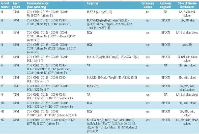
Table 1. Characteristics of the 12 patients suffering from blastic plasmacytoid dendritic cell neoplasm (BPDCN) who provided primary BPDCN cells.
Patient Age/ Immunophenotype Karyotype Cutaneous Pathology Sites of disease number gender (flow cytometry) lesions diagnosis involvement
#1 75/M CD4+CD56+CD123++CD303+CD304+ 45,XY,-5 [3], 46XY [10] no NA LN, BM blood,
My-B-CD7+(others T-) spleen
#2 59/M CD4+CD56+CD123++CD303-CD304+ 46,XY,del(6q)(q16q25),der(7)t(7;12) yes BPDCN LN, BM skin
CD33+(others My-) B-CD7+(others T-) (p11;q1?3),?del(7)(p21),-8x2,-9x2,-12x2,
+mar [4], 46XY [11]
#3 81/M CD4+CD56+CD123++CD303+CD304+ 46XY yes BPDCN LN, BM, skin, blood
CD33+(others My-) CD22+(others B-)CD2+
(others T-)
#4 69/M CD4+CD56+CD123++CD303+CD304+ 46XY yes BPDCN skin, BM
CD33+(others My-)CD22+(others B-) CD7+
(others T-)
#5 63/M CD4+CD56+CD123++CD303+CD304+ 44,X,-Y,-13[3]/44,sl,i(7)(q10)[15]/45,XY,-15[3] yes BPDCN LN, BM skin blood,
TCL1+My-B-T- spleen
#6 56/M CD4+CD56+CD123++CD303-CD304+ NA yes NA BM, skin, blood
TCL1+ILT7-CD33+CD117+(others My-)
CD22+(others B-) CD7+(others T-)
#7 33/M CD4+CD56+CD123++CD303-CD304+ 44,X,Y,13[3]/44,sl,i(7)(q10)[15]/45,XY,-15[3] yes BPDCN BM, skin, blood
TCL1+ILT7-My-B-T
-#8 70/F CD4+CD56+CD123++CD303+CD304+ 46,XX i(7q) yes BPDCN LN, BM, skin,
TCL1+ILT7- My-B-T- blood, spleen
#9 72/M CD4+CD56+CD123++CD303-CD304+ NA yes NA LN, BM, skin, blood
TCL1+ILT7-My-B-CD2+CD7+(others T-)
#10 63/M CD4+CD56+CD123++CD303+CD304+ 46XY yes BPDCN BM, skin, blood
TCL1+ILT7-My-B-CD2+CD7+(others T-)
#11 58/M CD4+CD56+CD123++CD303+ 46XY yes BPDCN LN, BM, skin,
CD304+TCL1+ILT7+CD33+(others My-) B-T- spleen
#12 80/M CD4+CD56+CD123++CD303-CD304+TCL1+ 43-44,XY,der(2) t(2;?)(q23;?),der(4)t(4;?) yes BPDCN LN, BM, skin, blood,
ILT7-My-B-CD7+(others T-) (q35;?),5,der(7)t(7;?)(q31;?),-9,-10,-13,-15, spleen
-16,del(17)(p11),+1-4mar[17];82 83,idemx2 [12];46,XY
Age (years)/gender; Results of phenotypic analysis performed on blood or bone marrow samples (flow cytometry); karyotype; presence of cutaneous lesions; histopathological diagnosis. My: myeloid markers (including myeloperoxidase, CD13, CD33, CD117, CD15, CD65, CD14, CD64); T : T lymphoid markers (including membrane CD3, intracytoplasmic CD3, CD7, CD5, CD2, CD8); B: B lymphoid markers (including intracytoplasmic CD79a, intracytoplasmic CD22, intracytoplasmic Ig m chains, CD19, CD20, CD22, surface immunoglobulin); +: positive expression; ++: high expression; -: absence of expression; NA: not available; LN: lymph nodes; BM: bone marrow.
the concentrations obtained in vivo in patients enrolled in clinical
trials.23,24 The effects of chemotherapy agents used in acute
leukemia were also evaluated against BPDCN cells (Online
Supplementary Appendix). BPDCN cells were incubated at 3x10⁵
cells/mL in RPMI 1640 glutamax medium (Invitrogen, Cergy Pontoise, France) supplemented with 10% fetal calf serum (Invitrogen), 1% penicillin/streptomycin (PAA Laboratoires, Velizy Villacoublay, France) with or without SL-401 or the relevant
drugs under 5% CO2for 18 h at 37°C.
Cytotoxicity evaluation by flow cytometry
Flow cytometry was performed using a CANTO II cytometer (BD Biosciences, San Jose, CA, USA) and DIVA 6.2 software (BD Biosciences). The cytotoxic effects of SL-401 and the various drugs were evaluated using annexin-V and 7-amino actinomycin D (AV/7AAD) and a panel of different monoclonal antibodies to gate the blastic population described in the Online Supplementary
Appendix. In the mouse model, anti-mouse and anti-human CD45
plus anti-human CD123, CD4, CD56, CD304 were used to iden-tify BPDCN human cells (Online Supplementary Appendix). A defined number of calibrated 3-mm latex beads (Flowcount beads, Beckman Coulter) was added to each sample to obtain the absolute number of circulating BPDCN cells in mice, as previously
described.25
Cytotoxicity evaluation by the MTT assay
The percentage of viable cells obtained after incubation with or without SL-401 was assessed using the MTT assay (3-(4,5-dimethylthiazol-2-yl)-2,5-diphenyltetrazolium bromide, Sigma Aldrich, Saint Quentin Fallavier, France). Details of the culture and following analysis are provided in the Online Supplementary
Appendix.
Mice
NOD-SCID IL2Rγc-deficient (NSG) mice were irradiated (2 Gy)
and inoculated intravenously 24 h later with 1x10⁶GEN2.2 cells
and treated intraperitoneally 8 days later with five daily injections
of SL-401 (2 mg/mouse/injection, total experimental dose 100
mg/kg) or with phosphate-buffered saline (PBS) only. Mice were monitored weekly by blood cell counts and flow cytometry (Online Supplementary Appendix).
Statistical analysis
Statistical analyses were performed using Statel software 2.6 (Adscience, Paris, France) (Online Supplementary Appendix).
Results
SL-401 is cytotoxic against blastic plasmacytoid
den-dritic cell neoplasm cell lines and primary cells
The viability of the two CD123pos BPDCN cell lines,
GEN2.2 and CAL-1, decreased from 62 ± 6% (range, 44-96) to 5 ± 2% (range, 0-17) after treatment with SL-401 at the experimental dose of 365 pM (21 ng/mL) for 18 h (n=9, Figure 1A). Moreover the viability of these two BPDCN cell lines decreased in a dose-dependent manner (Figure 1B). Similarly, after treatment with SL-401 at the experi-mental dose of 365 pM (21 ng/mL), the viability of 12 sam-ples of freshly isolated BPDCN primary cells decreased significantly from 50 ± 4% (range, 31-71) to 10 ± 1% (range, 3-17). As expected, the CD123neg(Daudi) cells were
not sensitive to treatment with SL-401 concentrations as high as 365 pM for 18 h [viability was unchanged from
65% (range, 88-41) to 61% (range, 83-41) in cells untreated or treated with SL-401, respectively, n=3; Figure 1A,B].
SL-401 is cytotoxic against blastic plasmacytoid
dendritic cell neoplasm primary cells in a
concentration-dependent manner
The viability of primary malignant cells obtained from 12 BPDCN patients treated with SL-401 for 18 or 48 h decreased in a concentration-dependent manner, as assessed by flow cytometry and MTT (Figure 1C,D). As expected, CD123negDaudi cells were resistant to SL-401
treatment (Figure 1C,D). For patient #10, primary BPDCN cells were obtained at both the time of diagnosis and di -sease relapse following treatment with chemotherapy [including CHOP (cyclophosphamide, hydroxydaunoru-bicin, vincristine, and prednisone), methotrexate, and Lasparaginase]. Interestingly, BPDCN cells obtained at di -sease recurrence were slightly more sensitive to SL-401 than cells obtained at diagnosis (IC50, 6 fM versus 79 fM,
respectively; P=0.049; Figure 1E). Thus, relapsing cells were still sensitive to SL-401 in a concentration-dependent manner, indicating that BPDCN cells retain their sensitivi-ty to SL-401 following treatment with various cytotoxic agents, thereby suggesting a lack of cross-resistance.
SL-401 is more efficient than other tested
chemotherapeutic drugs − except idarubicin −
at killing blastic plasmacytoid dendritic cell
neoplasm cells
In order to appreciate the cytotoxic effect of SL-401 better, we also assessed the effects of other chemothera-peutic drugs. Primary malignant cells obtained from three BPDCN patients (#7, #9 and #11) were significantly more sensitive to SL-401 than to a wide variety of cyto-toxic agents commonly used for the treatment of hema-tologic malignancies, including cytosine arabinoside, cyclophosphamide, vincristine, dexamethasone, methotrexate, Erwinia L-asparaginase, and asparaginase (Figure 2; P<0.05 for all agents). Only idarubicin was found to be more efficient than SL-401 (viability <1%, n=5; Figure 2).
Blastic plasmacytoid dendritic cell neoplasm primary
cells are more sensitive to SL-401 than acute myeloid
or lymphoblastic leukemia primary cells in vitro
We next assessed the viability after SL-401 treatment of primary malignant cells isolated from three previously untreated patients suffering from acute lymphoblastic leukemia and six untreated patients suffering from AML. We compared these results to those achieved following SL-401 treatment of primary BPDCN cells. The viability of the leukemic cells decreased slightly after treatment with SL-401 (365 pM for 18 h). We observed an average decrease of 13% in viability for acute lymphoblastic leukemia cells [33±20% (range, 6-73) to 26±20% (range, 4-69), n=3] and 16% for AML cells [40±8% (range, 18-75) to 36±8% (range, 11-70), n=6]. BPDCN cells were signifi-cantly more sensitive to SL-401 and had a 75% decrease in viable cells [50±5% (range, 20-71) to 11±1%, (range, 4-17) n=11; P<0.001; Figure 3].
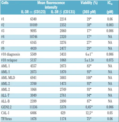
CD123 expression correlates with SL-401
cytotoxicity in vitro
We next compared the relative expression levels of IL-3Ra (CD123) and β (CD131) chains in primary malignant
BPDCN cells, acute leukemic cells, as well as established BPDCN cell lines to the respective sensitivities to SL-401 (Table 2). Overall, sensitivity of primary BPDCN cells was related to CD123 expression, as demonstrated by the inverse relationship between cell viability and CD123 expression (Spearman test: r = -0.58, P<0.012).
The high dependence of plasmacytoid dendritic cell line-age cells (including normal or leukemic plasmacytoid dendritic cells) to IL-3 may also contribute to the high sensitivity of BPDCN cells to deprivation of IL-3 signal-ing dursignal-ing SL-401 exposure. Importantly, no such rela-tionship was observed for CD131 (Spearman test: r =
F. Angelot-Delettre et al.
Figure 1. Sensitivity of BPDCN cells to SL-401-mediated death. (A) A representative experiment is shown. Upper panels: dot plots showing the
staining of annexin V (AV FITC) and 7-AAD, on the x- and y-axes, respectively, as assessed by flow cytometry (FC), on IL-3R non-expressing Daudi
cells (negative control) and CAL-1 cells (established BPDCN patient-derived cell line) after treatment with SL-401 (365 pM) for 18 h; lower pan-els: primary blasts from BPDCN patient #1 after treatment with 401 (365 pM, right hand side panel) for 18 h or no drug treatment (no
SL-401, left hand side panel). The percentages indicated in each dot plot represent viable cells (AV-/7-AAD-cells). (B) The percent viability (mean
± SEM) of the BPDCN cell lines CAL-1 (n=3) and GEN 2.2 (n=6) after treatment with SL-401 at different concentrations ranging from 0.08 fM
to 365 pM (0-21 ng/mL) for 18 h. The Daudi cell line was used as a negative control. (C) Each gray line represents the percentage viability of
primary blasts (AV-/7-AAD-cells) isolated from different BPDCN patients (#1-12) according to different SL-401 concentrations (from 0.08 fM
to 365 pM = 0-21 ng/mL) for 18 h. The black line represents the mean of BPDCN patients’ samples as a function of SL-401 concentration;
the Daudi cell line was used as a negative control. (D) Viability assessed using the MTT assay: the percentage viability of primary blasts from
BPDCN patients #3 and #4 and of CAL-1 and GEN 2.2 BPDCN cell lines is dependent on SL-401 concentrations. The Daudi cell line was used as a negative control and is insensitive to 401-mediated death whatever the concentration of the drug used. Cells were treated with
SL-401 for 48 h. The values represent the results of one experiment for patients’ samples or three independent experiments for cell lines. (E)
Percentage (mean ± SEM) of viable primary BPDCN cells (AV-/7-AAD- cells) from patient #10 at diagnosis (n = 3) and at relapse (n=6) after
incubation for 18 h with different concentrations of SL-401 or without any drug, as assessed using annexin V and 7-AAD staining and flow cytometry (FC). Untreated cells were considered as 100% viable (P=0.049).
A B C D E Daudi CAL-1 Patient #1 /AAD GEN 2.2 CAL-1 Negative control #1 #2 #3 #4 #5 #6 #7 #8 #9 #10 #11 #12 mean Negative control Diagnosis Relapse #4 #3 GEN 2.2 CAL-1 Negative control 100 80 60 40 20 0 100 80 60 40 20 0 100 80 60 40 20 0 150 100 50 0 % viabilit y (FC ) % viabilit y (FC ) % viabilit y (M TT) % viabilit y (FC ) SL-401 concentrations (fM) SL-401 concentrations (fM) 0 0.08 0.7 6 60 490 450040000 365000 SL-401 concentrations (fM) 0 0.08 0.7 6 60 490 4500 40000 365000 SL-401 concentrations (fM) 0 0.08 0.7 6 60 490 4500 40000 365000 0 0.08 0.7 6 60 490 4500 40000 365000 A V FI TC
0.01, P<0.93). For patient #10, the MFI of CD123 was quite similar both at diagnosis and relapse (5509 versus 5137, respectively).
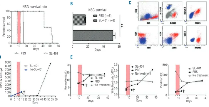
Treatment with SL-401 significantly increases the
over-all survival of NSG mice inoculated with blastic
plas-macytoid dendritic cell neoplasm cells
Irradiated NGS mice were inoculated with the GEN2.2 cell line (1x10⁶cells per mouse) through the tail vein. Eight
days after inoculation, mice were treated with a daily intra-peritoneal injection of SL-401 (2 mg/day) for 5 days or with PBS as controls (n=8 mice in 3 independent experi-ments). In PBS-treated control mice, the mean overall sur-vival was 17±1 days. Treatment with five daily injections of SL-401 significantly increased the overall survival of mice compared to that of control mice (58 ± 2 days; P<0.001; Figure 4A,B). Circulating BPDCN cells were identified as human CD45pos, CD123pos, BDCA4pos, CD4pos, CD56pos,
CD3neg, and CD34neg cells (Figures 4C). Nearly all of the
BPDCN cells expressed CD123 when mice developed the BPDCN (Figure 4C). This suggests that one course of SL-401 (2 µg/day for 5 days) is not sufficient to kill all of the BPDCN cells, rather that CD123negBDPCN cells emerge in
response to SL-401. In PBS-treated control mice, the num-ber of BPDCN cells progressively increased until death, whereas treatment for 5 days with SL-401 successfully reduced circulating BPDCN cells to undetectable levels for 15±3 days after treatment (Figure 4D). We monitored hemoglobin and platelet counts in mice to assess leukemic cell bone marrow involvement. In PBS-treated control mice inoculated with BPDCN, hemoglobin and platelet counts progressively decreased until death. In contrast, in treated mice, these hematologic parameters reached the levels observed in irradiated control mice that were not inocula ted
Figure 2. Sensitivity of primary BPDCN cells to SL401 and other rele -vant chemotherapeutic agents. The mean percentages of viable
cells (AV-/7-AAD-cells) from three patients (# 7, 9 and 11) after
treat-ment with with SL-401 (365 pM), Erwinia L-asparaginase (L-ASP, 10 IU/mL), asparaginase (ASP, 10 IU/mL), methotrexate (MTX, 9.9 mM), cyclophosphamide (CYC, 100 IU/mL), cytosine arabinoside (CYT, 0.329 mM), dexamethasone (DEX, 0.637 mM), vincristine (VIN, 0.0242 mM) and idarubicin (IDA, 0.158 mM). Untreated cells were considered as 100% viable. Histograms represent the mean ± SEM of three independent experiments (*P<0.05 between SL-401 and all other drugs pairwise except IDA).
Figure 3. Comparison of sensitivity of primary blasts from patients with BPDCN, acute lymphoid leukemia (ALL), and acute myeloid leukemia (AML) to SL-401-mediated death. Leukemia blasts sam-pled from three patients with ALL (2 B-ALL and 1 T-ALL) and six patients with AML (1 case of AML with multilineage dysplasia, 2 cases of M1 AML, 1 case of M0 AML, and 2 cases of M2 AML) and blasts from 11 BPDCN patients (#1-10 and 12) were cultured with or without SL-401 (365 pM) for 18 h. The viability was assessed by flow
cytometry (AV-/7-AAD-cells). The bars represent the mean ± SEM of
the percentage viable blastic cells, with untreated cells considered 100% viable. **P<0.001.
Table 2. Expression of IL-3Ra and β chains on primary malignant cells from patients suffering from BPDCN, acute myeloid leukemia (AML) cells, acute lymphoid leukemia (ALL) cells and cell lines −as assessed by mean fluores-cence intensity − was compared with the viability after treatment with SL-401.
Cells Mean fluorescence Viability (%) IC50
intensity IL-3R a (CD123) IL-3R β (CD131) (365 pM) (pM) #1 6340 2214 29▪ 0.06 #2 10189 2352 38▪ 0.083 #3 9095 2060 17* 0.006 #4 10746 2320 17* NA #7 6165 3276 27* NA #9 4439 2477 29* NA #10 diagnosis 5509 3433 6±1° 0.006 #10 relapse 5137 1068 5±1.3# 0.075 AML 1 4557 2073 83▪ NA AML 1 2073 1870 93▪ NA AML MLD 6941 3003 100▪ NA AML 2 3700 1473 93▪ NA AML 2 1068 2749 93▪ NA ALL-T 3049 2761 94▪ NA ALL-B 2399 2890 87▪ NA GEN2.2 11336 5378 0,45* 0.006 CAL-1 6406 629 12,5* 0.05 TF/hras 1478 1174 75* 0.06
Expression of IL-3Ra and β chains (CD123 and CD131) −measured by flow cytometry− in BPDCN cells from patients (# 1-4, 7, 9 and 10), or in different AML, ALL and cell lines (results rep-resent the mean of 2 determinations). Percentage of viability after culture with SL-401 (experi-mental dose 365 pM= 21 ng/mL) in one experiment (▪), mean±SEM of two (*), three (°) or six (#) indepen dent evaluations and IC50are indicated. AML MLD: AML with multi lineage dysplasia;
NA: not available.
100 80 60 40 20 0
**
100 80 60 40 20 0 Drugs*
% viabilit y % viabilit y ALL No S L-401 S L-401 L-A SP ASP MTX CYC CY T DEX VIN IDA ALL+S L-401 AM L+S L-401 BPD CN BPD CN +S L-401 AM Lwith BPDCN cells and not treated (Figure 4E). Regression of cytopenia under treatment indicates an absence, or at least, a lower level of bone marrow involvement by BPDCN cells in SL-401 treated mice. Overall, SL-401 is effective in con-trolling BPDCN cells in vivo.
Discussion
SL-401 is a biologic agent corresponding to IL-3 geneti-cally fused to truncated DT via a cleavable linker. This agent induces cytotoxicity by inhibiting ribosomal func-tion, and thereby, inhibiting protein synthesis, a mecha-nism that is distinct from all other anticancer therapeutics.20SL-401 has been demonstrated to induce a
profound cytotoxicity at picomolar and subpicomolar concentrations in AML cell lines,26as well as in a model of
human AML inoculated into immunocompromised mice.27 Moreover, SL-401 is cytotoxic in vivo, in patients
with advanced AML and myelosdysplastic syndrome,23
suggesting that SL-401 targets leukemic stem cells, as well as more mature tumor cells. In contrast, SL-401 is not cytotoxic to normal hematopoietic progenitor cells,28
which has translated into a paucity of myelosuppression in clinical trials to date.23,28-31
The present study was performed to evaluate and quan-tify the effects of SL-401 on various preclinical models of BPDCN, a malignancy that ubiquitously expresses high levels of the IL-3Ra chain, which is the target of SL-401.
The results reported here demonstrate that SL-401 is high-ly potent against BPDCN cell lines and primary BPDCN blasts obtained from patients. Although AML cell lines and primary AML cells have demonstrated sensitivity to SL-401 with IC50values in the picomolar range (10-12M),23
which are lower than plasma concentrations achieved in leukemic patients undergoing treatment with SL-401, BPDCN blasts are more sensitive, with IC50values in the
femtomolar range (10-15 M, experimental dose 0 to 21
ng/mL).32In addition, SL-401 produced a robust antitumor
effect in an in vivo xenograft model using human BPDCN cells. This also indicates a potential good therapeutic index, as well as systemic activity, since mice survived more than 40 days after SL-401 treatment. The high sensi-tivity of BPDCN to SL-401 and the potential good thera-peutic index of this agent likely reflect the high specificity of the IL-3 ligand component of SL-401 for CD123, in addition to the mechanism of action and potency for its DT payload. Since the IL-3 component of SL-401 is bound
via an amino acid linker to a DT for which the receptor
binding site is truncated, free DT is essentially inert from a toxicity standpoint. DT can only be delivered intracellu-larly following the binding of SL-401 to the IL-3R via IL-3 and internalization. Since IL-3Ra expression is limited to only a few normal tissues (plasmacytoid dendritic cells and basophils) and, in contrast, the receptor is over-expressed by BPDCN cells, SL-401 can potentially confer high therapeutic indexes for patients. Additionally, SL-401 is not a substrate for p-glycoprotein and other efflux
F. Angelot-Delettre et al.
Figure 4. In vivo efficacy of SL-401 in a NSG mouse model inoculated with BPDCN cells. NSG mice were irradiated with 2 Gy and then
inocu-lated intravenously with 1x10⁶GEN2.2 BPDCN cells on day 0. (A) Overall survival (OS) of BPDCN inoculated-mice treated with SL-401 (solid
line; n=4) or with PBS (dotted line; n = 3). Treatment with SL-401 (2 mg/mouse intraperitoneally, experimental dose 100 mg/kg) performed
daily for 5 days, was begun on day 7 (pink bar). OS from one representative experiments out of three is shown (P=0.3). (B) Mean OS of BPDCN
inoculated-mice treated with SL-401 (n=8) or PBS (n=8) from three independent experiments (**P<0.001). (C) one example of the immuno
-staining of circulating peripheral blood mononuclear cells performed at day 53 prior to sacrifice. Murine (blue) and human GEN 2.2 (red) cells are distinguishable due to specific CD45 antibody expression. Human GEN2.2 BPDCN cells express CD123, BDCA4, CD4, CD56, but not CD34
or CD3. (D) BPDCN cell count values in the blood of a mouse following treatment with SL-401 (dotted line, black circles) or with PBS (blue
solid line, blue square). (E) Means of hemoglobin, as well as white blood cell and platelet counts in the blood of mice following treatment with
SL-401 (n=4) or saline control (n=3). The gray line represents blood parameter values in irradiated mice that were not treated with SL-401. †: means that the mice died.
A B C D E 100 80 60 40 20 0 900 800 700 600 500 400 300 200 100 0 NSG survival rate NSG survival PBS (n=8) SL-401 (n=8) SL-401 no-SL-401 SL-401 PBS No treatment SL-401 PBS No treatment SL-401 PBS No treatment 0 20 40 80 Days 0 10 20 30 40 50 60 Days 0 5 10 15 20 25 30 35 40 45 50 55 60 Days 0 10 20 30 40 Days 0 10 20 30 40 Days 0 10 20 30 40 Days 1500 1000 500 0 2.5 2.0 1.5 1.0 0.5 0.0 20 15 10 5 0 P la te le t (x 1 0 9/L ) H e m o g lo b in ( g /d L ) W h it e b lo o d c e ll s ( x 1 0 9/L ) PBS SL-401 P er cent sur vival B P D C N cells /m L
pumps, and thus, cannot be excluded from the blastic cells. Moreover, its cytotoxic mechanism, binding to ADP-ribosylated elongation factor 2, thereby uncoupling pro-tein synthesis blockade, does not overlap with other agents currently used.22IL-3 has also been shown to be a
critical survival factor for plasmacytoid dendritic cells.33
Thus, interference of this pathway by SL-401 may explain the high sensitivity of BPDCN compared with the sensi-tivity of other myeloid and lymphoid leukemic cells.
There is still no consensus on the best therapeutic approach for BPDCN,3,12-15,34-38 and, overall, BPDCN
remains a chemotherapy-resistant disease and may also resist the graft-versus-leukemia effect, since 32% of patients relapse after allogeneic hematopoietic cell trans-plantation according to a recent study.13Thus, targeted or
immune-based therapies are alternative strategies to treat this aggressive leukemia.39-41Here, we propose that SL-401
is an efficient target-based therapy available for clinical tri-als and has already been shown to have favorable effects in patients with refractory or relapsed AML or myelodys-plastic syndrome, although expression of CD123 is lower on myeloid blasts than on BPDCN cells.23 We recently
published data from a phase I/II clinical study involving 11 BPDCN patients. These data showed that a single cycle of SL-401 induced major responses in 78% of the patients.24
The way to use SL-401 in BPDCN patients must, howe ver, be discussed in the light of data from literature obtained in such patients. SL-401 can be used to consolidate the effects of first-line chemotherapy, reducing the number of relapses, which always occur after chemothe rapy treat-ment. The combination of SL-401 with chemotherapy should make it possible to reduce chemotherapy doses, and consequently, their adverse effects, which are signifi-cant in elderly patients with comorbidities (i.e., most of the patients with BPDCN). We showed here that idaru-bicin, at the dose we tested, induces a relevant level of cytotoxic activity in vitro whereas cytosine arabinoside does not. This confirms recent data showing that the BPDCN cell line CAL-1 is resistant to cytosine arabi-noside.41 Intrathecal injection of SL-401 could also be of
interest since there are frequently patients with central nervous system relapse42and the molecular weight of
SL-401 (57 KDa) predicts no diffusion across the blood-brain barrier. For patients who undergo allogeneic hematopoiet-ic cell transplantation, SL-401 treatment can be used before allografting to minimize the level of minimal
resid-ual disease, which is the most important prognostic factor in a recent study on allografted BPDCN patients,13or as a
consolidation treatment after allogeneic hematopoietic cell transplantation.13 In support of this latter use, we
observed that, in vitro, the blastic cells from a relapsing BPDCN patient were still sensitive to SL-401-mediated death. Moreover, the IL-3Ra chain (CD123) was still expressed on the surface at relapse (CD123 MFI at diagno-sis: 5509 versus 5137 at relapse, patient #10). In the mouse model after one course of SL-401, all the BPDCN cells at relapse also expressed CD123. This supports the hypoth-esis that patients suffering from BPDCN can be treated with SL-401 as first-line or second-line therapy and, maybe, even with several courses of SL-401. Overall, immune-based therapy using SL-401 appears to be an appropriate way to treat BPDCN patients. New approach-es based on immunomodulators41 or demethylating
agents40 must be further evaluated and compared − or
associated − with SL-401.
In conclusion, we demonstrate that clinical grade SL-401, which specifically targets IL-3R, efficiently kills pri-mary BPDCN cells in culture and significantly improves the overall survival of mice inoculated with BPDCN receiving a single cycle of SL-401. This provides a strong rationale for the use of SL-401 in the treatment of patients suffering from BPDCN. As BPDCN is a rare subtype of leukemia, an international clinical trial using SL-401 should now be conducted to validate these results prospectively.
Acknowledgments
This work was supported by grants from the University of Franche-Comté (BQR25JC), La Ligue Contre le Cancer (116AD.2010), the Agence Nationale de la Recherche (Labex LipSTIC, ANR-11-LABX-0021) and the Conseil Régional de Franche-Comté (“Soutien au LabEX LipSTIC” to PS). We would like to thank Sophie Perrin and the Pharmacy Department (CHRU Besançon) for their support in providing the chemother-apeutic drugs; Laboratory of Cytology (EFS BFC, Dr Françoise Schillinger); Dr Francis Bonnefoy and all the biologists and physicians who participate in the French BPDCN network.
Authorship and Disclosures
Information on authorship, contributions, and financial & other disclosures was provided by the authors and is available with the online version of this article at www.haematologica.org.
References
1. Chaperot L, Bendriss N, Manches O, et al. Identification of a leukemic counterpart of the plasmacytoid dendritic cells. Blood. 2001;97(10):3210-3217.
2. Swerdlow SH, Campo E, Harris NL, et al. World Health Organisation Classification of Tumors. 4th ed. Lyon; 2008.
3. Jegalian AG, Buxbaum NP, Facchetti F, et al. Blastic plasmacytoid dendritic cell neo-plasm in children: diagnostic features and clinical implications. Haematologica. 2010;95(11):1873-1879.
4. Sakashita K, Saito S, Yanagisawa R, et al. Usefulness of allogeneic hematopoietic stem cell transplantation in first complete remission for pediatric blastic plasmacytoid
dendritic cell neoplasm with skin involve-ment: a case report and review of literature. Pediatr Blood Cancer. 2013;60(11): E140-142.
5. Julia F, Petrella T, Beylot-Barry M, et al. Blastic plasmacytoid dentritic cell neo-plasm: clinical features in 90 patients. Br J Dermatol. 2013;169(3):579-586.
6. Petrella T, Meijer CJ, Dalac S, et al. TCL1 and CLA expression in agranular CD4/CD56 hematodermic neoplasms (blas-tic NK-cell lymphomas) and leukemia cutis. Am J Clin Pathol. 2004;122(2):307-313. 7. Angelot-Delettre F, Biichle S, Ferrand C, et al.
Intracytoplasmic detection of TCL1--but not ILT7-by flow cytometry is useful for blastic plasmacytoid dendritic cell leukemia diagno-sis. Cytometry A. 2012;81(8):718-724. 8. Garnache-Ottou F, Feuillard J, Ferrand C, et
al. Extended diagnostic criteria for plasma-cytoid dendritic cell leukaemia. Br J Haematol. 2009;145(5):624-636.
9. Marafioti T, Paterson JC, Ballabio E, et al. Novel markers of normal and neoplastic human plasmacytoid dendritic cells. Blood. 2008;111(7):3778-3792.
10. Petrella T, Bagot M, Willemze R, et al. Blastic NK-cell lymphomas (agranular CD4+CD56+ hematodermic neoplasms): a review. Am J Clin Pathol. 2005;123(5): 662-675.
11. Dalle S, Beylot-Barry M, Bagot M, et al. Blastic plasmacytoid dendritic cell neo-plasm: is transplantation the treatment of choice? Br J Dermatol. 2009;162(1):74-79. 12. Dietrich S, Andrulis M, Hegenbart U, et al.
Blastic plasmacytoid dendritic cell neopla-sia (BPDC) in elderly patients: results of a
treatment algorithm employing allogeneic stem cell transplantation with moderately reduced conditioning intensity. Biol Blood Marrow Transplant. 2011;17(8):1250-1254. 13. Roos-Weil D, Dietrich S, Boumendil A, et al. Stem cell transplantation can provide durable disease control in blastic plasmacy-toid dendritic cell neoplasm: a retrospective study from the European Group for Blood and Marrow Transplantation. Blood. 2013;121(3):440-446.
14. Gilis L, Lebras L, Bouafia-Sauvy F, et al. Sequential combination of high dose methotrexate and L-asparaginase followed by allogeneic transplant: a first-line strategy for CD4+/CD56+ hematodermic neoplasm. Leuk Lymphoma. 2012;53(8):1633-1637. 15. Pagano L, Valentini CG, Pulsoni A, et al.
Blastic plasmacytoid dendritic cell neo-plasm with leukemic presentation: an Italian multicenter study. Haematologica. 2013;98(2):239-246.
16. Piccaluga PP, Paolini S, Sapienza MR, Pileri SA. Blastic plasmacytoid dendritic cell neoplasm: is it time to redefine the stan-dard of care? Expert Rev Hematol. 2012;5 (4):353-355.
17. Ramanathan M, Cerny J, Yu H, Woda BA, Nath R. A combination treatment approach and cord blood stem cell transplant for blas-tic plasmacytoid dendriblas-tic cell neoplasm. Haematologica. 2013;98(3):e36.
18. Blalock WL, Weinstein-Oppenheimer C, et al. Signal transduction, cell cycle regulatory, and anti-apoptotic pathways regulated by IL-3 in hematopoietic cells: possible sites for intervention with anti-neoplastic drugs. Leukemia. 1999;13(8):1109-1166. 19. Miyajima A, Kitamura T, Harada N, Yokota
T, Arai K. Cytokine receptors and signal transduction. Annu Rev Immunol. 1992;10:295-331.
20. Frankel AE, Ramage J, Kiser M, Alexander R, Kucera G, Miller MS. Characterization of diphtheria fusion proteins targeted to the human interleukin-3 receptor. Protein Eng. 2000;13(8):575-581.
21. Rapoport AP, Luhowskyj S, Doshi P, DiPersio JF. Mutational analysis of the alpha subunit of the human interleukin-3 receptor. Blood. 1996;87(1):112-122. 22. Kreitman RJ. Recombinant immunotoxins
containing truncated bacterial toxins for the treatment of hematologic malignancies. BioDrugs. 2009;23(1):1-13.
23. Frankel A, Liu JS, Rizzieri D, Hogge D. Phase I clinical study of diphtheria toxin-interleukin 3 fusion protein in patients with acute myeloid leukemia and myelodyspla-sia. Leuk Lymphoma. 2008;49(3):543-553. 24. Frankel AE, Woo JH, Ahn C, et al. Activity of
SL-401, a targeted therapy directed to the interleukin-3 receptor, in patients with blas-tic plasmacytoid dendriblas-tic cell neoplasm patients. Blood. 2014;124(3):385-392. 25. Perruche S, Kleinclauss F, Lienard A,
Robinet E, Tiberghien P, Saas P. A single-platform approach using flow cytometry and microbeads to evaluate immune recon-stitution in mice after bone marrow trans-plantation. J Immunol Methods. 2004;294(1-2):53-66.
26. Frankel AE, McCubrey JA, Miller MS, et al. Diphtheria toxin fused to human inter-leukin-3 is toxic to blasts from patients with myeloid leukemias. Leukemia. 2000;14(4):576-585.
27. Black JH, McCubrey JA, Willingham MC, Ramage J, Hogge DE, Frankel AE. Diphtheria toxin-interleukin-3 fusion pro-tein (DT(388)IL3) prolongs disease-free sur-vival of leukemic immunocompromised mice. Leukemia. 2003;17(1):155-159. 28. Feuring-Buske M, Frankel AE, Alexander
RL, Gerhard B, Hogge DE. A diphtheria toxin-interleukin 3 fusion protein is cyto-toxic to primitive acute myeloid leukemia progenitors but spares normal progenitors. Cancer Res. 2002;62(6):1730-1736. 29. Alexander RL, Ramage J, Kucera GL,
Caligiuri MA, Frankel AE. High affinity interleukin-3 receptor expression on blasts from patients with acute myelogenous leukemia correlates with cytotoxicity of a diphtheria toxin/IL-3 fusion protein. Leuk Res. 2001;25(10):875-881.
30. Hogge DE, Yalcintepe L, Wong SH, Gerhard B, Frankel AE. Variant diphtheria toxin-interleukin-3 fusion proteins with increased receptor affinity have enhanced cytotoxicity against acute myeloid leukemia progenitors. Clin Cancer Res. 2006;12(4):1284-1291.
31. Su Y, Li SY, Ghosh S, Ortiz J, Hogge DE, Frankel AE. Characterization of variant diphtheria toxin-interleukin-3 fusion pro-tein, DTIL3K116W, for phase I clinical tri-als. Biologictri-als. 2010;38(1):144-149. 32. Angelot-Delettre F, Frankel A, Liu J, et al.
The IL-3Ra-targeted drug SL-401
selective-ly kills blastic plasmacytoid dendritic cell neoplasm cells. Blood. 2011;118:2588. 33. Grouard G, Rissoan MC, Filgueira L, Durand
I, Banchereau J, Liu YJ. The enigmatic plas-macytoid T cells develop into dendritic cells with interleukin (IL)-3 and CD40-ligand. J Exp Med. 1997;185(6):1101-1111. 34. Ben Amor R, Hicheri Y, Pautas C, et al.
Successful non-myeloablative allogeneic HLA-identical stem cell transplantation for CD4/CD56 positive acute leukemia. Transplantation. 2007;84(8):1066-1067. 35. Fontaine J, Thomas L, Balme B, et al.
Haematodermic CD4+CD56+ neoplasm: complete remission after methotrexate-asparaginase treatment. Clin Exp Dermatol. 2009;34(5):e43-45.
36. Gruson B, Vaida I, Merlusca L, et al. L-asparaginase with methotrexate and dex-amethasone is an effective treatment com-bination in blastic plasmacytoid dendritic cell neoplasm. Br J Haematol. 2013;163(4): 543-545.
37. Leitenberger JJ, Berthelot CN, Polder KD, P et al. CD4+ CD56+ hematodermic/plasma-cytoid dendritic cell tumor with response to pralatrexate. J Am Acad Dermatol. 2008;58(3):480-484.
38. Narita M, Kuroha T, Watanabe N, et al. Plasmacytoid dendritic cell leukemia with potent antigen-presenting ability. Acta Haematol. 2008;120(2):91-99.
39. Agliano A, Martin-Padura I, Marighetti P, et al. Therapeutic effect of lenalidomide in a novel xenograft mouse model of human blastic NK cell lymphoma/blastic plasma-cytoid dendritic cell neoplasm. Clin Cancer Res. 2011;17(19):6163-6173.
40. Menezes J, Acquadro F, Wiseman M, et al. Exome sequencing reveals novel and recur-rent mutations with clinical impact in blas-tic plasmacytoid dendriblas-tic cell neoplasm. Leukemia. 2014;28(4):823-829.
41. Sapienza MR, Fuligni F, Agostinelli C, et al. Molecular profiling of blastic plasmacytoid dendritic cell neoplasm reveals a unique pattern and suggests selective sensitivity to NF-kB pathway inhibition. Leukemia. 2014;28(8):1606-1616.
42. Hertler AA, Schlossman DM, Borowitz MJ, Poplack DG, Frankel AE. An immunotoxin for the treatment of T-acute lymphoblastic leukemic meningitis: studies in rhesus monkeys. Cancer Immunol Immunother. 1989;28(1):59-66.