HAL Id: hal-02473206
https://hal.archives-ouvertes.fr/hal-02473206
Submitted on 24 Nov 2020
HAL is a multi-disciplinary open access
archive for the deposit and dissemination of
sci-entific research documents, whether they are
pub-lished or not. The documents may come from
teaching and research institutions in France or
abroad, or from public or private research centers.
L’archive ouverte pluridisciplinaire HAL, est
destinée au dépôt et à la diffusion de documents
scientifiques de niveau recherche, publiés ou non,
émanant des établissements d’enseignement et de
recherche français ou étrangers, des laboratoires
publics ou privés.
ATPase RavA and the LdcI-RavA cage-like complex
Matthew Jessop, Benoît Arragain, Roger Miras, Angélique Fraudeau, Karine
Huard, Maria Bacia-Verloop, Patrice Catty, Jan Felix, Hélène Malet, Irina
Gutsche
To cite this version:
Matthew Jessop, Benoît Arragain, Roger Miras, Angélique Fraudeau, Karine Huard, et al.. Structural
insights into ATP hydrolysis by the MoxR ATPase RavA and the LdcI-RavA cage-like complex.
Communications Biology, Nature Publishing Group, 2020, 3 (1), pp.46. �10.1038/s42003-020-0772-0�.
�hal-02473206�
Structural insights into ATP hydrolysis by the MoxR
ATPase RavA and the LdcI-RavA cage-like complex
Matthew Jessop
1,3
, Benoit Arragain
1,3
, Roger Miras
2
, Angélique Fraudeau
1
, Karine Huard
1
,
Maria Bacia-Verloop
1
, Patrice Catty
2
, Jan Felix
1
*, Hélène Malet
1
* & Irina Gutsche
1
*
The hexameric MoxR AAA+ ATPase RavA and the decameric lysine decarboxylase LdcI form
a 3.3 MDa cage, proposed to assist assembly of specific respiratory complexes in E. coli.
Here, we show that inside the LdcI-RavA cage, RavA hexamers adopt an asymmetric spiral
conformation in which the nucleotide-free seam is constrained to two opposite orientations.
Cryo-EM reconstructions of free RavA reveal two co-existing structural states: an asymmetric
spiral, and a
flat C2-symmetric closed ring characterised by two nucleotide-free seams.
The closed ring RavA state bears close structural similarity to the pseudo two-fold symmetric
crystal structure of the AAA
+ unfoldase ClpX, suggesting a common ATPase mechanism.
Based on these structures, and in light of the current knowledge regarding AAA
+ ATPases,
we propose different scenarios for the ATP hydrolysis cycle of free RavA and the LdcI-RavA
cage-like complex, and extend the comparison to other AAA
+ ATPases of clade 7.
https://doi.org/10.1038/s42003-020-0772-0
OPEN
1Institut de Biologie Structurale, Univ. Grenoble Alpes, CEA, CNRS, IBS, 71 Avenue des martyrs, F-38044 Grenoble, France.2Laboratoire de Chimie et
Biologie des Métaux, Univ. Grenoble Alpes, CEA, CNRS, DRF, IRIG, UMR 5249, 17 rue des Martyrs, F-38054 Grenoble, France.3These authors contributed
equally: Matthew Jessop, Benoit Arragain. *email:jan.felix@ibs.fr;helene.malet@ibs.fr;irina.gutsche@ibs.fr
123456789
A
AA+ ATPases of the MoxR family are ubiquitous and
found in all major phyla of bacteria and archaea. They are
proposed to fulfil chaperone-like functions assisting the
maturation or assembly of metabolic protein complexes
1,2, and are
often found in an operon upstream of a gene encoding a von
Willebrand factor A (VWA) domain-containing protein. Recent
examples include the P. denitrificans genes norQ and norD, which
code for a MoxR ATPase and a VWA domain-containing protein
facilitating the insertion of the non-heme Fe
Bcofactor into nitric
oxide reductase
3, and the A. ferrooxidans MoxR-related protein
CbbQ which binds the VWA domain-containing CbbO to activate
Ribulose-1,5-bisphosphate carboxylase/oxygenase (Rubisco)
4,5.
The most well-characterised representative of the MoxR family is
the E. coli ATPase RavA, encoded by the ravAviaA operon,
together with the VWA domain-containing protein ViaA. These
two proteins were proposed to play a role in the maturation of
both respiratory Complex I and fumarate reductase
6,7. In addition,
RavA is involved in the E. coli acid stress response by binding
to the acid stress-inducible lysine decarboxylase LdcI
8,9. LdcI
catalyses the conversion of lysine into cadaverine, thereby
con-suming a proton and buffering both the intra- and extracellular
medium
10,11. Under conditions of combined acid and nutrient
stress, LdcI is inhibited by the stringent response alarmone
ppGpp, preventing excessive consumption of lysine
12. However,
binding of RavA to LdcI was shown to alleviate this inhibition
8.
Remarkably, RavA and LdcI together form a 3.3 megadalton
cage-like complex, consisting of two D5-symmetric decameric LdcI
rings located at the top and bottom of the cage, surrounded by
five
RavA hexamers
9.
Combined with crystal structures of the LdcI decamer (PDB
ID: 3N75) and the RavA monomer (PDB ID: 3NBX), our
first low
resolution cryo-electron microscopy (cryo-EM) map of the
LdcI–RavA cage, performed imposing the D5 symmetry of the
LdcI onto the whole assembly (EMD-2679), provided initial
insights into the elements involved in the complex formation
13.
Specifically, rotations of the C-terminal arms of RavA with
respect to the N-terminal AAA+ ATPase modules, and
accom-panying massive reorientation of the tip domains called LARA
(LdcI Associating domain of RavA), were shown to mediate RavA
binding to either LdcI or adjacent RavA monomers in the cage
13.
The lateral contacts observed between neighbouring RavA
hexamers in the LdcI–RavA complex are unique amongst AAA+
ATPases.
Building further upon these results, we now present a higher
resolution cryo-EM structure of the LdcI–RavA cage in the
pre-sence of ADP, obtained without symmetry imposition. We show
that the complex is built by
five RavA hexamers arranged
into spirals, with a prominent gap (or
“seam”) between two
LdcI-binding RavA monomers facing either the top or the bottom
LdcI decamer. Spiral conformations have recently been observed
for AAA+ ATPases such as katanin, Vps4, Hsp104, ClpB and
Lon
14–17, but have not yet been described for the MoxR family. In
addition, cryo-EM analysis of free RavA in the presence of ADP
reveals the presence of two distinct conformational states: a RavA
spiral containing a single seam, equivalent to the one inside the
LdcI–RavA cage, and a planar C2-symmetric ring with two
nucleotide-free seams at opposite positions in the RavA hexamer.
This second conformation may represent an intermediate state
between the
“seam up” and “seam down”-oriented RavA spirals
inside the LdcI–RavA complex. Moreover, it displays remarkable
structural similarity to the approximately two-fold symmetric
“dimer of trimers” arrangement of subunits in crystal structures
of the extensively studied AAA+ unfoldase ClpX
18,19and the
protein-remodeling AAA+ ATPase PCH2
20. Consequently, the
mechanism of the RavA ATPase cycle may be unexpectedly
similar to the meticulously dissected ATP hydrolysis cycle
of ClpX
19,21–23, although the respective families of these two
proteins belong to different clades of AAA+ ATPases
24–26.
Finally, we characterise the LdcI–RavA interaction using bio-layer
interferometry (BLI) binding studies and ATPase activity assays.
We demonstrate that while the affinity of LdcI for RavA is
pH-independent, LdcI-binding results in an increase in RavA ATPase
activity at acidic pH, at which this complex should be formed
inside the cell. Based on these results, we propose different
pos-sible scenarios for the ATP hydrolysis cycle of RavA, both alone
and in the context of the LdcI–RavA cage, and discuss their
functional implications.
Results
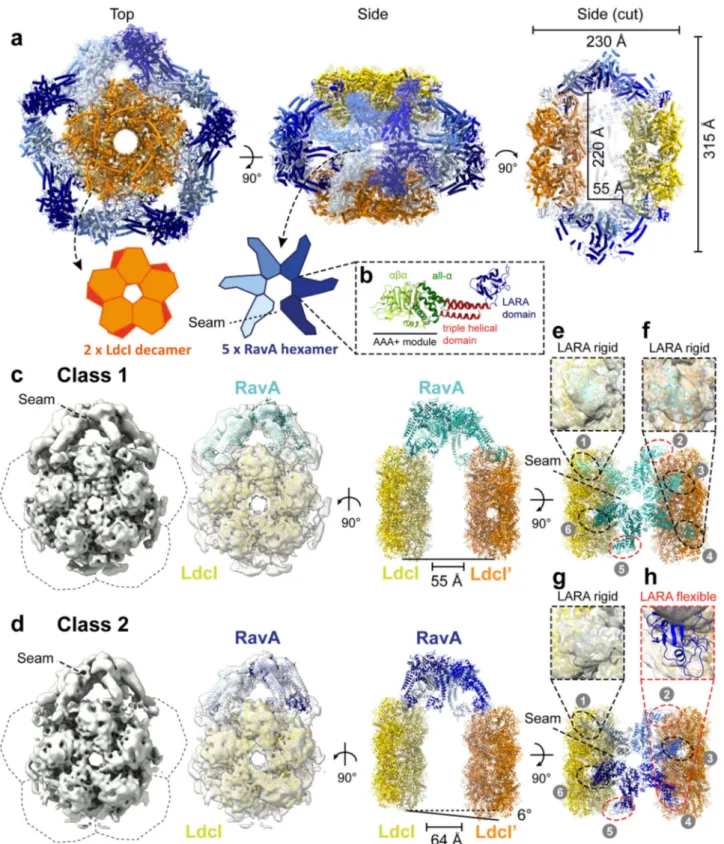
The LdcI–RavA cage is formed by spiral RavA hexamers. Initial
attempts to reconstruct the LdcI–RavA complex by imposing
C5 symmetry resulted in maps with visible heterogeneity for the
five RavA copies (Supplementary Fig. 1). Therefore, we applied a
symmetry expansion procedure (Supplementary Fig. 1, Methods),
followed by a masked 3D classification without angular search
using a soft mask focussing on one RavA hexamer and two LdcI
decamers. This resulted in two essentially identical classes, apart
from a 180° rotation around the centre of the RavA hexamer.
These classes displayed left-handed spiral RavA hexamers
con-taining a seam pointing either to the top (orientation A) or
bottom (orientation B) LdcI decamer in the cage (Fig.
1
,
Sup-plementary Fig. 1). The particles from orientation A and B were
grouped together after applying a 180° rotation to orientation B,
and used in a second masked 3D refinement. To account for
observed heterogeneity in the LARA domains of RavA, a
final
round of 3D classification was carried out followed by 3D
refinement. The resulting two classes, with an overall resolution
of 7.6 Å and 7.8 Å, respectively (Fig.
1
, Supplementary Fig. 2,
Supplementary Table 1), both display spiral RavA hexamers
bound to LdcI, but show clear differences in the presence (Class
1) or absence (Class 2) of density for one specific LARA domain
(Fig.
1
c and d, panels f and h). The distance between the two LdcI
decamers is about 10 Å larger in Class 2, which displays a 6° tilt
between opposite LdcI rings in contrast to the parallel position of
LdcI copies in Class 1. Both classes show a higher overall
reso-lution for LdcI (5–6 Å), compared to RavA (12 Å), likely
origi-nating from the inherent
flexibility of the RavA spirals in the cage
(Supplementary Fig. 2). The complete LdcI–RavA cage is formed
by two parallel LdcI decamers surrounded by
five hexameric
RavA spirals. Therefore, to illustrate the overall architecture of the
LdcI–RavA cage, we constructed a C5-symmetrised map of Class
1 (Methods). Each RavA hexamer harbours six binding interfaces:
two lateral interactions with neighbouring RavA monomers
mediated by the triple helical domain of RavA (Fig.
1
b,), and two
interactions per LdcI decamer mediated by the LARA domains at
the end of the four other RavA monomers (Fig.
1
). The resulting
map differs dramatically from the previously published one
13(EMD-2679, PDB ID: 4UPB) that was calculated with a
D5 symmetry inherent to LdcI, thereby leading to a distortion of
the RavA spirals into a C2-symmetrical assembly.
A pseudo-atomic model of the cage was then created by
flexibly
fitting crystal structures of LdcI and RavA into the maps of Class
1 and 2 using iMODFIT
27(Methods). The positions of the LARA
domains of RavA contacting LdcI were inferred from a cryo-EM
map of the LdcI-LARA complex (EMD-3206)
28. In contrast to
what was anticipated from the LdcI-LARA cryo-EM map, the
crystal structure of the LdcI decamers (PDB ID: 3N75)
12remained virtually unchanged upon
fitting into the map of the
LdcI–RavA complex, indicating that RavA binding does not affect
the LdcI conformation. As for the atomic model of RavA to be
used for
flexible fitting into the spiral RavA density inside the
Fig. 1 Cryo-EM structure of the LdcI–RavA cage-like complex. a Pseudo-atomic model of the LdcI–RavA complex, based on flexible fitting of crystal structures of RavA (PDB ID: 3NBX)8and LdcI (PDB ID: 3N75)12. The cryo-EM map used forfitting corresponds to the “Class 1” map after 3D classification (containing two
LdcI decamers and one RavA hexamer, see panelb) to which C5 symmetry has been applied. Two LdcI decamers (coloured yellow and orange) andfive spiral RavA hexamers (individually coloured light to dark blue) are shown as cartoons. Top and side views are shown, as well as a cut-away side view displaying the inner cavity of the cage. A dashed box (b) shows one RavA monomer with annotations for the different domains: AAA+ module (green), triple helical domain (red), and LARA domain (blue). The seam in the spiral RavA hexamer is indicated by a dashed line.c, d Classes 1 (c) and 2 (d) obtained after C5 symmetry expansion followed by a masked 3D classification without angular search in RELION-2.0, resulting in C1 asymmetric maps. For each class, a post-processed cryo-EM map is shown (left) along with afit of two LdcI decamers (yellow and orange) and one RavA hexamer (Class 1: cyan, Class 2: dark blue). Dashed lines indicate the positions of the masked-out RavA hexamers during symmetry expansion. On the right, side and top views of thefits are shown, with panels (e)–(h) (dashed boxes) focusing on specific LARA domains (numbered 1–6, black circles: rigid, red circles: flexible) contacting LdcI and their corresponding fits in the EM map. Class 2 displays a 6° tilted orientation of the second LdcI decamer (coloured orange) compared to Class 1.
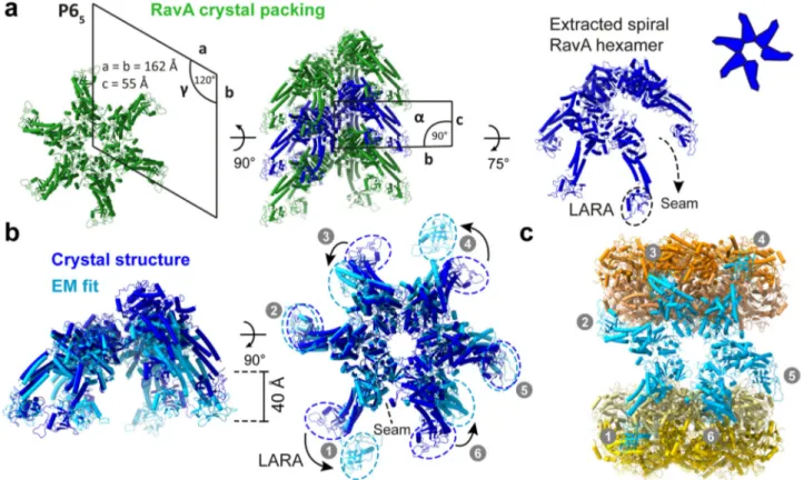
cage, we decided to reexamine the crystal packing in the RavA
structure (PDB ID: 3NBX)
8. Indeed, while RavA was reported to
crystallise as a monomer, analysis of the crystal packing in space
group P6
5reveals a continuous left-handed RavA helix (Fig.
2
a)
with ADP molecules bound at the intersubunit interface
(Supplementary Fig. 3). This interface is essentially equivalent
to the one that we originally inferred from a
fit of the RavA
monomer crystal structure into a low resolution C6-symmetric
negative stain EM map constrained by a comparison with other
AAA+ ATPases
8. Specifically, ADP is coordinated by the
Walker-A and -B residues A51, K52, S53 and D114 from one
RavA monomer and by Sensor 2 and R-finger residues (RavA
R251 and R170, respectively) from another RavA monomer
8(Supplementary Fig. 3). These observations favour the idea that
the crystallographic RavA–RavA interface constitutes (or at least
closely resembles) the biological interface. Therefore, we opted for
the usage of a spiral RavA hexamer generated from the RavA
crystal structure as a starting point for
flexible fitting in the
cryo-EM maps. Thus, despite the overall low local resolution of RavA
in the LdcI–RavA cryo-EM map, the combined use of crystal
structures of LdcI decamers, RavA spiral hexamers and a
cryo-EM map of LdcI-LARA allows us to confidently model the
LdcI–RavA complex.
A comparison between a RavA hexamer generated from the
crystal structure and RavA
fitted in the cryo-EM map of Class 1 is
shown in Fig.
2
b. While the RavA–RavA interface is retained
after
fitting, major differences are observed in the pitch of the
RavA spiral, and in the positions of the four LARA domains
contacting LdcI. Indeed, these LARA domains (numbered 1, 3, 4
and 6 in Fig.
2
b, c) undergo massive rotations when compared
to the crystal structure, whereas the LARA domains of the RavA
monomers involved in lateral RavA interactions only show
minor movements. The apparent
flexibility of RavA in our
cryo-EM maps is accentuated by the disappearance of density for
LARA domain 4 in Class 2, while it is clearly present in Class 1
(Fig.
1
c, d, panels e–h).
RavA seams are oriented up or down in the LdcI–RavA cage. In
the LdcI–RavA complex, the RavA spiral seam is oriented
between the two RavA monomers that have LARA domains
interacting with LdcI. This results in a seam that always faces
either towards the upper (orientation A:
“seam up”) or lower
(orientation B:
“seam down”) LdcI decamer, and never
towards adjacent RavA hexamers (Fig.
1
, Supplementary Fig. 1).
Generally, for AAA+ ATPases forming spiral assemblies, the
progressive movement of the seam around the hexameric ring
is shown to be functionally important
14,15,29–32. Therefore, the
occurrence of only two opposite seam orientations of RavA
spirals inside the LdcI–RavA cage may be explained as follows: (i)
the lateral RavA–RavA interactions impose local geometrical
constraints causing ATP hydrolysis to occur solely at the active
sites formed between RavA monomers 3–4, and 1–6 in the
hexamer (Fig.
2
c), or (ii) binding to LdcI stalls RavA in an
inactive form by preventing ATP hydrolysis from proceeding
Fig. 2 Comparison between a RavA hexamer generated from the RavA crystal structure and afit in the cryo-EM map of the LdcI–RavA complex. a Crystal structure ofE. coli RavA (PDB ID: 3NBX)12, displayed as cartoons, showing the helical crystal packing of RavA crystallised in spacegroup P65.
A top view along the helical screw axis of the assembly (left, with annotated unit-cell parameters) resembles a RavA hexamer. A side view of the helical assembly is shown as well (middle, with annotated unit-cell parameters), with one spiral RavA hexamer coloured dark blue. On the right, an extracted spiral hexamer (dark blue) is displayed with a slight tilt to allow visualisation of the seam, along with a schematic representation.b Comparison of a spiral RavA hexamer extracted from the crystal structure (dark blue) and afit of RavA in the cryo-EM map of Class 1 (light blue). Side (left) and top views (right) are shown, with dashed circles around the LARA domains (numbered 1–6) to highlight the differences between the crystal structure and EM fit. The position of the seam is indicated by a dashed line.c LdcI–RavA complex obtained after fitting of structures of RavA (light blue) and LdcI (yellow and orange) in the cryo-EM map of Class 1, with LARA domains numbered as in (b).
in a progressive fashion. The former hypothesis would imply a
switching between the two observed orientations of the RavA
spiral seam and would therefore contradict a strictly sequential
ATPase cycle for RavA. In addition, there remains a possibility
that other seam orientations of RavA spirals in the LdcI–RavA
cage also exist but are less stable and thus not resolved in our
analysis because of the limited number of particles used. In light
of the observed spiral conformation of RavA inside the
LdcI–RavA complex, we then revisited the structure of the free
RavA hexamer. Indeed, the previous negative stain EM
recon-struction of the hexameric RavA was performed with a
C6 symmetry imposed
8, based on planar symmetric hexamers
observed in numerous AAA+ ATPase crystal structures available
at the time
2,8,33–35.
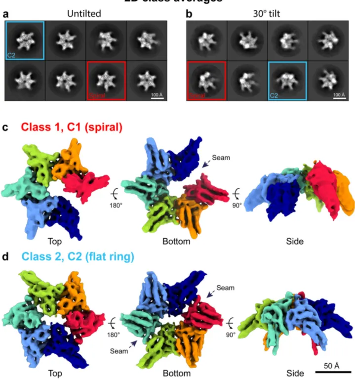
Free RavA has a spiral and a
flat C2-symmetric conformation.
Initial processing of the RavA-ADP dataset indicated a strongly
preferred top-view orientation of the particles on the cryo-EM
grid, resulting in a nonuniform distribution of angular
projec-tions. A second dataset was therefore collected at a 30° tilt
(see
“Methods”). 2D class averages from the combined dataset
revealed the presence of two distinct conformational states of the
RavA hexamer, containing either one (Fig.
3
a, b, blue squares) or
two gaps (Fig.
3
a, b, red squares). 3D classification and
refine-ment (see
“Methods”, Supplementary Figs. 4 and 5,
Supplemen-tary Table 1) resulted in two maps (at overall resolution of 7 and
6 Å, respectively), corresponding to an open spiral with a single
nucleotide-free seam (Fig.
3
c, Supplementary Figs. 4 and 5) and a
planar C2-symmetric closed ring characterised by two
nucleotide-free seams at opposite positions (Fig.
3
d, Supplementary Figs. 4
and 5). These medium resolution maps are sufficient to enable the
high-confidence flexible fitting of a RavA hexamer (for the spiral)
or a trimer (for the C2-symmetric ring) extracted from the RavA
helix generated from the crystal structure (Fig.
4
a, b,
Supple-mentary Fig. 6). Noteworthy, while caution in interpreting
fine
details at the level of the interfaces and loop regions should still be
exercised, rigid-body
fitting of RavA monomers into the two
maps without any prior knowledge leads to an interface virtually
identical to the one observed in the RavA crystal structure, further
validating the RavA intersubunit interface.
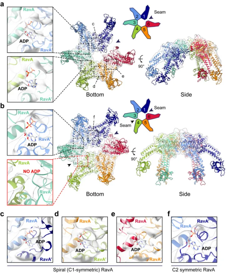
The spiral structure of free RavA is equivalent to its
conformation in the LdcI–RavA complex (Supplementary Fig. 6a),
except for the lack of a defined density for the LARA domains
that are
flexible in solution before binding to LdcI. The map
contains densities attributed to
five ADP molecules bound to the
interface between each contacting RavA monomer (Fig.
4
a, c–e).
In contrast, the C2-symmetric conformation contains only four
ADP molecules bound in the active sites between subunits 1–2,
2–3, 4–5 and 5–6 (Fig.
4
b, f). The 27 Å gap between subunits 1
and 6 in the spiral conformation is much wider than the seams in
the closed-ring conformation, meaning that rearrangements
between subunits 1–6 and 3–4 that destroy the
nucleotide-binding site are more subtle (Supplementary Fig. 6b). Loss of
nucleotide binding mainly results from a rigid-body rotation of
RavA monomers 1 and 4, accounted by shifts of helix
α3 and its
preceding loop, which contains the Walker-A residues A51, K52
and S53, and helix
α7, which contains residue M189. All of these
residues directly interact with ADP in the intact active site
interface (Supplementary Fig. 3, Supplementary Fig. 6c, d).
In an attempt to mimic the active ATP-bound state of the
RavA hexamer, a cryo-EM dataset was collected on free RavA in
the presence of ATPγS, a slowly-hydrolysable ATP analogue
often used to stabilise the ATP-bound state of ATPases. However,
2D class averages indicated that RavA-ATPγS displays even more
conformational heterogeneity than RavA-ADP (Supplementary
Fig. 7). Both the C2-symmetric and spiral conformations are
present, as well as a seemingly C6-symmetric ring and even a
C7-symmetric oligomer. The biological relevance of these two
additional states is uncertain (see for instance Sysoeva, 2017
36for review), and this heterogeneity, coupled with a strongly
preferential orientation, hampers successful 3D separation.
However, a comparison of 2D class averages displaying the
asymmetric spiral and C2-symmetric closed ring conformations
of RavA-ADP and RavA-ATPγS datasets does not reveal any
noticeable differences. Most importantly, this observation
sug-gests that, similarly to ADP, ATPγS binding does not fix RavA in
one particular structural state.
Structural insights into the ATPase cycle of RavA. The crystal
structure of the RavA monomer
8corroborated the former
phylogeny-based classification of the MoxR family as a member
of the AAA+ clade 7
24–26,37. This clade harbours in particular an
additional linker (termed the pre-Sensor 2 insertion) that
repo-sitions the C-terminal helical lid of the AAA+ module relative to
the N-terminal
αβα core domain
25,26. In such a spatial
config-uration, different from all other AAA+ clades and unique to
clade 7, the Sensor 2 motif cannot contribute to ATP binding
and hydrolysis in the same monomer (Supplementary Fig. 8).
However, based on the crystal structure of the
first crystallised
clade 7 member, the magnesium chelatase BchI monomer
(PDB ID: 1G8P)
38, aligned onto active hexamers from other
clades, AAA+ ATPases of clade 7 were proposed to rely on a
trans-acting Sensor 2 contributed by the neighbouring monomer
in the hexamer
25. The
first pseudo-atomic model of the RavA
hexamer, which was based on a
fit of the monomer structure into
a negative stain C6-symmetric EM map
2,8and guided notably by
relative positioning of Sensor 2, agreed with this hypothesis and
suggested that oligomerisation of MoxR ATPases is required for
completion of their ATP binding sites. Specifically, in MoxR-type
AAA+ ATPases, the ATP binding site was proposed to be located
not between the large (αβα) and small (all-α) AAA+ domains of
the same monomer, but between the large domain of one
monomer in a hexamer and the small domain of its neighbour
2,8.
The cryo-EM maps presented here and the resulting atomic
models of the RavA hexamer in spiral and C2-symmetric
con-formations provide strong experimental support to this model,
which is presently extended to all clade 7 members
25,26.
Viewed from this perspective, the planar double-seam
con-formation of the RavA hexamer is strikingly reminiscent of the
approximately two-fold symmetric
“dimer of trimers” arrangement
of subunits in hexamers of the AAA+ unfoldase ClpX
18,19, which
belongs to clade 5 AAA+ ATPases and thus lacks the pre-Sensor 2
insertion
25,26. In crystal structures of ClpX, hexamers are arranged
with an approximate two-fold symmetry, and contain four ClpX
subunits in a nucleotide loadable (L) and two in unloadable (U)
conformation on opposite sides of the hexamer. In the unloadable
ClpX subunits, the small and large AAA+ domains are positioned
in an
“open” conformation which destroys the nucleotide-binding
site
18,19. The resulting 4L-2U arrangement of ClpX contains a
characteristic seam which runs along the hexamer centre. A
comparison of the C2-symmetric closed ring conformation of
RavA with the 4L-2U ClpX crystal structure reveals highly similar
assemblies (Fig.
5
a, b). While ClpX binds nucleotides in the
interface formed between the large and small AAA+ domains
within one subunit, the nucleotide-binding interface in RavA
hexamers is formed in between adjacent monomers (Fig.
5
c, d).
Importantly, our structural comparison shows that similar
rigid-body like movements between the large and small AAA+
subdomains in a single ClpX subunit, or between the large or
small subdomains of adjacent RavA monomers, lie at the basis of
L to U subunit conversion, resulting in an impaired
nucleotide-binding site (Fig.
5
e, f). In addition, similar C2-symmetric
closed ring hexamer conformations have been observed for the
crystal structures of the T. maritima metalloprotease FtsH
39,
and the C. elegans protein-remodeling AAA+ ATPase PCH2,
a TRIP13 ortholog
20. Interestingly, a recent structure of human
TRIP13 solved by cryo-EM in both apo- and substrate-bound
states
40displays a right-handed spiral, but no closed-ring
conformation.
Previous studies have shown that conversion between L- and
U-states is necessary to couple ATP hydrolysis to ClpX
functioning, and provide evidence for a probabilistic model for
L to U subunit switching upon ATP hydrolysis. In the proposed
model, a 4L:2U ClpX hexamer converts to a 5L:1U hexamer in the
presence of nucleotide, followed by subunit switching between L
and U states in a non-sequential manner
19. The cryo-EM analysis
of RavA presented here reveals the presence of a mixture of both
a spiral (5L:1U) and C2-symmetric closed ring conformation
Fig. 3 Cryo-EM analysis of free RavA in the presence of nucleotide (ADP). a, b 2D classes of untilted (a) and 30° tilted (b) datasets of free RavA in the presence of ADP. Red squares highlight classes belonging to a spiral RavA conformation, while blue squares show classes belonging to a C2-symmetric closed ring conformation of RavA.c, d 3D reconstructions of the spiral (c) and closed ring (d) RavA conformations corresponding to class 1 and 2, respectively. Individual subunits in the maps are coloured according to a rainbow colour scheme. The nucleotide-free seams in the two maps are annotated using dotted arrows.
Fig. 4 Structural analysis of spiral and C2-symmetric closed ring RavA conformations. Fit of the spiral (a) and C2-symmetric closed ring (b) conformations of RavA in their respective EM maps, displayed as cartoons. Individual RavA subunits, labelled 1–6 in the accompanying schematic representations, are coloured according to a rainbow colour scheme. Zooms show the presence or absence of ADP in the nucleotide-binding site interface formed between subunits 2–3 and 3–4 in the spiral (a) and C2-symmetric closed ring (b) conformations of RavA. The nucleotide-free seams in the two maps are annotated using black arrows.c–f. Insets showing the nucleotide-binding site interface formed between subunits 1–2 (c), 4–5 (d) and 5–6 (e) of the spiral RavA conformation, and between subunits 1–2 (f) of the C2-symmetric closed ring RavA conformation.
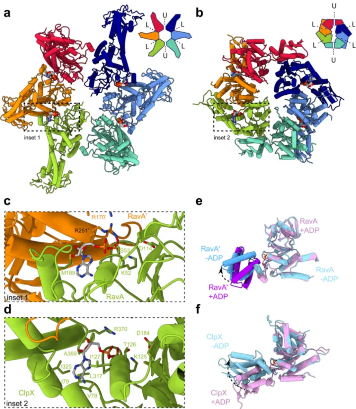
Fig. 5 Comparison between C2-symmetric closed ring conformations of RavA and ClpX. Comparison between closed ring conformations of RavA (a) and ClpX (b), shown as cartoons with accompanying schematic representations. RavA and ClpX subunits in equivalent positions around the hexamer are given identical colours following a rainbow colour scheme. In a RavA hexamer, the active site is formed in between the large and small AAA+ domains of adjacent RavA monomers, while in ClpX the nucleotide-binding site is formed between the large and small AAA+ domains of within a single ClpX subunit. Loadable and unloadable ATP binding sites in RavA and ClpX are annotated with L and U, respectively.c, d Zooms of the nucleotide-binding interface between adjacent RavA monomers (c, coloured orange and green) and the nucleotide-binding interface within one ClpX subunit (d, coloured green). Bound ADP molecules and interacting residues of RavA or ClpX are labelled and shown as sticks.e Superposition of the large and small AAA+ domains of adjacent RavA subunits (labelled RavA and RavA) from an interface in the C2-symmetric closed ring conformation with bound ADP (“closed”conformation, coloured pink and purple) and without bound ADP (“open” conformation, coloured light and dark blue). f Superposition of the large and small AAA+ domains within one ClpX subunit containing bound ADP (“closed” conformation, coloured pink) or without bound ADP (“open” conformation, coloured light blue). Movement of the small AAA+ domains of RavA and ClpX upon nucleotide binding is shown using black arrows.
(4L:2U) in solution. The presence of 4L:2U and 5L:1U RavA
states, and the similarity between the 4L:2U conformations of
ClpX and RavA, seems to suggest that RavA would function via a
similar ATP hydrolysis cycle.
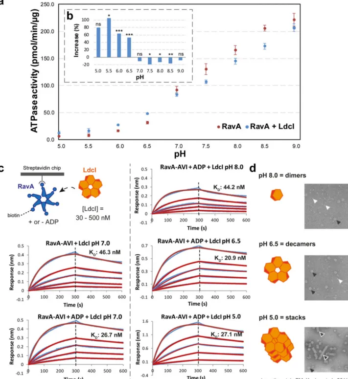
LdcI binding increases RavA ATPase activity at low pH. To
investigate whether or not the restricted orientations of RavA in
the LdcI–RavA complex would result in RavA being incapable of
hydrolysing ATP, we performed ATPase activity measurements
of RavA in the absence or presence of a three-fold molar excess of
LdcI at pH values between 5.0 and 9.0. While the RavA ATPase
activity increases with pH in the explored interval, LdcI increases
it further at pH below 7.0, and decreases it slightly at pH 7.5 or
higher (Fig.
6
a, b). At pH 7.0, LdcI exerts no apparent effect on
RavA ATPase activity. The observed bimodal effect of LdcI
on RavA is unexpected, and contrasts with previous results
which showed that LdcI increases the ATPase activity of RavA at
pH 7.5
9.
Given the pH-dependent effect of LdcI on RavA ATPase
activity, we investigated the LdcI–RavA interaction at different
pH values using BLI. In vitro biotinylated RavA-AviTag was
immobilised on streptavidin-coated biosensors, and subsequently
exposed to a concentration series of LdcI at pH between 5.0 and
8.0 (see
“Methods”). BLI measurements at different pH values did
not show a marked difference in the binding affinity of LdcI for
RavA, which ranged from K
D=20 nM to K
D=40 nM. However,
we observed an increase in the height of the BLI signal (response
in nm) with decreasing pH (Fig.
6
c). This indicates that the mass
of the bound ligand increases with lower pH, which can be
explained by the pH-dependent oligomerisation of LdcI. Indeed,
while LdcI is predominantly dimeric at pH 8.0, at pH 6.5 and
5.0 LdcI forms decamers and stacks of decamers, respectively
(Fig.
6
d)
12. Taken together, our BLI and ATPase activity studies
suggest that dimeric LdcI has a moderate inhibitory effect on
RavA, while LdcI decamers and stacks increase RavA ATPase
activity. Most importantly, when bound to LdcI, RavA can exert
its ATPase activity over a broad pH range. Thus, despite the
restricted orientations of the RavA spiral seam, RavA is still able
to efficiently hydrolyse ATP in the LdcI–RavA cage.
Discussion
AAA+ ATPases of the MoxR family have been suggested to play
a role as chaperones in the assembly of multi-protein complexes,
but in general the functions of MoxR family members are not well
characterised and their structures are scarce
2. Functionally, RavA
has been implicated in the assembly of E. coli respiratory
Com-plex I and modulation of the activity of fumarate reductase
6,7.
The crystal structure of E. coli RavA displays monomers that are
packed in a left-handed helix, and contains an ATP binding site at
the interface between adjacent monomers in the helix
8. In fact,
several other AAA+ ATPases crystallise as apparent monomers
in spacegroup P6
5, thereby forming continuous helices due to
crystal packing that resembles hexamers when viewed along the
helical screw axis
8,29,30,32,38,41–45. In some of these cases, the
interface between monomers in the helical crystal packing is very
similar to the interface elucidated by other structural methods.
For instance, the spiral RavA hexamers observed in our cryo-EM
reconstructions of the free RavA and the LdcI–RavA complex
display an interface which is equivalent to the helical RavA
assembly observed in the crystal structure. Likewise, the crystal
structure of Spastin forms a helical assembly in which the
monomer-monomer interface is compatible with a hexameric
model based on docking of monomers in an ab-initio small-angle
X-ray scattering envelope
42,45. In contrast, crystal structures of
Vps4 show an interface only partially similar to the interface
observed in cryo-EM maps of hexameric Vps4 spirals
30,31,46.
Moreover, the crystal structure of apo-katanin forms a helix with
a different handedness than the asymmetric hexamer found in a
cryo-EM reconstruction of ATP-bound katanin, and as such
does not retain the biologically relevant monomer-monomer
interface
29. In addition, these helices contrast with the planar,
symmetric hexamers observed in numerous available AAA+
ATPase crystal structures
5,33–35,47. Interestingly, a crystal
struc-ture of ClpX also shows helically arranged monomers
48, and the
interfaces formed are only slightly shifted compared to the ones
observed in hexameric ClpX structures
18.
Based on negative stain EM data and comparison with related
hexameric AAA+ ATPases, RavA was initially modelled as a
planar hexameric assembly
2,8. The cryo-EM analysis of free RavA
described here unexpectedly showcases a mixture of two distinct
conformational states: a spiral RavA hexamer, also observed in
the present cryo-EM structure of the LdcI–RavA cage, and a flat
C2-symmetric RavA hexamer characterised by two
nucleotide-free seams. Spiral hexameric assemblies with a seam devoid of
any bound nucleotide are common among different AAA+
ATPases
14,15,46,49. For several AAA+ ATPases that form spiral
hexamers, a sequential mechanism was proposed whereby ATP
hydrolysis causes the seam to move processively around the spiral
hexamer via a closed ring intermediate
14,29,49–51.
Besides a sequential ATP hydrolysis cycle, two other models
are put forward to explain how AAA+ ATPases couple ATP
hydrolysis to mechanical force to exert their function: the AAA+
lTag is suggested to act via concerted (all-or-none) nucleotide
binding and hydrolysis that occurs simultaneously in all
sub-units
52, while the AAA+ unfoldases HslU and ClpX are thought
to hydrolyse ATP via a probabilistic mechanism where ATP
hydrolysis is not strictly sequential around the hexamer
19,21,23,53.
Evidence for probabilistic L to U subunit switching in ClpX stems
from assays using individually mutated, disulfide-linked
19or
crosslinked
23subunits in covalently tethered ClpX
pseudohex-amers. These studies also demonstrate that ClpX hexamers with
one or more L or U locked subunits are able to hydrolyse ATP,
but are impaired in substrate binding and degradation. Thus,
blocking of L to U switching in a single ClpX subunit uncouples
ATP hydrolysis from mechanical work, supporting a probabilistic
but coordinated ATP hydrolysis mechanism in which
commu-nication between ClpX subunits is obligatory. Similar studies
performed on disulfide-crosslinked HslU pseudohexamers
53show that HslU pseudohexamers with different mixtures of active
and inactive subunits can unfold protein substrates and support
their degradation by HslV, albeit at a lower rate than
wild-type HslUV.
Remarkably, several very recent papers reopen the debate on
the exact ATPase mechanism of ClpX by showing high-resolution
cryo-EM structures of the ClpXP complex where hexameric ClpX
in a spiral 5L:1U state is bound to the tetradecameric ClpP
protease
54–56. Therefore, further studies are required to elucidate
whether ClpXP follows a probabilistic
56or rather processive
model
55of ATPase hydrolysis. Regardless, the existence of both
spiral (5L:1U) and a C2-symmetric closed ring (4L:2U)
con-formations of hexameric RavA, and the resemblance of the latter
to 4L:2U ClpX structures, suggests that RavA may follow a similar
ATPase mechanism as proposed for ClpX. Consequently,
different scenarios for RavA ATPase cycling upon LdcI binding
may be envisioned. The current cryo-EM reconstructions of the
LdcI–RavA complex in the presence of ADP contain five “seam
up” or “seam down” RavA spirals. One possible scenario is that
probabilistic L to U subunit switching allows the seam to be
transferred to an opposite position in the RavA hexamer. The
absence of any alternative observable seam positions other than
“seam up” or “seam down” could be the result of geometrical
Fig. 6 Characterisation of the LdcI–RavA interaction by ATPase assays and BLI binding studies. a Effect of LdcI on the ATPase activity of RavA. ATPase activity was measured at various pH values (ranging from 5 to 9) as described in“Methods”, either for RavA alone (red) or a mix containing RavA and a three-fold molar excess of LdcI (blue). Each data point represents the average of three independent measurements. Error bars correspond to the standard deviation. The dashed inset (b) shows the percent change of RavA activity when comparing the ATPase activity of RavA alone and RavA plus a three-fold molar excess of LdcI. The statistical significance is calculated using a 2-sided T-test (2-sample unequal variance). P ≥ 0.1: not significant (ns), P ≤ 0.1: *P ≤ 0.05: **P ≤ 0.001: ***.c BLI measurements of the LdcI–RavA interaction at different pH values ranging from 5 to 8 (blue curves: experimental data, red curves: calculated fit using a 1:1 interaction model.). For each experiment, biotinylated RavA-AVITAG was immobilised on a streptavidin-coated BLI biosensor (with or without prior incubation with 1 mM ADP) followed by binding measurements using different concentrations of LdcI (500 nM, 250 nM, 125 nM, 62.5 nM and 31.25 nM). Average values for the RavA-ADP:LdcI interaction measured at four different pH values (8, 7, 6.5, 5) are:KD= 29.7 nM ± 10.0, kon= 2.68 × 1041/Ms ± 2.13 ×
103, andk
dis= 8.11 × 10−41/s ± 3.41 × 10⁻⁴. TheKD,konandkdisvalues of the individual experiments can be found in Supplementary Table 2.d Negative-stain
EM micrographs of LdcI incubated at different pH values (reproduced from Kanjee et al.12, with permission of the EMBO Journal)12. At lower pH, LdcI mainly
constraints imposed by lateral contacts between the triple helical
domains of neighbouring RavA monomers in the LdcI–RavA
cage, thereby restricting the rotations between subunits needed
for release of ADP by RavA monomers making these lateral
contacts (schematically shown in Supplementary Fig. 9). If RavA
indeed follows a similar ATP hydrolysis mechanism to ClpX, this
would imply that conformational locking of RavA subunits in the
LdcI–RavA cage, as described above, would lead to RavA
hex-amers that can hydrolyse ATP, but are functionally inactive.
However, based on our data we cannot rule out the possibility
that other seam orientations of RavA hexamers in the LdcI–RavA
cage can occur, but are not observed in our cryo-EM analysis for
example because of their transient nature. In addition, alongside
the observed alternation of RavA seam states, a comparison of the
cryo-EM maps representing the two classes of the LdcI–RavA
complex reveals a near 10 Å difference in the distance between
opposite LdcI decamers forming the central cavity of the cage
(Fig.
1
). If, while bound to LdcI, RavA is able to exert its potential
protein-remodeling capacity, then the existence of these two
classes tends to suggest that ATP hydrolysis by RavA would cause
a breathing motion of the LdcI–RavA cage, thereby transferring
mechanical force to remodel substrates inside the complex.
Taken together, our synergistic approach, which combines data
from complementary structural techniques such as cryo-EM,
X-ray crystallography, modelling, as well as biochemical
char-acterisation, has provided insights into nucleotide-dependent
conformational changes of RavA during ATP hydrolysis, and the
possible ATPase mechanism of RavA in the LdcI–RavA complex.
Validation of the proposed interaction partners and
character-isation of interaction with substrates are necessary future steps in
the elucidation of the structure-function relationships of the
LdcI/RavA/ViaA triad, and in uncovering of the mechanism of its
action in sensitization of E. coli to aminoglycosides
6. Our work
adds to the growing number of AAA+ ATPase structures
cor-responding to snapshots of the ATP hydrolysis cycle. Clade 7
AAA+ ATPases encompass very divergent families, including
MoxR, Chelatase/YifB, the minichromosome maintenance
pro-tein MCM built by six different ATPase subunits, and even the
eukaryotic Dynein/Midasin where the six AAA+ subunits are all
covalently linked. It is tempting to suggest that the observations
and hypotheses based on the RavA cryo-EM structures described
here may be extended to all clade 7 AAA+ ATPases that
share a spatial arrangement of
αβα and all-α subdomain
resulting in an active site formed between adjacent monomers
(Supplementary Fig. 8).
Methods
LdcI–RavA complex formation. RavA and LdcI proteins were expressed and purified as previously described8,12,13, with the sole exception that LdcI was
expressed in a different ppGpp−/−E. coli strain (MG1655ΔrelA ΔspoT), gener-ously provided by Dr. Emmanuelle Bouveret. To promote optimal LdcI–RavA cage formation in vitro, purified LdcI and RavA were initially separately diluted to a respective concentration of 0.76 mg ml−1and 1.2 mg ml−1in a buffer containing 20 mM Tris pH 7.9, 300 mM NaCl, 2 mM ADP, 10 mM MgCl2, 0.1 mM PLP and
1 mM DTT. After 10 min at room temperature (RT), equal volumes of both pro-teins were mixed and incubated 10 min at RT. In thefinal mix, concentrations of LdcI and RavA were 0.38 mg ml−1(4.67 µM) and 0.6 mg ml−1(10.64 µM), respec-tively, resulting in a RavA monomer:LdcI monomer ratio of 10.64 µM:4.67 µM= 2.278, or approximately 4.5:2.
Cryo-electron microscopy on the LdcI–RavA complex. The quality of complex formation was checked by negative-stain electron microscopy (EM) using 5 times diluted 4.5:2 RavA:LdcI mix (see above). 4μl of sample was applied to the clear side of carbon on a carbon-mica interface and stained with 1% (w/v) uranyl acetate. Images were recorded under low-dose conditions with an FEI T12 microscope operated at 120 kV or FEI F20 microscope operated at 200 kV, at nominal mag-nifications ranging from 13,000x to 19,000x.
For cryo-EM grid preparation, 4μl of 4.5:2 LdcI–RavA mix was applied onto a glow-discharged quantifoil 400 mesh 1.2/1.3 grid (Quantifoil Micro Tools GmbH,
Germany), the excess solution was blotted for 3 s with a Vitrobot (FEI) using blot force 1, and the grid plunge-frozen in liquid ethane. Data collection was performed on an FEI Polara microscope operated at 300 kV. Movies of 40 frames were collected with a total exposure time of 8 s and a total dose of 40 e−Å−2on a K2 summit direct electron detector (Gatan) at a magnification of 41,270x, corresponding to 1.21 Å pixel−1at the specimen level. Specimen motion during data collection was evaluated and corrected with MotionCor257,58. Frames 3–40 of
each movie were dose-weighted, summed and kept for further processing. The contrast transfer function (CTF) of each micrograph was determined with GCTF59.
1819 best micrographs were selected based on visual quality control and CTF inspection. As previously noticed13, and despite the high affinity of RavA for the
LdcI12, the LdcI–RavA cage is extremely sensitive to the cryo-EM grid preparation
process, which results in a very low amount of intact particles per image. This difficulty in sample preparation limits the number of particles available for further analysis, complicates the particle selection process and hinders obtainment of a high-resolution structure. Eventually, 18,902 particles were manually picked using EMAN2 e2boxer60and subjected to two rounds of 2D classification with
RELION-2.061to yield a cleaned dataset containing 15,771 particles. For 2D classification
and all further steps, CTF-amplitude correction was performed starting from the first peak of the CTF. Visual analysis of 2D class averages immediately revealed considerable heterogeneity in RavA conformations/positions while LdcI appeared more rigid. The initial 3D model based on 2D class averages was calculated with sxviper (SPARX)62imposing D5 symmetry and appeared similar to our previously
published map13. Subsequent 3D classification in RELION-2.0 was performed with
C1 symmetry in order to remove the remaining incomplete cages containing 3 or 4 RavA hexamers. This led to a clean dataset containing 11,866 particles. 3D refinement with Relion auto-refine, using the initial sxviper 3D model low-pass filtered to 40 Å as a reference and imposing C5 symmetry, led to a 7.3 Å resolution map.
Albeit already much better than our previous map13, the resulting map showed
a lower resolution of RavA in comparison to LdcI, which again pointed to structural heterogeneity of RavA particles inside the LdcI–RavA complex. Thus, a soft mask was created from afit of one hexameric RavA8and two LdcI decamers12
into the map, and the dataset was expanded by replicating each particle from the C5 consensus refinement and adding n*72° with n = 1,…,5 to its first Euler angle. A masked 3D classification was then conducted with RELION-2.0 without angular search. This procedure enabled a reliable separation of the dataset into two classes, containing 47 and 53% of the data, respectively. Unexpectedly, both classes showed left-handed RavA spirals, with a clearly defined seam facing either the upper (orientation A) or the lower (orientation B) LdcI decamer and related exactly by a 180° rotation (Supplementary Fig. 1). In order to be able to combine images of both orientations, we applied a -phi, 180°-theta, 180°+psi transformation to the Euler angles of orientation B to bring it into orientation A. Images corresponding to orientation A and rotated orientation B were then subjected to a masked local 3D auto-refinement (RELION-2.0), with the mask that again included two LdcI decamers and one RavA hexamer. This masked reconstruction had a global resolution of 7.3 Å, with local resolution ranging from 5 to 14 Å. The B-factor sharpening, using a B-factor value of−270 Å2, was performed in RELION-2.0 as
described63.
To further address eventual conformational variability of the RavA spiral inside the LdcI–RavA complex, we undertook a final 3D classification using the same mask as before, including two LdcI decamers and one RavA hexamer. This classification allowed separation of two defined states (Class 1 and 2) containing 32 and 28% of particles, respectively, and a third state corresponding to more mobile/ flexible RavA containing 40% of particles (Class 3). Refinement of Class 1 and Class 2 gave respective resolutions of 7.6 and 7.8 Å, with local resolution ranging from 5 to 14 Å for both classes. In Class 1, the densities corresponding to all RavA domains contacting LdcI (i.e. LARA domains) are well resolved. In contrast, in Class 2 as well as in the preceding 7.3 Å map from the masked refinement containing all particles, the density of one LARA domain is missing.
Post-processing of LdcI–RavA Cryo-EM maps and fitting of structures. Local resolution estimation and subsequentfiltering of maps were performed in RELION-3.0, using B-factors of−200, −250 and −300 Å2for the masked 3D
refinement containing all particles, or particles from Class 1 and 2, respectively. For fitting of atomic models in the resulting filtered maps, we used the previously-determined crystal structures of LdcI (PDB ID: 3N75)12and RavA (PDB ID:
3NBX)8. Careful analysis of the RavA crystal packing revealed that RavA was
crystallised as a continuous helix. In each map, two decameric LdcI molecules extracted from PDB 3N75 and one spiral RavA hexamer extracted from a con-tinuous RavA helix generated from PDB 3NBX werefirst manually placed using Chimera64, and thenfitted separately using iMODFIT27, followed by a single round
of B-factor (ADP) refinement in Phenix65,66.
Cryo-electron microscopy on free RavA. Purified RavA (see above) was diluted to afinal concentration of 0.1 mg ml−1in the presence of 1 mM ADP and incu-bated at room temperature for 10 min. 3μL of RavA:ADP was applied to glow-discharged (20 mA, 45 s) R2/1 400 mesh holey carbon copper grids (Quantifoil Micro Tools GmbH). Grids were plunge-frozen in liquid ethane with a Vitrobot
Mark IV (FEI) operated at 100% humidity using blot force 1 and a blot time of 2 s. Data collection was performed on an FEI Polara microscope operated at 300 kV.
A total of 2944 movies comprising 40 frames were recorded at a tilt angle of 0° on a K2 summit direct electron detector (Gatan Inc) operated in counting mode. Movies were collected with a total exposure time of 6 s and a total dose of 40 e−Å−2. Preliminary processing suggested that RavA adopted a strongly preferred orientation on the grid. To overcome preferred orientations of RavA, a further 1083 micrographs were recorded with a 30° tilt, with a total exposure time of 6 s and a total dose of 44 e−Å−2. All movies were recorded at a magnification of 41,270x, corresponding to a pixel size of 1.21 Å pixel−1at the specimen level, with a target defocus range of 1.8–3.8 µm.
RavA:ATPγS grids were prepared as for RavA:ADP, except with a 10 min incubation with ATPγS instead of ADP. Data collection was performed on a Glacios microscope (Thermo Scientific) operated at 200 kV. A total of 2809 movies (1224 of which were tilted to 30°) comprising 29 frames were recorded on a Falcon II direct electron detector (Thermo Scientific) at a magnification of 116,086×, corresponding to a pixel size of 1.206 Å pixel−1at the specimen level. Movies were collected with a total exposure time of 6 s and a total dose of 41 e−Å−2,with a target defocus range of 1.5–3.5 µm.
Image processing and 3D reconstruction of free RavA. Motion correction on both RavA:ADP datasets was carried out using MotionCor257. After discarding the
first two frames, the remaining frames were aligned, dose-weighted and summed. CTF parameters were determined on aligned dose-weighted sums using CTFFIND467, and micrographs with an estimated resolution by CTFFIND4 of
better than 8 Å were kept for further processing. Because of the observation that RavA was present as a spiral in the LdcI–RavA cage, particles were picked from all micrographs using the particle-picking software FPM68using a spiral hexamer
extracted from the RavA crystal structure (PDB ID: 3NBX)8filtered to a resolution
of 20 Å as a reference. Per-particle CTF estimation was then carried out on selected particles using GCTF59to account for variations in defocus across the tilted
micrographs. A total of 924,000 particles were picked, and particles were extracted with a box size of 256 × 256 pixels. Particles from untitled and tilted micrographs were separately subjected to several rounds of 2D classification in RELION-2.0, then combined prior to 3D classification resulting in a cleaned dataset of 562,000 particles. 3D classification without imposed symmetry was subsequently carried out with four classes, using an asymmetric initial model generated in RELION-2.1 filtered to 40 Å as a reference. This resulted in three classes displaying asymmetric spirals (comprising 416,000 particles) and unexpectedly, one class showing a 2-fold symmetric closed ring (corresponding to 146,000 particles).
Particles from the three classes displaying asymmetric spirals were grouped together and subjected to a second round of 3D classification into two classes, resulting in one junk class and one good class (comprising 216,000 particles). However, density for the sixth monomer in the spiral was weak, most likely due to partial occupancy orflexibility. To resolve this monomer, a final round of 3D classification was carried out into 5 classes. Particles from the best class were then subjected to 3D refinement, resulting in a map with a final resolution of 6.94 Å after post-processing and sharpening with a B-factor of−400 Å2.
Due to the fact that the micrographs were originally picked using a spiral hexamer as a reference, it was possible that side views corresponding to the 2-fold symmetric closed ring were missed during the picking process. To overcome this, micrographs were re-picked using the closed-ring mapfiltered to a resolution of 15 Å. 1,072,943 picked particles were subjected to per-particle CTF correction followed by several rounds of 2D classification. The resulting 721,000 particles were imported into CryoSPARC69, and divided randomly into four subsets, each
containing ~180,000 particles. For each subset, particles were subject to ab-initio 3D classification (using the Ab-initio Reconstruction algorithm) into five classes with no imposed symmetry. 257,000 particles which classified into closed-ring classes were combined, and subject to a further round of asymmetric ab-inito 3D classification into two classes, resulting in one volume with visibly less stretching in the z-direction, corresponding to 72,000 particles. Particles from this class underwent a homogeneous refinement against the resulting volume, resulting in a map with a resolution of 5.96 Å after post-processing in RELION- 2.1, which was sharpened with a B-factor of−350 Å2.
For the RavA:ATPγS dataset, micrographs were motion corrected using MotionCor2, after discarding thefirst two frames. CTF estimation was carried out using GCTF. Particles from the best 1044 micrographs after manual screening were picked using Gautomatch (http://www.mrc-lmb.cam.ac.uk/kzhang/), using a.mrcs stack containing projections of both the C2-symmetric and spiral RavA hexamers as a reference. The resulting ~477,000 particles were subject to per-particle CTF estimation using GCTF. Particles were imported into CryoSPARC and 2D classification was then carried out. Classes showed significant heterogeneity and a strongly preferred orientation, even more so than for the RavA:ADP dataset (see Supplementary Fig. 6).
Fitting of structures and refinement. Local resolutions of 3D reconstructions were calculated in RELION-3.070. All resolution estimates are calculated using
the 0.143 gold-standard Fourier shell correlation (FSC) criterion63. Forfitting of
atomic models, a RavA hexamer (for the spiral RavA conformation) and two RavA trimers (for the C2-symmetric RavA conformation) were extracted from a
continuous RavA helix generated from the crystal structure (PDB ID: 3NBX)8,
and werefitted into the corresponding maps using iMODFIT27. The two
resulting models were then subjected to a single round of ADP refinement in Phenix, followed by geometry minimisation65,66. Considering the medium
resolution of the cryo-EM maps for both LdcI–RavA and RavA alone, we took particular caution not to interpret the models at atomic level. Rather, we focus on large-scale conformational changes such as the orientation of the RavA seam, the distance between two LdcI decamers forming the LdcI–RavA complex, the movement of the LARA domains, the spiral and the C2-symmetric conformation of RavA and the presence or absence of the nucleotide in the RavA intersubunit interface.
BLI binding studies. For BLI binding studies, a C-terminal AviTag was added to RavA cloned in the p11 vector (N-terminal cleavable HIS-tag). The AviTag-containing RavA was expressed and purified using the same protocol as described for RavA8with the exception that 100μM of D-biotin was added to the LB medium
during expression in E. coli BL-21 DE3 cells (overnight expression, 20 °C). Bioti-nylated RavA-AviTag was purified to homogeneity, concentrated to 9 mg ml−1,
aliquoted andflash-frozen for later use. BLI experiments were performed in either 1x TBS pH 8 (25 mM Tris, 300 mM NaCl, 10 mM MgCl2, 10% glycerol), 1× HBS
pH 7 (25 mM HEPES, 300 mM NaCl, 10 mM MgCl2, 10% glycerol), 1× MES pH
6.5 (25 mM MES, 300 mM NaCl, 10 mM MgCl2, 10% glycerol) or 1× MES pH
5 supplemented with 1× kinetics buffer (0.1% w/v BSA, 0.02% v/v Tween-20), 1 mM ADP, 1 mM DTT and 0.1 mM PLP.
Experiments were performed using an Octet RED96 instrument (FortéBio), operated at 293 K. Before the start of each BLI experiment, RavA-AviTag was incubated with 1 mM ADP for 10 min. Streptavidin-coated Octet biosensors (FortéBio) were functionalised with biotinylated RavA-AviTag, quenched with 10μg ml−1biocytin, and dipped in wells containing 500, 250, 125, 62.5, 31.25 or
0 nM LdcI. To check for nonspecific binding during the experiments, non-functionalised biosensors were used to measure the signal from the highest ligand concentration as well as running buffer. All data werefitted with the FortéBio Data Analysis 9.0 software using a 1:1 interaction model. Average values and standard deviations for the RavA-ADP:LdcI interaction measured at four different pH values (8, 7, 6.5, 5) are: KD= 29.7 ± 10.0 nM, kon= 2.68 × 10⁴± 2.13 × 10³M−1s−1, and
kdis= 8.11 × 10⁻⁴± 3.41 × 10⁻⁴s−1(Supplementary Table 2).
RavA ATPase activity measurements. RavA ATPase activity was measured at 25 °C on a Infinite® 200 microplate reader (TECAN). The reaction was performed in 100 µl of 50 mM MES/Tris pH 5–9, 10 mM MgCl2, 2 mM ATP, 0.02% (v/v)
Triton X-100, 1 mM DTT, 17μg ml−1RavA. Experiments were initiated by the
addition of RavA with or without a three-fold molar excess of LdcI, and stopped after 45 min by addition of 50 µl of 3% (w/v) lithium dodecyl sulfate. The amount of inorganic phosphate produced by ATP hydrolysis was assessed using the col-orimetric Fiske and Subbarow method71. Inorganic phosphate standards were used
at each of the experimental conditions to calculate the specific activity of RavA. All measurements were performed in triplicate (technical replicates).
Statistics and reproducibility. For BLI binding studies, the reported average value and standard deviation for the RavA:LdcI interaction were calculated using four measurements at pH 8, 7, 6.5 and 5.
For RavA ATPase activity measurements, the reported average values and standard deviations were calculated using three technical replicates per measurement.
Reporting summary. Further information on research design is available in the Nature Research Reporting Summary linked to this article.
Data availability
Cryo-EM maps of the two classes of the LdcI–RavA complex in ADP-bound state, and the spiral and C2-symmetric closed ring conformations of free RavA in the presence of ADP, together with their correspondingfitted atomic structures have been submitted to the EMDB and PDB with accession codes EMD-4469 and PDB-6Q7L for LdcI–RavA Class 1, EMD-4470 and PDB-6Q7M for LdcI–RavA Class 2, EMD-10351 and PDB-6SZA for the C2-symmetric closed ring RavA conformation, and EMD-10352 and PDB-6SZB for the spiral RavA conformation. Source Data for Fig.6a–c can be found in Supplementary Data 1.
Received: 22 November 2019; Accepted: 11 January 2020;
References
1. Snider, J. & Houry, W. A. MoxR AAA+ ATPases: a novel family of molecular chaperones? J. Struct. Biol. 156, 200–209 (2006).