HAL Id: tel-02615585
https://tel.archives-ouvertes.fr/tel-02615585
Submitted on 23 May 2020
HAL is a multi-disciplinary open access archive for the deposit and dissemination of sci-entific research documents, whether they are pub-lished or not. The documents may come from teaching and research institutions in France or abroad, or from public or private research centers.
L’archive ouverte pluridisciplinaire HAL, est destinée au dépôt et à la diffusion de documents scientifiques de niveau recherche, publiés ou non, émanant des établissements d’enseignement et de recherche français ou étrangers, des laboratoires publics ou privés.
Temporal and spatial control of fungal filamentous
growth in Candida albicans
Patricia Maria de Oliveira E Silva
To cite this version:
Patricia Maria de Oliveira E Silva. Temporal and spatial control of fungal filamentous growth in Candida albicans. Cellular Biology. COMUE Université Côte d’Azur (2015 - 2019), 2018. English. �NNT : 2018AZUR4030�. �tel-02615585�
Contrôle spatio-temporel de la croissance
filamenteuse chez
Candida albicans
Temporal and spatial control of fungal
filamentous growth in
Candida albicans
Patrícia Maria de Oliveira e Silva
Institut de Biologie ValrosePrésentée en vue de l’obtention du grade de docteur en Sciences de l’Université Côte d’Azur
Spécialité: Interactions Moléculaires et Cellulaires
Dirigée par: Robert Arkowitz Soutenue le: 22 Mai 2018
Devant le jury, composé de:
Dr Robert Arkowitz, CNRS DR, iBV, Université Côte d’Azur Dr Martine Bassilana, CNRS DR, iBV, Université Côte d’Azur Pr Neil Gow, University of Aberdeen UK
Pr Sophie Martin, University of Lausanne CH Pr Joachim Morschhäuser, Universität Würzburg DE Dr Agnese Seminara, CNRS HDR, Université Côte d’Azur
THÈSE DE DOCTORAT
filamenteuse chez
Candida albicans
Temporal and spatial control of fungal
filamentous growth in
Candida albicans
Jury:
Président du Jury: Dr Martine Bassilana, CNRS DR, iBV, Université Côte d’Azur Directeur de Thèse: Dr Robert Arkowitz, CNRS DR, iBV, Université Côte d’Azur Rapporteuse: Pr Sophie Martin, University of Lausanne CH
Rapporteur: Pr Neil Gow, University of Aberdeen UK
Examinatrice: Dr Agnese Seminara, CNRS HDR, Université Côte d’Azur Examinateur: Pr Joachim Morschhäuser, Universität Würzburg DE
Sing with me Sing for the year Sing for the laughter And sing for the tear Sing with me, if it’s just for today Maybe tomorrow the good Lord will take you away
Dream On Dream On Dream On Dream until the dream comes true
– Steven Tyler Aerosmith, 1972
Printed by
CPNU UNS – Centre de Production Numérique Universitaire Impressions Nice, France
© 2018 CNRS
Abstract ... vii
Résumé ... ix
Acknowledgements ... xi
List of Abbreviations ... xiii
Introduction ... 1
I – Polarized growth and morphogenesis ... 1
II – Candida albicans, an opportunistic pathogen ... 7
III – The small Rho-GTPase Cdc42 in fungi ... 16
IV – Cell reorganization during polarized growth in filamentous fungi ... 24
V – Protein recruitment systems ... 31
VI – Objective of this work ... 39
Results ... 41
A dynamic polarity axis is established in the absence of directional growth ... 45
Additional Results ... 77
Discussion ... 81
i) How is the initial polarized growth site disrupted? ... 82
ii) How is new growth initiated? ... 83
iii) What influences the location of new growth? ... 84
iv) What is the relationship between the Spitzenkörper and the new cluster of secretory vesicles? ... 85
v) What is the Spitzenkörper? ... 85
vi) How does the new cluster of secretory vesicles form, move, and settle? ... 87
Conclusions and Future Perspectives ... 87
Materials & Methods ... 89
I – Molecular biology ... 89
II – Plasmids ... 91
III – Strains ... 94
IV – Growth conditions... 95
V – Yeast transformation ... 95
VI – Actin cytoskeleton staining and actin cables quantification ... 96
VII – Microscopy ... 97
Annex ... 107
Temporal and spatial control of fungal filamentous growth in Candida albicans Candida albicans is a fungal human pathogen that can cause life-threatening infections in
immunocompromised patients, in part, due to its ability to switch between an oval budding form and a filamentous hyphal form. The small-Rho GTPase Cdc42 is crucial for filamentous growth and, in its active form, localizes as a tight cluster at the tips of growing hyphae. I have used a light-activated membrane recruitment system comprised of the Arabidopsis thaliana Cry2PHR-CibN domains to control the recruitment of constitutively active Cdc42 to the plasma membrane. I have determined how photorecruitment of constitutively active Cdc42 perturbs filamentous growth and where, when and how new filamentous growth is subsequently initiated. My results demonstrate that, upon photorecruitment of constitutively active Cdc42, filament extension is abrogated and a new growth site can be established in the cell. Location of a new filamentous growth site correlated with the length of the initial filament. I have investigated the molecular mechanisms that underlie the disassembly of an initial growth site and the specific location of the new filamentous growth site. In growing hyphae a cluster of vesicles, referred to as a Spitzenkörper, is localized at the tip of the filament. Upon photorecruitment of constitutively active Cdc42, a new cluster of vesicles, with a similar composition to that of the initial Spitzenkörper, appears in the mother cell. I have followed the dynamics of the Spitzenkörper, active Cdc42, sites of endocytosis, secretory vesicles and actin cables subsequent to disruption of the initial growth site in the filament. Taken together, my results suggest that there is competition for growth between the Spitzenkörper and the cluster of vesicles that forms immediately after the photorecruitment of constitutively active Cdc42 and that a dynamic polarity axis can be established in the absence of directional growth.
I have presented this work at 2 international meetings and received an Elsevier Outstanding Young Investigator Award for an elevator talk. This work will be submitted shortly to a peer reviewed journal (Silva P.M., Puerner C., Seminara A., Bassilana M. & Arkowitz R.A., A dynamic polarity axis is established in the absence of directional growth). In addition, I am co-inventor of a patent (N. Minc, V. Davì, H. Tanimoto, P. Silva & R. Arkowitz) entitled « PROCEDE DE MESURE EN TEMPS REEL DE L'EPAISSEUR DE LA PAROI ET SES APPLICATIONS » n° PCT/EP2017/070729.
Key Words
Contrôle spatio-temporel de la croissance filamenteuse chez Candida albicans
Candida albicans est un pathogène fongique opportuniste de l’Homme, qui peut causer des
infections superficielles mais aussi systémiques chez les patients immunodéprimés. Sa virulence est associée à sa capacité de changer d’une forme bourgeonnante à une forme hyphale. La petite GTPase de type Rho, Cdc42, est critique pour la croissance filamenteuse et, sous forme activée, sa localisation est restreinte à l’extrémité des hyphes. J’ai utilisé un système photoactivable, constitué des domaines d’Arabidopsis thaliana Cry2PHR-CibN, pour contrôler le recrutement de Cdc42 constitutivement actif à la membrane plasmique. J'ai déterminé comment le photo-recrutement de Cdc42 constitutivement actif perturbe la croissance filamenteuse et où, quand et comment une nouvelle croissance filamenteuse est ré-initiée. Mes résultats démontrent que, lors du photo-recrutement de Cdc42 constitutivement actif, l'extension du filament cesse puis un nouveau site de croissance s’établit dans la cellule. La localisation de ce nouveau site de croissance est corrélée à la longueur du filament. J'ai étudié les mécanismes moléculaires qui sous-tendent le désassemblage du site de croissance initial et l'emplacement spécifique du nouveau site de croissance filamenteuse. Dans les hyphes en croissance, un « cluster » de vésicules, appelé Spitzenkörper, est localisé à l'extrémité du filament. Lors du photo-recrutement de Cdc42 constitutivement actif, un nouveau « cluster » de vésicules, de composition similaire à celui du Spitzenkörper initial, apparaît dans la cellule mère. J'ai suivi la dynamique du Spitzenkörper et la localisation de Cdc42 sous forme activée, des sites d'endocytose, des vésicules de sécrétion et des câbles d’actine suite à la perturbation du site de croissance initial dans le filament. Dans l’ ensemble, mes résultats indiquent qu'il existe une compétition pour la croissance entre le Spitzenkörper et le « cluster » de vésicules qui se forme immédiatement après le photo-recrutement de Cdc42 constitutivement actif et qu'un axe de polarité dynamique peut être établi en l'absence de croissance directionnelle.
Mots Clés:
My father once told me a story, many many years ago, from his university years when he was presented with the opportunity to choose the teacher he wanted for some disciplines, among a couple or three. Every student would rush to choose the teachers who had the reputation of being soft and gentle on their students. My dad didn’t rush to choose, he wanted the teachers who were demanding, who challenged their students and who made them think! Back then, when I listened to this, I didn’t understand what he meant, but the story stuck with me…
I will never forget the day that I knew I had been accepted in Rob and Martine’s lab – I rushed to Google “Nice France”… wooooow! I couldn’t believe I was going to live in this amazing place and with such a good scholarship! Tomorrow will be exactly 4 years that I arrived in this sunny blue corner of France and today my dad’s story makes every sense to me. I am very grateful for having been a student in Robert (Rob) Arkowitz and Martine Bassilana’s lab. I want to thank both of you for welcoming me in your lab, teaching me, guiding me, discussing the project with me, believing in me even when I couldn’t. It has been a pleasure to be your student and I wish great success to the Arkowitz lab!
During this amazing journey I have met incredible people from all corners of the world, whom I will always remember with a smile: Rocio Garcia Rodas, Rohan and Archana Wakade, Hayet Labbaoui, Stephanie (Stephy) Bogliolo, Miguel Basante, Danièle Stalder, Martina Iapichino, Charles (Charlie) and Maddy Puerner, Alon Weiner, Darren Thomson, Marjorie Heim, Jeshlee Cyril, Anup Shegaonkar, Madhu Hedge, Taras Ostapchuk, Yumiko Sugita, Alex Bisbal, and of course the coolest housemate/cat owner Nadiaaaaaa Formicola (and Tatini, whom I love, even if she doesn’t love me back)! You are my family from Nice and you have filled my stay here with smiles and laughter, you are the reason why I love this city. People, everything is about people, everything in this life that’s worth a damn.
Thank you for always being there for me - and for laughing at my silly jokes!
Charlitos and Miguelito, who will take this great desk? By the window, great sun light, you can aaaaalmost see the Mediterranean AAAAAND there is a stand for the
Acknowledgements
computer, built by a *great* engineer (pffffhhh me, of course…)! Don’t drive Stephy crazy, please, guys!! =)
I couldn’t forget of course the Fungibrain family, Valeria Davì, Paola Bardetti, Hugo Amoedo Machi, Cassandre Kinnaer, Luigi (Gigi, the future of Science) di Vietro, Pavlos Geranios, Stefania Vitale, Patricia Ortiz, Mariana Almeida, Tânia Fernandes, Saskia du Pré, Klara Junker and Antonio Serrano! What a great pleasure it was to share this experience with you, starting from the moment we met in Copenhagen (and Salamanca)! Every year, I couldn’t wait for the chance to see you again in a different city (Paris, Aubergine or Berlin), eating all the burgers, steaks and fries that one person can have XD love you all! I want to thank as well to all the PIs who made this project possible, Nick Read, Antonio Di Pietro, Alex Brand, Nicolas Minc, Gerhard Braus, André Fleißner, Jürgen Wendland, Jason Oliver,
José Pérez-Martín, Philippe Perret, and specially Sophie Martin and Neil Gow for accepting the invitation to be a part of my defense jury. And of course last but not least, Colette Inkson, the best Marie Curie Program Manager!
I would like to extend my acknowledgements to the other members of my defense jury, Agnese Seminara and Joachim Morscchäuser.
To all the PRISM team, Magali Mondin, Sébastien Schaub, Maéva Gesson and Simon Lachambre, thank you for your patience, for your tremendous help with image acquisition and analysis. Thank you, Corinne Fiorucci and Brigitte Grlj for technical support in the lab. I am grateful to the Marie Curie ITN Fungibrain and Fondation pour la Recherche Médicale (FRM) for funding.
E claro que não poderia deixar de agradecer à minha família, que apesar de estar longe, está sempre no meu coração. À minha mãe Maria Filomena Oliveira, ao meu pai Rogério Silva, à minha mana Verónica Silva, ao meu mano João Silva e avózinha Emília Franco! Obrigada pelas vossas palavras de encorajamento e por acreditarem em mim! E por fim quero agradecer ao meu companheiro Pedro Fonseca, por me dar todo o apoio sempre, por ter estado presente ao longo desta etapa tão importante para mim. Dás-me imensa força, txi bom! Amo-vos todos muito!
Patrícia Silva
The following table describes the significance of the most relevant abbreviations and acronyms used throughout the thesis. The page where each one is defined/used for the first time is also given.
Abbreviation Meaning Page
Abp1 CAAX cAMP-PKA Cdc42 Cdc42[G12V,C188S] Cdc42•GTP Cdc42•GDP Cib1 CibN CRIB Cry2 Cry2PHR DH/PH F-actin FCS GAP GDI GDP/GTP GEF GFP GTPase
Actin Binding Protein 1
Carboxyl-terminal tetrapeptide motif
(C = cysteine, A = aliphatic amino acid, X = terminal residue)
Cyclic AMP protein kinase A Cell division control protein 42
Mutated Cdc42, constitutively active (G12V) and cytoplasmic (C188S)
Active, GTP-bound Cdc42 Inactive, GDP-bound Cdc42
Cryptochrome-interactive basic helix-loop-helix 1 Truncated version of Cib1 – aminoacids 1-170 Cdc42/Rac1-interactive binding domain Chryptochrome 2
Chryptochrome 2 photolyase homology region Dbl homology and Pleckstrin homology domain Filamentous actin
Fetal calf serum
GTPase-activating protein
Guanine nucleotide exchange inhibitors Guanosine diphosphate/triphosphate Guanine nucleotide-exchange factor Green fluorescent protein
GTP binding/hydrolyzing protein 26 17 14 5 21/38 6 6 35 35 17 35 35 16 24 68 2 18 2 2 22 2
List of Abbreviations MAPK mCh Mlc1 PAK Sec4 SNARE
Mitogen-activated protein kinase mCherry, red fluorescent protein Myo2-regulatory light chain 1 p21-activated kinase
Secretion protein 4
Soluble NSF(N-ethylmaleimide-sensitive factor) Attachment Protein Receptor
14 43 29 6 27 3
Introduction
I – Polarized growth and morphogenesis
a) Cell Polarity
polarity (noun)►Biology
the tendency of living organisms or parts to develop with distinct anterior and posterior (or uppermost and lowermost) ends, or to grow or orientate in a particular direction.
The Oxford English Dictionary
During development, a cell differentiates into a specific cell type as it receives signals and the asymmetric localization of internal components coordinates its behaviour as a single cell or as part of a tissue. As a result of differentiation, the cell develops distinct ends, which can be distinguished according to shape, physical and mechanical properties, molecular gradients and structure. This asymmetric organization of cellular components and properties, which is termed cell polarity, is crucial for normal tissue function and its misregulation can result in developmental disorders and cancers (Wodarz and Näthke, 2007; Lee and Vasioukhin, 2008). Neurons and fungal hyphae are examples of highly polarized and specialized cells. Polarization enables long-range communication by neurons, as axons carry the chemical and electrical signals that allow us to feel a hot surface and carry the same type of signals to our muscles, to react to the elevated temperature (Pogorzala et al., 2013). Escherichia coli grows as a rod-shaped cell before it divides (Bramkamp and van Baarle, 2009) – this type of cell shape is also present in Archaea species (Ettema et al., 2011). Pollen tubes grow inside the carpel to deliver the male gametes to the ovules (Feijó, 2010); fungi form hyphal filaments that invade and infect the host tissues (e.g., Candida and Aspergillus species) (Mayer et al., 2013; Croft et al., 2016). The
I – Polarized growth and morphogenesis
ability to polarize transiently or in a stable fashion is an important structural and functional specialization for a range of species. From a reductionist point of view, there is a signal that triggers an internal response to be integrated in a signal transduction pathway, leading to the redistribution of cellular contents - polarization.
The coordination of a range of conserved and specialized molecular mechanisms such as i) sensing mechanisms, ii) signalling pathways, iii) membrane trafficking mechanisms, iv) cytoskeletal distribution/dynamics and v) organelle positioning, ensures proper polarization. There are two observations supporting this idea: first, all eukaryotic cells share common cellular machineries for protein trafficking and compartmentalization (Palade, 1975; Gurkan et al., 2007); second, cells adopt different shapes and functions in response to specific physiological conditions, e.g., Saccharomyces cerevisiae and
Schizosaccharomyces pombe grow as budding and fission yeasts, respectively, but during the
mating process they grow towards a pheromone gradient produced by partner cells (chemotropism) (Arkowitz, 2013; Merlini et al., 2013); cancerous epithelial cells lose polarity (through epithelial-mesenchymal transition – EMT), arrest multicellular interactions, migrate and undergo structural and functional reorganization to integrate into a new tissue (Halaoui and McCaffrey, 2015).
Cell polarization is achieved through spatially restricted cytoskeleton remodelling. The remodelling is induced by signalling of several Rho GTPases, which are highly conserved cellular switches that are, in turn, activated by cortical multi-protein complexes such as the PAR, SCRIBBLE and CRUMBS complexes in animals (Campanale et al., 2017). A key multiprotein complex involved in regulating the actin cytoskeleton and secretory machinery required for polarized growth in fungi, is the polarisome (Köhli et al., 2008; Jones and Sudbery, 2010; Lichius et al., 2012). Rho GTPases cycle between an active GTP-bound state and an inactive GDP-GTP-bound state with guanine nucleotide-exchange factors (GEFs) facilitating their activation and GTPase-activating proteins (GAPs) promoting inactivation. Many Rho GTPase effectors, which typically bind to the active form of Rho GTPases, bring downstream signalling pathways into proximity and regulate, as well as remodel, the cytoskeleton. A loss of activity of specific Rho GTPases leads to an impairment of the cytoskeletal organization and hence the polarization process (Mellman and Nelson, 2008). The model yeasts S. cerevisiae and S. pombe have been very useful in the
study of the molecular mechanisms responsible for cell polarity and of the role of Rho GTPases as modulators of polarized growth (reviewed in Perez & Rincón, 2010).
Cell polarity can be divided into polarity establishment, which can be referred to as symmetry breaking, and polarity maintenance. Symmetry breaking, i.e., establishment of polarity in a round symmetric yeast cell, is driven by a reorganization of the cytoskeleton, which forms a network that allows the transmission of forces inside the cell and vesicular transport (reviewed in Mullins, 2010). The actin cytoskeleton is essential for cell shape change and motility and organelles organization and dynamics (reviewed in Rottner et al., 2017). In yeasts, the actin cytoskeleton marks the location of exocytosis and endocytosis. In contrast, the microtubule cytoskeleton is not used as a major vesicular transport route but is crucial for nuclear migration and division in S. cerevisiae and S. pombe (reviewed in Martin and Arkowitz, 2014).
In addition to the cytoskeleton, other cellular components that contribute to cell polarity are the plasma membrane (Hammond and Hong, 2017) and endomembranes – endoplasmic reticulum, Golgi and endosomes (Baschieri and Farhan, 2015; Navarro and Miller, 2016). Transport between different membrane compartments is sequential and is mediated by sorting vesicles that are selected or excluded via recognition of cargo proteins. Sorting proteins as they travel through the endomembrane system is a mechanism to generate different protein distribution in polarized cells. This protein sorting mechanism must be coupled with the cytoskeleton, for delivery of the vesicles, and protein complexes – Rab-GTPases, vesicle-tethering complexes and SNAREs (Soluble NSF(N-ethylmaleimide-sensitive factor) Attachment Protein REceptor) (Rothman, 1994; Grosshans et al., 2006; Papadopulos, 2017) – that ensure the docking and fusion to different membrane domains.
b) Polarized Growth in Fungi
The fungal kingdom is extremely diverse and is estimated to have 3.5-5.1 million species (O’Brien et al., 2005), with species that play important roles in health, ecology and industry. Of all the estimated species of fungi, only a few hundred are human pathogens, such as Aspergillus fumigatus, Cryptococcus neoformans, Histoplasma capsulatum and Candida albicans (Kӧhler et al., 2015; Kim, 2016). The Ascomycota phyla are the most extensively studied at
I – Polarized growth and morphogenesis
the molecular level and include the yeasts S. cerevisiae, S. pombe and C. albicans. The latter is a diploid polymorphic fungus that is a well-known causative agent of opportunistic infections, which can be deadly depending on the context of infection. In this study, I have examined the initiation and maintenance of polarity in the human fungal pathogen C.
albicans, specifically during filamentous growth. This introduction focuses mainly on the
known aspects of polarized growth in C. albicans, with an introduction of the yeast models
S. cerevisiae and S. pombe. Other fungal species are mentioned where relevant.
The budding yeast S. cerevisiae can be easily maintained in the laboratory as a haploid or diploid (Morin et al., 2009). After cell division, each daughter cell is round and grows by budding, placing a new bud next to the previous division site or at the opposite pole. Division occurs between the mother cell and daughter bud. Initially, the bud grows apically, extending in length. Then a transition to isotropic growth occurs and with growth throughout the bud, so it expands spherically (Fig. 1a).
The fission yeast S. pombe grows as haploid but it can diploidize by conjugation and initiate meiosis when starved for nutrients, especially nitrogen (Yamashita et al., 2017). These rod shaped cells maintain a constant diameter, growing in length by tip extension and dividing by medial fission. Fission yeast cells exhibit several polarity transitions during a mitotic cycle: - a daughter cell initially grows at the “old” pole, which existed before cell division (Fig. 1b); then, in the G2 cell cycle phase, the second “new” pole initiates growth in a process called NETO (New End Take-Off); at the end of G2, the cell stops growing and enters mitosis, redirecting the growth machinery to building the septum in the centre of the cell in the transverse plane.
Figure 1. Location of growth sites during mitotic cell division. Red arrows indicate the local addition of new
plasma membrane material (proteins, lipids, and cell wall biosynthetic enzymes), which alternates between single and multiple sites in both yeasts. Green indicates bud landmark (Martin and Arkowitz, 2014).
Small GTPases, particularly the conserved small Rho GTPase Cdc42, play an important role at many levels in polarized growth in virtually all eukaryotes (Etienne-Manneville, 2004; Park and Bi, 2007; Bi and Park, 2012). Cdc42 was first discovered in yeast (Adams et al., 1990) and since then it has been shown to be required for cell polarization in many eukaryotic organisms (Etienne-Manneville, 2004). S. cerevisiae cdc42 mutants continue to grow but are unable to bud at the restrictive temperature (Adams et al., 1990; Johnson and Pringle, 1990) and depletion of Cdc42 produce large, round unbudded cells (Gladfelter et al., 2001). In contrast, Cdc42 depletion in S. pombe cells results in small and round cells that do not appear to grow substantially (Miller and Johnson, 1994). Cdc42 will be introduced in more detail in the section III of the Introduction – The Small Rho-GTPase Cdc42 in fungi.
S. cerevisiae cells exhibit a unique axis of polarization, with polarized growth
occurring only at one location in a cell at any given moment (Wu et al., 2013). Haploid cells form new single bud in an axial pattern, adjacent to the previous division site, whereas diploid cells form the new bud in a bipolar pattern, at the opposite pole from the previous division site (Chant and Pringle, 1995). A unique axis of polarization is determined by the activation and clustering of Cdc42 in response to internal cues that dictate where the new bud will emerge (Pringle et al., 1995; Johnson et al., 2011), but these internal signals can also be overridden in response to external cues that trigger the formation of mating projections (Herskowitz, 1988; Chang and Peter, 2003; Arkowitz, 2009; Turrà et al., 2015). The location of the active GTP-bound Cdc42 cluster is determined by upstream cortical markers formed by the bud site selection machinery or the site of ligand bound mating pheromone receptors (Bender and Pringle, 1989; Chant and Herskowitz, 1991; Kang et al., 2010). The bud site selection machinery is controlled by the Ras-family GTPase Rsr1/Bud1 (Wu et al., 2013).
In wild-type yeast cells, landmark transmembrane proteins are deposited at specific places during bud formation and then inherited by daughter cells. These proteins include Axl2/Bud10 (forming a ring on both sides of the site of cleavage), Bud8 and Bud9 (localized at the distal and proximal poles of new born cells, respectively), Rax1 and Rax2 (localized as a ring around bud scars) (Chant and Pringle, 1995; Chen et al., 2000, 2004; Harkins et al., 2001; Kang et al., 2004; Gao et al., 2007). These landmark proteins interact
I – Polarized growth and morphogenesis
with Bud5, which is a GEF that activates Rsr1/Bud1 locally (Park et al., 1999, 2002; Kang
et al., 2001; Marston et al., 2001). Rsr1/Bud1-GTP interacts with Cdc24 recruiting it to the
incipient bud site, promoting the activation of Cdc42 and the formation of a cluster of Cdc42•GTP, leading to a new site of polarized growth influenced by the location of landmark proteins (Shimada et al., 2004). Cells lacking Rsr1/Bud1 form a bud at a random location (Bender and Pringle, 1989; Chant and Herskowitz, 1991). The same strain has been reported to have the ability to sustain more than one cluster of Cdc42•GTP at the same time, but then the clusters compete with each other and a single winner emerges (Howell et al., 2012).
Bem1 is a scaffold protein that forms a complex with Cdc24 (the sole Cdc42 GEF) (Sloat et al., 1981; Zheng et al., 1994; Ziman and Johnson, cdc421994; Irazoqui et al., 2003), Cdc42•GTP and a Cdc42 effector (PAK, p21-activated kinase). When this complex forms, Cdc24 enables the activation of neighbouring molecules of Cdc42•GDP, creating a positive feedback loop that promotes the growth of a cortical cluster of Cdc42•GTP (Goryachev and Pokhilko, 2008; Kozubowski et al., 2008; Howell et al., 2009; Johnson et al., 2011).
The formation of a polarized hypha is a defining feature of filamentous fungi, allowing them to efficiently colonize and exploit new substrates. Filamentous fungi extend over long distances and invade multiple substrates (including soil and host tissues) by hyphal growth (Sheppard and Filler, 2014; Yang et al., 2014; Zeilinger et al., 2016). Fungal hyphae can extend at rates at least two-fold greater than yeast buds or mating projections (Trinci, 1974) by exocytosis at the apex (Bartnicki-Garcia and Lippman, 1969; Wessels, 1988; Pantazopoulou et al., 2014; Guo et al., 2015; Sánchez-León et al., 2015; Takeshita, 2016). Exocytosis is balanced with endocytic uptake of soluble and membrane-bound material to be recycled within the cell (Peñalva, 2010). Another role for the coupling of these two processes could be to enable the spontaneous generation of local asymmetries that potentiate a polarity axis (Steinberg et al., 2017).
Hyphal tip growth, similar to bud growth, is initiated by establishment of a growth site and the subsequent maintenance of the growth axis, while growth supplies are delivered to the apex by motors, along the cytoskeleton (Steinberg, 2007; Schultzhaus and Shaw, 2015). But unlike budding growth, the identity of landmark/marker systems
involved in the establishment of filamentous growth in fungi remains unknown to date, and it is still unclear if such a system is indeed required. S. pombe displays two polarized growth modes: an intrinsic vegetative growth, determined by an internal positioning mechanism, and an extrinsic shmooing growth, activated by external pheromone (Merlini et
al., 2013, 2017).
Hyphal growth is distinguished by the presence of a structure localized at the tip of growing hyphae, the Spitzenkörper – from German Spitze (“point”) + Körper (“body”), which name derives from light microscopy observations by Brunswick, in 1924 (Brunswick, 1924; Girbardt, 1969; Harris et al., 2005; Virag and Harris, 2006). This accumulation of vesicular components was confirmed in 1969 in fixed cells by ultrastructural studies using electron microscopy (Girbardt, 1969; Grove and Bracker, 1970). Since then, it is still unknown whether this vesicle accumulation has functional importance or rather represents the transient gathering of individual vesicles before they fuse with the plasma membrane. Until this question is resolved, the term Spitzenkörper should be retained (Girbardt, 1969). Despite the Spitzenkörper being considered a defining feature of hyphae, a Spitzenkörper-like structure has been observed in mating projections of C. albicans and S. cerevisiae and in the constitutive pseudohyphal phenotype of S. cerevisiae mutants containing various septin assembly defects (Chapa-y-Lazo et al., 2011; Kim and Rose, 2015).
II –
Candida albicans
, an opportunistic pathogen
a) Species and Genome
Species from the Candida genus are typically harmless eukaryotic commensal yeasts, which colonize environmental, human and other mammalian sources. The currently understood position of the genus Candida within the fungal kingdom is shown in Fig. 2. These fungi belong to the Ascomycota phylum, which has 3 sub-phyla, one of which is the sub-phylum Saccharomycotina. The Saccharomycotina sub-phylum contains the class Saccharomycetes
and the order Saccharomycetales (Suh et al., 2006). This order comprises approximately 16 families, two of which diverged approximately 170 million years ago (Wolfe and Shields, 1997; Massey et al., 2003). One of these families, the Saccharomycetales incertae sedis family, contains a subgroup known as the CTG clade, the members of which unusually
II – Candida albicans, an opportunistic pathogen
translate the CTG codon as serine instead of leucine. The transfer RNA Ser-tRNACAG first
appeared approximately 270 million years ago but the mechanism(s) of this codon reassignment is still unclear (Massey et al., 2003; Miranda et al., 2009; Gomes et al., 2012). This clade contains the majority of the medically relevant Candida species. Another clade within the order Saccharomycetales consists predominantly of the Saccharomyces, which are species for which the genomes have undergone complete duplication (Wolfe and Shields, 1997; Diezmann et al., 2004; Fitzpatrick et al., 2006), referred to as the whole genome duplication (WGD) clade.
Figure 2. Summary of the current understanding of the ancestry and phylogeny of Candida albicans. The evolutionary pathway of C. albicans is indicated in bold italicised typeface on a darker grey background. Taxonomic classifications are indicated in plain typeface on a lighter grey background. The genus Candida is currently classified with the Saccharomycetales incertae sedis until its family classification is more accurately resolved. The divergence of
C. albicans and C. dubliniensis from its C. tropicalis ancestor, thought to have occurred approximately 20 million years ago
is illustrated, as is the separation of the whole genome duplication (WGD) and CTG lineages (McManus and Coleman, 2014).
These eukaryotes are part of the commensal microbiota, reside on mucosal surfaces of the gastrointestinal and genitourinary tracts (Kumamoto, 2011) of healthy individuals and cause infection when the host immune defenses become compromised. Only a relatively small number of Candida species are of clinical importance for humans, such as
Candida albicans, Candida glabrata, Candida tropicalis, Candida parapsilosis and Candida dubliniensis. C. albicans is the most prevalent and the most pathogenic of the Candida species
and is responsible for the majority of oral and systemic candidiasis cases and nosocomial candidaemias (Moran et al., 2004; Pfaller et al., 2010; Thompson et al., 2010; Zomorodian et
al., 2011). Nosocomial infections are associated with elevated costs and candidaemia –
presence in the blood of fungi of the genus Candida – is the most common clinical manifestation of invasive candidiasis. Although species prevalence has been shifting in the past decades, C. albicans still remains the predominant agent of infection of its genus, accounting for 50% of all cases (Quindós, 2014).
The C. albicans genome consists of eight pairs of chromosomal homologs comprising 16 Mb in total (Chibana et al., 2000). The species is predominantly diploid, but can exhibit a high degree of genome plasticity and frequent losses of heterozygosity, as well as massive chromosomal rearrangements, resulting in aneuploidy in azole-resistant strains (Selmecki et al., 2006, 2009, 2010). C. albicans does not undergo meiosis, however it can go through a parasexual cycle with the formation of tetraploid progeny following the mating of diploid parents and subsequently diploidy is then restored by chromosome loss (Bennett and Johnson, 2003; Bennett, 2015; Hickman et al., 2015). The parasexual cycle occurs rarely in nature and is thought to occur only under stress conditions (Berman and Hadany, 2012).
b) Morphogenic States
C. albicans is widely referred to as a dimorphic fungi, due to its proliferation in either
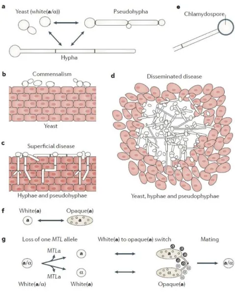
a budding yeast form or a hyphal form (Fig. 3a). In the former state, growth occurs similar to S. cerevisiae budding growth, each daughter cell is round and grows by budding, placing a new bud next to the previous division site or at the opposite pole. The budding form is associated with commensalism (Fig. 3b). During hyphal growth, a germ tube emerges in the mother cell in response to external cues and polarized growth is sustained to grow long hyphae - tube-like filaments with completely parallel sides and no constrictions at the site of septation (Sudbery, 2011) - that are important for tissue invasion (Fig. 3c). This study examines the transition between isotropic and polarized types of growth, focusing predominantly on the hyphal growth.
In spite of the term dimorphic being widely used to describe C. albicans, yeast and hyphae are not the only morphological states of C. albicans: i) pseudohyphae – a form of filamentous growth that involves cell elongation without the formation of true hyphae and
II – Candida albicans, an opportunistic pathogen
of a Spitzenkörper, with constrictions separating the cells. This form of growth is stimulated by environmental conditions, such as phosphate or alkane rich medium, and by the mutation of genes involved in the cell cycle regulation (Fig. 3a) (Sorkhoh et al., 1990; Hornby et al., 2004; Bensen et al., 2005; Li et al., 2006; Mukaremera et al., 2017; Noble et al., 2017). The pseudohyphal form often coexists with the yeast and hyphal forms during infection (Fig. 3d) (Sudbery et al., 2004; Noble et al., 2017); ii) the chlamydospores – the ability to form chlamydospores has been extensively used to distinguish between Candida species in the clinic. These large cells with thick walls, high lipid and carbohydrate content, develop in environments low in oxygen, light, temperature and nutrients, which are thought to be somehow related to the commensal/opportunistic lifestyle of these species (Fig. 3e) (Fabry et al., 2003; Palige et al., 2013; Böttcher et al., 2016); iii) the opaque cells. The white-opaque switch is a phenotypic switch, intimately coupled to the sexual mating process in C. albicans, exclusive to strains that are homozygous for the MTL locus that controls cell type (Fig. 3f-g) (Morschhäuser, 2010; Zhang et al., 2015; Li et al., 2016).
In both C. albicans budding and hyphal growth, cells exhibit polarized growth, yet polarity is more extreme in the hyphal state, as the switch from polarized to isotropic growth, characteristic of budding growth, is replaced by the maintained apical growth of the hypha. The differences between the distinct morphological forms of C. albicans include the extent of polarized growth, nuclear migration, position of septation and ability to separate after cytokinesis (Sudbery et al., 2004). In the yeast form, budding occurs either next to the bud scar from the previous cell cycle (axial pattern) or at the opposite end of the cell from where the previous bud formed (bipolar pattern) in a temperature-dependent manner (Chaffin, 1984). A septin ring appears before bud emergence, marking where a bud will emerge (Warenda and Konopka, 2002). Septins are highly conserved structural components that have important roles in C. albicans polarization, cell division and virulence (Warenda and Konopka, 2002; Warenda et al., 2003; Blankenship et al., 2014). This septin ring will be the plane across which mitosis will occur between mother and daughter cells (Sudbery et al., 2004). After mitosis is complete, the septin ring separates in two rings and a primary septum, composed of chitin (made by chitin synthase 1, CHS1, or CHS3) forms between them (Lenardon et al., 2010). An actomyosin ring then forms, contracts and cells separate (González-Novo et al., 2009).
Figure 3. Candida albicans cell type transitions. a | C. albicans switches reversibly between yeast (also known as
white(a/α)), hyphal and pseudohyphal cell types under different environmental conditions. b,c | In mucocutaneous infection models, such as oropharyngeal candidiasis, yeasts are associated with commensalism (part b), whereas the filamentous forms (hyphae and pseudohyphae) are associated with tissue invasion and damage (part c). d | Yeasts, hyphae and pseudohyphae all seem to have roles in disseminated disease — for example, in abscesses within internal organs of the host. e | Chlamydospores are produced by terminal (suspensor) cells of mycelia (multicellular hyphae or pseudohyphae) under adverse growth conditions. f | Mating-type-like (MTL) loci MTLa (‘a’) or MTLα (‘α’) cells can undergo an epigenetic switch between white(a or α) and opaque(a or α) phenotypes. White (a or α) cells have the same appearance as typical budding yeasts, whereas opaque (a or α) cells are elongated and have ‘pimple’ structures on their cell surface. g | Mating in C. albicans requires three events: loss of one allele of the MTL locus (MTLα or MTLa) to generate white (a) or white (α) strains; an epigenetic switch from white (a or α) to opaque (a or α); and pheromone sensing by opaque (a or α) cells of the opposite mating type, which triggers sexual filament production and mating (figure from Noble et al., 2017).
The incipient point of evagination of germ tubes is marked by a septin patch, which then forms a band at the base of the germ tube and a cap at the tip (Warenda and Konopka, 2002). As the germ tube elongates, the septin cap at the tip generates a ring in the germ tube that remains fixed in position as the tip continues to elongate. The nucleus then migrates out of the mother cell and mitosis occurs within the germ tube. After
II – Candida albicans, an opportunistic pathogen
mitosis, one daughter nucleus migrates back to the mother cell and the other nucleus migrates to the apical side of the septin ring (Sudbery, 2001; Finley and Berman, 2005). The septum of hyphal cells forms similarly to the septum of yeast or pseudohyphae, with a contraction of the actomyosin ring, yet cytokinesis does not result in a constriction, so the characteristic long tube-like structure is maintained (Sudbery, 2011).
c) Virulence
During both superficial and systemic infections, C. albicans relies on a wide arsenal of virulence factors and fitness attributes. Morphological transition between yeast and hyphal forms, expression of adhesins and invasins on the cell surface, and thigmotropism, capacity to form biofilms, phenotypic switching and secretion of hydrolytic enzymes are considered virulence factors (reviewed in Mayer et al., 2013). In addition, the ability to adapt rapidly to environmental changes contributes to this fungus increased fitness (Nicholls et al., 2011). During hyphal formation, a subset of genes is expressed. These genes encode virulence factors that are not directly involved in hyphal formation but provide an adaptative advantage as C. albicans invades the host tissues and causes infection (Mayer et al., 2013). The most highly expressed genes encode the extensively studied adhesin agglutinin-like sequence protein 3 (ALS3), the GPI-anchored hyphal wall protein 1 (HWP1), secreted aspartyl protease (SAP) family proteins, such as SAP4, SAP5 and SAP6, and Extent of Cell Elongation protein 1 (ECE1), a fragment of which is now referred to as candidalysin – a fungal peptide toxin (Fan et al., 2013; Hoyer et al., 2014; Orsi et al., 2014; Modrezewka and Kurnatowski, 2015; Moyes et al., 2016; Richardson et al., 2018). These proteins become of primary importance in the interaction of C. albicans with host cell surface receptors. Specifically, targeting virulence factors has been proposed as an alternate antifungal strategy (Gauwerky et al., 2009), hence, the understanding the pathogenicity mechanisms used by C. albicans during infection is of utmost importance.
C. albicans ability to switch from yeast to hypha has drawn interest because of its
relevance to pathogenicity; it is believed that each form of growth provides critical functions important for the pathogenic lifecycle (Sudbery et al., 2004; Jacobsen et al., 2012). Hyphae and pseudohyphae are postulated to promote tissue penetration during the early stages of infection, whereas the yeast form is thought to be more suited for dissemination
in the bloodstream (Saville et al., 2003; Jacobsen et al., 2012; Noble et al., 2017). However, it is important to note that most dimorphic fungi that infect humans exhibit growth by budding in infected tissues and exist as filamentous mycelial fungi in the external environment, such as H. capsulatum or Sporothrix schenkii (Lorenz et al., 2004; Gauthier, 2015). This fact suggests that filamentous growth is not mandatorily coupled with tissue invasion. However, C. albicans mutants that are unable to form or sustain hyphal growth in
vitro, are attenuated in virulence (Lo et al., 1997; Iranzo et al., 2003; Kavanaugh et al., 2014;
Labbaoui et al., 2017). This observation supports the view that the ability to form hyphae is an important virulence attribute for this fungus.
d) Polarized Filamentous Growth
As mentioned previously, the ability to switch from yeast to hyphal growth is a striking characteristic of C. albicans, which is clinically relevant (Mayer et al., 2013). C.
albicans is well adapted to growth in its human host and forms hyphae under a varied range
of environmental conditions. For example, hyphae form in response to the presence of serum (Taschdjian et al., 1960), neutral pH (Buffo et al., 1984), 5% CO2 (Mardon et al.,
1969), N-acetyl-glucosamine (GlcNAc) (Simonneti et al., 1974), and generally requires a temperature of 37 ºC. The combination of serum and 37 ºC provides a powerful signal for germ tube formation from yeast-form cells. The ability to form hyphae can be assessed in liquid or in solid media. Liquid media conditions are used to test the ability to initiate hyphal growth and the solid media conditions are used to test whether the organism can maintain invasive hyphal growth in a complex and changing environment as the colony develops in the agar.
The interaction with the microflora, which C. albicans encounters in its environment, acts as a regulator for the morphological switch. Communication with other cells, not only other C. albicans cells but also bacteria, relies on quorum sensing (Shareck and Belhumeur, 2011). For example, farnesol, a sesquiterpene that is secreted by C. albicans inhibits hyphal formation (Hornby et al., 2001; Lu et al., 2014). In contrast, the aromatic alcohol tyrosol promotes hyphal formation in biofilms (Chen et al., 2004). C. albicans can also form mixed infections with bacterial species, such as Pseudomonas aeruginosa, as both species are often recovered from lung infections of cystic fibrosis patients or infections in burned patients (De Sordi and Muhlschlegel, 2009). The bacteria attach specifically to the hyphae, leading
II – Candida albicans, an opportunistic pathogen
to the death of the fungal cell. As a response, C. albicans senses the presence of bacteria, via 3-oxo-C(12)-homoserine lactone (HSL) - a component of the P. aeruginosa quorum sensing mechanism – and hyphal growth is repressed in favour of budding growth (Hogan and Kolter, 2002; Hogan et al., 2004; Hogan, 2006; De Sordi and Muhlschlegel, 2009).
The expression
of hyphal-specific genes is negatively controlled by the general
transcriptional co-repressor Tup1, which forms a complex together with Nrg1 or
Rfg1
(Braun and Johnson, 1997; Braun et al., 2001; Murad et al., 2001; Kadosh and Johnson, 2005). Positive regulation of hyphal-specific genes is carried out by a variety of transcription factors, including Efg1 (Stoldt et al., 1997), Cph1 (Liu et al., 1994; Leberer etal., 1996), Cph2 (Lane, Zhou, et al., 2001), Tec1 (Lane, Birse, et al., 2001), Flo8 (Simonneti et al., 1974), Czf1 (Cao et al., 2006), Rim101 (Fig. 4; Davis et al., 2000; El Barkani et al.,
2000). Efg1 is thought to be the major regulator of hyphal formation under most conditions (Braun and Johnson, 2000; Sohn et al., 2003; Nobile et al., 2012; Jakubovics, 2017). Efg1 and Cph1 are activated via different upstream signalling pathways (Biswas and Morschhäuser, 2005). The former is activated through the cyclic AMP protein kinase A (cAMP-PKA) pathway (Tebarth et al., 2003), whereas the latter depends on the mitogen-activated protein kinase (MAPK) signalling pathway (Liu et al., 1994; Malathi et al., 1994). The guanine nucleotide binding protein Ras1 is involved in both pathways: it activates Cyr1 in the cAMP-PKA pathway and Cdc42 in the MAPK pathway (Feng et al., 1999; Leberer et al., 2001). These signalling pathways are the most studied in C. albicans hyphal growth. They share several upstream components and respond to multiple host environmental conditions including serum, body temperature (37 ºC), nitrogen starvation, high CO2 levels, GlcNAc, certain amino acids and quorum sensing molecules (Sudbery,
2011). Other pathways have been described to regulate C. albicans hyphal growth. These include the high osmolarity glycerol (Hog), the protein kinase C (Pkc) 1 cell wall integrity, the regulation of Ace2 and morphogenesis (RAM), the Rac1 activation and the Rim101 pathways (Alonso-Monge et al., 1999; Brown et al., 1999; Davis et al., 2000; Kullas et al., 2004; Li et al., 2004; Kumamoto, 2005; Bassilana and Arkowitz, 2006; Song et al., 2008; Hope et al., 2008; Shapiro et al., 2011; Saputo et al., 2012; Calderón-Noreña et al., 2015).
C. albicans has a single adenylyl cyclase encoded by CYR1. This gene has the role of
for hyphal formation (Rocha et al., 2001; Zou et al., 2010). Serum-mediated hyphal induction stimulates Cyr1 through the interaction with Ras1 with a RAS-associated domain in Cyr1 (Fang and Wang, 2006; Piispanen et al., 2011, 2013). Elevated temperature is required for all hypha-inducing conditions (except growth in embedded matrix). Temperature is sensed by the heat shock protein 90 (Hsp90) (Shapiro et al., 2009). Inhibition of Hsp90 using geldanamycin leads to hyphal growth, and mutant strains with a reduction in Hsp90 levels form hyphae in a media with serum at 30 ºC instead of 37 ºC (Shapiro et al., 2009). Hsp90 has been shown to activate hyphal growth via the cAMP-PKA pathway, although an efg1 mutant still forms hyphae when Hsp90 is inhibited, which suggests an alternative downstream target for Hsp90 (Shapiro et al., 2009). This pathway is also activated by the transmembrane ammonium permease Mep2, under nitrogen starvation conditions (Biswas and Morschhäuser, 2005). The previously mentioned quorum sensing molecules farnesol and HSL both inhibit C. albicans Cyr1 activity, inhibiting the cAMP-PKA signalling cascade and hyphal growth (Lindsay et al., 2012).
Ras1 activates the Cdc42 Rho GTPase via its GEF Cdc24 (Piispanen et al., 2011). Cdc42 then activates the MAPK signalling pathway, and the terminal MAPK of this cascade, Cek1, is important to activate Cph1 (Csank et al., 1998) - the downstream transcription factor for this cascade and an homolog of S. cerevisiae Ste12 (involved in mating pheromone response) (Liu et al., 1994). Cph1, in turn, controls the level of activation of some hyphal-specific target genes (Leng et al., 2001; Brown et al., 2007; Shapiro et al., 2011). During serum-induced hyphal growth, both Cdc42 and Cdc24 localized to the hyphal tip (Hazan and Liu, 2002; Bassilana et al., 2005).
Hypha formation requires both sustained apical growth and inhibition of cell separation. Strains with reduced expression levels of Cdc42 and its GEF Cdc24 are viable but unable to switch to polarized hyphal growth in response to serum (Bassilana et al., 2005). While Cdc42 and Cdc24 both localize at the tip of growing hyphae (Hazan and Liu, 2002; Bassilana et al., 2005), one of the two Cdc42 GAPs, Rga2, no longer localizes at the hyphal tip upon CDK/Hgc1 phosphorylation (Court and Sudbery, 2007; Zheng et al., 2007). During budding growth in S. cerevisiae, a number of proteins associate with the septin ring and cell separation is ensured by enzymes coded by genes from the RAM pathway (Roncero and Sanchez, 2010). In C. albicans, deletion of genes of the RAM pathway results in cell septation defects (Kelly et al., 2004; Song et al., 2008; Saputo et al., 2012)
III – The small Rho-GTPase Cdc42 in fungi
a) Rho GTPases
The Ras superfamily of GTPases is particularly interesting, as its members are master regulators of many varied aspects of cell behaviour, such as regulation of gene expression, cell proliferation, differentiation, cell polarity, cell movement, cell-cell interactions, endocytosis and exocytosis, vesicle formation and fusion with the acceptor compartment, vesicle transport and nucleocytoplasmic transport of RNA and proteins (Wennerberg et al., 2005). This superfamily of monomeric GTPases, with over 60 representatives in mammals, is divided in 5 major families: Ras, Rho, Rab, Arf and Ran (Wennerberg et al., 2005). The first reports of the cellular functions of Rho GTPases date from 1990, when they were identified in yeast and human cells (Bender and Pringle, 1989; Adams et al., 1990; Johnson and Pringle, 1990; Munemitsu et al., 1990; Shinjo et al., 1990). Here, I focus specifically on Rho GTPases, emphasizing the role of the master regulator of polarized growth, Cdc42, in fungi. Other Rho GTPases play a role in hyphal growth, Rho3 is required for actin polarization, Rho1 is required for invasive filamentous growth and Rac1 and its GEF, Dck1, are required for matrix embedded filamentous growth (Bassilana and Arkowitz, 2006; Hope et al., 2008; Corvest et al., 2013).
Rho GTPases are highly conserved molecular switches in eukaryotes involved in the regulation of morphogenesis (Etienne-Manneville and Hall, 2002; Etienne-Manneville, 2004; Arkowitz and Bassilana, 2015; Hervé and Bourmeyster, 2015). There are 2 distinct RhoGEF families, the DH/PH (Dbl homology and Pleckstrin homology)-domain-containing proteins (Rossman et al., 2005) and DOCK (Dedicator of cytokinesis) homology domain (Meller, 2005). These proteins can hydrolyze GTP and hence cycle between two conformational states: a GTP-bound active state and a GDP-bound inactive state (Fig. 4). In the active GTP-bound state, they interact with effector proteins and, in contrast, in the inactive GDP-bound state, they typically do not interact with their targets, thereby interrupting signaling (Vetter and Wittinghofer, 2001; Etienne-Manneville and Hall, 2002; DerMardirossian and Bokoch, 2005).
Figure 4. The Rho GTPase cycle. Rho GTPases cycle between an active (GTP-bound) and an inactive
(GDP-bound) conformation. In the active state, they interact with one of over 60 target proteins (effectors) in mammalian cells. The cycle is highly regulated by three classes of proteins: the guanine nucleotide exchange factors (GEFs) catalyze nucleotide exchange and thereby mediate activation; the GTPase-activating proteins (GAPs) stimulate GTP hydrolysis, leading to inactivation; and the guanine nucleotide exchange inhibitors (GDIs) extract the inactive GTPase from membranes. All Rho GTPases are prenylated at their C terminus, a process required for their function (Etienne-Manneville and Hall, 2002).
Rho GTPases share a common G-domain fold, which consists of 5 α-helixes and 6-strand β-sheet (Vetter and Wittinghofer, 2001). The formation of the active GTP-bound state occurs with a conformational change in the N-terminal regions, known as switch I and switch II (Fig. 5). These two regions not only constitute the nucleotide binding pocket but also engage with their regulators (GEFs, GAPs and GDIs) and downstream effectors (for example kinases) (Ihara et al., 1998; Burridge and Wennerberg, 2004; Miyazaki et al., 2006; Parri and Chiarugi, 2010). Rho GTPase GEFs contain the catalytically active Dbl homology (DH domain) followed by an adjacent pleckstrin homology (PH) domain (Erickson and Cerione, 2004). These two domains interact with switch I and switch II regions inducing a conformational rearrangement that promotes nucleotide ejection and is the defining mechanism of activation and inactivation of the GTPases, termed the loaded-spring mechanism (Vetter and Wittinghofer, 2001). GTP can be hydrolysed from the Rho GTPase by an intrinsic reaction that can be stimulated through the interaction of the GTPase with GAPs; GAPs have a conserved catalytic domain, which is sufficient for GTPase binding and for the stimulation of the GTP-hydrolysis reaction (Vetter and Wittinghofer, 2001). Effectors containing a Cdc42/Rac1-interactive binding (CRIB) domain bind to Cdc42/Rac1 at the switch I domain (Abdul-Manan et al., 1999; Morreale et al., 2000). At the C-terminus of Rho GTPases is the CAAX box, where post-translational isoprenylation on the cysteine residue facilitates the specific binding to the plasma membrane, which is essential for their biological activity (Olofsson, 1999; McTaggart, 2006; Park and Bi, 2007).
III – The small Rho-GTPase Cdc42 in fungi
Figure 5. Structure of guanine nucleotide binding proteins.
Ribbon plot of the minimal G domain, with the conserved sequence elements and the switch regions in different colors as indicated. The nucleotide and Mg21 ion are shown in ball-and-stick representation (Vetter and Wittinghofer, 2001).
GEFs interact with Rho proteins and alter the nucleotide-binding site, facilitating the release of the nucleotide and, since the cytoplasmic concentration of GTP is higher than that of GDP, it is more likely that Rho proteins will bind with GTP (Bos et al., 2007). In addition, GAPs promote the hydrolysis of GTP from Rho proteins (Bos et al., 2007). There are more Rho GEFs and GAPs than Rho proteins - one Rho protein can be regulated by more than one GEF and GAP, and these regulators can interact with more than one Rho protein (García et al., 2006). For example, S. pombe Cdc42 has two GEFs, Scd1 and Gef1, which regulate apical growth and cytokinesis, respectively (Coll et al., 2003; Hirota et al., 2003). It is thought that this promiscuity accounts for a process-specific regulation of a Rho protein. Rho GEFs and GAPs also have the ability to bind other proteins and membranes due to their multidomain configuration. In turn, these proteins change the activation state of Rho proteins and can also act as scaffold proteins, aiding Rho protein localization and coupling upstream signals with downstream effectors (Toenjes et al., 1999; Bose et al., 2001; Ito et al., 2001; Gimona et al., 2002; Bos et al., 2007; Lemmon, 2008; Yohe et al., 2008). Other regulators of Rho proteins are GDIs, which control the access of Rho GTPases to GEFs and GAPs. GDIs interact only with the GDP-bound state and sequester the GTPase from the membrane into the cytoplasm (DerMardirossian and Bokoch, 2005).
b) Cdc42-Cdc24 Module in fungi
In the fungal kingdom, polarized growth depends on a number of different small GTPases of the Ras family. Bud site selection in the budding yeast S. cerevisiae is one of the best studied cell polarization systems. Wild-type yeast cells use landmark-directed cues and a GTPase cascade that transduces these signals, which is initiated with Rsr1/Bud1, to choose the polarization axis in a process called symmetry breaking (Singh et al., 2017). However, cells can also polarize in the absence of spatial information, i.e., in an rsr1/bud1 deletion mutant (Wedlich-Söldner and Li, 2003). In S. cerevisiae, the activation at the bud landmark of the essential Rho GTPase Cdc42 requires the GEF Cdc24, which binds the GTP bound form of Rsr1/Bud1 (Park and Bi, 2007; Kang et al., 2010). C. albicans mutants that lack Rsr1/Bud1 or Bud2 form wider hyphae than wild type cells and are unable to maintain hyphal growth in one direction (Hausauer et al., 2005). Rsr1/Bud1 C. albicans mutants have also been demonstrated to be less virulent, this can attributed to the reduced germination, shorter hyphae and defects in thigmotropism and galvanotropism (hyphal turning in response to changes in substrate topography and imposed electrical fields, respectively) and penetration into semisolid substrates (Yaar et al., 1997; Brand et al., 2008).
The current model of symmetry breaking suggests that Cdc42•GTP accumulates stochastically and induces a positive feedback loop mediated by Cdc42•GTP, the Cdc24 scaffold protein Bem1 and the PAK kinase Cla4, (Howell et al., 2012; Rapali et al., 2017). This protein complex is required for polarity establishment and in vivo and computational model analysis of symmetry breaking are consistent with positive feedback via local Cdc42 activation (Chenevert et al., 1992; Holly and Blumer, 1999; Bose et al., 2001; Butty et al., 2002; Ozbudak et al., 2005; Goryachev and Pokhilko, 2008). When this complex forms, Cdc24 GEF activity is increased, enhancing the activation of neighbouring molecules of Cdc42•GDP. A positive feedback loop is initiated and promotes the formation of a cortical cluster of activated Cdc42 at the polarization site (Gulli et al., 2000; Bose et al., 2001; Goryachev and Pokhilko, 2008; Kozubowski et al., 2008; Howell et al., 2009; Johnson
et al., 2011; Woods et al., 2015). On the other hand, Bem1 triggers a negative feedback loop
as it brings Cdc24 and Cla4 together, resulting in subsequent Cdc24 phosphorylation by Cla4, leading to the disruption of its GEF activity, and release from the Bem1 scaffold complex (Howell et al., 2012; Rapali et al., 2017). An actin-dependent process has also been
III – The small Rho-GTPase Cdc42 in fungi
implicated in a second positive feedback loop for symmetry breaking, via local delivery of Cdc42 (GDP or GTP bound). Actin nucleation depends upon the formation of a Cdc42•GTP cluster at the cortex and, in turn, the accumulation and stabilization of the Rho GTPase relies on actin-dependent vesicle transport and endocytosis (Wedlich-Söldner
et al., 2003; Marco et al., 2007; Slaughter et al., 2009, 2013). These two models are not
mutually exclusive, however findings from different laboratories lead to contradictory conclusions regarding their relative importance.
Cdc42 is a central regulator of the polarisome, a complex of the proteins localized at sites of growth that is required for actin polarization and polarized growth. This complex was initially identified in budding yeast and is composed of the proteins Spa2, Pea2 and Bud6 (Sheu et al., 1998). The polarisome has been observed in several filamentous fungi, such as Neurospora crassa, Aspergillus nidulans, Ashbya gossypii and C. albicans, where it localizes to a crescent at the tip of growing hyphae (Crampin et al., 2005; Köhli et al., 2008; Jones and Sudbery, 2010; Lichius et al., 2012).
Bud initiation and growth are tightly coordinated with the cell cycle. In the budding yeast model, cell polarization occurs only once per cell cycle, being dependent on signals that are triggered by the cyclin-dependent kinase 1 (CDK1) Cdc28 and its cyclin partners (Enserink and Kolodner, 2010). Cdc28 activity has been suggested to be required for proper localization of the GEF of Cdc42, Cdc24, to the presumptive bud site (Gulli et al., 2000; Moffat and Andrews, 2004). Although Cdc28 phosphorylates Cdc24 in vitro (McCusker et al. 2007), Cdc24 function in vivo is not affected by mutation of predicted phosphorylation sites (Gulli et al., 2000; Wai et al., 2009; Rapali et al., 2017). The PAK-like kinase Cla4 is thought to phosphorylate Cdc24 (Gulli et al., 2000), and Cla4 has been implicated in a Cdc28-Clb-dependent pathway that promotes the switch from polar to isotropic bud (Tjandra et al., 1998).
During the G1/S transition of the cell cycle of S. cerevisiae, Cdc42•GTP localizes to the polarization site which becomes the bud tip. When bud growth switches from apical to isotropic, Cdc42 redistributes from a tip cortical location to the septin ring in late anaphase (Lew and Reed, 1993). The uniqueness of a site of growth at any given time in S. cerevisiae is controlled by the Cdc28/G1 CDK-cyclin complex regulation of Cdc42, so when Cdc42 is
no longer under the control of the cyclin complex, e.g., upon overexpression of a constitutively active mutant (Cdc42[G12V] or Cdc42[Q61L), and in the absence of all G1 cyclins, polarization occurs at multiple sites (Gulli et al., 2000; Wedlich-Söldner and Li, 2003). Mathematical modelling and studies on artificially rewired cells suggest that there is a fast competition between polarization clusters, which is ultimately responsible for restricting polarization to a single site (Goryachev and Pokhilko, 2008; Howell et al., 2009). Increased expression of a constitutively active form of Cdc42 at the plasma membrane led to an increased number of cells initiating polarization at two or more sites (Wedlich-Söldner et al., 2003). Mutations (dominant negative, Cdc42[T17N], and constitutively active, Cdc42[G12V] and [Q61L]) in the Cdc42 putative GTP binding and hydrolysis domains negatively impact cell proliferation (Ziman et al., 1991; Vanni et al., 2005). FRAP experiments of the inactive Cdc42[D57Y] mutant and the constitutively active Cdc42[Q61L] mutant revealed a much slower recovery of fluorescence compared to wild-type Cdc42 (Wedlich-Söldner et al., 2004). Taken together, these results suggest that the ability of Cdc42 to cycle between the active and inactive states plays an important role in the exchange of Cdc42 between the polarization site and the cytosol, hence proper function (Ziman et al., 1991; Wedlich-Söldner et al., 2004; Vanni et al., 2005).
Cdc42 is essential for polarized growth in the fission yeast S. pombe, and the spatial control of its activation determines cell width (Kelly and Nurse, 2011). Active Cdc42 localizes at the cells tips, where it cycles between the active and inactive states, and unlike what has been observed in S. cerevisiae, the expression of constitutively active Cdc42 promotes a non-polarized phenotype, resulting in round cells (Miller and Johnson, 1994; Bendezú et al., 2015). S. pombe has two known Cdc42 GEFs, Scd1 and Gef1, which are essential and localize to the cell tip and division site (Coll et al., 2003; Rincon et al., 2007), where active Cdc42 is observed (Miller and Johnson, 1994; Tatebe et al., 2008). Cells lacking Scd1 are also round and have endocytosis defects (Murray and Johnson, 2001), while deletion of Gef1 results in slightly thinner cells with defects in bipolar growth and cytokinesis (Coll et al., 2003). The formin For3 is activated by Cdc42 and is responsible for the formation of the actin cables that stabilize the axis of polarity by directing secretion towards the tips (Bendezú and Martin, 2011; Estravís et al., 2011; Kelly and Nurse, 2011; Bonazzi et al., 2015). Both GAPs, Rga4 and Rga6, localize to non-growing tips and lateral areas of the plasma membrane, spatially restricting active Cdc42 at the cell tips to maintain