HAL Id: hal-01965639
https://hal.archives-ouvertes.fr/hal-01965639
Submitted on 26 Dec 2018
HAL is a multi-disciplinary open access
archive for the deposit and dissemination of
sci-entific research documents, whether they are
pub-lished or not. The documents may come from
teaching and research institutions in France or
abroad, or from public or private research centers.
L’archive ouverte pluridisciplinaire HAL, est
destinée au dépôt et à la diffusion de documents
scientifiques de niveau recherche, publiés ou non,
émanant des établissements d’enseignement et de
recherche français ou étrangers, des laboratoires
publics ou privés.
Computational Performance of Time-Resolved Diffuse
Optical Tomography in a Two-Layer Brain Model
David Orive-Miguel, Lionel Hervé, Jerome Mars, Laurent Condat, Jean-Marc
Dinten, Sophie Morales
To cite this version:
David Orive-Miguel, Lionel Hervé, Jerome Mars, Laurent Condat, Jean-Marc Dinten, et al..
Com-putational Performance of Time-Resolved Diffuse Optical Tomography in a Two-Layer Brain Model.
OSA Biophotonics Congress: Biomedical Optics, Apr 2018, Hollywood, Florida, United States.
�hal-01965639�
Computational Performance of Time-Resolved
Diffuse Optical Tomography in a Two-Layer Brain
Model
David Orive-Miguel1,2, Lionel Herv´e1, J´erˆome Mars2, Laurent Condat2, Jean-Marc Dinten1and Sophie Morales1
1: CEA, LETI, MINATEC Campus, F-38054 Grenoble, France
2: Univ. Grenoble Alpes, CNRS, Grenoble INP, GIPSA-lab, 38000 Grenoble, France [email protected]
Abstract: We evaluate Mellin-Laplace algorithm in a two-layer brain model with Monte-Carlo generated data. Results show accurate absorption estimation and the flexibility of the algorithm to detect each layer thickness.
OCIS codes: 170.6920, 110.6955, 110.0113, 030.5260, 100.3190
1. Introduction
Traumatic brain injury (TBI) is one of the leading causes of disability and injury-related deaths. Although magnetic resonance imaging or computerized x-ray tomography are standard techniques in TBI treatment protocols there is still a need of a technology that could allow to perform bedside, continuous and non-invasive brain monitoring. Time-resolved diffuse optical tomography (TR-DOT) is an emerging photonics technology that could potentially permit to continuously measure important brain physiological values in TBI cases such as blood oxygenation levels.
Even though TR-DOT has already been applied, for example, to flap viability assessment [1] with very promising results there is still a lack of accurate optical properties estimation for brain imaging cases. Nonlinear Monte-Carlo fitting methods have been used in computational analysis [2] but their performance depends significantly on the knowl-edge of the geometry being scanned and is time consuming. In this work we computationally evaluate an algorithm based on Mellin-Laplace moments and we analyze its accuracy and adaptation to different layer thicknesses.
2. Method
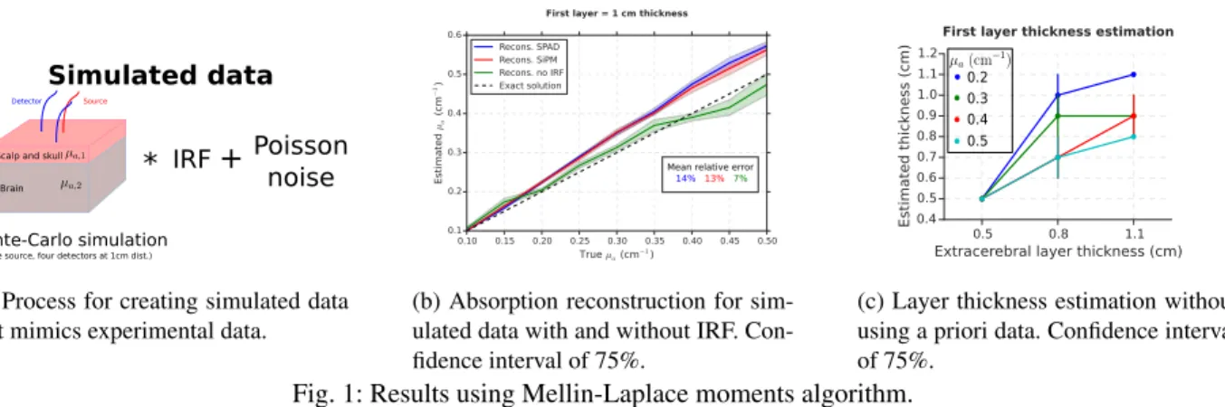
The computational phantom consists of a two-layer model (8×8×5 cm3) containing an extracerebral and cerebral layer with one light source and four detectors placed at 1 cm distance at the top boundary, see Figure1a. Monte-Carlo simulations [3] were performed on the phantom, signals were convolved with the instrumental response function (IRF) of single-photon avalanche diode (SPAD) and silicon photomultipliers (SiPM) detectors, and were corrupted with Poisson noise.
For the reconstruction of the optical properties, Mellin-Laplace moments algorithm [4] with twelve moments and p = 9 was used. This algorithm is based on finite element method and computes the moments of time-point spread functions in order to achieve a good balance between accuracy and computational time. The algorithm can be used on experimental data by performing measurements in a reference phantom; moreover, knowledge of the shape of the IRF is not necessary.
3. Results and Discussion
The first experiment consists in evaluating the Mellin-Laplace algorithm performance for estimating the absorption at cerebral layer. Monte-Carlo simulations were done with a constant absorption of µa,1= 0.1 cm−1at the extracerebral
layer and an increasing absorption from µa,2= 0.1 to 0.5 cm−1 on steps of 0.05 cm−1 for the cerebral layer. The
scattering coefficient was maintained homogeneous along both layers because of its little sensitivity at brain layer [2]. Extracerebral and cerebral layer thicknesses were 1 cm and 4 cm respectively; homogeneous absorption through each layer was assumed. An homogeneous phantom of µa= 0.1 cm−1was taken as reference measurement. Results are
plotted in Figure1bfor different types of detectors; they show that Mellin-Laplace algorithm reconstructs linear ab-sorption estimations with low relative error. It is also seen that IRFs influence the results by yielding an overestimation
Scalp and skull Brain
Monte-Carlo simulation
(one source, four detectors at 1cm dist.)
*
PoissonnoiseSimulated data
Detector Source
+
IRF
(a) Process for creating simulated data that mimics experimental data.
Mean relative error 14% 13% 7%
(b) Absorption reconstruction for sim-ulated data with and without IRF. Con-fidence interval of 75%.
0.3 0.4 0.2
0.5
(c) Layer thickness estimation without using a priori data. Confidence interval of 75%.
Fig. 1: Results using Mellin-Laplace moments algorithm.
at absorption values much higher than the reference measurement making the relative error to increase twice with respect to free IRF data. No significant differences are seen in the performance with SPAD or SiPM detectors.
In the second experiment we aim to evaluate whether the algorithm is able to detect the thickness of the two layers; that is, whether the algorithm has the flexibility to adapt to the two-layer geometry when thickness of each layer is not given in advance. The domain was segmented in 0.3 cm-thick layers each of them with an homogeneous absorption. The two-layer geometry interface was estimated as the layer interface with largest absorption difference between above and below layers. Monte-Carlo simulations were performed for cases were extracerebral layer thickness ranged from 0.5 to 1.1 cm on steps of 0.3 cm; cerebral layer absorption ranged from µa,2= 0.2 to 0.5 cm−1 on steps of
0.1 cm−1. Results in Figure1cshow that accurate extracerebral layer thickness is estimated in 0.5 and 0.8 cm cases. Nevertheless, thickness is underestimated in 1.1 cm case because the algorithm has less sensibility to deeper layers. Higher absorption values tend to underestimate the thickness because the algorithm tries to compensate the high absorption by decreasing the extracerebral layer thickness.
4. Conclusions and Future Work
We studied the performances of a Mellin-Laplace algorithm for two-layer brain models with data that mimics realistic conditions. Accurate results are obtained if each layer thickness is given in advance although the algorithm is also capable of doing good estimations without that a priori information.
In the near future, we will extend the analysis by performing Monte-Carlo simulations on ATLAS head models and doing reconstructions of the absorption at the brain layer.
5. Acknowledgements
This project has received funding from the European Union’s Horizon 2020 Marie Skodowska-Curie Innovative Train-ing Networks (ITN-ETN) programme, under grant agreement no 675332 BitMap. The authors thank to the photonics lab at Politecnico de Milano for providing the instrumental response functions of the detectors.
References
1. L. Di Sieno, G. Bettega, M. Berger, C. Hamou, M. Aribert, A. Dalla Mora, A. Puszka, H. Grateau, D. Contini, L. Herv´e, J.-L. Coll, J.-M. Dinten, A. Pifferi, A. Planat-Chr´etien, “Time-resolved diffuse optical tomography for non-invasive flap viability assessment: pre-clinical tests on rats” in European Conferences on Biomedical Optics 2015Proceedings Volume 9538, Diffuse Optical Imaging V; 95380N (2015).
2. J. Selb, T.M. Ogden, J. Dubb, Q. Fang, D.A. Boas, “Comparison of a layered slab and an atlas head model for Monte Carlo fitting of time-domain near-infrared spectroscopy data of the adult head” in J. Biomed. Opt 2014 Jan; 19(1): 01601.
3. Q. Fang, “Mesh-based Monte Carlo method using fast ray-tracing in Plcker coordinates” in Biomed. Opt. Ex-pressVol. 1, Issue 1, pp. 165-175 (2010).
4. J. Zouaoui, L. Di Sieno, L. Herv´e, A. Pifferi, A. Farina, A. Dalla Mora, J. Derouard, J.-M. Dinten, “Quantifi-cation in time-domain diffuse optical tomography using Mellin-Laplace transforms” in Biomed. Opt. Express Vol. 7, Issue 10, pp. 4346-4363 (2016).