HAL Id: tel-01310047
https://tel.archives-ouvertes.fr/tel-01310047v3
Submitted on 31 Aug 2016HAL is a multi-disciplinary open access archive for the deposit and dissemination of sci-entific research documents, whether they are pub-lished or not. The documents may come from teaching and research institutions in France or abroad, or from public or private research centers.
L’archive ouverte pluridisciplinaire HAL, est destinée au dépôt et à la diffusion de documents scientifiques de niveau recherche, publiés ou non, émanant des établissements d’enseignement et de recherche français ou étrangers, des laboratoires publics ou privés.
application to oesophagus
Anant Suraj Vemuri
To cite this version:
Anant Suraj Vemuri. Inter-operative biopsy site relocalization in gastroscopy : application to oe-sophagus. Other [cs.OH]. Université Nice Sophia Antipolis, 2016. English. �NNT : 2016NICE4018�. �tel-01310047v3�
UNIVERSITY OF NICE SOPHIA ANTIPOLIS
DOCTORAL SCHOOL STIC
(INFORMATION AND COMMUNICATIONTECHNOLOGIES ANDSCIENCES)
T H E S I S
to obtain the title of
PhD of Science
of the University of Nice - Sophia Antipolis
Specialty : AUTOMATION, SIGNAL ANDIMAGEPROCESSING
Prepared by
Anant Suraj V
EMURI
Inter-Operative Biopsy Site Relocalization in
Gastroscopy: Application to Oesophagus
Thesis Advisors: Nicholas A
YACHEand Luc S
OLERprepared at IHU, Strasbourg and INRIA, Sophia Antipolis, A
SCLEPIOSTeam
defended on April 26th, 2016Jury :
Reviewers :
Lena Maier-Hein
- Deutsches Krebsforschungszentrum (DKFZ)
Tom Vercauteren - Univeristy College London
President :
Nassir Navab
- Technische Universität München
Advisor :
Nicholas Ayache
- INRIA (Asclepios)
Co-Advisor :
Luc Soler
- IRCAD (Strasbourg)
Examiners :
Stéphane Nicolau - IRCAD (Strasbourg)
Invited :
Michel Delvaux
- CHRU (Strasbourg)
UNIVERSITÉ DE NICE SOPHIA ANTIPOLIS
ÉCOLE DOCTORALE STIC
(SCIENCES ET TECHNOLOGIES DE L’INFORMATION ET DE LA COMMUNICATION)
T H È S E
pour obtenir le titre de
Docteur en Sciences
de l’université de Nice - Sophia Antipolis
Mention : AUTOMATIQUE, TRAITEMENT DUSIGNAL ET DESIMAGES
Preparé par
Anant Suraj V
EMURI
Relocalisation de site de biopsie en gastroscopie:
application à l’œsophage
Thèse dirigée par Nicholas A
YACHEet Luc S
OLERpréparée à IHU, Strasbourg et INRIA, Sophia Antipolis, projet A
SCLEPIOS soutenue le 26 Avril, 2016Jury :
Rapporteurs :
Lena Maier-Hein
- Deutsches Krebsforschungszentrum (DKFZ)
Tom Vercauteren - Univeristy College London
Président :
Nassir Navab
- Technische Universität München
Directeur :
Nicholas Ayache
- INRIA (Asclepios)
Co-Directeur :
Luc Soler
- IRCAD (Strasbourg)
Examinateurs :
Stéphane Nicolau - IRCAD (Strasbourg)
Invités :
Michel Delvaux
- CHRU (Strasbourg)
iii
Acknowledgments
Foremost, I would like to thank my advisor, Prof. Luc Soler for providing the opportunity to work with IHU/IRCAD and establishing the environment for conducting my work. Luc, you energy levels never seized to amaze me. Despite an intense schedule you always took time to follow my work. Your guidance, advice and motivation were invaluable. Your vision of the future and ability to present those ideas, inspire me tremendously.
Then, I would like to express my gratitude to Dr. Stéphane Nicolau, my immediate supervisor and my dear friend. Thank you for taking such an large amount of time to guide my work. You channelled my energies in the right direction, whenever I exploded with ideas. It was a pleasure working with you.
To my advisor, Prof. Nicholas Ayache, it was an honour working with you. Thank you for accepting me into the asclepios team. Although much of the project related work, you advanced to Luc and Stéphane; from time to time you provided the necessary environment to stimulate my thinking. Whether it was through organizing visits to INRIA to meet the team or the wonderful Auron retreats, where I met the most brilliant scientific minds every year.
To Dr. Adrien Sportes, my partner in crime from the clinical front, your contribution in my work cannot be understated. Thank you for those long and fruitful discussions on gastro-intestinal procedures, I gained a lot of insight into the clinical aspect through you. You are the right clinical partner an engineer could have wished for.
The reviewers of my thesis, Dr. Lena Maier-Hein and Dr. Tom Vercauteren; your insights and comments were invaluable. Thank you for taking the time to make yourselves available for the review process. To Dr. Michel Delvaux and Dr. Lee Swanstrom for their clinical assessment of my work. In medical technology, the opinions of key medical experts, such as yourselves, who are the end users of our work, is most important. Thank you all for accepting to be part of my thesis Jury.
To the IRCAD/IHU development team (current and past); Julien Waechter, Emilie Harquel, Pascal Monnier, Nicolas Philipps, Jessica Gromer, Johan Moreau, Frédéric Champ, Arnaud Charnoz, Marc Schweitzer and Flavien Bridault. I would like to extend my deepest gratitude for working with me to transfer the approach presented in this thesis to clinical application and building a professional looking software that could be used in the operating room. I would like to also thank Jessica especially, for taking the time to participate in the clinical experiments. Your efforts were very much appreciated. Pascal, you are a very good friend and I will miss our morning tea and coffee sessions. To Dr. Oscar Garcia, I would like to say thank you. Your presence helped me understand the mathematics behind the Lie algebra and our discussions on calibration, error computation and propagation were very insightful. Your critiques made me delve deeper into the subject. Jalal Chaabane, who worked as an intern for an year and is an amazing programmer. You made my job of interfacing optical tracker over the network with my desktop easier through your application. Without it, the quantitative evaluation of the system would have been impossible. Thank you so much. Dr. Vincent Agnus, my neighbour at Virtual Surg for 3 years, I cannot understate your contribution through discussions, suggestions and guidance. You spent nearly 8 hours over a span of 10 days to help formulate my slides for the defence, even when you were
busy with things. Thank you very much Vince, I will miss your company.
To Dr. Xavier Pennec, a tireless mathematician. Your ability to model the most intricate parts of the surgical application was amazing. You helped me formulate a simplified trajectory based registration approach and understand the concept of error propagation in presence of isotropic noise.
Pamela Lhote, you are an amazing person and were super helpful throughout. From your arranging meetings with potential rentals when I first arrived in Strasbourg, to coordinating with everyone until my final defence, your role in this thesis completion, though behind the scenes, did not go unnoticed. Clélia Kinderstuth, you are another one of those behind the scenes person who organized so many things during my employment with IHU. Thank you! From the IHU radiology team Rodrigo Cararo, Mourad Bouhadjar and Gael Fourré, and , who organized the experiment room professionally and facilitated Adrien and I, to perform experiments with ease, I appreciate the work you guys do, thank you! To the staff at the endoscopy unit at NHC, Strasbourg for cooperating with us to conduct our data collection. It would have been quite challenging without your support.
Then to Prof. Jacques Marescaux and Jean-Luc Dimarcq for supporting my research through IRCAD and IHU foundation.
I would like to thank the patients who consented to us for collecting data using the electromagnetic tracker. To the 5 pigs on whom I perform the animal trials, I apologize for the pain you may have been put through. I extend my sincere gratitude for your sacrifice. To the printer, the trees (for the paper) and to Google scholar for supporting my work silently. To the members of the asclepios team, thank you for making me feel home every time I visited. Dr. Chloé Audigier, thank you for being so helpful in clarifying questions about UNice from time to time and just being such a nice person. Isabelle Strobant, you were amazing in coordinating things with UNice and INRIA for me. I really appreciate your help and support. To Regine Saelens from EDSTIC, thank you for being patient with me and replying to my “english” emails with care. My dear friend, Dr. Debarshi Dey, thank you for the last minute clarification for the calculation of statistics for my presentation.
To my family, for their never ending support, through the toughest of times. My Mom, Sister, Uncles, Aunts, Grandma and brother-in-law who have stood by me and given me the confidence to continue striving. My Dad and Grandpa, who passed on before they could see me complete, you have both been pillars in my life and your presence will be sorely missed. Last and the most important acknowledgement of all, is the contribution and sacrifice of my wife, Archana Vemuri, who gave up so much to be with me and help me complete my work. Without your support, this would not have been possible. Thank you from the bottom of my heart.
v
Abstract
Minimally invasive surgery in gastrointestinal (GI) endoscopy has evolved from being a diagnostic tool to a therapeutic solution. It is common that GI procedures involve periodic monitoring or surveillance of the internal anatomy. Specifically in oesophageal procedures (the target of this thesis), surveillance interventions involve obtaining biopsies at different regions along the oesophagus. The tracking and relocalization of these biopsy sites “inter-operatively” poses a significant challenge for providing targeted treatments.
This thesis, clarifies the concept of relocalization, and analyses the need for a platform to aide GI endoscopy. Then, based on the understanding of the clinical context in oesophageal procedures; a novel framework to use electromagnetic tracking system is proposed, which is used to perform a “recording” of an intervention. This framework and the recording is then used to provide a guided navigation to the GI expert, during a follow-up surveillance endoscopy; for accurate re-positioning of the endoscope at previously targeted sites. This is achieved using inter-operative video synchronization, and the various steps involved in achieving this are described in this thesis.
Following the description of system design and the methodology, a careful analysis of noise affecting the system is performed. Using a Gaussian noise model, quantitative evalua-tion of the system is performed with synthetic and real (porcine) data. The results indicate that the relocalization was achieved with an uncertainty in depth inside the oesophagus of ±10mm, which was considered acceptable for the GI expert. Additionally, qualitative experiments were performed using data from pigs, to simulate the real task of biopsy site relocalization, which was evaluated by 10 GI experts. The results of these experiments showed an improvement in biopsy site relocalization rate from 47.5% to 94%, thus clearly demonstrating the benefits of the proposed system towards assisted guidance. Furthermore, an incremental improvement in inter-operative video synchronization is proposed, that uses additional information obtained during the course of the intervention. Synthetic experiments indicated, inclusion of this additional information reduced the error in video synchronization by ∼ 50%.
This framework, was then extended by proposing a constrained inter-operative image matching, for further improvement in quality of video synchronization. Within this context, the effect of, the choice of feature descriptors and colour-space, filtering of uninformative frames and the endoscopic modality in use, are investigated and shown that further improvement is achieved in image synchronization to [92%, 87%] from [73%, 76%] for both narrow band imaging and white-light endoscopic modalities.
This research work has been implemented in a software (at IHU and IRCAD, Strasbourg), allowing us to validate our results in clinical conditions. This work was supported by IHU Strasbourg, through a grant# ANR-01AHU-02.
Keywords: Gastro-Intestinal Endoscopy, Inter-operative relocalization, Electro-magnetic tracking
vii
Résumé:
La chirurgie mini-invasive en endoscopie gastro-intestinale a évolué d’un outil de diagnostic à une solution thérapeutique. En règle générale, les procédures gastro-intestinales impliquent un contrôle ou une surveillance périodique de l’anatomie interne. Dans le contexte des interventions de l’œsophage, les exploration de de surveillance implique la réalisation de multiples biopsies régulièrement le long de l’œsophage. Ainsi, les défis les plus importants auxquels il faut faire face au cours de ces procédures sont le suivi et la relocalisation inter-opératoires de ces sites de biopsies (pour un même patient opéré plusieurs fois). L’objectif de cette thèse est de proposer une solution informatisée afin de guider le gastroentérologue pendant de telles procédures.
Cette thèse précise tout d’abord le concept de relocalisation et analyse la nécessité d’une plate-forme pour aider l’endoscopie gastro-intestinale. Ensuite, après une analyse des procédures de l’œsophage et de leurs besoins, nous proposons un cadre novateur utilisant un système de suivi électromagnétique pour réaliser des enregistrements d’intervention de l’œsophage, couplant la vidéo à la profondeur de l’endoscope inséré. Ces enregistrements sont utilisés pour fournir une navigation aux gastroentérologues pendant des procédures de surveillances afin de repositionner l’endoscope de façon précise sur des sites de biopsie préalablement ciblés. Cette navigation consiste en une synchronisation vidéo entre la vue endoscopique courante et celles des surveillances endoscopiques précédentes enregistrées. Une première version de notre système est présentée, associée à une analyse rigoureuse des bruits affectant le système. Cette évaluation incrémentale est réalisée sur des données d’abord synthétiques puis réelles recueillies sur des cochons. Les résultats montrent que la relocalisation est obtenue avec une précision de l’ordre de ±10mm, considérée comme largement acceptable par les experts. En outre, une expérience qualitative a été conçue à partir de données provenant de cochons pour simuler une tâche réelle de relocalisation de site de biopsie, et a été évaluée par 10 gastroentérologues. Celle-ci a clairement démontré l’avantage du système de guidage assisté en améliorant le taux de prélèvement de sites de biopsie de 47,5% à 94%. Une seconde version est alors proposée, utilisant la trajectoire 3D complète de l’œsophage acquise pendant l’intervention pour synchroniser les vidéos. Cette version permet d’éviter les erreurs importantes dues à des facteurs humains et permet de réduire l’erreur globale du système qui est améliorée d’environ 50%.
Ce cadre est finalement étendue afin d’améliorer encore la précision de la relocalisation à partir d’une sélection optimale de l’image vidéo pré-enregistrée dont le point de vue est le plus proche de celui de l’image endoscopique courante. Dans ce contexte d’appariement d’images, l’influence du choix des descripteurs, de l’espace de couleurs, de la présence d’une étape de filtrage d’images peu informatives et de la modalité d’images (lumière blanche ou NBI) sont examinés et démontrent qu’une amélioration encore plus significative peut être obtenue pour la synchronisation des images.
Contents
1 Introduction 1
1.1 A Historical Perspective . . . 1
1.2 Endoluminal Endoscopy . . . 2
1.3 Gastrointestinal cancer . . . 3
1.4 Guidance in Endoluminal Interventions . . . 4
1.5 Objectives of the thesis . . . 5
1.6 Manuscript Organization . . . 5
2 Analysis and Problem Statement 9 2.1 Oesophageal Adenocarcinoma . . . 9
2.2 Colorectal Cancer . . . 11
2.3 Rationale for Relocalization . . . 13
2.3.1 Biopsy Site Relocalization . . . 13
2.3.2 Temporal Differential Surveillance . . . 13
2.4 Current solutions . . . 14
2.4.1 Intra-Operative relocalization . . . 14
2.4.2 Inter-Operative relocalization . . . 15
2.5 Challenges. . . 16
2.5.1 EMTS in Clinical Applications. . . 18
2.6 Problem Analysis and Proposed Solution. . . 20
3 System Description and Methodology 23 3.1 System Setup . . . 24
3.2 Data Acquisition . . . 25
3.2.1 Notation . . . 25
3.3 Inter-Operative Registration: Basic Model Using 3-points . . . 27
3.3.1 Video Synchronization . . . 28
3.4 Orientation Difference Estimation . . . 28
3.5 Application Interface . . . 31
3.5.1 BSR Interface. . . 31
3.5.2 TDS Interface. . . 33
3.6 Conclusion . . . 34
4 System Evaluation 37 4.1 Analysis of Error Sources. . . 38
4.1.1 Measurement of Uncertainty in D . . . 39
4.1.2 Measuring Uncertainty in the Endoscope Tip . . . 40
4.2 Quantitative Evaluation . . . 40
4.2.1 Generation of Synthetic Data. . . 41
4.2.3 Results . . . 42
4.2.4 Evaluation on Real Data . . . 43
4.3 Qualitative Evaluation . . . 48
4.3.1 Results . . . 50
4.4 Discussion. . . 51
5 Inter-operative Registration: Using Complete Oesophagus Trajectory 55 5.1 Formulation of Model of Oesophagus Trajectory . . . 56
5.2 Registration of Point Clouds . . . 57
5.3 Registration Methods Considered for Comparison . . . 60
5.4 Comparative Evaluation of Registration . . . 62
5.5 Conclusion . . . 63
6 Endoscopic Image Analysis 69 6.1 Feature Detectors . . . 71
6.2 Feature Descriptors . . . 72
6.2.1 Shape Descriptors . . . 73
6.2.2 Spectra Descriptors . . . 73
6.2.3 Local Binary Descriptors . . . 74
6.2.4 Basis Space Descriptors . . . 74
6.3 Matching and Classification . . . 78
6.4 Discussion and Conclusion . . . 79
7 Image classification and Fine Positioning 81 7.1 Constrained Scene Matching . . . 83
7.2 Uninformative Frame Removal . . . 85
7.3 Experiments and Results . . . 86
7.4 Discussion and Conclusion . . . 90
8 Conclusion and Future Perspectives 103 8.1 Summary . . . 103
8.2 Contributions . . . 104
8.3 Perspectives . . . 106
A Endoscopic Imaging 111
B Optimal Rotation and Translation Between Corresponding 3D Points 113
Chapter 1
Introduction
Contents 1.1 A Historical Perspective . . . 1 1.2 Endoluminal Endoscopy . . . 2 1.3 Gastrointestinal cancer . . . 31.4 Guidance in Endoluminal Interventions . . . 4
1.5 Objectives of the thesis . . . 5
1.6 Manuscript Organization . . . 5
1.1 A Historical Perspective
Since its inception in early 1800’s minimally invasive surgery (MIS) has become the de-facto standard today. It reduces the operative trauma for the patient and has significantly improved postoperative recovery. The earliest descriptions of endoscopic examinations from the era of Hippocrates described the use of rigid tubes supported by natural lighting to examine the insides of the patient to perform diagnosis. In 1805 Phillipe Bozzini developed an instrument for inspecting the bladder and rectum with candle light reflected by mirrors which kick-started a new era in endoscopic diagnosis. The introduction of rigid telescopic instruments and improvement in artificial lighting using an incandescent light bulb (developed by Edison), revolutionized endoscopic diagnosis. But, it was Boisseau du Rocher, in 1889, who introduced a separate channel in a telescopic instrument, that established the potential for modern endoscopy and endoscopic surgery was realised. The journey to flexible instrumentation began in 1881 when Johann Von Mickulicz, designed an instrument that could be angled at 30 degrees towards its lower third section. However it was not until 1936 that Wolf and Schindler developed the first semi-flexible gastroscope which ultimately initiated the field of flexible endoscopy.
Until the 1950’s, a key area in endoscopic technology, the choice of an ideal light source was lacking. Endoscopic illumination was provided by a small tungsten filament lamp positioned at the tip of the viewing instrument which was subsequently augmented by the use of telescopic lenses. This arrangement, however, was less than satisfactory as it provided poor illumination and introduced significant colour distortion. Heinrich Lamm had demonstrated in 1930 that fine threads of glass fibres could be bundled together to act as a conduit for a light source, and that the bundle could be flexed or bent without losing its transmission capabilities. However, it remains a mystery why this idea languished for
nearly 25 years, until 1954. Thus, changes in the light source from the distal electric bulb to the external light unit and Heinrich‘s light-conducting fibreglass technology eliminated these problems. The first gastrocamera was envisioned by Lange and Meltzing in 1898. 62 years later, the first prototype of the modern day gastroscope was developed at the University of Michigan, School of Medicine by Basil Hirschowitz, Wilbur Peters, and Lawrence Curtis. This initiated the modern era of endoscopy which has evolved from looking through a rigid tube to viewing a high definition image of the anatomy using a flexible scope on a digital screen. With the invention of flexible instrumentation, the access to the internal organs with minimal or no external incisions, while negotiating the natural curves of the human anatomy, paved the way for modern diagnostic and therapeutic endoscopy; thereby establishing the field of Gastrointestinal (GI) endoscopy. We refer the reader to [Sliker 2014,Menciassi 2014] which provides a review of flexible and other instrumentation currently in use or under clinical evaluation for screening, diagnosis and treatment in the GI tract.
1.2 Endoluminal Endoscopy
The development of flexible endoscopes with fibre-optics has allowed therapeutic procedures to be performed throughout the GI tract. GI endoscopy is a non-invasive procedure that allows a endoscopist to look at the lining of the oesophagus, stomach, biliary system, pancreas, small and large intestine, rectum and anus, using a thin flexible viewing tool called an endoscope shown in Figure1.1.
Figure 1.1: Karl Storz Gastroscope.
In GI endoscopy, the tip of the scope is inserted through the mouth or the anus to view internal structure. Over the years, GI endoscopy has evolved from being a purely diagnostic tool for endoscopists to a minimally (or non-) invasive surgical tool. The advancements in high-definition imaging in laparoscopy has been extended to endoluminal gastric surgery. The word endoluminal literally means “within the lumen” and is synonymous with incision-less, transluminal and natural orifice transluminal endoscopic surgery. Operations performed within the lumen of the GI tract using an endoscope, include simple procedures such as foreign body removal, dilation of strictures, and excision of polyps, first performed through
1.3. Gastrointestinal cancer 3 rigid endoscopes in the early 20th century. Endoluminal procedures have since combined the techniques of flexible GI endoscopy with MIS to provide therapeutic treatment of diseases such as gastro-oesophageal reflux disease (GORD), morbid obesity, ablation of pre-malignant tissue etc. Endoluminal approaches are also being used in conjunction with laparoscopy to drain pseudocytes and necrosis of the pancreas and to excise stromal tumours. Likewise, a new generation endoluminal surgical techniques such as, transanal endoscopic microsurgery, transgastric endoscopic surgery etc., are being investigated. The use of endoscopic ultrasound has enabled assessment of nature and depth of penetration of lesions in the GI tract. With needle biopsy and other techniques its utility has been extended to areas outside the GI tract, bridging the gap between laparoscopic and endoscopic techniques.
In the upper GI tract, oesophageal varices are routinely treated with banding, injection therapy, or both, in most cases obviating the need for emergency surgery. Diagnosis and treatment of oesophagitis, gastritis, chronic inflammation, GORD, and Barrett’s oesophagus are being routinely performed using endoluminal procedures. Treatments for Barrett’s, a pre-malignant condition that can lead to oesophageal cancer is being closely studied under different imaging methods (Appendix A). Figure 1.2 illustrates the oesophagus under three different endoscopic modalities. For therapy of Barrett’s mucosa, ablation and excision of the suspicious tissue are routinely performed. However, initial evidence ([Wang 2008a,Fitzgerald 2014]) suggests that regular screening and biopsies have improved the median survival rate. For colonic endoscopy as well, aggressive resection of polyps and regular surveillance has decreased the need for surgery [Karlen 1998]. Thus, the need for routine surveillance using different imaging modalities has become integrated into most healthcare systems.
(a) (b) (c)
Figure 1.2:(a)Endoscopic view under standard white-light imaging. (b)Endoscopic view under Narrow band imaging.(c)Histopathology of an extracted tissue sample Source:http: //pathology2.jhu.edu/beweb/fig1.htm. All images are from the human oesophagus.
1.3 Gastrointestinal cancer
GI cancer refers to malignant conditions of the GI tract and accessory organs of digestion, including the oesophagus, stomach, biliary system, pancreas, small and large intestine, rectum and anus. The symptoms relate to the affected organ and can include obstruction (leading to difficulty in swallowing or defecating), abnormal bleeding or other associated
problems. The diagnosis usually requires endoscopy to allow, biopsy of the suspicious tissue. The treatment depends on the location of the tumour, as well as the type of cancer cell and whether it has invaded other tissues or spread elsewhere, which then determines the prognosis. Such procedures often lead to regular surveillance to be performed on patients, which motivate the need for additional navigational aides. Surveillance of pre-malignant conditions, refers to endoscopic follow-up of individuals who are at an increased risk for malignancy or in whom a neoplastic lesion has been identified and removed [Hirota 2006,
Laine 2015].
1.4 Guidance in Endoluminal Interventions
Flexible endoscopic interventions are often impeded by the difficulty to orient in the endo-scopic view. This is due to the small field of view, the inhomogeneous illumination and the deformation of organs in the presence of complex movements. This presents difficulties for the orientation and localization of target structures. The rationale for a guidance platform is to be able to provide the endoscopist with adequate visual feedback about the location and orientation of the endoscope in order to improve instrument navigation and facilitate in the recognition of anatomical structures. It has been proven to have statistically significant benefits in enabling smooth navigation and increased confidence during the procedure [Córdova 2013,Fernández-Esparrach 2010,Azagury 2012].
Several methods for navigation in laparoscopic procedures have been presented in the literature; Baumhauer et al. [Baumhauer 2008] reviews some of these methods. A more recent review of methodologies in urological procedures is presented in [Rassweiler 2014]. Most of these techniques use registration between an alternate intra-operative imaging such as intra-operative ultrasound or CT in combination with an existing pre-operative model. The most significant challenges for navigation in endoluminal endoscopy are; (a) Pre-operative imaging is seldom available; and is usually not sufficiently discriminative to identify the sus-picious tissue structure that the GI experts require. (b) In cases where pre-operative imaging is performed, the deformation in the GI tract during the surgery, relative to the condition at the stage of pre-operative imaging, makes localization quite challenging. (c) Tracking of flexible instrumentation, which is necessary to combine with any pre-operative data, if present. (d) Furthermore, the use of a flexible scope prevents application of traditionally used optical tracking techniques. There are several commercially available navigation systems in laparoscopic surgery, for example, BrainLab for neurosurgery, CAScination for hepatic surgery, PercuNav for interventional radiology and HipSextantTMfor orthopaedic,
to name a few.
For flexible endoscopy, alternate tracking platform have been employed; in [Mori 2007,
Leong 2012,Grand 2011] the authors have used registration with pre-operative CT using an electromagnetic tracker for guidance in bronchoscopy. It has also been developed into a commercial product SPiNView® by Veran technologies. Olympus has pioneered the
inclusion of electromagnetic sensors in the colonoscope called ScopeGuide®. However,
their device is limited to providing guidance to avoid loops during the procedure and it’s use has been restricted to clinical studies thus far. But, there exists no commercially available
1.5. Objectives of the thesis 5 navigation platform for GI procedures.
Owing to the fact that many endoluminal interventions follow dedicated guidelines, calling for systematic surveillance and biopsy; navigation in this context can be differentiated as, “Intra-operative” (during a single surgical intervention) and “Inter-operative” (between two surveillance interventions). Besides the traditional navigational guidance, two very important paradigms exist in inter-operative methods; (1) Biopsy site re-localization: to reposition the endoscope at a previously biopsied site, during a follow-up endoscopy; (2) Temporal differential surveillance: to perform a comparative assessment of tissue evolution between two surveillance endoscopies for an informed diagnosis. A detailed description is provided in Section2.3. The challenge in the inter-operative methodologies require the tackling of both the above mentioned navigational issues in the presence of important clinical hurdles (Section2.5), whereas, intra-operative methods deal only with biopsy site re-localization. These two paradigms form the chief aspects of navigational guidance for endoluminal procedures. Providing a solution in this context is one of the motivations for this thesis.
1.5 Objectives of the thesis
This section identifies the main goals proposed to be achieved in this thesis. The next section then provides the connection between these objectives and the organization of the thesis by chapter.
1. To provide an initial proposition for a navigation system for endoluminal surgery, without using a pre-operative model. The proposed solution uses an electromagnetic tracking system. The characteristics of the approach are its ease of set up and integration with the clinical environment. Additionally, the approach must be invariant to the endoscopic modality employed during the procedure.
2. Development of an interface to suit the GI clinical workflow and enable a clear understanding for its users. This is significant, because, in order to fit into the clinical workflow, the information provided must utilize the existing tools in GI procedures. 3. Performing a complete quantitative and qualitative analysis of the system, to provide
a conservative margin of error in live cases that is quantifiable for the user.
4. Exploring the extent to which the image based relocalization approaches presented in the literature, are applicable for endoluminal procedures and to justify the need for an EM based solution. Then, extending the initial proposition from item (1) in order to perform a constrained image based navigation.
1.6 Manuscript Organization
• Chapter2begins with a study of the clinical conditions in upper and lower GI pro-cedures, that use surveillance based methodologies for long-term treatments. By analysing the various scenarios, the rationale for relocalization is identified. Further
review of the literature, revealed two solutions to the relocalization problem. Studying the specifics of these solution methodologies, determined the various challenges encountered and the shortcomings of the existing methods for solving them. This motivated the need for proposing an alternate approach to relocalization based on the electromagnetic tracking system (EMTS), which is presented in Chapter3. Fur-thermore, discussions with clinicians and a careful analysis of the problem domain, identified, two important paradigms of relocalization from a technical standpoint. These, in turn provided the guidelines for development of the desired software inter-face to be presented to the clinicians.
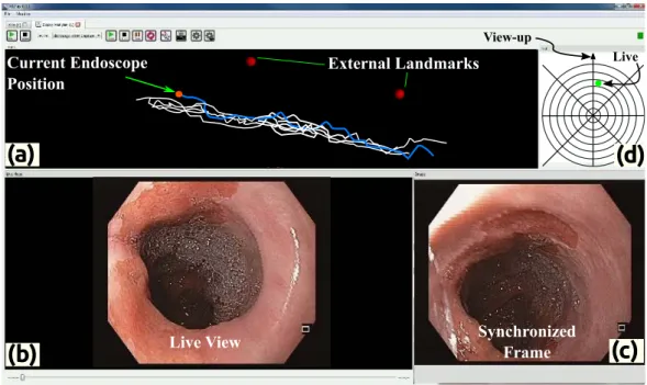
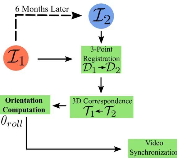
• Chapter3realises the conceptual model presented in2; describing the various ele-ments of the system design and set up for using an EMTS. It introduces the notion of “recording an intervention”, to be used in the proposed framework and builds the re-quired mathematical notation for the rest of the thesis. Broadly speaking, the proposed methodology, aims to provide inter-operative video synchronization between two surveillance procedures performed at different times on the patient. In this context, the application of inter-operative registration is explained and a approach using three landmarks is presented. Using the two paradigms of relocalization introduced in Chapter2, this chapter, then describes the design of, a first version of the software interface to be presented to the clinician during an intervention.
• Chapter4performs the evaluation of the proposed system described in Chapter3. The purpose of these evaluations is to assess the performance of the system both quantitatively and qualitatively. This chapter begins with a careful analysis of the various sources of uncertainty in the system, followed by a series of experiments to empirically measure them. These measurements were used to generate synthetic data-sets for the first set of quantitative evaluations. A second set of experimental evaluations were performed on interventions using pigs. Both these experiments compute the error in average depth during relocalization inside the oesophagus, as the reported metric to measure the system’s performance. Then, a set of experiments to quantify the subjective assessment of the clinical experts using the proposed conceptual and software framework is presented. The experiments in this chapter demonstrated benefit to the end user.
• Chapter4computed the error in relocalization caused by uncertainties in the system. In extreme cases, due to human factors or otherwise, large error values were ob-served. This motivated the need for an improvement in the inter-operative registration approach, which is the focus of Chapter5. The approach in this chapter extends the 3-point inter-operative registration, discussed in Chapter3to use the complete oesophagus trajectory, and then comparing the results to demonstrate an improvement in performance, even in extreme cases.
• Chapter6extends the methodology presented in until Chapter5, in which the relocal-ization was performed by video synchronrelocal-ization; only used the information obtained from the EMTS. This chapter highlights that such a video synchronization, may not
1.6. Manuscript Organization 7 always provide a qualitative visual match from a GI expert’s point of view. This could be due to a difference in view-point of the synchronized image or due to an image being uninformative. To alleviate this, the available image information must be utilized. Hence, this chapter reviews the application of computer vision concepts in the GI endoscopy literature for scene understanding and matching; in order to identify a few key methods that would be compared in7for an image based video synchronization.
• Chapter7follows the discussion in Chapter6and introduces the concept of “view-point” selection, using inter-operative image matching. Broadly, a constrained scene matching framework, extending the work-flow presented in Chapter3is proposed. It draws on the techniques selected in Chapter 6 to firstly, propose an alternate approach to detection of uninformative frames and secondly, provide a view-point match for inter-operative relocalization. Experiments conducted using images from interventions recorded on 7 patients indicated that, the constrained image-based match provided a significant improvement in video synchronization’s qualitative visual match score.
• Chapter8 summarizes the thesis, followed by an analysis of perspectives on the proposed framework and a discussion on the possible extensions for future research.
Chapter 2
Analysis and Problem Statement
Contents
2.1 Oesophageal Adenocarcinoma . . . 9 2.2 Colorectal Cancer . . . 11 2.3 Rationale for Relocalization. . . 13 2.3.1 Biopsy Site Relocalization . . . 13 2.3.2 Temporal Differential Surveillance. . . 13 2.4 Current solutions . . . 14 2.4.1 Intra-Operative relocalization . . . 14 2.4.2 Inter-Operative relocalization . . . 15 2.5 Challenges . . . 16 2.5.1 EMTS in Clinical Applications. . . 18 2.6 Problem Analysis and Proposed Solution . . . 20
The previous chapter indicated the lack of a commercially available guidance platform for endoluminal endoscopy and highlighted the importance of regular screening in GI endoscopy. This chapter further describes the clinical context for a navigational system to aide in such surveillance procedures. Specifically, the focus here is on two pathological conditions: (1) Oesophageal adenocarcinoma and; (2) Colorectal cancer. The context for surveillance endoscopy is explained; describing the need for a navigational platform that can provide inter-operative relocalization.
Sections2.1and2.2present the various pathological conditions in GI endoscopy that demand periodic surveillance. Section2.3discusses the rationale for relocalization in such procedures. Section2.4reviews the state of the art and Section2.5presents the primary challenges in the context of “inter”-operative relocalization. Then, in Section2.6, there is a presentation on the use of electromagnetic tracking in clinical applications and finally there is a brief overview of the proposed approach, based on the problem analysis.
2.1 Oesophageal Adenocarcinoma
There are two primary types of oesophageal cancers: squamous cell cancer and oesophageal adenocarcinoma (OAC). Squamous cell cancer occurs most commonly in people who smoke cigarettes and drink alcohol excessively. Whereas, OAC occurs most commonly in people with gastro-oesophageal reflux disease (GORD). The latter condition has seen an increase in
frequency in the last two decades. GORD is a benign complication caused when the stomach acid escapes into the lower part of the oesophagus. When this disorder becomes a chronic condition, it can lead to changes in the oesophageal lining, causing the tissue to resemble the intestinal lining. This pathological condition is termed as Barrett’s oesophagus (BO). The British society of gastroenterology provides a working definition of BO [Fitzgerald 2014]:
BO is defined as an oesophagus in which any portion of the normal distal squa-mous epithelial lining has been replaced by metaplastic columnar epithelium, which is clearly visible endoscopically (≥ 1cm) above the gastro-oesophageal junction and confirmed histopathologically from oesophageal biopsies.
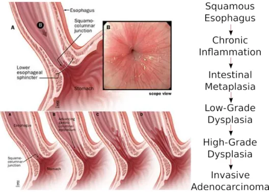
Several studies have indicated a direct link of BO with OAC. OAC appears to arise from the Barrett’s mucosa through progressive degrees of dysplasia [Conteduca 2012,Evans 2012] observed in the cells of the lower oesophagus as shown in Figure2.1. The possibility of being able to perform staging of the precancerous tissue, provides room for early diagnosis and targeted treatments, avoiding emergency surgical interventions such as oesophagectomy.
Figure 2.1: Progression of dysplasia in the oesophagus. The observation of many stages provides room for early detection and treatment.
The presence of BO is associated with increased risk of developing OAC. Adeno-carcinoma in BO develops in a sequence of changes, from non-dysplastic (metaplastic) columnar epithelium, through intermediate-grade, low-grade and then high-grade dysplasia (precancerous change detected microscopically) and finally into OAC. This makes early
2.2. Colorectal Cancer 11
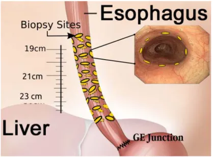
Figure 2.2: Seattle protocol based quadrant biopsies.
detection and treatment a possibility through surveillance. The guidelines [Wang 2008b] prescribe different levels of surveillance intervals depending on the degree of dysplasia, with a minimum of two surveillance endoscopies with biopsy per year. According to the Seattle protocol [Levine 1993,Levine 2000] a typical surveillance procedure involves taking four quadrant biopsies every 2 cms towards the distal end of the oesophagus and in suspicious regions. The biopsied tissue is sent to the pathology for evaluation. For most treatment procedures involving high-grade dysplasia, a 3-month follow-up for 1 year and an yearly follow-up thereafter is recommended. With the introduction of new imaging modalities (AppendixA), in-vivo evaluation of the tissue is now possible.
2.2 Colorectal Cancer
All colorectal cancers (CRC) develop from dysplastic precursor lesions. This is true either in the presence of a predisposing factor such as in inflammatory bowel diseases (IBD) or lack thereof, with lesions occurring sporadically. The fact that there is a pre-malignant phase in CRC, allows a window of opportunity for early detection and cure through planned surveillance. Macroscopically the shape of lesions observed in the colon have been classified as follows [Laine 2015,Inoue 2003];
1. Visible dysplasia: Dysplasia identified on targeted biopsies from a lesion visualized during colonoscopy.
(a) Polypoid: Lesion protruding from the mucosa into the lumen ≥2.5 mm Figure (a)and(b).
i. Pedunculated: Lesion attached to the mucosa by a stalk
ii. Sessile: Lesion not attached to the mucosa by a stalk: entire base is con-tiguous with the mucosa
(b) Non-polypoid: Lesion with little (<2.5 mm) or no protrusion above the mucosa Figure(c)-(f).
i. Superficial elevated: Lesion with protrusion but <2.5 mm above the lumen (less than the height of the closed cup of a biopsy forceps)
ii. Flat: Lesion without protrusion above the mucosa.
iii. Depressed: Lesion with at least a portion depressed below the level of the mucosa.
(c) General descriptors
i. Ulcerated: Ulceration (fibrinous-appearing base with depth) within the lesion.
ii. Borders
A. Distinct border: Lesion’s border is discrete and can be distinguished from surrounding mucosa.
B. Indistinct border: Lesion’s border is not discrete and cannot be distin-guished from surrounding mucosa.
2. Invisible dysplasia: Dysplasia identified on random (non-targeted) biopsies of colon mucosa without a visible lesion.
(a) (b) (c) (d) (e) (f)
Figure 2.3: Macroscopic classification of lesions in the colon:(a)Polypoid pedunculated, (b)Polypoid sessile,(c)Non-polypoid flat,(d)Non-polypoid depressed,(e)and(f) Non-polypoid Elevated. Source: [Soetikno 2008].
In some CRC where the dysplastic precursor lesion is of polypoid type; polypectomy is performed and the CRC risk is localized. In contrast, for patients with long standing IBD, dysplasia can be polypoid or flat, localized or multi-focal and, once found, marks the colon at high risk for CRC [Rembacken 2000,Soetikno 2008]. In addition, these types of lesions are significantly more difficult to identify, which makes the surveillance procedures, tissue characterization and disease evolution in IBD more challenging. The typical surveillance protocol involves endoscopic mucosal resection (EMR) of the suspicious regions and biopsies in the surrounding mucosa along with biopsies every 10 cm. The guidelines [Lieberman 2012,Lutgens 2008] prescribe repetition of surveillance endoscopy at least once every 1-3 years in moderate to high risk patients.
2.3. Rationale for Relocalization 13
2.3 Rationale for Relocalization
Broadly, the relocalization in endoscopic procedures can be classified as; (1) “Intra”-operative(IAO) relocalization methods focus on tracking and mapping the biopsy sites, as they move in and out of the field-of-view of the endoscope frame, during a single intervention. This is to enable the GI expert a global view of all the biopsy locations during a single procedure. (2) “Inter”-operative (IRO) methods, on the other hand, aim to provide guided navigation between two surveillance endoscopies. Relocalization in surveillance endoscopies can be considered to serve two main purposes; which are discussed in the following subsections.
2.3.1 Biopsy Site Relocalization
This is the process of finding the biopsy sites either during the same intervention or those from an earlier diagnostic endoscopy, which were identified as suspicious during patholog-ical analysis. Although the use of advanced imaging techniques has improved detection of suspicious regions; during a follow-up inspection, the GI specialist may be required to locate the previously biopsied or surveyed locations precisely with limited prior knowledge. Typically, the GI specialist uses the scale markings on the endoscope, to return the scope to previously surveyed region. For oesophageal procedures this distance is measured from the mouth (in trans-oral) and nasal opening (for transnasal endoscopy). In colorectal procedures the distance is marked from the anus. This information can be highly unreliable, especially when relocating previous biopsy sites and while using microscopic imaging devices such as CLE or OCT. Due to the lack of deterministic tools for providing such inter-operative relocalization, the GI specialist has to survey or biopsy a large mucosal region in the ab-sence of clear markers on the tissue, which prevents targeted treatments. In both upper and lower GI procedures, relocalization is performed based on the clinician’s experience, prior anatomic description and identification of tissue scaring. In colorectal procedures an additional marker using tattoo ink injection can be performed [Cho 2007]. However, this approach has its disadvantages; (1) Ink fades over time after several months; (2) Tattooing is an invasive procedure and damages the tissue; and (3) It is used mainly post-EMR and not a recommended guideline for all surveillance procedures.
2.3.2 Temporal Differential Surveillance
Visualizing the temporal changes in the tissue structure of the oesophagus or the colon is a significant challenge. Even in the presence of sufficient anatomical information (or tattoos), allowing localization during follow-up surveillance procedures, the clinician does not have access to an approach to visually compare the temporal changes in the tissue structure. This limits the clinician’s ability to effectively track the disease evolution, which is an important goal for surveillance endoscopies. It is especially significant since, there is a considerable misdiagnosis owing to the lack of clear understanding of diseased tissue appearance [Wang 2013b, Lieberman 2012]. In addition, the anatomic location of the diseased tissue may be significantly correlated with risk of cancer [Triantafillidis 2009];
making it important to augment the current endoscopic procedures with knowledge from previously conducted surveillance endoscopies.
From the medical application view-point, this thesis aims to introduce a new framework for “inter”-operative relocalization for flexible endoscopy. From the discussions in Sections 2.1and2.2it is clear that many GI protocols involve regular surveillance. Thus, there is a need to provide additional tools for navigational guidance in these procedures. In addition, inter-operative relocalization for endoscopic procedures should aide the endoscopist not only in biopsy site relocalization (BSR), but also in providing temporal differential surveillance (TDS) defined earlier in this section. This manuscript focuses on the relocalization in oesophageal interventions. The oesophagus was chosen as the target organ since; (1) It is relatively fixed on both ends and hence, does not exhibit strong deformation unlike the colon; (2) and the external anatomical landmarks can be used for providing inter-operative registration which will be discussed in Section2.6. (3) In addition, the clinical protocols for oesophageal surveillance are fairly well laid out as opposed to colonoscopic procedures, which are subject to change based on the disease condition and experience of the GI specialist.
2.4 Current solutions
Following the identification of the two important paradigms of relocalization in surveillance procedures this section presents the state-of-the-art on the techniques published in literature for two types of tasks depending on when the relocalization is actually performed.
2.4.1 Intra-Operative relocalization
The IAO based approaches as defined earlier in Section2.3focus on detecting, tracking and localizing biopsy sites during a single procedure. Primarily, these approaches focus on BSR. One of the first methods in IAO relocalization, was published by Allain et al. [Allain 2009,Allain 2010]. In their approach, the authors proposed to compute feature points in scale-space around the biopsy location and then extracted descriptors for these points using scale invariant feature transform (SIFT) for the two endoscopic views to be matched. Then employing the epipolar constraint, a fundamental matrix was computed between the two views, that mapped the biopsy site to facilitate re-targeting. In [Allain 2012] a framework for characterizing and propagation of the uncertainty in the localization of the biopsy points was presented. Mountney et al. [Mountney 2007] performed a review of various feature descriptors applied to deformable tissue tracking and in [Mountney 2006] proposed an Extended Kalman filter (EKF) framework for simultaneous localization and mapping (SLAM) based method for feature tracking in deformable scene, such as in laparoscopic surgery. This EKF framework was then extended in [Mountney 2009] for maintaining a global map of biopsy sites for endoluminal procedures, intra-operatively. The authors presented an evaluation of the EKF-SLAM on phantom models of stomach and oesophagus. Giannarou et al. [Giannarou 2009] presented an affine-invariant anisotropic region detector robust to soft tissue deformations. This was used by [Atasoy 2009] along with SIFT descriptors. The feature matching problem was then modelled as a global optimization of
2.4. Current solutions 15 an Markov Random Field (MRF) labelling. Recently, Ye et al. [Ye 2013,Ye 2014,Ye 2016] addressed the biopsy site re-targeting in three stages. First using the Tracking-Learning-Detection (TLD) method proposed by Kalal et al. [Kalal 2012]. TLD was used for tracking multiple regions around the selected biopsy site. Under the assumption that the regional tissue deformations can be approximated using local affine transformations, a local homography between matched region centres was estimated. In this way multiple regions around the biopsy sites are tracked, which were then used for homography estimation and mapping the biopsy sites. Wang et al. [Wang 2014a] proposed to learn a graph (atlas) from a sequence of images from several gastroscopic interventions. Considering that the stomach’s deformation is not large between similar frames, the nodes of the learnt graph atlas were connected by an estimated rigid transformation. Thus, the mapping of the biopsy sites from a single (reference) frame to subsequent frames for any given intervention was reduced to a graph search problem. First, for the reference frame and the moving frame their corresponding matching nodes in the graph were computed. Using Dijkstra‘s algorithm, the shortest path between these matched nodes was obtained. Hence, the transformation between the reference frame and moving frame was obtained as the associated combination of rigid transforms along the shortest path between the corresponding matched nodes of the graph.
2.4.2 Inter-Operative relocalization
In contrast, the IRO methods attempt to provide localization between interventions. In [Atasoy 2011,Atasoy 2012b], Atasoy et al. proposed to formulate the relocalization as a image-manifold learning process. The method involved, building an adjacency graph between the images of a surveillance intervention. Normalized cross-correlation was used as the similarity measure between image frames to compute the adjacency graph. Then using laplacian eigenmaps decomposition that was initially proposed in [He 2005], a linear projection matrix was computed. This approximation for projection on to the manifold was used to compute the low-dimensional representation for all the images in the intervention. Then, two separate methodologies for performing inter-operative relocalization was proposed using scene association. In [Atasoy 2012b], the scene association is performed by computing the nearest neighbour directly over the low-dimensional representation from an earlier surveillance endoscopy. However, in [Atasoy 2011] a two-run surveillance endoscopy was suggested, in which a dummy surveillance is initially performed, that is then used for scene association with the actual surveillance. The authors claimed that the modified approach in [Atasoy 2011] allowed for scene association in presence of significant structural changes in the tissue.
[Liu 2014] proposed the use of electromagnetic tracking system (EMTS) for local-izing the biopsy sites in the stomach. They construct a 3D model of the stomach using SLAM and map the biopsy points tracked using the EMTS on to the 3D model. The inter-operative registration was performed by selecting five reference points manually, during each intervention.
For colonoscopic procedures, the need to provide navigational assistance is substantial. One of the earliest approaches involved a combination of 3D reconstruction from
pre-operative CT with endoscopic video known as virtual colonoscopy. The chief aspect of this involved the computation of optical flow to estimate the ego-motion of the colonoscope. Ego-motion or visual odometry involves first, extracting features from the image and computing optical flow fields. Then, using the flow fields, the camera motion would be estimated. In [Puerto-Souza 2014] the authors presented a comparison of two ego-motion estimation schemes, supervised and unsupervised. Supervised methods, as shown in [Bell 2013] require training data to be available in the form of optical-flow measurements and corresponding camera motion data. Unsupervised approaches, however, used image correspondences between video frames and multiple-view geometry to estimate endoscope motion, as was shown in [Liu 2008].
Pre-operative CT scans are uncommon in routine GI endoscopies, as they are not cost effective. Intra-operative X-rays are sometimes used for guidance, to areas in the GI tract that are difficult to access, but they do not provide sufficient information for 3D reconstruction. Theoretically, the first endoscopy can be used to compute a 3D model of the target lumen, which can be used in the follow-up surveillance procedures. But, the video based 3D reconstruction in GI procedures is still an open are for research.
2.5 Challenges
This section presents some of the problems associated with the existing methods in the literature and the various challenges encountered in clinical settings of endoluminal surgeries which ultimately motivated the direction of the proposed methodology of this thesis. There are several hurdles involved in both of the relocalization schemes discussed in Section2.4. An exhaustive list of these challenges that were identified during the course of this research is provided below.
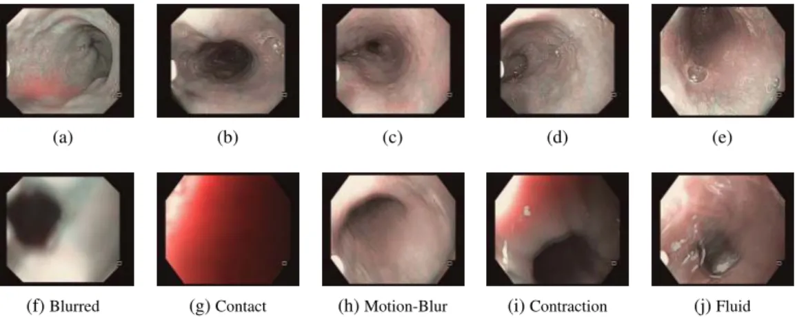
Uninformative (UI) Frames: A typical endoscopic exploration lasts for several minutes and may suffer from large number of uninformative frames, as shown in Figure2.4. Relocaliza-tion method requires the robustness to UI frames during the procedure. It should be noted that seldom are the images completely informative or UI. The presence of fluid in part of the image or contraction, in only a section of the tissue can give us a frame that is usable but that which is not completely UI. This is addressed in detail in Chapter7. It should be noted that the presence of UI frames affects both IAO and IRO relocalization.
Endoscope and tissue motion: An obvious challenge in IAO relocalization is to accounting for tissue deformation while tracking and mapping the biopsy sites. Scene recognition in endoscopic videos is a challenging task, not only due to the tissue movement but also due to the motion of the endoscope, causing a change in view-point.
Evolution of tissue structure: Surveillance procedures in BO are performed at an interval of at least 3-6 months. One of the most important constraints as a result of this is the evolution of the tissue structure due to progression of the disease. There has been no study conducted thus far, which unequivocally demonstrates the deterministic nature of the image-based
2.5. Challenges 17
(a) (b) (c) (d) (e)
(f)Blurred (g)Contact (h)Motion-Blur (i)Contraction (j)Fluid
Figure 2.4:(a)-(e): Informative frames.(f)-(j)Uninformative frames.
methods described in Section2.4. However, an IRO relocalization must be robust to these changes in scene understanding. Chapter6presents a complete survey of image analysis methods applied to detection, classification and tracking in GI endoscopy.
Localization of the endoscope: For manual IRO relocalization, the endoscopist uses markings on the outer sheath of the endoscope to re-position. This can be affected by the patient position on the table (decubitus lateral or supine) and also by the position and orientation of the neck, which introduces an additional source of uncertainty.
Large search area: With the introduction of new imaging devices (AppendixA), in-vivo visualization of the tissue has improved. With devices such as the confocal microscope and the optical coherence tomography, it is now possible to make an initial diagnosis before sending tissue to the pathologist. However, the area of exploration using these devices is in the micro metric range and the area of a typical biopsy is ≈ 0.25 ∼ 4mm2;
whereas, the search area extends over a region of ≈ 450mm2. In presence of uncertainty in
the localization of the endoscope, this area can be larger. This is closely linked with the view-point problem mentioned earlier. However, owing to a good IRO relocalization, IAO methodologies can be applied to map the regions of interest to reduce this search area. Orientation difference: An aspect which has not been clearly identified in IRO literature has been to account for the orientation difference during relocalization. When IRO relocalization is performed using video synchronization, the synchronized frames need not be at the same orientation as the endoscope inside the lumen. Clinically, the visual disparity does not provide an ideal view for comparison to the GI expert and is an important point that needs to be clearly addressed. From a technical standpoint, the approaches discussed in Section2.4.2, rely heavily on, either the development of orientation invariant global image description or the estimation of this difference through matched features; which would be hindered by the challenges discussed previously.
2.5.1 EMTS in Clinical Applications
The approach presented in this thesis is inspired by the need to be independent of the assumptions about, the evolution of tissue structure, rotation invariance of descriptor etc. in image-based methodologies; that were described in the previous section. Moreover, with the development of different endoscopic modalities, it is essential that the IRO framework integrates with all of them, without the need for significant modifications. To this effect, an EMTS based approach for tracking the endoscope in the lumen is proposed here. An EMTS based method offer a distinct advantage over the optical tracking methods in situations where line-of-sight is an issue and where flexible instruments are in play such as in the GI endoscopy. Additionally, multiple EM sensors can be integrated together without significant increase in the endoscope dimensions. There are several commercial EMTS available for medical applications: Ascension trakSTAR/driveBAY®, Northern Digital Inc (NBI)
Aurora®, Polhemus Fastrak®and Calypso (Figure2.5); are a few. EMTS is the only tracker
that enables real-time tracking of objects without the line-of-sight restrictions. This has permitted its extensive use in MIS applications to provide navigation to reach deep-seated anatomy.
Broadly, the application of EMTS in clinical procedures can be classified as, (a) In-terventional radiology: Typically, such procedures are percutaneous. They involve either obtaining biopsies for diagnosis or ablative procedures for therapeutic treatment. Such as in, [Grand 2011], the authors performed a clinical evaluation of the EMTS guided lung biopsies on 60 patients under CT imaging and in [Franz 2012], where a modified ultra-sound probe fitted with a compact EMFE for navigation and guidance is proposed, also for percutaneous procedures. (b) Catheter interventions: These are procedures that use the human circulatory system for guiding the instrument. Tracking of electrophysiological catheters in cardiological interventions allows the creation of high-resolution spatial maps of the electrical activity of the heart [Linte 2012,Manstad-Hulaas 2012]. (c) Endoscopic: In pulmonary and GI procedures, researchers have considered tracking the tip of the scope using an EMTS to provide accurate localization. This approach has been applied to bron-choscopy by [Mori 2007, Leong 2012, Grand 2011]. For GI endoscopy, Olympus has designed a colonoscope (ScopeGuide®) with multiple embedded EM sensors that would
track the position of the tip, and additionally provide the shape of a proximal section of the scope. (d) Laparoscopic: EMTS has been used in tracking of absolute and relative poses of laparoscopic ultrasound and video for procedures involving kidney, liver, gall bladder, lymph nodes etc. In [Jolles 2004] the authors employed EM tracking to perform computer assisted total hip arthroplasty to improve accuracy of cup placement. EMTS has been used for navigation in neuroendoscopy [Suess 2001, Peng 2002] as well. (e) Miscellaneous: There are several other clinical applications where EMTS has been tested for guidance, such as in the placement of feeding tubes [October 2009,Krenitsky 2011].
Franz et al. [Franz 2014] have provided a comprehensive overview of the application of EMTS in clinical scenarios. They provide a detailed review of the commercially available devices and cite various clinical evidence presented in literature, validating the methodolo-gies. Further research is under-way to design new devices such as the one presented by [Lucarini 2013] that use wireless capsule endoscope with an EMTS for localization.
2.5. Challenges 19
Figure 2.5: Commercial Electromagnetic trackers: (a) Northern Digital Inc Aurora®[NDI],
(b) Calypso EM tracker [Varian], (c) Cortrack [Corpack-Medical], (d) Fastrack by Polhemus [Polhemus], (e) Ascension TrackSTAR and driveBAY [Ascension-tech].
This thesis does not study the GI endoscopic workspace for assessment of adaptability of EMTS in GI endoscopy. However, it relies on several such studies already conducted on phantom models and in-vivo in clinical environment. A few of the most significant studies have been listed here; [Wilson 2006,Frantz 2003,Fischer 2005,Wang 2013a,Lugez 2015,
Cleary 2005, Peters 2008, Yaniv 2009, Krücker 2007, Franz 2014]. It should be noted that, apart from environment, the complex work-flow of the surgical procedure introduces additional sources of noise in the usage of EMTS. Since the use of EMTS in the clinical environment varies depending on the kind of intervention, no fixed evaluation methodology can be employed. Although some of the above mentioned methods can be employed for distortion correction, it is out of scope of this work to incorporate these models.
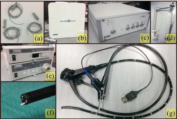
During endoscopic procedures it was observed that the following devices were employed routinely,
(1) Endoscope: Plastic or Non-ferromagnetic outer sheath with some electronic components and very low current devices.
(2) Biopsy catheter: also typically with a plastic sheath with a small gauge wire connecting to biopsy forceps. These are used in between the procedure and will not stay in place to continuously affect tracking.
(3) Radio frequency ablation probes and Coagulation forceps: These are high current devices and can affect the tracking. However typically when using these device the
tracking would generally not be employed and in case it is required to use tracking, the positioning would be completed using the navigation before using them.
Hence, for the purposes of this application, since the workspace for oesophageal procedures is limited to about 25 × 10 × 10 cm3. This is well within the prescribed bounds of the EMFE
specifications, and the distortions introduced during the procedure, can be neglected without significant loss of quality. Thus no distortion correction measures have been employed here, however, a complete assessment of it’s effect with other instrumentation in the operating room must be definitely studied in the future.
2.6 Problem Analysis and Proposed Solution
Let us consider that the intervention performed a few months prior was termed as the “diagnostic endoscopy” (DE). During the DE procedure several biopsies are taken and samples sent for histopathological analysis. Depending on the outcome of their analysis, the patient is asked to return for a repeat intervention. This follow-up is termed as the “surveillance endoscopy” (SE). Broadly speaking, the proposed approach aims to achieve video synchronization between the SE, with image frames recorded during the DE or between two successive SE procedures. This is conceptually inspired, in part, by the method presented in [Atasoy 2012a]. However, their approach is constrained by the implicit assumptions made for image-based methodologies, which were discussed in Section2.5.
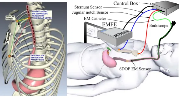
To perform video synchronization of two interventions, a correspondence between the images of these interventions must be made. Over the length of the lumen in the oesophagus, the tissue texture, is quite uniform and visually does not exhibit strong variation. Thus, using the image matches to synchronize frames leads to a strong uncertainty and low confidence in obtaining the right match. To overcome this, the proposed framework includes an EM sensor that is inserted in the endoscopic channel to track its position in the oesophagus. A synchronized capture of the pose of the embedded EM sensor and the endoscopic image frame is performed, to generate a database. So, each captured image has a corresponding 3D pose associated with it.
Considering an ideal scenario, if the DE and SE were performed in succession, without disturbing the patient and the EMTS set up in the operating room, a 3D position corre-spondence from the EM sensor trajectory, would directly lead to video synchronization of SE with DE. However, since typical surveillance interventions are performed 3-6 months apart, to establish such a correspondence, registration of the EMTS reference frames for the two interventions must be performed. One approach that was presented in [Vemuri 2013], involved using the gastro-oesophageal junction (GOJ) as an anatomical landmark, along with the trajectory of EM sensor in the oesophagus to perform registration.
However, this approach poses difficulties in clinical settings. It requires the expert to necessarily reach the GOJ first before receiving any guidance. This however is not always possible; in case of oesophagus stenosis due to a tumour, for instance. It is also a constraint that since the clinician may come across lesions before reaching the GOJ, and may prefer to treat them immediately. Additionally, the combined variation in head and neck orientation introduces an uncertainty that is difficult to model. In fact, the patient is not always laying in
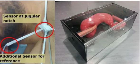
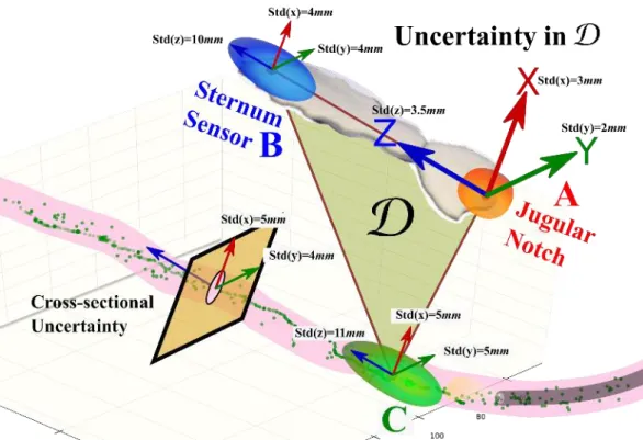
2.6. Problem Analysis and Proposed Solution 21 a supine position and can be set in the lateral decubitus position, depending on the surgical conditions making the orientation compensation for estimating the 6-DOF registration from the 3D endoscope trajectory alone unreliable. Hence, an alternative method using two additional sensors attached to the sternum of the patient is considered, with details provided in Chapter 3. It will be explained how, the three sensor set up is used to perform IRO registration.
Two important paradigms of relocalization, were identified in Section2.3. The impor-tance of this distinction between BSR and TDS, when providing a software framework for relocalization, cannot be understated; and this has not been clearly delineated in the literature. For BSR, the guidance platform requires identification and tagging of the biopsy sites during DE, thus providing relocalization to specific points in the oesophagus during SE interventions. Whereas, for TDS, the GI expert must be provided with the synchronization of the complete endoscopic intervention from DE (that may or may not contain any specific tags for biopsy sites). It is important to note that the interface difference though subtle adds substantial value to the expert under varying clinical conditions and will also be discussed further in the following chapter.
Chapter 3
System Description and Methodology
Contents
3.1 System Setup . . . 24 3.2 Data Acquisition . . . 25 3.2.1 Notation . . . 25 3.3 Inter-Operative Registration: Basic Model Using 3-points . . . 27 3.3.1 Video Synchronization . . . 28 3.4 Orientation Difference Estimation . . . 28 3.5 Application Interface . . . 31 3.5.1 BSR Interface. . . 31 3.5.2 TDS Interface. . . 33 3.6 Conclusion . . . 34
This chapter is based on work published in [Vemuri 2013,Vemuri 2015b].
This chapter describes the system framework and methodology of the proposed approach to provide Inter-operative (IRO) video synchronization, using an electromagnetic tracking system (EMTS). The goal is to provide a seamless integration of the hardware into the endoscopy suite with minimal additional time for set up. As was briefly described in Section 2.6, the endoscopic intervention is recorded, it is thus important, to clearly identify the various elements of such a recording, to provide a consistent mathematical structure to the problem. For the interventions to be synchronized; diagnostic endoscopy (DE) and surveillance endoscopy (SE), are performed several weeks or months apart, to provide video synchronization, a global registration between the EMTS reference frames for SE and DE, must be performed. Additionally, the synchronized frames could have been captured at different orientations of the endoscope camera w.r.t a reference plane, such as the patient table. This can happen due to two factors; (a) orientation of the patient (decubitus lateral or supine); (b) orientation of the endoscope (sensor) in the oesophagus at the synchronized frames. . This can lead to ambiguity in visual correlation of the scenes for the GI expert. Thus, an approach to reliably estimate this orientation difference must also be provided.
Section3.1describes the components of the system set up and highlights the need for, the two supplementary sensors that were initially discussed in Section2.6. Section3.2outlines the description of the various elements recorded during an intervention and lists the notation that would be used in the rest of the thesis. Section3.3depicts the inter-operative registration
