bASM Archéologie des Sociétés Méditerranéennes, UMR 5140, Université Paul Valéry Montpellier, CNRS, MCC, 34000, Montpellier, France cUMR-IMBE, Université d'Avignon, ERASMUS MUNDUS MASTER IN ARCHaeological MATerials Science Student, Ireland
A R T I C L E I N F O Keywords: Iron Age Celts Embalming Severed heads Chemical analyses A B S T R A C T
Ancient texts described that one of the most impressive ritual practices of the Celts during the Iron Age was to remove the heads of enemies killed in battle and to embalm them for display in front of the victors dwellings. An archaeological settlement excavation site in Le Cailar, in southern France, has revealed a considerable number of examples of this practice. It was documented by Classical authors and later by the archaeological recording of iconographic representations and skeletal remains of human heads. Weapons were also exhibited alongside the severed heads. Here we report the results of chemical investigations for the characterization of the biomarkers of embalming that are likely to be present in eleven fragments of these human cranial remains. These results may lead to answers to some of the archaeometric questions related to the subject of embalming in 3rd century BC Transalpine Gaul, thus advancing the knowledge of these ritual practices, documented by Greek Classical au-thors as part of the wider research into the proto-historic societies of the Mediterranean coastal region.
1. Introduction
In the 3rd century BC, the number of wars and battles seems to increase in almost the whole of Western Europe. Indeed, hundreds of weapons have been found in Iron Age sanctuaries and sacred places since they weren't display there before. In many of these sites, human remains have been discovered with both metal artifacts and fauna re-mains associated with the sacrifice of animals (Buchsenschutz, 2017; Brunaux, 2004; Barral et al., 2006). The Classical textual sources document the practice of the Celts cuttingboff their enemies'head after the battle, to transport them to their settlements by hanging the de-capitated heads around their horse's necks. This very precise picture of this practice is known through two fragments of ancient texts, written in the 1st century BC respectively by Strabo and by Diodorus of Sicily, both recording the testimony of an ancient Greek, named Poseidonios, who travelled in the south of Gaul around 100 BC (Strabo, IV, 4, 5 in Lasserre, 1966). Other classical texts mention this practice, such as Polybius and Livy and much of the archaeological record corroborates with the descriptions of this practice (Ciesielski et al., 2011; Armit, 2012;Boulestin and Henry Gambier, 2012). The Iron Age settlement of Entremont in Provence, which was one of thefirst archaeological ex-cavations in south of France, revealed much sculpting of decapitated
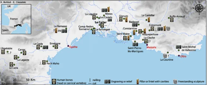
heads, with one particular sculpture representing a warrior mounted on a horse, with a sword and a spear at their side, and a severed head suspended from the horse's neck (Arcelin, 2011), just as testified by Classical textual sources. This practice of decapitation as depicted in both the Classical commentary and aforementioned surviving sculpting is further evidenced throughout the south of France by the cranial re-mains, engraved and cut stone iconography of other sites of the Second Iron Age (Fig. 1), such as Roquepertuse and Glanum. In some places, archaeologists found human skulls with iron nails inside them and in other places they found pillars or lintels with cavities of the approx-imate dimensions and shape of human skulls (Fig. 1).
Thus, displaying severed heads was a well-known practice, but it had never been observed in recent excavations in the South of France. Therefore the discovery in Le Cailar is of great importance and provides us with a significant amount of new data (Roure et al., 2006), and the opportunity to make new analysis, such as chemical analysis to verify if the decapited skulls have been prepared as the Greeks testified it. In-deed, Strabo and Diodorus both wrote that the Celts embalmed dec-apited heads, with‘cedar oil’; however, this may be a misspecification of a local Pinacea oil that Greeks named‘cedar’ because the smell was close. This is the reason why this paper's study aimed at verifying the presence of possible embalming remnants in archaeological cranial
https://doi.org/10.1016/j.jas.2018.09.011
Received 17 June 2018; Received in revised form 10 September 2018; Accepted 27 September 2018
∗Corresponding author. UMR5140-ASM, site Saint-Charles Université Paul Valéry Montpellier 3, Route de Mende, F-34199, Montpellier Cedex 5, France.
E-mail address:rejane.roure@univ-montp3.fr(R. Roure).
Available online 07 November 2018
fragments from Le Cailar. Chemical analyses using GC-MS were per-formed in order to characterise organic components likely to be present in eleven of these human cranial fragments.
Embalming and other mummification phenomena are well-docu-mented, worldwide with much of both the scientific and the archaeo-historic academic literature documenting the best surviving examples of embalming from pharaonic dynasties of Egypt (Łucejko et al., 2012; Ménager et al., 2014;Nicholson and Shaw, 2000), and mummification has also been evidenced in Bronze Age in Britain (Parker Pearson et al., 2005). Our paper will present another example of embalming practice.
2. Experimental section 2.1. Sample description
The Iron Age fortified settlement of Le Cailar was situated near a wide lagoon connected to the Rhône River. The site was occupied from the 6th century BC until the Roman period in Gaul (1st century AD). The fortified settlement was located on a small hill and was also a harbour for Mediterranean traders (Etruscans and Greeks con-temporaneous). The excavation of weapons and human skulls located proximal to the interior of the walls and possibly one of the gates of the Iron Age settlement, has been interpreted as a large area where these objects were displayed to the inhabitants on the interior of the settle-ment (Fig. 2). This archaeological context is very clear: there is no Fig. 1. Map of Iron Age decapited remains and iconography in the South of France.
Fig. 2. Maps of the Le Cailar excavation with the distribution of each type of remains and location of samples (on the left) and only the metal artifacts and the human bones (on the right) (all the deposits mapped together).
doubt that this context was an open area where the aforementioned heads and weapons were displayed. There is stratigraphical and chronological evidence for each archaeological level that contained skeletal remains and metal artifacts (Roure et al., 2017). The remnants of metal (mainly weapons), potteries and faunal bones, were inter-mingled with the human skulls in this intramural area. The ceramic and metallic remains allowed a precise chronology of this embalming ac-tivity to be assigned to the 3rd century BC which was further reinforced by the numismatic dating from the discovery of forty Massilian coins in this same area (thus radiocarbon dating was not used). Amphora and vessels from the Greek city of Massalia and vases from Italy and Spain all belong to this period and the metal artifacts are all linked to the latenian typology (La Tène B2-C1). The stratigraphy of the Le Cailar
excavation and its artifacts showed that several deposits occured: the first deposits correspond to the end of the 4th century or to the very beginning of the 3rd century BC, the decapited heads and weapons were displayed throughout the 3rd century BC until circa 200 BC, when the area was covered by soil.
Approximately 2700 fragments of human bones were recorded during ten excavation seasons: all the fragments are part of the skull, except for six small pieces of cervical vertebrae (Fig. 3). Many of the skulls bore cut marks, not only of decapitation, but also to preparing the heads for display by the removal of cervical vertebrae and aperture of the postero-inferior portion of the cranium, probably - to remove the brain; and tongue ablation, or at least the scraping of the muscles under the mandible (Ciesielski and Bohbot, 2014).
The discovery of these marked skulls was immediately related to the afore-mentioned ancient texts from Strabo and Diodorus of Sicily. These texts indicated that the Gauls, to quote Strabo -“embalmed the head of the most famous enemy with cedar oil” (IV, 4, 5 inLasserre, 1966). This is the reason why chemical analysis, as opposed to other types of analyses, was undertaken. The preliminary chemical analyses on the human bones were carried out in search for traces of biological pro-ducts which could have been used to embalm these heads for their display, despite no macroscopic remains were visible.
Eleven cranial fragments were selected for analyses (Table 1,Fig. 4), from each of which two powder samples (100 mg–150 mg each) were taken,first from the exterior and then from the interior surfaces. A total of twenty-two samples were analysed. The eleven samples were re-trieved at random from the skulls: frontal, zygomatic, parietal, all ori-ginating from different deposits and moreover the assemblage of 2676 Fig. 3. Table of human remains and map of cranial MNI from Le Cailar (2003–2011).
Table 1
Description of cranial fragments.
Alpha-numeric context number General bone identification
CLR 07 Mandible (central section)
CLR 04 X5 Mandible (right side)
CLR 03 X103 Mandible (left side)
CLR 04 X11 Parietal CLR 03 X29 Parietal CLR 03 X44 Parietal CLR M18 R6 570 Frontal CLR N17 R0 8 Frontal CLR K15 R5 340 Frontal CLR K16 R9 286 Zygomatic (left) CLR N17 R3 53 Zygomatic (left)
bones had no visible residue, with which to discriminate between for the analyses. All the human remains were precisely recorded with three coordinates (x, y, z) and registered by number (excluding the bones which were excavated before the establishment of this numbering protocol, at the beginning of the excavation).
Five faunal remains (Table 2), discovered in the same context and level as the human bones, were also analysed using identical protocols, in order to assess if biomarkers would be taphonomically biased or associated with embalming.
2.2. Materials
The dichloromethane used in preparation for GC-MC analysis was of GC grade and purchased from Merck (Darmstadt, Germany). The high purity water (18.3 MΩ.cm) used in the preparation was obtained from a Milli-Q purification system (Millipore).
Derivatisation was made using BSTFA (N,O-bis(trimethylsilyl) tri-fluoroacetamide) with 1% TMC (trimethylchlorosilane) purchased from Sigma Aldritch.
2.3. GC-MS analyses
Each powdered cranial sample was added to 6 mL of di-chloromethane and sonicated (2 min, 70% amplitude) using an ultra-sonic probe (Vibra-Cell model 75186). This volume was necessary in order to obtain the immersion of the probe in the extraction solvent. The samples were then centrifuged (30 min, 6000 rpm). The super-natant corresponding to the organic extract was split into two, in order to perform GC-MS analyses.
Before the GC-MS analyses were performed, the organic extracts were derivatised by trimethylsilylation. For this purpose, the solutions were evaporated to until dry under nitrogen and mixed with 100μL of BSTFA with 1% TMC for 30 min at 70 °C. The trimethylsilylated extracts were dried under nitrogen and dissolved in 500μL of hexane. All of the samples werefiltered through a 0.45 μm PTFE filter before injection.
GC-MS analyses were carried out using a Thermo Scientific™ Focus gas chromatographic system mounted with a Thermo Scientific Al 3000 auto-injector and coupled with a ITQ™ 700 Series GC-Ion Trap Mass Spectrometer (Thermo Fisher Scientific Inc.). GC separation was per-formed on a fused silica capillary TG-5MS column (Thermo Fisher Scientific, alvc), with a stationary phase (5% diphenyl-95% dimethyl-polysiloxane phase).
A volume of 1μL for each sample was injected in splitless mode an injector set at a temperature of 250 °C. Molecular components were eluted using helium at a constantflow of 1.2 mL/min. The following temperature programme was used: with an initial temperature of 50 °C for 2 min, 50–220 °C at 8 °C/min, 220–260 °C at 2 °C/min and 260–330 °C at 10 °C/min.
The mass spectra were recorded in electron impact mode with an electron ionisation voltage of 70 eV, an ionisation time of 25,000μs and a mass range of 50–650 m/z. The ion trap and interface transfer line were respectively set at 250 °C and 300 °C.
Thermo Xcalibur™ 2.2 software (Thermo Fisher Scientific Inc.) was used for instrumental control and data acquisitions.
The assignment of mass spectra peak was based on a comparison with an internal mass spectrum database (from commercial standards, from fresh and artificially-aged resins and oils) and finally the NIST database (NIST MS Search 2.0).
3. Results and discussion
All of the components were identified according to their specific mass data (base and molecular ions), their retention times in compar-ison with standard molecules and specialized literature (Table 3).
For an accurate interpretation of the GC-MS results, the contribution of lipids from bone cannot be disregarded. Fresh bones contain sig-nificant amounts of cholesterol and a lesser concentration of fatty acids associated with bone marrow (Colonese et al., 2015;Evershed et al., 1995). Previous analyses of archaeological bones revealed only the presence of cholesterol, together with its diagenetic degradation pro-ducts, especially cholest-5-en-3ß-ol-7-one (Collins et al., 2002; Colonese et al., 2015;Evershed et al., 1995;Stott et al., 1997). How-ever, traces of saturated fatty acids (primarily C14:0, C16:0and C18:0), a
lesser concentration of oleic acid (C18:1) and a low quantity of linoleic
acid (C18:2) were recently detected in the analyses of human bones for
archaeological purposes (Colonese et al., 2015).
The total ion current of the lipid extract from CLR K16 R9 286 sample is presented inFig. 5and shows that almost all lipid extracts from the analyzed bones exhibited the presence of saturated fatty acids Fig. 4. Pictures of a. Total assemblage b. CLR K16 R9 286 exterior surface c. CLR N17 R3 53 interior surface cranial fragments (after Ghezal and Gosnell).
Table 2
Description of faunal remains.
Alpha-numeric context number K15 R5 361
K15 R5 354 K16 R9 304 M18 R6 535 A M18 R6 538
C9:0, C14:0, C16:0and C18:0, monoacylglycerols, cholesterol and its
de-gradation products: cholest-5-en-3β,7β-diol and cholest-5-en-3ß-ol-7-one.
The fatty acid composition of the different samples is presented in Fig. 6, showing a chromatogram of the internal CLR K16 R9 286 sample
with extracted signals at m/z 117 and the base peak of main saturated fatty acids. The chromatogram shows that almost all lipid extracts from analysed bones exhibited the presence of saturated fatty acids C9:0,
C14:0, C16:0and C18:0. Some unsaturated fatty acids were also detected
(C14:1, C16:1, C17:1, C18:1). C17:1 – – – – – – – – – – – – – – – – + – + – – – C17:0 – – – – – – – – – – – – – – – – + – + + – – Cis,Cis C18:2Δ 9,12 – – – – – – – – – – – – – – – – + – + + – – Cis C18:1Δ 9 – – – – – – – – – – – – – – – – – – + + – – C18:0 + + + + + + + + + + + + + + + + + + + + + + C19:0 – – – – – – – – – – – – + + – – + + + + – – Monoacylglycerol MAG C14:0 – – – – – – – – – – – – + + – – + + + + – – MAG C16:0 + + + + + + + + + + + + + + + + + + + + + + MAG C18:0 + + + + + + – – + + – + + + + + + + + + + + Steroidal compounds Cholesterol + + + + + + + + + + + + + + + + + + + + + + β-Sitosterol + + + + + + + + + + + + + + + + + + + + + + Cholest-5-en-3β,7β-diol – – – – + – + + – – – – + + + + + + + + – – Cholest-5-en-3ß-ol-7-one + + + + + + – – – – – – + + – – + + + + + + Glycerol – – + – + + – – + – + + – – + + – – – – + + n-Alkanes C23 – – – – – – – – – – + – – – – + – – – – – – C24 – – – – – – – – – – + – – – – – + + + + – – C25 – – + – – – + + – – + – – – – + + + + + – – C26 – – + – – – + + – – + – – – – + + + + + – – C27 + + + – – – + + – – + – – – – + + + + + – – C28 + + + – – – + + – – + – – – – + + + + + – – C29 + + + – – – + + – – + – + + – + + + + + – – C30 – – + – – – + + – – + – + + – + + + + + – – C31 + + + – – – + + – – + – + + – – + + + + – – C32 + + – – – – + + – – + – + + – + + + + + – – C33 + + – – – – – – – – + – + + – – – – – – – – n-alkan-1-ol C12 OH – – – – + + – – – – – – + + + + + + + + – – C14 OH – – – – + + – – – – – – + + – – + + + + – – C15 OH + + – – – – – – – – – – + + + + + + + + + + C16 OH – – – – + + – – – + – – + + + + + + + + + + C17 OH – – – – + + + + – – – – – – + + – – + + + + C18 OH + + + + + + + + – + – – + + + + + + + + + + C19 OH – – – – + + – – – – – – + + – – + + + + + + C20 OH + + – – + + – – – + – – + + + + + + + + + + C21 OH + + – – + + – – – – – – + + – – + + + + + + C22 OH + + – – + + + + – – – – + + + + + + + + + + C23 OH + + – – + + – – – – – – + + + + + + + + + + C24 OH + + – – + + + + – – – – + + + + + + + + + + C25 OH + + – – – – – – – – – – + + – – – – – – – – C26 OH + + – – + + + + – – – – + + + + + + + + – – Diterpenoids Palustric acid – – – – – – – – – – – – + + – – + + + + – – Isopimaric acid – – – – – – – – – – – – + + – – + + + + – – DHA – – – – – – – – + – + + + + + + + + + + – – 7-Oxo-DHA – – – – – – – – – – + – + – – – + tr tr tr – – Retene – – – – – – – – – – + – – + – – + – – – – –
AbbreviationsCn:x, Monocarboxylic acid with n carbon atoms and x unsaturations; MAG Cn:x, Monoacylglycerols with n carbon atoms and x unsaturations; Cn, alkane with n carbon atoms, CnOH, alkan-1-ol with n carbon atoms; DHA, Dehydroabietic acid; +, presence; -, absence; tr, traces.
The high amount of saturated fatty acids, monoacylglycerol (MAG), glycerol, cholesterol and its observed degradation products, are char-acteristic of degraded animal fats (Mottram et al., 1999). The high ratio of palmitic acid compared with that of stearic acid, in addition to the presence ofβ-sitosterol are indicative of fats, possibly of plant origin. However, the contribution from endogenous bone fats cannot be dis-counted.
Unfortunately, it is not possible to assign a precise plant origin on the basis of fatty acids alone using conventional chromatographic methods, because of the thermal degradation of the lipids leading to changes in the saturated fatty acid proportions (Nawar, 1969).
Six of the eleven human samples (CLR 03 X44; CLR M18 R6 570; CLR N17 R0 8; CLR K15 R5 340; CLR K16 R9 286; CLR N17 R3 53) contained diterpenoic compounds, degradation products of abietic acid and biomarkers of conifer resin. The gas chromatogram of the internal K15 R5 340 sample is presented inFig. 7with extracted signals at m/z
239, m/z 253, m/z 219 and m/z 241 base peaks respectively of: dehy-droabietic acid, 7-oxo-dehydehy-droabietic acid, retene, palustric acid (26.24 min) and isopimaric acid (26.66 min).
The dehydrogenation of abietic acid leads to dehydroabietic acid and this compound was the most abundant diterpenoid detected in our samples, followed by its oxidation product, 7-oxo-dehydroabietic acid. Retene is the final product of the thermal degradation of abietane skeleton diterpenoids. The detection of such aromatic compounds in these samples is characteristic of intense heating of the resin from the tree belonging to the Pinaceae family (Marchand-Geneste and Carpy, 2003).
The traces of linear n-alkanes (C23-C33) and n-alkanols (C12-C26)
which were detected are probablycaused by soil contamination. In fact, these compounds were already detected in significant amounts in the soil (Poirier et al., 2005).
Interestingly, in the lipid extracts from the faunal samples only Fig. 5. Total ion current gas chromatogram of internal CLR K16 R9 286 sample lipid extract. Excluding for retene, all the compounds were detected in their trimethylsilylated form. AbbreviationsCn:x fatty acids with n carbon atoms and x unsaturations. DHA, Dehydroabietic acid; MAG Cn:x, Monoacyl glycerol with n carbon atoms and x unsaturations; u, unknown.
Fig. 6. Gas chromatogram of internal CLR K16 R9 286 sample with extracted signals of m/z 117. Cn:x fatty acids with n carbon atoms and x unsaturations. All the fatty acids were detected in their trimethylsilylated form.
cholesterol was preserved (data not shown). Fatty acids initially present in the bones seemed degraded and terpenoid compounds were not de-tected. The results suggest that lipids observed in the human skull ex-tracts originate not only from human bones, but also from vegetal or animal fats. This allowed the elimination of the hypothesis of external contaminations for all the detected substances excluding linear n-al-kanes and n-alkanols.
4. Conclusion
Chemical analyses using GC-MS were performed in order to char-acterise organic components likely to be present in eleven of the human cranial fragments discovered at the Le Cailar archaeological site in the south of France.
Thanks to this study, we demonstrated that some of the severed heads displaying in this Iron Age fortified settlement were embalmed. This corroborated with the documentation by both literary sources and archaeology that the Celtic people removed the heads of their enemies slain on the battlefield and that they exhibited them in public spaces. This is possibly an expression of the bravery and strength of the com-munity and of its warriors (Boulestin and Henry Gambier, 2012; Ciesielski et al., 2011).
This paper mentions ‘mumification’ because Ancient Greek texts clearly assert that Celts used to embalm heads with cedar-oil– or a local pinacea oil that Greeks named‘cedar’ – to preserve those heads for a long time. Moreover, both Strabo and Diodorus of Sicily wrote:“They never gave back the head belonging to the most famous and brave person, even for an equal weight of gold” (Strabo, IV, 4, 5 inLasserre, 1966). Therefore, according to this quote, the severed head must have been facially identifiable for some time and thus necessitated the use of embalming to preserve the facial tissue.
In fact, analyses highlighted the presence of saturated and un-saturated fatty acids, monoacylglycerols, sterols, alkanes, alkanols and biomarkers of conifer resins. In the past, resins were usually heated and
mixed with plant oil, which may explain the presence of retene and the high amount of fatty acids in these samples, notably palmitic and stearic acids. The use of a mixture of resin and plant oil is documented, in many societies and at different periods in antiquity, for their anti-bacterial, anti-oxidative and aromatic properties (Langenheim, 2003). The effects of using this oleo-resin mixture are in the short-term - the anti-odour properties and in the longer term the anti-bacterial proper-ties preserving the head. None of the fauna remains analysed contained the aforementioned biomarkers, thus soil contamination bias from the contexts of the cranial samples can be excluded, therefore it can be deduced that these compouds must have been deliberately applied to these bones, as an embalming practice by these Celtic people.
The precise process of embalmment in the Iron Age is quite difficult to deduce: possibly the heads were dipped in cedar-oil or the local pi-nacea oil; possibly the heads were covered with the pipi-nacea mixture with a tool which has decomposed over time. As noted above, biological study of the human bones remains has showed many cut marks linked to preparation of the heads for mummification – probably by tongue ablation and the removal of the brain (Ciesielski and Bohbot, 2014). The process of the embalming material absorbing into the bone may have been effected by the initial use of a large amounts of embalming material and/or by environmental weathering over a long period of time. It is also possible that Pinaceae oil was applied several times, during the lifetime of the heads display, in order to continue to preserve the head. In either case, the reason why it is the parietal/frontal where the Pinaceae biomarkers are found is likely because this was the most visible and exposed part of the skulls.
A question for further research arising from this study is whether or not this specific practice actually began in the early 3rd century BC or at the end of the 4th century BC. Further analyses should be carried out in order to answer this question. We also have to question if the skulls came solely from the enemies or also from the ancestors at the same time (Ciesielski, 2017), as is recorded in head hunting societies (Boulestin and Henry Gambier, 2012). Finally, we have to determine if Fig. 7. Gas chromatogram of internal CLR K15 R5 340 sample with the extracted signals of m/z 239, 253, 219 and 241. Excluding retene, all the terpenoid compounds were detected in their trimethylsilylated form.
the process was used for all the heads or only part of them. Conflicts of interest
The authors declare that they have no conflict of interest. Acknowledgment
The authors thank all the research teams who worked on the Le Cailar site (especially Michel Py and Henri Duday), as well as the in-stitutions that contributed to the progress of the archaeological ex-cavations and research. This work was supported by the Labex Archimede « Archéologie et Histoire de la Méditerranée et de l'Egypte anciennes » [ANR-11-LABX-0032-01].
References
Arcelin, P., 2011. Entremont. In: In: Roure, R., Pernet, L., (dir) (Eds.), Des Rites et des Hommes. Les pratiques symboliques des Celtes, des Ibères et des Grecs en Provence, en Languedoc et en Catalogne, Catalogue de l’exposition, Musée Archéologique Lattara, Lattes, Errance Archéologie de Montpellier Agglomération, pp. 63–71.
Armit, I., 2012. Headhunting and the Body in Iron Age Europe. Cambridge University Press.
L’âge du Fer dans l’arc jurassien et ses marges. In: Barral, P, Daubigney, A., Dunning, C., Kaenel, G., Roulière-Lambert, M.-J. (Eds.), Dépôts, lieux sacrés et territorialité à l’âge du Fer. Actes du XXIXe colloque international de l’AFEAF, Bienne, 5-8 mai 2005. Presses Universitaires de Franche-Comté, Besançon 2006 (Annales littéraires ;Série "Environnement, sociétés et archéologie »).
Boulestin, B., Henry Gambier, D., 2012. Crânes trophées, crânes d'ancêtres et autres pratiues autour de la tête: problèmes d'interprétation en archéologie. In: Actes de la table-ronde pluridisciplinaire, musée national de Préhistoire, Les Eyzies-de-Tayac (Dordogne, France), 14-16 octobre 2010, BAR International Series 2415.
Brunaux, J.-L., 2004. Guerre et religion en Gaule, essai d'anthropologie celtique, Errance.
Buchsenschutz, O. dir, 2017. L'Europe celtique à l’âge du Fer, VIIe – Ier siècles, Nouvelle Clio, L’histoire et ses problèmes.
Ciesielski, E., 2017. La pratique celtique des « têtes coupées » en France méditerranéenne: l’exemple du site du Cailar (Gard) au IIIe s. av. n.-è. Approche archéothanatologique et traitements informatiques des données », PhD, Montpellier, 20 décembre 2017.
Ciesielski, E., Bohbot, H., 2014. Analyses of bone modifications on human remains: a GIS approach. In: Giligny, F., Djindjan, F., Costa, L., Moscati, P., Robert, S. (Eds.), Proceedings of the 42ndAnnual Conference on Computer Applications And
Quantitative Methods into Archaeology, CAA– 21stCentury Archaeology,
Archaeopress, pp. 423–429.
Ciesielski, E., Duday, H., Girard, B., Roure, R., Martin, A., Agusti, B., 2011. La pratique des têtes coupées et les dépôts d'armes en Gaule méditerranéenne et dans le nord-est de la Péninsule Ibérique. In: Roure, R., Pernet, L., (dir) (Eds.), Des Rites et des Hommes. Les pratiques symboliques des Celtes, des Ibères et des Grecs en Provence, en Languedoc et en Catalogne, Catalogue de l’exposition, Musée Archéologique Lattara, Lattes, Errance. Archéologie de Montpellier Agglomération, pp. 113–145.
Collins, M.J., Nielsen-Marsh, C.M., Hiller, J., Smith, C.I., Roberts, J.P., Prigodich, R.V.,
Wess, T.J., Csapo, J., Millard, A.R., Turner-Walker, G., 2002. The survival of organic matter in bone: a review. Archaeometry 44, 383–394.https://doi.org/10.1111/ 1475-4754.t01-1-00071.
Colonese, A.C., Farrell, T., Lucquin, A., Firth, D., Charlton, S., Robson, H.K., Alexander, M., Craig, O.E., 2015. Archaeological bone lipids as palaeodietary markers: lipids as dietary markers. Rapid Commun. Mass Spectrom. 29, 611–618.https://doi.org/10. 1002/rcm.7144.
Evershed, R.P., Turner-Walker, G., Hedges, R.E., Tuross, N., Leyden, A., 1995. Preliminary results for the analysis of lipids in ancient bone. J. Archaeol. Sci. 22, 277–290.
Langenheim, J.H., 2003. Plant Resins: Chemistry, Evolution, Ecology, and Ethnobotany. Timber Press Portland, OR.
Lasserre, 1966. Strabon, Géographie, Tome II: Livres III-IV, texte établi et trad. par François Lasserre. Les Belles Lettres.
Marchand-Geneste, N., Carpy, A., 2003. Theoretical study of the thermal degradation pathways of abietane skeleton diterpenoids: aromatization to retene. J. Mol. Struct. THEOCHEM 635, 55–82.https://doi.org/10.1016/S0166-1280(03)00401-9. Ménager, M., Azémard, C., Vieillescazes, C., 2014. Study of Egyptian mummification
balms by FT-IR spectroscopy and GC–MS. Microchem. J. 114, 32–41.https://doi.org/ 10.1016/j.microc.2013.11.018.
Mottram, H., Dudd, S., Lawrence, G., Stott, A., Evershed, R., 1999. New chromatographic, mass spectrometric and stable isotope approaches to the classification of degraded animal fats preserved in archaeological pottery. J. Chromatogr. A 833, 209–221.
https://doi.org/10.1016/S0021-9673(98)01041-3.
Nawar, W.W., 1969. Thermal degradation of lipids. J. Agric. Food Chem. 17, 18–21.
https://doi.org/10.1021/jf60161a012.
Nicholson, P.T., Shaw, I., 2000. Ancient Egyptian Materials and Technology. Cambridge University Press.
Parker Pearson, M., Chamberlain, A.T., Collins, M.J., Craig, O.E., Marshall, P., Mulville, J., Smith, H., Chenery, C., Cook, G., Craig, J., Evans, J., Hiller, J., Montgomery, J., Schwenniger, J.-L., Taylor, G., Wess, T., 2005. Evidence for mummification in Bronze Age Britain. Antiquity 79, 529–546.
Poirier, N., Sohi, S.P., Gaunt, J.L., Mahieu, N., Randall, E.W., Powlson, D.S., Evershed, R.P., 2005. The chemical composition of measurable soil organic matter pools. Org. Geochem. 36, 1174–1189.https://doi.org/10.1016/j.orggeochem.2005.03.005.
Roure, R., Duday, H., Gardeisen, A., Girard, B., Lenorzer, S., Marchand, G., Piques, G., Schwaller, M., 2006. Armes et têtes coupées au Cailar (Gard): premiers éléments de réflexion sur un dépôt rituel en Gaule méditerranéenne, dans L’âge du Fer dans l’arc jurassien et ses marges (est de la France, Suisse, sud de l'Allemagne). Dépôts, lieux sacrés et territorialité à l’âge du Fer. In: Actes du XXIXe colloque international de l’AFEAF, Bienne, 5-8 mai 2005. Presses Universitaires de Franche-Comté, Besançon, pp. 653–658.
Roure, R., Creuzieux, A., Girard, B., 2017. Fonder un lieu de culte en Gaule à l’âge du Fer: l’exemple du site du Cailar (Gard). In: Quand naissent les dieux. Fondation des sanc-tuaires antiques: motivations, agents, lieux, sous la direction de S. Agusta-Boularot, S. Huber et W. Van Andringa. Ecole Française de Rome, pp. 277–298.
Stott, A.W., Evershed, R.P., Tuross, N., 1997. Proceedings of the 17th International Meeting on Organic GeochemistryCompound-specific approach to the δ13C analysis of cholesterol in fossil bones. Org. Geochem. 26, 99–103.https://doi.org/10.1016/ S0146-6380(96)00132-5.
Łucejko, J.J., Lluveras-Tenorio, A., Modugno, F., Ribechini, E., Colombini, M.P., 2012. An analytical approach based on X-ray diffraction, Fourier transform infrared spectro-scopy and gas chromatography/mass spectrometry to characterize Egyptian em-balming materials. Microchem. J. 103, 110–118.https://doi.org/10.1016/j.microc. 2012.01.014.