HAL Id: hal-02956346
https://hal.archives-ouvertes.fr/hal-02956346
Submitted on 18 Nov 2020
HAL is a multi-disciplinary open access
archive for the deposit and dissemination of
sci-entific research documents, whether they are
pub-lished or not. The documents may come from
teaching and research institutions in France or
abroad, or from public or private research centers.
L’archive ouverte pluridisciplinaire HAL, est
destinée au dépôt et à la diffusion de documents
scientifiques de niveau recherche, publiés ou non,
émanant des établissements d’enseignement et de
recherche français ou étrangers, des laboratoires
publics ou privés.
Nicolas Leconte, Solène Gentil, Florian Molton, Christian Philouze, Alan Le
Goff, Fabrice Thomas
To cite this version:
Nicolas Leconte, Solène Gentil, Florian Molton, Christian Philouze, Alan Le Goff, et al.. Complexes
of the Bis(di-tert-butyl-aniline)amine Pincer Ligand: The Case of Copper. European Journal of
Inorganic Chemistry, Wiley-VCH Verlag, 2020, 2020 (28), pp.2691-2699. �10.1002/ejic.202000379�.
�hal-02956346�
Redox Noninnocent Ligands
Complexes of the Bis(di-tert-butyl-aniline)amine Pincer Ligand:
The Case of Copper
Nicolas Leconte,*
[a]Solène Gentil,
[a,b]Florian Molton,
[a]Christian Philouze,
[a]Alan Le Goff,*
[a]and Fabrice Thomas*
[a]Abstract: The N,N-bis(2-amino-3,5-di-tert-butylphenyl)amine
pincer ligand was coordinated to copper. Depending on the copper source, a mononuclear complex 1+or a trimer 2 could
be isolated and were structurally characterized. Complex 1+
consists of two deprotonated iminobenzoquinone ligands coor-dinated to a Cu(I) center. Complex 2 is trinuclear with a (3:3) (M:L) stoichiometry, featuring a three-fold repetition of a unit made of a Cu(II) center coordinated to a tridentate ligand radi-cal-dianion. In 2, the metal ions are bridged by an anilido nitro-gen. The coordination sphere of each copper is completed to four by a neighboring iminosemiquinone moiety. Complex 1+
belongs to an electron-transfer series. The paramagnetic
com-Introduction
Redox noninnocent ligands constitute a fascinating class of che-lators whose spectroscopic, chemical, magnetic and coordina-tion properties can be modulated by external redox stimuli.[1]
The corresponding complexes find applications in number of fields, including optics,[1c]magnetism,[1e,1h]catalysis.[1a,1b,1d,1f,1h]
The N2O chelate N,N-bis(3,5-di-tert-butyl-2-phenoxide)amide
(H3LO) is one of the most richly studied redox noninnocent
li-gands. This pincer ligand was originally designed by Balch and Girgis during the 70's,[2] and was extensively studied by
Pier-pont and other groups.[3] Its tridentate nature makes it a
versatile ligand, capable of coordinating metals of the d-block,[2,3,3h–3j,3l,3n,3p–3s]f-block[3k,3r]as well as post-transition
ele-ments.[3g,3m,3o,3r]Once engaged in coordination in its partially
or fully deprotonated form, it was demonstrated to produce persistent radicals, which could be characterized and even
crys-[a] Dr. N. Leconte, Dr. S. Gentil, F. Molton, Dr. C. Philouze, Dr. A. Le Goff, Prof. F. Thomas
Univ. Grenoble Alpes,
CNRS, DCM, 38000 Grenoble, France E-mail: Nicolas.Leconte@univ-grenoble-alpes.fr
alan.le-goff@univ-grenoble-alpes.fr Fabrice.Thomas@univ-grenoble-alpes.fr [b] Dr. S. Gentil
Univ. Grenoble Alpes,
CEA, CNRS, Laboratoire de Chimie et Biologie des Métaux, 38000 Grenoble, France
Supporting information and ORCID(s) from the author(s) for this article are available on the WWW under https://doi.org/10.1002/ejic.202000379. This manuscript is part of the Special Collection Pincer Chemistry and Catal-ysis.
plexes 1 and 12+were generated and characterized by EPR and
Vis-NIR spectroscopy. Complex 1 exhibits an isotropic resonance at g = 2.00, which is reminiscent of Cu(I) iminosemiquinone species. The dication 12+exhibits a metal-based ground spin
state and hence is described as a Cu(II) iminobenzoquinone complex. Both 1 and 1+show a NIR band (954, 980 nm) of high
intensity (> 20 mM–1cm–1) assigned to ligand-based charge
transfer transitions. Two-electron reduction of 1 produces 2 via ligand release and disproportionation. Conversely, oxidation of
2 affords 1+. Finally, carbon-nanotube-supported complex 2 is
active towards electrocatalytic reduction of H2O2.
tallized. Amongst the many metals investigated the cobalt ion[3a,3c,3d,3l]and to a lesser extend the zinc ion[3i]were shown
to promote valence tautomerism phenomena: In the first case an electron redistribution is observed between the metal and the ligand as a function of the temperature, whereas in the latter case the redistribution occurs between the ligands. An-other metal of great interest is copper since a related copper-radical unit could be identified in the active site of an enzyme, galactose oxidase.[4]In this biomimetic context Wieghardt et al.
demonstrated that the pincer ligand H3LO[3j]as well as related
architectures[5]could be used to prepare functional models of
the enzyme.
We recently described the N3chelate analog of H3LO, namely
H3LN[6a]and investigated its coordination chemistry with several
transition metal ions (Scheme 1).[6,7]The easier oxidation of
ani-lines compared to phenols allowed us to prepare complexes with unprecedented oxidation states. As an example, a bench stable octahedral MnV radical species ([MnV(LN
IBQ)(LNISQ)]2+,
could be isolated and even crystallized. By X-ray diffraction we established that the ligand coordinates in a tridentate fashion in [MnV(LN
IBQ)(LNISQ)]2+.[6a]Our previous work also highlighted
that H3LNcan behave as a bidentate ligand after reaction with
group 10 metal ions, leading to complexes of general formula [MII(HLN
ISQ)2].[6b]These species feature an uncoordinated
pend-ing NH2 residue, offering an anchoring fragment or a proton
relay for further catalytic applications.
Herein the coordination chemistry of H3LNis further
investi-gated with copper. In agreement with our expectations, the N3
ligand offers unique properties, distinct from that of H3LO. Two
Scheme 1. Structure of the ligands H3LOand H3LNas well as previously
re-ported complexes of H3LN.[6]
crystals (complexes 1+ and 2, respectively). We demonstrate
that an interconversion can be triggered by controlled electron transfer. We disclose that complex 1+ belongs to an electron
transfer series and establish the electronic structure of each member by spectro-electrochemistry combined with DFT calcu-lations. Furthermore, the electrocatalytic activity of carbon-na-notube-supported complex 2 towards H2O2activation was
as-sessed in water.
Results and Discussion
Synthesis
We described the synthesis of the ligand H3LN in previous
work.[6a]The copper radical species were tentatively prepared
from CuCl2, using a classical protocol, i.e. refluxing a solution of
H3LN(2 molar equiv.) and the metal ion (1 molar equiv.) in the
presence of a base (4 molar equiv.) Such a strategy proved to be efficient for Mn, Co and group 10 metal complexes.[6,7]
Un-fortunately, the isolation of any Cu-containing compound failed under identical conditions (methanolic solution). Finally, the ad-dition of KSbF6to the reaction mixture was the key parameter
to observe the precipitation of a finely divided crystalline pow-der. The isolated complex (1+(SbF
6–), see below) was found to
be diamagnetic at r.t., with a1H NMR spectrum displaying
reso-nances in the range 7.83–1.12 ppm. As demonstrated below it is a Cu(I) complex, showing that a redox reaction occurred be-tween the ligand in overstoichiometry and the metal. Complex
1+(SbF
6–) shows three distinct N–H stretching bands at 3528,
3417, 3379 cm–1.
Complex 2 was prepared by refluxing equimolar amounts of the copper(I) salt [Cu(CH3CN)4] (PF6–) and H3LN, in acetonitrile.
An excess of NEt3was added to enforce full deprotonation of
the ligand. Similarly to 1+(SbF
6–), complex 2 is diamagnetic, as
demonstrated by1H NMR in CD
2Cl2. Its IR spectrum however
shows only two N–H stretchings at 3361 and 3317 cm–1,
sug-gesting a different oxidation/protonation state of the ligand within 2. Strikingly, complex 2 was found to be a Cu(II) complex,
Eur. J. Inorg. Chem. 2020, 2691–2699 www.eurjic.org 2692 © 2020 Wiley-VCH Verlag GmbH & Co. KGaA, Weinheim
revealing an subtle control of the redox reaction between the ligand and the metal by the reaction conditions.
X-ray Crystal Structures
Complex 1+(SbF
6–) was crystallized by vapor diffusion at r.t. of
MeOH into a benzene solution of the compound, while com-plex 2 was crystallized by vapor diffusion at r.t. of MeCN into a benzene solution of the complex. Their crystal structures are depicted in Figure 1. The quality of the structure of 1+(SbF
6–)
(Figure 1a) was found to be lower than that of 2 due to twin, resulting in significant standard deviations of the bonds within the ligands. The crystal structure contains two distinct mol-ecules A and B, which both show a tetra-coordinated copper ion. The geometry of the central metal ion is intermediate be-tween square planar and tetrahedral, with a τ4index of 0.39–
0.42 (0 for a square plane, 1 for a tetrahedron),[8]as observed
in the homoleptic 1:2 (Cu:L) complex of the 2-(2-thiomethyl)ani-lino-4,6-di-tert-butylphenol.[9]The copper center is chelated by
two ligands, which coordinate in a bidentate fashion by the N1A/B, N2A/B, N4A/B and N5A/B atoms (Figure 1, Table 1). One terminal amino group of each ligand (N3A/B and N6A/B) re-mains pending under its NH2 form. Interestingly, the NH2
groups of the two ligands adopt a cis configuration, in sharp contrast with [NiII(HLN·)
2], whereby they lie in trans
configura-tion.[6b] Both NH
2 groups point almost orthogonal to the
Cu,N1,N2,N4,N5 plane and subsequently on the same side of the complex. The SbF6–counterion is located above the copper
ion and interacts through hydrogen bonding with both pend-ing NH2 (shortest F–N distance 2.95(1)-3.45(1) Å). The C1–N1
and opposite C13–N4 bond lengths are short (1.28(1)-1.31(1) Å) and mainly correspond to double bonds. The neighboring C2– N2 and C14–N5 are also relatively short (1.31(1)-1.34(1) Å), which altogether support a neutral HLN
IBQformulation of both
Figure 1. ORTEP (30 % probability level) and schematized views of complexes (a) 1+(SbF
6–) and (b) 2. The hydrogen atoms are omitted for clarity in the
ligand molecules.[6b,10]Hence, a (+I) oxidation state of the
cop-per is expected, which is consistent with tetrahedral distortion at the metal center.
Table 1. Selected bond lengths in 1+and 2.[a]
Bond 1+ 2 Bond 1+ 2 length length Cu1–N1 1.906(8) 1.937(7) Cu1–N5 1.945(8) – 1.918(9) 1.899(7) 1.976(7) 1.947(9) Cu1–N2 1.977(7) 1.930(9) Cu1–N3′ – 1.99/1.97(1) 1.967(7) 1.925(9) 1.98(1) 1.941(8) 1.94(1) Cu1–N3 – 1.972(8) Cu1–Cu1 – 2.92(1) 1.965(6) 2.91(1) 1.958(9) 2.88(1) Cu1–N4 1.894(7) – 1.919(9)
[a] In Å. More than one value indicated means that distinct molecules are present in the crystal cell.
The structure of 2 shows a trinuclear copper complex of stoichiometry Cu:L (3:3). Three independent molecules A, B and C are present, whose structures do not differ significantly. Each copper center is four-coordinate, bound to the N1, N2, N3 of one ligand and the N3′ atom of a different ligand. Hence all the N3 atoms act as μ-anilido bridges. The copper ion is less tetrahedrally distorted than in 1+, as judged by the τ
4index of
0.24–0.28. The C1–N1 bond length is 1.32(1) Å, while the C7– N3 one is 1.42(1)-1.44(1) Å, indicative of distinct oxidation states for each aniline counterpart within a ligand. The C1–N1 bond length is close to that measured in [Ni(HLN·)
2] and [Pt(HLN·)2]
(1.339 Å), supporting an iminosemiquinone description of each non-bridging ring.[6b,7,10]The other (bridging) rings show
anil-ido features, which is demonstrated by the significant displace-ment (average 0.58 Å) of the N3 atom above the Cu–C7–Cu cores that contrasts with the expected planarity for a nitrido (N–) or iminosemiquinone binding. One consequence is the
chair conformation of the Cu1–N3–Cu1′–N3′–Cu1′′–N3′′ core. Fi-nally the intermetallic distance is 2.88(1)-2.92(1) Å, which is in the expected range for doubly bridged copper(II) μ-phenolato dimers,[11] further supporting the (+II) oxidation of the metal
ions within 2.
Electrochemistry
The electrochemical behaviour of 1+and 2 has been
investi-gated in CH2Cl2containing tetra-n-butylammonium perchlorate
(TBAP) as supporting electrolyte. The cyclic voltammetry (CV) curves recorded under Ar (glove box) are depicted in Figure 2, while the potentials given relative to the Fc+/Fc reference are
summarized in Table 2.
The CV of complex 1+shows four redox events in the
poten-tial range –2 to 1 V (Figure 2a, Scheme 2 and Scheme 3). Two reversible reduction waves are observed at E1/2red1= –0.75 and
E1/2red2= –1.43 V, which correspond to two successive HLNIBQ/
(HLN
ISQ)–redox couples. The first oxidation wave is
character-ized by a large peak separation (Epa1= 0.29, Epc1= –0.15 V),
which demonstrates that oxidation is accompanied by a large
Figure 2. CV curve of (a, black) 1+and (b, black) 2 in a 0.5 mMCH
2Cl2solution
containing 0.1MTBAP at a carbon electrode. The red line in a) represents the RDE curve, the red line in b) the CV curve of complex 2 after electrolysis at 0.2 V. The red stars denotes the features of complex 1+, the black arrows
denote the features of complex 2. Scan rate, 0.1 V/s; T, 298 K; ref. Fc+/Fc.
Table 2. Electrochemical properties of the complexes.[a]
Complex E1/2red2 E1/2red1 E1/2ox1 [b] Epa2 Epa3 Epa4
1+ –1.43 –0.75 0.06 0.70 – –
2 – – –0.40 –0.13 0.78 0.97
[a] In CH2Cl2containing 0.1MTBAP. All the potentials are given in V, relative
to Fc+/Fc reference. [b] Calculated as the average between E
pa1et Epc1. The
ΔE is 0.44 V for 1+because of structural rearrangements (see the text).
Scheme 2. Relevant oxidation and protonation states with nomenclature for H3LNonce coordinated to copper.
reorganization of the complex. Hence we assign this wave to a metal-centered redox event, whereby the tetrahedrally dis-torted Cu(I) center is oxidized into a square pyramidal Cu(II) center (see below). The second oxidation wave is irreversible and occurs at Epa2 = 0.70 V. It is tentatively assigned to the
oxidation of one pending aniline moiety.
Scheme 3. Redox chemistry of complex 1+ (herein summarized as
[CuI(HLN
IBQ)2]+), showing the metal and ligand oxidation states.
The CV of complex 2 exhibits two anodic peaks at Epa1 =
–0.37 and Epa2= –0.13 V vs. Fc+/Fc (Figure 2b), the first being
associated to twin cathodic peaks at Epc1= –0.39 and Epc1′ =
–0.44 V. These redox waves are assigned to ligand-centered processes. Two irreversible oxidation waves are additionally de-tected at Epa3= 0.78 and Epa4= 0.97 V.
The stability of the anion 1–, neutral 1 and dication 12+, as
well as the nature of the first oxidation waves of 2 were investi-gated by bulk electrolysis at 298 K under Ar.
Both 1 and 12+are stable on the time scale of electrolysis,
as verified by the similarities between their CV curves and that of starting 1+. A slight but significant difference is observed
between the CV of 12+and that of 1+, whereby E
pc1(CuII/CuI
redox couple) is observed at –0.25 V instead of –0.15 V on the first scan (see ESI). This reflects an increased stability of the dication, presumably by more favorable solvation and/or a reor-ganization of the coordination sphere during electrolysis.
The two-electron reduction of 1+ produces significant
changes in the CV (see ESI), suggesting that the anion 1–
evolves during experiment. It is significant that the so-obtained CV shows the distinct features of 2. Hence the anion 1–likely
evolves towards 2 during electrolysis. This conversion is also supported by spectro-electrochemical measurements (see be-low). Given the absence of dioxygen (glove box) we ruled out an air-promoted rearrangement of the complex. A possible mechanism would thus involve the initial formation of 1–, which
evolves by releasing one (HLN
ISQ)– ligand molecule. This latter
molecule may then act as an oxidant towards the (1:1) complex to give 2. Related disproportionation reactions were docu-mented for some complexes of H3LO.[3f ]
Strikingly, the electrochemical oxidation of 2 at 0.4 V results in the growing of the waves reminiscent of 1+in the CV curve
(Figure 2b, red line). Hence the trimer is dissociated upon oxid-ation. Most probably oxidation of the ligand decreases its charge and subsequent coordinating ability making it unable to stabilize oligomeric structures.
DFT Calculations
The electronic structures of 1+, 2 and the corresponding
acces-sible oxidation states have been investigated by DFT calcula-tions, at the TPSSh level of theory.
Eur. J. Inorg. Chem. 2020, 2691–2699 www.eurjic.org 2694 © 2020 Wiley-VCH Verlag GmbH & Co. KGaA, Weinheim
For the cation 1+ we considered three putative electronic
structures: A closed-shell singlet that corresponds to a Cu(I) metal ion coordinated to two HLNIBQligands, a triplet resulting
from a Cu(II) center ferromagnetically exchange coupled to one (HLNISQ)–ligand (the other adopting the HLNIBQform), as well as
the associated broken symmetry singlet. The closed-shell sin-glet is greatly favored, as it lies 11.5 kcal/mol below the triplet, and also below the broken symmetry singlet. This clearly shows that 1+is a genuine Cu(I) complex. It is worth noting that the
large tetrahedral distortion at the metal center, which manifests by a τ4index of 0.43 in the optimized structure of the singlet,
compares fairly well with the experimental value.
For the dication 12+we considered only a doublet state, in
agreement with EPR data (see below). In the optimized struc-ture of 12+the metal ion is pentacoordinate and adopts a
geo-metric intermediate between square pyramidal and trigonal bi-pyramid (Figure 3). This result corroborates the electrochemical data which points to a significant rearrangement upon oxid-ation of 1+to 12+. The Mulliken spin population in 12+is mainly
located at the metal (0.65), with smaller spin population onto the chelating nitrogen atoms due to the covalency of the coor-dination bonds. This demonstrates that 12+ has a Cu(II)
bis(HLNIBQ) character (Scheme 2).
Figure 3. Optimized structures of (a) 1+and (b) 12+, with selected bond
lengths. Selected H as well as the tert-butyl groups are omitted for clarity.
The neutral complex 1 can be described as either a [CuI(HL
NISQ)(HLNIBQ)] complex (doublet spin state) or a diradical
[CuII(HL
NISQ)2] species (quartet or broken symmetry doublet
spin state). The energetic analysis points to a doublet ground spin state, with a doublet-quartet gap of 9.3 kcal/mol. Notably the <S>2 in the doublet is 0.85, with a slightly negative spin
population at the copper center (–0.16) and a positive spin pop-ulation at the coordinating nitrogen atoms (+0.13 and +0.17). The spin contamination, as well as this small spin density at the copper point to a small energy gap between the genuine dou-blet [CuI(HL
NISQ)(HLNIBQ)] and the broken symmetry singlet
[CuII(HL
NISQ)2], the first being the ground spin state.
The hypothetical 1– complex was also computed. Both the
triplet and singlet spin states were calculated, and the energy gap was found to be 2.8 kcal/mol. Due to this small value it is not possible to unambiguously assign the ground spin state of the anion on the sole basis of an energetic analysis. Since 1–
evolves once generated (see above) we did not investigate fur-ther its detailed electronic structure.
The electronic structure of 2 was first regarded by using a frozen structure (coming from X-ray crystallography) because of the prohibitive computational cost for DFT calculations on such cluster. Single point energy calculations were first undertaken
on the heptet, closed-shell singlet and open-shell singlet. They demonstrate that the singlet lies 17 kcal/mol below the heptet, but its wavefunction shows an UHF/RHF instability. Hence the open-shell singlet, which corresponds to a broken symmetry state is the lowest energy state. We secondly optimized the heptet and computed the associated broken-symmetry state. Despite that mono-determinant DFT is not the most appropri-ate method to investigappropri-ate such complicappropri-ated systems, some meaningful information can be extracted from calculations. Pos-itive spin population is indeed found at each copper center and negative spin population is predicted on the coordinating nitrogen atoms of the three ligands in the open-shell doublet. This suggests antiferromagnetic interactions between each rad-ical ligand (LN
ISQ)2–and the coordinated copper centers, which
all adopt a (+II) oxidation state in 2.
EPR Spectroscopy
The starting complexes 1+and 2 are EPR silent in CH
2Cl2due
to their diamagnetic ground spin states. Conversely, 1 and 12+
show (S = 1/2) resonances (Figure 4), which provide important information regarding their electronic structure. Each complex exhibits a four line pattern that can be interpreted by the hy-perfine interaction of the electronic spin with the copper nu-cleus (ICu = 3/2). The following spin Hamiltonian parameters
were determined for complex 1: giso = 1.998, |A1| = |A2| =
22 MHz, |A3| = 73 MHz for a single copper nucleus. The fact
that the g value is isotropic and close to the free electron value (ge= 2.0023) demonstrates a predominant radical character of
the complex. Furthermore, the hyperfine coupling constants are much lower than those classically observed for Cu(II) ions.[2,3c]
In other terms this spectrum can be interpreted by the coordi-nation of a ligand radical to a diamagnetic Cu(I) center. In order to confirm this interpretation, we computed the spin Hamilto-nian parameters by DFT. We obtained a reasonable agreement with experiment, as the computed parameters are gx= 2.000,
gy = 1.997, gz = 1.965 (giso= 1.988) and ACu,x= –92 MHz, A Cu,y= –52 MHz and ACu,z= 126 MHz.
Figure 4. EPR spectra of (a) 1, (b) 12+in 0.5 mMCH
2Cl2solution containing
0.1MTBAP. Microwave Freq. 9.636 GHz; power, 5 mW. Mod. Freq. 100 kHz; Amp. 0.4 mT. T = 18 K. Black lines are experimental spectra, red lines are simulations using the parameters given in the text.
The spectrum of the dication 12+was simulated by
consider-ing both an unresolved copper signal and an isolated mononu-clear copper complex, the latter exhibiting the following spin
Hamiltonian parameters: gx= gy= 2.050, gz= 2.214, with Ax=
Ax= 20 MHz, Az= 517 MHz. The higher g-tensors, the
signifi-cant anisotropy and the larger ACu point to a metal centered
magnetic orbital. Hence the dication features a Cu(II) center co-ordinated to two closed-shell ligands. The spin Hamiltonian pa-rameters were again computed by DFT, giving gx= 2.015, gy=
2.065, gz= 2.100 (giso= 2.060) while the ACuare Ax= 199 MHz,
Ay= –135 MHz and Az= –414 MHz. As previously documented
for copper complexes the computed giso is smaller than the
experimental value due to the tendency of DFT to overestimate the covalency of the coordination bonds.[12] Apart from this
expected behavior, the trend of g and A between 1 and 12+is
correctly predicted by DFT, further validating our theoretical approach.
UV/Vis-NIR Spectroscopy and TD-DFT Calculations
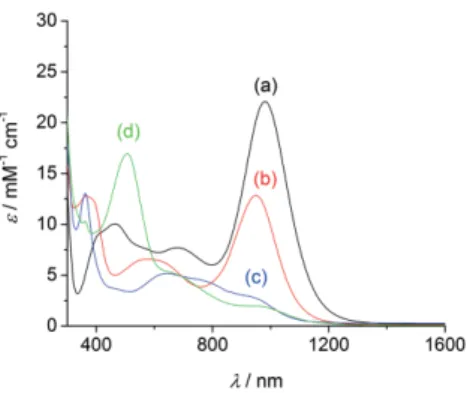
Complexes 1+and 2 are deeply colored and show intense
ab-sorptions over both the visible and NIR regions (Figure 5, Fig-ure 6, Table 3). Complex 1+exhibits a remarkable NIR band at
980 nm (22 080 M–1cm–1), that is assigned by TD-DFT to HOMO
→ LUMO and corresponds mainly to a ligand-to-ligand charge transfer transition from a delocalized ligand orbital to the imin-obenzoquinone cores (λcalcd= 796 nm, fosc= 0.349). Upon
oxid-Figure 5. UV/Vis-NIR spectra of (a, black) 1+, (b, red) 1, (c, blue) “two-electron
reduced” 1–, (d, green) 12+in 0.5 m
MCH2Cl2solution containing 0.1MTBAP (except d, which was generated by adding one molar equiv. of AgSbF6). For
the two-electron reduction the molar extinction coefficient is based on the total copper concentration.
Figure 6. UV/Vis-NIR spectra of 2. (a, blue) before electrolysis, (b, black) after exhaustive electrolysis at +0.2 V, (c, green) exhaustive electrolysis at +0.4 V in 0.5 mMCH2Cl2solution containing 0.1MTBAP.
ation to 12+this intense band vanishes, replaced by a broad
unresolved NIR feature of lower intensity with shoulders at ca. 640 and 960 nm, as well as a predominant band at 505 nm (16 960M–1cm–1). Consistently, TD-DFT calculations do not predict
electronic excitations with oscillator force greater than 0.05 above 560 nm. The most intense excitation is predicted at 552 nm (fosc= 0.1239), and accounts for the experimental band
at 551 nm. Several electronic excitations contribute to this exci-tation, which is principally due to an intraligand CT within the tris-ligated ligand. Reduction into neutral 1 is accompanied by a shift of the NIR band towards 948 nm, whose intensity de-creased by one half (12 840M–1cm–1). It is assigned to β-HOMO
→ β-LUMO and corresponds mainly to CT from the whole li-gands to the iminobenzoquinone or iminosemiquinone cores (λcalcd= 914 nm, fosc= 0.211). Further reduction of 1 results in
a severe broadening of the Vis-NIR features, which expand from 500 to 1000 nm with intensity lower than 5500M–1cm–1. The
main peak is at 645 nm (5170M–1cm–1), with shoulders at 770
and 930 nm. By TD-DFT we recalculated the spectrum of the anion 1–. An intense NIR band is predicted for the singlet
(λcalcd = 1017 nm, fosc = 0.178), which contrasts sharply with
experiment. We take this as further evidence for the rearrange-ment of the monoanion into 2 (see above).
Table 3. UV/Vis-NIR properties of the complexes.[a]
Complex λ [nm] (ε [mM–1cm–1]) 1red[b] 362 (13 060), 645 (5170), 770sh (4460), 930sh (2900) 1 366 (12 790), 578 (6590), 948 (12 840) 1+ 464 (10 052), 676 (7710), 980 (22 080) 12+ 505 (16 960), 640sh (5400), 960sh (1950) 2 361 (28 860), 653 (10 980), 770sh (9790) 2ox[c] 460 (4241), 679 (2210), 980 (14 860) 2ox′[c] 523 (13 890), 650 sh (700)
[a] In CH2Cl2containing 0.1MTBAP. [b] After exhaustive electrolysis of 1+at
–1.6 V. [c] After exhaustive electrolysis of 2 at +0.2 V (2ox) or +0.4 V (2ox′).
The spectrum of 2 consists of a band at 653 nm (10 980 M–1cm–1), with a shoulder at 770 nm
(Fig-ure 6a). DFT calculations predict several low energy excitations (fosc< 0.05), which account for the NIR tail. Its single oxidation
produces a chromophore that absorbs strongly at 980 nm (14 860M–1cm–1, Figure 6b), similarly to 1+, while double
oxid-ation affords a species that shows a dominant band at 523 nm (13 890M–1cm–1, Figure 6c) associated to a low intensity NIR
tail and hence compares with 12+. These spectroscopic data
are thus consistent with electrochemistry, demonstrating that oxidation of the trimer 2 produces a mononuclear complex, either 1+or 12+, depending on the applied potential.
Electrocatalytic Reduction of Dioxygen and Hydrogen Peroxide
Tricopper centers are found in laccases,[13]which catalyze the
four electron reduction of dioxygen into water. This has prompted us to investigate the electrocatalytic properties of 2 towards dioxygen and its two-electron reduced form, hydrogen peroxide. We exploited the presence of multiple hydrophobic
tert-butyl groups and aromatic rings to adsorb complex 2 onto
Eur. J. Inorg. Chem. 2020, 2691–2699 www.eurjic.org 2696 © 2020 Wiley-VCH Verlag GmbH & Co. KGaA, Weinheim
multiwall carbon nanotubes (MWCNT). Complex 2 does not cat-alyze dioxygen reduction but exhibits electrocatalytic activity regarding reversible hydrogen peroxide reduction. The CV for MWCNT functionalized by complex 2 both under argon and in the presence of 10 mMH2O2is depicted in Figure 7. For
com-parison the CV of MWCNT electrodes functionalized with horse-radish peroxidase (HRP), a well-known H2O2-reducing
metallo-enzyme is also included. This metallo-enzyme was immobilized by bor-onic-ester covalent binding with a pyrene-boronic-acid linker, as previously reported.[14]
Figure 7. Cyclic voltammetry for (blue straight line) complex 2-functionalized MWCNT electrodes (acetate buffer, pH 5) and HRP-functionalized MWCNT electrodes (phosphate buffer, pH 7) under Ar before and after addition of 10 mMH2O2(v = 10 mV s–1, ref. SHE); (inset) Tafel plot for complex
2-function-alized MWCNT electrodes (acetate buffer, pH 5).
First, CV under argon confirms the stable adsorption of the complex 2 in water. This is indicated by the presence of a poorly-reversible redox system, which is preserved after several repeated CV scans and corresponds to the oxidation of immobi-lized complex 2. This stable adsorption might be favored by the strong hydrophobic pi–pi interaction brought about by the three polycyclic ligands with CNT sidewalls. A surface coverage of Γmax= 76 pmol cm–2for complex 2 was estimated by
integra-tion of the charge of the anodic peak under argon. After addi-tion of 10 mMH2O2, the 2-functionalized MWCNTs exhibit
elec-trocatalytic activity towards both H2O2reduction and oxidation.
It is noteworthy that pristine MWCNT electrodes do not show any electrocatalytic activity towards H2O2in the same potential
window. Tafel analysis indicates an onset potential of Eonset =
+0.56 V vs. SHE. Maximum current density of 0.22 mA cm–2was
measured with an apparent turnover frequency (TOF) of 15 s–1
by taking into account the surface coverage by complex 2. A plausible electrocatalytic mechanism might involve the oxid-ation of complex 2 by H2O2, generating the catalytically active
species. It is noteworthy that the Tafel slopes were measured at 130 mV per decade, indicative of a rate-determining one-elec-tron. However, exhaustive studies will be required in order to identify if copper-peroxo intermediate are involved, as well as potential ligand exchange in complex 2. For comparison, HRP is able to reduce H2O2 at higher onset potentials of Eonset =
+0.83 V vs. SHE (pH 7) at modified MWCNT electrodes.[14,15] A
deca-heme cytochrome exhibits onset potential of +0.72 V (pH 6.5) on a porous ITO electrode,[16]while bioinspired
iron-por-phyrin complexes exhibits onset potentials of 0.7 V at pH 7.[15,17]It must be stressed that we recently immobilized mono
and dicopper-phenolato complexes which exhibit smaller onset potentials of + 0.33 V (pH 4) towards both O2and H2O2
reduc-tion.[11c,18]Thus, despite the fact that only 150 mV are still to be
gained in order to reach the best iron-porphyrin-based catalyst efficiency, these tricopper complexes represent a novel family of promising molecular electrocatalysts based on earth abun-dant metals for H2O2reduction under mild conditions.
Conclusion
In summary, we demonstrate that the pincer ligand H3LNcan
stabilize copper in both its +I and +II oxidation states. Depend-ing on the copper source used for the synthesis and the poten-tial applied during electrolysis two complexes with distinct stoi-chiometries form: A mononuclear (1:2) or trinuclear (3:3) com-plex. None of the structures herein reported were observed for the copper complexes of the related pincer ligand H3LO
(Scheme 4).[3f,j,s]
Scheme 4. Structure of -crystallized copper complexes of H3LO[3f,j]containing
one fully oxidized arm (benzoquinone, shown in red) or one iminosemiquin-one radical arm (blue).
We establish that the distinct basicity of the peripheral do-nors of H3LN allowed for a bis(bidentate) ligation, with one
amine of each ligand remaining in its NH2form. The NH2groups
adopt a cis-configuration and each is involved in H-bonding interactions with a SbF6–molecule. The steric clash induced by
the bulky ligands enforce strong tetrahedral distortions favora-ble to monovalent copper. In the H3LO chelate the terminal
carbonyl groups are coordinating, giving an octahedral geome-try with Jahn–Teller distortion that is suitable for Cu(II).[3f ]
Nota-bly the carbonyl groups bind in trans configuration, at each apical position. Another striking difference is the oxidation state of the loosely bound (or unbound) unit: In the case of H3LO, it
is the fully oxidized (quinone) unit, which is a poor donor, whereas in 1+it is unoxidized diaminobenzene unit.
Complex 2 is trinuclear, which is an unprecedented stoichio-metry for this family of pincer ligands (N3 or N2O chelates). The fourth coordination in the fully deprotonated (1:1) complex is occupied by the anilido nitrogen of a neighboring ligand, de-spite the excess of Et3N used for the synthesis.
We did not isolate a monomer with one bound triethyl-amine, as reported by Wieghardt for the H3LOcounterpart,[3j]
which may be another consequence of the distinct basicity of the peripheral donors.
Another striking point is the peculiar redox properties the H3LNcomplexes: First the potentials are in general much lower
than for the copper complexes of H3LO, allowing for the
isola-tion of a caisola-tionic and not a neutral monomeric species. Sec-ondly, the oxidation potentials in the H3LO chelates are >
+0.13 V and correspond to ligand-centered processes,[3b,c,j]
whereas the first oxidation is observed as low as –0.37 for 1+
and corresponds to a metal-centered process. Furthermore, we evidenced an unprecedented redox interconversion for H3LN,
whereby the monomeric species can be converted upon reduc-tion into the trimer 2 and vice versa.
Finally, owing to a stable immobilization at nanostructured electrodes, complex 2 exhibits electrocatalytic activity towards reversible H2O2reduction at high potentials of 0.53 V. This work
demonstrates the first example of an electrode functionalized with a tricopper complex active towards high potential H2O2
reduction in mild aqueous conditions. Further studies are in progress in order to decipher the electrocatalytic mechanism as well as study their use in peroxidase-like sensors or in H2O2
-based fuel cells where high potentials are required towards H2O2reduction.
Experimental Section
General
Unless otherwise noted, all operations were performed under anae-robic conditions under a pure argon atmosphere using standard Schlenk techniques. Anhydrous triethylamine was distilled from CaH2under an argon atmosphere prior to use. Anhydrous
aceto-nitrile and methanol were purchased from Acros. NMR spectra were recorded on a Bruker Avance 400 (1H at 400 MHz,13C at 100 MHz).
Chemical shifts are given relative to solvent residual peaks. Mass spectra were recorded on a Bruker Esquire 3000 (ESI/Ion Trap) equipment. Microanalysis were performed by using an apparatus designed by the Service Central d'Analyse du CNRS (Lyon, France). UV/Vis spectra were recorded on a Perkin-Elmer Lambda 1050 spec-trophotometer in quartz cells (Hellma) of 1.00 mm path length. X-band EPR spectra were recorded on an EMX plus spectrometer equipped with an Oxford Helium cryostat. Spectra were simulated using the Easyspin software package.[19]Electrochemical
measure-ments were carried out using a BioLogic SP300 potentiostat or a CH Instruments 620 potentiostat. Experiments were performed in a standard three-electrode cell under argon atmosphere in CH2Cl2
solutions containing 0.1Mtetrabutylammonium perchlorate (TBAP)
as supporting electrolyte. An Ag/AgNO3(0.01M) reference electrode
was used. All the potentials given in the text are referred to the regular Fc+/Fc redox couple used as an internal reference. A glassy
carbon disc electrode (5 mm diameter), which was polished with 1 mm diamond paste, was used as the working electrode. RDE ex-periments were performed by using a Radiometer CTV101 unit. Electrolyses were conducted at constant applied potential by using a carbon plate as working electrode.
Electrocatalysis
Commercial grade thin multi-walled carbon nanotubes (MWCNT, 9.5 nm diameter, purity >95 %) were obtained from Nanocyl and
used as received without any purification step. The electrochemical experiments in aqueous media were performed in 0.1 M acetate
buffer pH 5 or 0.1 Mphosphate buffer pH 7 in a three-electrode
electrochemical cell using an Autolab pgstat100 potentiostat. The surface of GC electrodes was polished with a 2 μm diamond paste purchased from Presi (France) and rinsed successively with water, acetone, and ethanol. A Pt wire placed in a separated compartment was used as counter electrode, and the Ag/AgCl (KCl sat) served as a reference electrode. Horseradish peroxidase/MWCNT electrode was prepared according to a previously-described procedure.[15] GC
electrode was drop-casted with 20 uL of MWCNT suspension (5 mg/ mL in NMP). A homogeneous CNT film was obtained after drying under vacuum for 1 hour, affording a 0.2 cm2MWCNT electrode.
MWCNT electrodes were soaked in a solution of 3 mMof complex
2 for 1 hour, then rinsed with DMF and dried. Electrochemical
char-acterizations were carried in a 0.1Macetate buffer pH 5 under
ar-gon to prevent any contribution of dissolved oxygen on the re-corded measures. 10 mMof H2O2was then added to the electrolyte
to assess the catalytic activity of the immobilized copper complex.
Crystal structure analysis
A single crystal was coated with a parafin mixture, picked up with nylon loops and mounted in the nitrogen cold stream of a Nonius 4 circles diffractometer at 200 K. The Mo-Kαradiation (λ = 0.71073Å)
from an Incoatec micro Mo-target X-ray source equipped with Mon-tel optics was used. The data were collected with a Bruker APEXII detector. Final cell parameters were obtained from refinements us-ing the whole data. Intensities were integrated and corrected for Lorentz and polarization factor using EVAL14 then corrected for ab-sorption using a multiscan method implemented with the program SADABS and then finally merged using XPREP. The structures were solved and refined by charge flipping methods and subsequent difference Fourier techniques. The OLEX software was used for the refinement.[20]All non-hydrogen atoms were anisotropically refined
and hydrogen atoms were placed at calculated positions and re-fined as riding atoms with isotropic displacement parameters. Both compounds crystallized as twins. For 1+(SbF
6–) the two lattices
could have been identified and integrated separately with a rate of 60.15 % vs. 39.85 %. The numerous overlaps in the data prevented the obtaining of a really accurate structure. For compound 2 we simply used the following twin law:
The rate between the two lattices is of 63.48 % vs. 36.16 %. Deposition Number 1989039 and 1989040 contain the supplemen-tary crystallographic data for this paper. These data are provided free of charge by the joint Cambridge Crystallographic Data Centre and Fachinformationszentrum Karlsruhe Access Structures service http://www.ccdc.cam.ac.uk/structures.
Computational details
Full geometric optimizations were performed with the Gaussian 9.0 program.[21]The TPSSh functional[22]was used together with the
6-31g* basis set for the C,H,N atoms.[23]Frequency calculations were
systematically performed in order to ensure that the optimized structures correspond to a real energy minimum and not a saddle point. The relative energies were obtained from single-point calcu-lations and corrected from the ZPE using the same level of theory
Eur. J. Inorg. Chem. 2020, 2691–2699 www.eurjic.org 2698 © 2020 Wiley-VCH Verlag GmbH & Co. KGaA, Weinheim
(functional and basis sets). Electronic transition energies for all com-plexes were calculated using time-dependent DFT (TD-DFT).[24]The
solvent effect was considered by using a polarizable continuum model[25]and the 40 lowest energy excited states were calculated.
Synthesis
The synthetic procedure for the preparation of H3LNwas reported
elsewhere.[6a] [Cu(HLN
IBQ)2](SbF6). CuCl2(16 mg, 0.12 mmol, 0.5 equiv.) and Et3N
(66 μL, 0.47 mmol, 2.0 equiv.) were added to a solution of H3LN
(100 mg, 0.24 mmol, 1.0 equiv.) in MeOH (12 mL). The mixture was immediately heated to 80 °C and rapidly turned black. After 15 min, KSbF6(199 mg, 0.72 mmol, 3.0 equiv.) was added and the mixture
was refluxed during 45 min. While cooling the reaction to r.t., a black precipitate formed. The suspension was cooled to 5 °C with an ice bath and the precipitate was filtered through a frit and washed with cold MeOH. Yield: 68 %;1H NMR (400 MHz, CD
2Cl2): δ
(ppm)= 7.83 (br s, 2H, NH), 7.38 (s, 2H, HAr), 7.15 (s, 2H, HAr), 6.96
(d, J = 2.4 Hz, 2H, HAr), 6.88 (br s, 2H, HAr), 4.67 (br s, 4H, NH), 1.42
(s, 18H, t-Bu), 1.34 (s, 18H, t-Bu), 1.17 (s, 18H, t-Bu), 1.12 (br s, 18H,
t-Bu); 13C NMR (100 MHz, CD
2Cl2): δ (ppm)= 162.3, 155.7, 144.2,
140.5, 140.4, 137.2, 135.6, 128.4, 125.4, 118.2, 115.1, 36.8, 35.30, 35.27, 34.9, 31.9, 30.8, 30.1, 29.2. MS (ESI): m/z= 606 [M – SbF6]+. IR:
ν (cm–1) 3528, 3417, 3379, 2955, 2908, 2867, 1610, 1477, 1461, 1277,
1261, 761, 748. Anal. Calcd for C56H86N6CuSbF6: C, 58.85; H, 7.60; N,
7.36; found C, 59.13; H, 7.71; N, 7.67.
[Cu3(LNISQ)3]·CH3CN. Under argon, to a degassed solution of
Cu(CH3CN)4·PF6(880 mg, 2.36 mmol, 1.0 equiv.) in MeCN (12 mL)
were added the ligand H3LN(1.0 g, 2.36 mmol, 1.0 equiv.) and
tri-ethylamine (2.3 mL, 16.50 mmol, 7.0 equiv.). The mixture was re-fluxed for 1 hour and subsequently exposed to air while cooling to r.t. After an additional hour whilst stirring at r.t., the mixture was cooled with an ice bath. The black precipitate that formed was fil-tered through a frit, abundantly washed with cold MeCN and dried under vacuum to afford a greenish black microcrystalline powder (1.01 g, 0.68 mmol, yield= 86 %). An analytically pure sample was obtained from a CH2Cl2solution layered with CH3CN. Single crystals
suitable for X-ray analysis were obtained by vapor diffusion at r.t. of MeCN into a benzene solution of the complex.1H NMR (400 MHz,
CD2Cl2): δ (ppm)= 8.28 (s, 3H, HAr), 7.74 (s, 3H, HAr), 7.46 (s, 3H, HAr),
7.22 (s, 3H, HAr), 5.77 (br s, 3H, NH), 2.16 (br s, 3H, NH), 1.97 (s, 3H,
CH3CN), 1.71 (s, 27H, t-Bu), 1.40 (s, 27H, t-Bu), 1.21 (s, 27H, t-Bu),
0.95 (s, 27H, t-Bu).13C NMR (100 MHz, CD
2Cl2): δ (ppm)= 155.9,
146.0, 145.0, 144.0, 143.7, 143.2, 142.4, 140.7, 119.2, 116.6, 114.9, 114.2, 36.9, 35.8, 35.4, 35.2, 32.4, 31.8, 31.3, 29.9. MS (ESI): m/z= 551 [M + H]+. IR: ν (cm–1) 3361, 3317, 2954, 2899, 2867, 1457, 1362,
1151. Anal. Calcd for C84H126Cu3N9, CH3CN, 0.5 CH2Cl2: C, 67.53; H,
8.53; N, 9.12; found C, 67.54; H, 8.64; N, 8.92.
Acknowledgments
The authors thank the Labex ARCANE (CBH-EUR-GS/ANR-17-EURE-0003) for financial support. This work was supported by the Ministère de l'Environnement, de l'Energie et de la Mer and the Agence Nationale de la Recherche through the LabEx AR-CANE programe (ANR-11-LABX-0003-01). The authors are grate-ful to Dr. Pierre Girard for technical assistance for the DFT calcu-lations and the Centre de Calcul Intensif en Chimie de Grenoble (CECIC) for providing the computational resources. The
Nano-bio-ICMG Platform (FR 2607) is acknowledged for analytical sup-port.
Keywords: Copper · Phenylenediamine · Radicals ·
Electronic structure · Iminosemiquinones
[1] a) P. J. Chirik, K. Wieghardt, Science 2010, 327, 794–795; b) O. R. Luca, R. H. Crabtree, Chem. Soc. Rev. 2013, 42, 1440–1459; c) F. Novio, E. Evan-gelio, N. Vazquez-Mera, P. González-Monje, E. Bellido, S. Mendes, N. Keha-gias, D. Ruiz-Molina, Sci. Rep. 2013, 3, 1708; d) D. L. J. Broere, R. Plessius, J. I. van der Vlugt, Chem. Soc. Rev. 2015, 44, 6886–6915; e) S. Demir, I.-R. Jeon, J. R. Long, T. D. Harris, Coord. Chem. Rev. 2015, 289–290, 149–176; f) B. de Bruin, P. Gualco, N. D. Paul in Redox Non-innocent Ligands, (Ed.: M. S. a. R. J. Lundgren), 2016, pp. 176–204; g) I.-R. Jeon, L. Sun, B. Negru, R. P. Van Duyne, M. Dincă, T. D. Harris, J. Am. Chem. Soc. 2016, 138, 6583– 6590; h) J. A. DeGayner, K. Wang, T. D. Harris, J. Am. Chem. Soc. 2018, 140, 6550–6553.
[2] A. Y. Girgis, A. L. Balch, Inorg. Chem. 1975, 14, 2724–2727.
[3] a) S. K. Larsen, C. G. Pierpont, J. Am. Chem. Soc. 1988, 110, 1827–1832; b) C. L. Simpson, S. R. Boone, C. G. Pierpont, Inorg. Chem. 1989, 28, 4379– 4385; c) B. G. Maiya, Y. Deng, K. M. Kadish, J. Chem. Soc., Dalton Trans.
1990, 3571–3576; d) C. G. Pierpont, C. W. Lange in The Chemistry of
Transition Metal Complexes Containing Catechol and Semiquinone Ligands,
1994, pp. 331–442; e) S. N. Lyubchenko, V. A. Kogan, L. P. Olekhnovich,
Russ. J. Coord. Chem. 1996, 22, 534–540; f) G. Speier, J. Csihony, A. M. Whalen, C. G. Pierpont, Inorg. Chem. 1996, 35, 3519–3524; g) M. A. Brown, J. A. Castro, B. R. McGarvey, D. G. Tuck, Can. J. Chem. 1999, 77, 502–510; h) C. Camacho-Camacho, G. Merino, F. J. Martínez-Martínez, H. Nöth, R. Contreras, Eur. J. Inorg. Chem. 1999, 1999, 1021–1027; i) P. Chaudhuri, M. Hess, K. Hildenbrand, E. Bill, T. Weyhermüller, K. Wieghardt, Inorg. Chem. 1999, 38, 2781–2790; j) P. Chaudhuri, M. Hess, T. Weyher-müller, K. Wieghardt, Angew. Chem. Int. Ed. 1999, 38, 1095–1098; Angew. Chem. 1999, 111, 1165; k) N. G. Furmanova, S. N. Lyubchenko, V. Kogan, L. P. Olekhnovich, Cryst. Rep. 2000, 45, 439–443; l) D. Ruiz-Molina, J. Veci-ana, K. Wurst, D. N. Hendrickson, C. Rovira, Inorg. Chem. 2000, 39, 617– 619; m) A. Piskunov, I. Smolyaninov, O. Yu. Sukhoshkina, Russ. J. Gen. Chem. 2010, 80, 790–799; n) A. V. Piskunov, O. Y. Trofimova, S. Y. Ketkov, G. K. Fukin, V. K. Cherkasov, G. A. Abakumov, Russ. Chem. Bull. 2011, 60, 2522–2530; o) G. Szigethy, A. F. Heyduk, Dalton Trans. 2012, 41, 8144– 8152; p) G. Szigethy, D. W. Shaffer, A. F. Heyduk, Inorg. Chem. 2012, 51, 12606–12618; q) J. L. Wong, R. H. Sánchez, J. G. Logan, R. A. Zarkesh, J. W. Ziller, A. F. Heyduk, Chem. Sci. 2013, 4, 1906–1910; r) A. A. Maleev, O. Y. Trofimova, A. P. Pushkarev, N. V. Somov, V. V. Travkin, G. L. Pak-homov, A. V. Piskunov, M. N. Bochkarev, Nanotechnol. Russ. 2015, 10, 613–620; s) J. Jacquet, K. Cheaib, Y. Ren, H. Vezin, M. Orio, S. Blanchard, L. Fensterbank, M. Desage-El Murr, Chem. Eur. J. 2017, 23, 15030–15034. [4] a) N. Ito, S. E. V. Phillips, C. Stevens, Z. B. Ogel, M. J. McPherson, J. N. Keen, K. D. S. Yadav, P. F. Knowles, Nature 1991, 350, 87–90; b) B. P. Branchaud, B. E. Turner in 30 - Galactose Oxidase: Probing Radical Mecha-nism with Ultrafast Radical Probe, Vol. 354 (Ed.: D. L. Purich), Academic Press, 2002, pp. 415–425; c) J. W. Whittaker, Chem. Rev. 2003, 103, 2347– 2364; d) D. Rokhsana, D. M. Dooley, R. K. Szilagyi, J. Am. Chem. Soc. 2006, 128, 15550–15551; e) F. Thomas, Eur. J. Inorg. Chem. 2007, 2007, 2379– 2404; f) D. Yin, S. Urresti, M. Lafond, E. M. Johnston, F. Derikvand, L. Ciano, J.-G. Berrin, B. Henrissat, P. H. Walton, G. J. Davies, H. Brumer, Nat. Commun. 2015, 6, 10197; g) A. K. Chaplin, C. Bernini, A. Sinicropi, R. Basosi, J. A. R. Worrall, D. A. Svistunenko, Angew. Chem. Int. Ed. 2017, 56, 6502–6506; Angew. Chem. 2017, 129, 6602.
[5] a) P. Chaudhuri, M. Hess, U. Flörke, K. Wieghardt, Angew. Chem. Int. Ed.
1998, 37, 2217–2220; Angew. Chem. 1998, 110, 2340; b) T. K. Paine, T.
Weyhermüller, K. Wieghardt, P. Chaudhuri, Dalton Trans. 2004, 2092– 2101.
[6] a) N. Leconte, J. Moutet, K. Herasymchuk, R. M. Clarke, C. Philouze, D. Luneau, T. Storr, F. Thomas, Chem. Commun. 2017, 53, 2764–2767; b) N. Leconte, J. Moutet, T. Constantin, F. Molton, C. Philouze, F. Thomas, Eur. J. Inorg. Chem. 2018, 2018, 1752–1761.
[7] N. Leconte, B. Baptiste, C. Philouze, F. Thomas, Dalton Trans. 2018, 47, 11303–11307.
[8] L. Yang, D. R. Powell, R. Houser, Dalton Trans. 2007, 955–964.
[9] S. Ye, B. Sarkar, F. Lissner, T. Schleid, J. van Slageren, J. Fiedler, W. Kaim, Angew. Chem. Int. Ed. 2005, 44, 2103–2106; Angew. Chem. 2005, 117, 2140.
[10] a) H.-Y. Cheng, C.-C. Lin, B.-C. Tzeng, S.-M. Peng, J. Chin. Chem. Soc. 1994, 41, 775–781; b) D. Herebian, E. Bothe, F. Neese, T. Weyhermüller, K. Wie-ghardt, J. Am. Chem. Soc. 2003, 125, 9116–9128; c) E. Bill, E. Bothe, P. Chaudhuri, K. Chlopek, D. Herebian, S. Kokatam, K. Ray, T. Weyhermüller, F. Neese, K. Wieghardt, Chem. Eur. J. 2005, 11, 204–224; d) K. Chłopek, E. Bill, T. Weyhermüller, K. Wieghardt, Inorg. Chem. 2005, 44, 7087–7098; e) K. Chłopek, E. Bothe, F. Neese, T. Weyhermüller, K. Wieghardt, Inorg. Chem. 2006, 45, 6298–6307; f) K. M. Clark, J. Bendix, A. F. Heyduk, J. W. Ziller, Inorg. Chem. 2012, 51, 7457–7459; g) N. Leconte, J. Ciccione, G. Gellon, C. Philouze, F. Thomas, Chem. Commun. 2014, 50, 1918–1920; h) J. Ciccione, N. Leconte, D. Luneau, C. Philouze, F. Thomas, Inorg. Chem.
2016, 55, 649–665; i) K. D. Spielvogel, E. J. Coughlin, H. Petras, J. A. Luna,
A. Benson, C. M. Donahue, A. Kibasa, K. Lee, R. Salacinski, S. C. Bart, S. K. Shaw, J. J. Shepherd, S. R. Daly, Inorg. Chem. 2019, 58, 12756–12774. [11] a) F. Michel, F. Thomas, S. Hamman, E. Saint-Aman, C. Bucher, J.-L. Pierre,
Chem. Eur. J. 2004, 10, 4115–4125; b) F. Michel, S. Torelli, F. Thomas, C. Duboc, C. Philouze, C. Belle, S. Hamman, E. Saint-Aman, J.-L. Pierre, Angew. Chem. Int. Ed. 2005, 44, 438–441; Angew. Chem. 2005, 117, 442; c) S. Gentil, D. Serre, C. Philouze, M. Holzinger, F. Thomas, A. Le Goff, Angew. Chem. Int. Ed. 2016, 55, 2517–2520; Angew. Chem. 2016, 128, 2563; d) S. Torelli, C. Belle, I. Gautier-Luneau, J. L. Pierre, E. Saint-Aman, J. M. Latour, L. Le Pape, D. Luneau, Inorg. Chem. 2000, 39, 3526–3536. [12] F. Neese, J. Biol. Inorg. Chem. 2006, 11, 702–711.
[13] N. Hakulinen, J. Rouvinen, Cell Mol. Life Sci. 2015, 72, 857–868. [14] B. Reuillard, A. Le Goff, M. Holzinger, S. Cosnier, J. Mater. Chem. B 2014,
2, 2228–2232.
[15] B. Reuillard, S. Gentil, M. Carrière, A. Le Goff, S. Cosnier, Chem. Sci. 2015, 6, 5139–5143.
[16] B. Reuillard, K. H. Ly, P. Hildebrandt, L. J. C. Jeuken, J. N. Butt, E. Reisner, J. Am. Chem. Soc. 2017, 139, 3324–3327.
[17] Y. Yamada, S. Yoshida, T. Honda, S. Fukuzumi, Energy Environ. Sci. 2011, 4, 2822–2825.
[18] S. Gentil, J. K. Molloy, M. Carrière, A. Hobballah, A. Dutta, S. Cosnier, W. J. Shaw, G. Gellon, C. Belle, V. Artero, F. Thomas, A. Le Goff, Joule 2019, 3, 2020–2029.
[19] S. Stoll, A. Schweiger, J. Magn. Reson. 2006, 178, 42–55.
[20] O. V. Dolomanov, L. J. Bourhis, R. J. Gildea, J. A. K. Howard, H. Puschmann, J. Appl. Crystallogr. 2009, 42, 339–340.
[21] M. J. Frisch, G. W. Trucks, H. B. Schlegel, G. E. Scuseria, M. A. Robb, J. R. Cheeseman, G. Scalmani, V. Barone, B. Mennucci, G. A. Petersson, H. Na-katsuji, M. Caricato, X. Li, H. P. Hratchian, A. F. Izmaylov, J. Bloino, G. Zheng, J. L. Sonnenberg, M. Hada, M. Ehara, K. Toyota, R. Fukuda, J. Hase-gawa, M. Ishida, T. Nakajima, Y. Honda, O. Kitao, H. Nakai, T. Vreven, J. A. Montgomery Jr., J. E. Peralta, F. Ogliaro, M. Bearpark, J. J. Heyd, E. Broth-ers, K. N. Kudin, V. N. Staroverov, R. Kobayashi, J. Normand, K. Raghava-chari, A. Rendell, J. C. Burant, S. S. Iyengar, J. Tomasi, M. Cossi, N. Rega, J. M. Millam, M. Klene, J. E. Knox, J. B. Cross, V. Bakken, C. Adamo, J. Jaramillo, R. Gomperts, R. E. Stratmann, O. Yazyev, A. J. Austin, R. Cammi, C. Pomelli, J. W. Ochterski, R. L. Martin, K. Morokuma, V. G. Zakrzewski, G. A. Voth, P. Salvador, J. J. Dannenberg, S. Dapprich, A. D. Daniels, Ö. Farkas, J. B. Foresman, J. V. Ortiz, J. Cioslowski, D. J. Fox, Gaussian 09, Revision D.01, Gaussian, Inc., Wallingford CT, 2009.
[22] a) J. Tao, J. P. Perdew, V. N. Staroverov, G. E. Scuseria, Phys. Rev. Lett. 2003, 91, 146401; b) V. N. Staroverov, G. E. Scuseria, J. Tao, J. P. Perdew, J. Chem. Phys. 2003, 119, 12129–12137.
[23] G. A. Petersson, M. A. Al-Laham, J. Chem. Phys. 1991, 94, 6081–6090. [24] M. E. Casida in “Recent Advances in Density Functional Methods” (Ed.:
D. P. Chong), World Scientific, Singapore, 1995.
[25] S. Miertuš, E. Scrocco, J. Tomasi, Chem. Phys. 1981, 55, 117–129.

![Table 1. Selected bond lengths in 1 + and 2. [a]](https://thumb-eu.123doks.com/thumbv2/123doknet/12935316.374489/4.895.519.754.98.509/table-selected-bond-lengths.webp)

![Table 3. UV/Vis-NIR properties of the complexes. [a]](https://thumb-eu.123doks.com/thumbv2/123doknet/12935316.374489/7.895.524.754.293.524/table-uv-vis-nir-properties-of-the-complexes.webp)
