ORIGINAL RESEARCH
GRP78-targeted nanotherapy against castrate-resistant
prostate cancer cells expressing membrane GRP78
Florence Delie&Patrick Petignat&Marie CohenReceived: 21 May 2012 / Accepted: 8 October 2012 / Published online: 23 October 2012 # Springer-Verlag France 2012
Abstract Glucose-regulated protein 78, GRP78, is a chap-erone protein mainly located in the endoplasmic reticulum (ER) of normal cells. In stress conditions, GRP78 is overex-pressed and in different cancer cell types, it is exoverex-pressed at the cell surface, whereas it stays intracellular in non-cancerous cells. Therefore, it appears as a strategic target to recognize malignant cells. Prostate cancer is one of the most diagnosed cancers in men. The development of castrate resistant tumors and the resistance to chemotherapy frequently occur. The carboxy-terminal ER retention domain is defined by the KDEL amino acid sequence. We developed anti-KDEL func-tionalized polymeric nanoparticles (NPs) loaded with pacli-taxel (Tx) to specifically target prostate cancer cells expressing GRP78. The sensitivity to Tx in different formu-lations was compared in three prostate cell lines: PNT1B, a normal cell line, PC3, a cancer cell line faintly expressing GRP78 at its surface, and DU145, a cancer cell line expressing GRP78 at its cell surface. Our results show that the targeted formulation significantly increases Tx sensitivity of cell line
expressing GRP78 at its surface compared to other treatments suggesting the added value of GRP78 targeted therapy for castrate resistant tumor which expresses GRP78 at its cell surface.
Keywords Prostate . Cancer . Targeting . KDEL . GRP78 . Nanoparticle . Paclitaxel
Introduction
Prostate cancer is the most diagnosed cancer in men in Europe. Despite advances in therapies, the occurrence of castrate resistant prostate cancer (CRPC) and resistance to chemotherapy are still challenging.
GRP78 is an endoplasmic chaperone which plays a crit-ical role in prostate cancer. this protein is primarily located in the endoplasmic reticulum (ER) where it is a regulator of the unfolded protein response. GRP78 expression is induced under stress conditions such as hypoxia and glucose depri-vation which are frequently observed in tumor microenvi-ronment. In many type of cancer cells, GRP78 is expressed at the cell surface, a fact not observed in normal cells [1]. Furthermore, GRP78 expression has been associated with malignancy and drug resistance [2–6]. It has also been shown that GRP78-knock-down greatly induces chemosen-sitivity of growth arrested dormant cancer cells and tumor-associated endothelial cells [7,8]. These observations sug-gest that inhibition or downregulation of GRP78 will be particularly relevant to eradicate residual tumor by over-coming drug resistance in proliferating and dormant cancer cells, as well as in cells supporting tumor growth [1]. In cancer cells, several proteins have been identified to bind membrane GRP78 in its N- or C-terminus [9–14]. In pros-tate cancer cells, activated alpha2-macroglobulin binding to membrane GRP78 activates several signaling pathways in F. Delie
School of Pharmaceutical Sciences, University of Geneva, Quai Ernest Ansermet 30,
1211 Geneva 4, Switzerland F. Delie
University of Lausanne, Lausanne, Switzerland P. Petignat
:
M. CohenDepartment of Obstetrics and Gynecology, University of Geneva, Boulevard de la Cluse 30,
1211 Geneva, Switzerland M. Cohen (*)
Laboratoire d’Hormonologie, HUG Maternité, 30 bd de la Cluse,
1211 Genève 14, Switzerland e-mail: [email protected]
favor of proliferation and cell survival [11]. These pathways can be suppressed by ligation of cell surface GRP78 with antibodies directed against its COOH-terminal domain [15,16].
GRP78 is therefore, a good pharmacological target to inhibit cancer cell growth and spreading. Furthermore, as exclusively expressed at cancer cell surface, GRP78 may be a good candidate to specifically deliver drug to the cells of interest. Targeted therapy is based on the specific recogni-tion of a molecular entity characteristic of a designated organ or cells to concentrate large amount of drug to the target without inducing adverse effects related to a non-specific distribution. To this end, a drug or a drug-loaded carrier may be chemically linked to a ligand specific to a molecular target overexpressed or ideally exclusively expressed at the cells of interest. Polymeric nanoparticles (NPs) are solid drug carriers in which the drug is encapsu-lated in a polymeric matrix. NPs offer the advantages of high compound loading rates, enabling the formulation of poorly soluble drugs as fine injectable suspensions. The surface of the nanocarriers may be functionalized with spe-cific ligands such as antibodies, aptamers, glycoproteins, lectines or peptides to trigger the recognition of a defined target. Paclitaxel (Tx) is a chemotherapeutic drug of the taxane family widely used in the treatment of advanced and recurrent prostate cancers. It suffers from a low water solubility necessitating the use of cosolvents such as Cre-mophor®, responsible for severe side effects. NP formula-tion has been proposed to circumvent the solubility problem related to Tx. Recently; Tx-loaded NPs were functionalized with Herceptin®, as a targeting moiety, for the delivery to ovarian cancer cells overexpressing HER2 specific antigens. This drug delivery system proved to be efficient in vitro and in vivo against SKOV-3 cell model [17,18].
In this study, we focused on the interest of anti-KDEL antibodies as a recognition moiety for the COOH-terminal domain of GRP78 present at cancer cell surface to specifi-cally target cancer cells in CRPC and to increase response to chemotherapy. Tx-loaded nanoparticles (NPs-Tx) were cou-pled to antibodies against the KDEL sequence and studied on different prostate cell lines viability: normal cell line, PNT1B, castrate resistant cancer (CRC) cell line which does not or faintly express GRP78 on its surface, PC3 and castrate resistant cell line which expresses GRP78 at its cell surface, DU145 [11,19].
Materials and methods Reagents
Poly (DL-lactic acid) (100DL 4A, MW 57 kD) was provid-ed by Lakeshore Biomaterials, Inc (Birmingham, AL,
USA). 1-Ethyl-3-(3-dimethylaminopropyl)-carbodiimide (EDAC), D(+)-Trehalose dihydrate, phosphate buffer saline (PBS), poly-L-Lysine solution, 0.1 % (w/v) were from Sig-ma (Buchs, Switzerland). m-Sig-maleimidobenzoyl-N-hydroxy- m-maleimidobenzoyl-N-hydroxy-sulfosuccinimide ester (sulfo-MBS), Tris (2-carboxyethyl)-phosphine hydrochloride (TCEP) and D-salt dextran plastic columns were supplied by Pierce (Rockford, IL, USA). Dioctadecyloxacarbo-cyanine perchlorate (DiO) was from Molecular Probes (Leiden, The Netherlands). Poly(vinyl alcohol) (Mowiol 4–88) was purchased from Hoechst (Frankfurt/M, Germany). Paclitaxel (Tx) was obtained from Cfm Oskar Tropitzsch (Marktredwitz, Germany).
Rabbit anti-GRP78 antibodies (GL-19) were from Sigma (Buchs, Switzerland) and mouse anti-KDEL antibodies from Assay Designs (Ann Arbor, MI, USA). Secondary antibodies were goat anti-mouse IgG-HRP conjugated from Santa Cruz Biotechnology (Santa Cruz, CA, USA) and goat anti-rabbit IgG-HRP conjugated from BIO-RAD (Munich, Germany).
Dulbecco’s modified Eagle’s medium (DMEM), Hank’s balanced salts, gentamycin, were products of Invitrogen (Basel, Switzerland). Fetal bovine serum (FBS) was from Biochrom AG (Oxoid AG, Basel, Switzerland).
Cell proliferation reagent (WST-1) was provided by Roche Diagnostics GmbH (Indianapolis, IN, USA). Nanoparticles
The nanoparticles were prepared by a salting-out process and characterized as previously described [20,21]. Briefly, Tx was added to a solution of poly (DL-lactic acid) (PLA) in acetone (drug to polymer ratio: 1:10). The organic phase was mixed under vigorous stirring at 45 °C with an aqueous phase containing 15 % (w/w) poly (vinyl alcohol) (PVAL) used as surfactant and 60 % (w/w) magnesium chloride hexahydrate. Then, 20 ml of pure water was added to the NPs suspension and stirring was maintained for 10 min. NPs were recovered by centrifugation (Optima® XL-100K, rotor 70.1 Ti Centrifuge, Beckman Coulter Inc., Fullerton, CA, USA), washed twice with pure water to remove unencapsu-lated drug, PVAL and magnesium salt. NPs were lyophilized (Edwards, Modulyo, Oberwil, Switzerland) in presence of trehalose (30 % w/w) stored at 4 °C until use.
The anti-KDEL antibodies were grafted using a carbodii-mide method as already described [20,21]. In a first step, the free carboxyl groups of the polymer at the NPs surface were thiolated to allow the covalent attachment of antibod-ies via a sulfo-MBS (m-maleimidobenzoyl-N-hydroxy-sul-fosuccinimide ester) cross-linker. Anti-KDEL antibodies were, then, activated in PBS (pH 7.4) with sulfo-MBS. After removing non-reacted sulfo-MBS by centrifugation (3000 rpm for 15 min) using Amicon Ultra-4 centrifugal filter devices, the activated ligand was incubated with NPs and
gently shaken at room temperature. Unconjugated ligand was removed by centrifugation.
The size and morphology of NPs were analysed by photon correlation spectroscopy using a Zetasizer 3000 HS (Malvern instruments Ltd, Worcestershire, UK), and scan-ning electron microscopy performed on gold-coated freeze-dried samples (Balzers SCD 004 Sputter Coater) with a JEOL JSM-6400 microscope (JEOL, Tokyo, Japan) at an accelerating voltage of 10 or 15 kV. Zeta potential of the particles was obtained in a 10−3M NaCl solution using the electrophoretic mode of the Zetasizer 3000 HS. Drug load-ing was determined by reverse-phase HPLC with UV detec-tion after dissoludetec-tion of the polymeric matrix in acetone [20]. The drug loading was expressed as the amount of drug encapsulated per mg of NPs. The amount of anti-KDEL antibodies conjugated to NPs was determined indirectly by measuring uncoupled antibodies in the supernatant by spec-trophotometry (λ0280 nm) (Hewlett Packard, Model 8453, Germany) after centrifugation.
Cell culture
Prostate cell lines DU145, PC3 and PNT1B were kindly provided by Dr P. Rocchi (Inserm 624, Marseille, France), and grown in DMEM medium supplemented with 10 % (v/v) foetal bovine serum, and 0.1 mg/ml gentamicin under 5 % CO2, at 37 °C.
Cell proliferation
Prostate cancer cells were seeded in 96-well plates at a density of 30 000 cells/well (PNT1B) or 10 000 cells/well (PC-3 and DU145) and incubated for 24 h before treatment. Cell culture medium was then replaced by 100μl of the different formulations (Tx, NPs, NPs-Tx, NPs-Tx-KDEL, anti-KDEL or anti-KDEL with NPs-Tx) at dif-ferent concentrations (0, 0.6, 1.2, 3.6, 6 and 60 ng/ml equivalent Tx and/or equivalent concentration of anti-KDEL) for 5 h before washing and replacement with culture medium for 24 h or 48 h. In vitro cytotoxicity was then tested on cells using 100 μl of WST-1 proliferation assay. Plates were analyzed after 1 h using a microplate reader at λ0 450 nm. Cell viability was calculated in percent compared to control samples incubated with normal culture medium and set at 100 %. Each experiment was run four times in triplicate.
Statistical analysis
Results were measured and expressed as mean ± SE. The differences between samples were evaluated by the Student’s t test and p value< 0.05 was considered significant.
Results-discussion
GRP78 is overexpressed and relocated at cell surface in many cancer cells. It, thus, represents a very interesting target to be associated with drug delivery systems, as it is specifically expressed at cell surface of cancer cells and not in healthy cells. Different peptide ligands of GRP78 have already been tested to bind to the surface expressed GRP78 to be used as drug delivery systems [22–25]. The peptides interacted with cancerous cells whereas only limited recog-nition was observed with normal cells. The peptide-drug conjugates were internalized in cancer cells, concentrated in the tumor tissue without accumulation in other organs, demonstrating the specificity of the binding and the interest of targeting the GRP78.
Direct binding of peptide to a drug molecule is a common way to achieve targeting. However, chemical modifications of the active compound and the further release of the drug may compromise the pharmacological efficiency. Therefore, other approaches involving synthetic drug carriers, such as liposomes and nanoparticles (NPs) have been developed. The drug is encapsulated or associated with a polymer or lipid core and the surface is functionalized with a recogni-tion moiety. For instance, pegylated liposomes were surface-modified by Katanasaka et al. [26] with a GRP-recognizing peptide developed by Arap et al. [22]. It was shown in vitro that the liposomes were able to target VEGF-activated HUVEC cells as well as DU145, a prostate cancer cell line [26]. This study confirms the interest of using GRP78 as a target element to bring large amount of drug to a tumor via colloidal carriers and shows that GRP78 can be also a target for cancer antineovascularisation therapy.
So far, to our knowledge, only peptides have been used as GRP78-recognizing entity. Despite the fact that some GRP78 antibodies have been described as potent anti-tumor agent [15, 16,27], they have never been tested for targeting therapy. As a proof of concept, Tx-loaded NPs were functionalized with antibodies against the KDEL ami-no acid sequence present in C-terminus of GRP78. The influence of these particles on cell survival was compared with other Tx formulations. Different recognition moieties have been tested for prostate cancer nanotargeting [28–34]. Compared to the other ligands, GRP78 offers the advantage of not only being expressed in prostate cancer cells [35] but
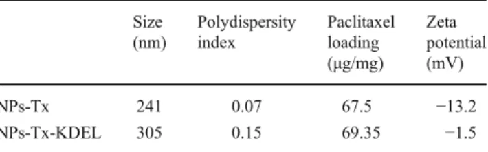
Table 1 Characterization of the NPs Size (nm) Polydispersity index Paclitaxel loading (μg/mg) Zeta potential (mV) NPs-Tx 241 0.07 67.5 −13.2 NPs-Tx-KDEL 305 0.15 69.35 −1.5
also on other tumors such as neuroblastoma, lung adenocar-cinoma, colon adenocaradenocar-cinoma, ovarian tumor cells [36], proliferating endothelial cells and, more generally, stressed tumor cells [10]. Therefore, it is more versatile target; im-plying that, if successful, this strategy may be applied to other cancer therapy. Furthermore, as antibodies against the -COOH end of the GRP78 present anti-cancer activity, a
synergistic effect is expected between the loaded drug and the targeting ligand.
The characteristics of the different NPs batches are de-scribed in details in (Biochimie, submitted) and are summa-rized in Table1.
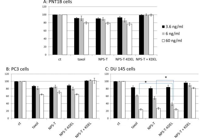
Figure1shows the effect of the different formulations of Tx on the viability of three prostate cell lines expressing Fig. 1 Cell viability after 5 h of incubation with NPs at different
concentrations (0, 3.6, 6, 60 ng/ml Tx equivalent), PNT1B (A), PC3 (B) and DU145 (C) cells were washed and cultured for 48 h before proliferation assay was performed. Unloaded NPs were used as controls for the possible cell toxicity of the polymer. In all cells and all concen-trations, cells viability was not altered by the presence of unloaded NPs
(data not shown). Viability was assessed by a WST1 assay and calculated in percent compared to control sample incubated with normal culture medium and set at 100 %. Each experiment was run four times in triplicate. Tx: paclitaxel; NPs-Tx: paclitaxel-loaded NPs, NPs-Tx-KDEL: anti KDEL conjugated-paclitaxel-loaded NPs; NPs-Tx + NPs-Tx-KDEL: paclitaxel-loaded NPs in the presence of free anti-KDEL antibodies
Fig. 2 DU145 cell viability after 5 h incubation with NPs at different concentrations (3.6, 4.5, 6, 9, 60 ng/ml Tx equivalent). Cells were washed and cultured for 48 h before proliferation assay was performed. Approximate IC50
for each molecule was determined. Tx: paclitaxel; NPs-Tx: paclitaxel-loaded NPs, NPs-Tx-KDEL: anti-KDEL conjugated-paclitaxel-loaded NPs
differently the GRP78 at their surface: the non-cancerous cell line, PNT1B; a CRC cell line not or faintly expressing GRP78 at its surface, the PC3 cells and the DU145 cells, a CRC cell line expressing GRP78 at its surface.
In the non-cancerous cell line, Tx either free or loaded in NPs did not induce a significant toxicity and no difference was seen between the formulations (Fig.1a).
As expected, cancerous cells were more sensitive to the treatment with Tx due to their higher proliferation rates. Free Tx induced a dose-related cytotoxicity which was more noticeable on DU145 compared to PC3 in these conditions of treatment.
Similar effects were observed when Tx was incubated as a suspension in NPs coated or not with KDEL anti-bodies in PC-3 cells which do not express GRP78 at their surface (Fig.1b). In the opposite, in DU145 cell line which expresses GRP78 at cell surface, NPs-Tx-KDEL significantly altered cell viability compared to Tx or NPs-Tx(Fig.1c). In this cell line, IC50of Tx was decreased by about 2-fold when
Tx is loaded in NPS, and more than 4-fold when Tx-loaded NPs were coated with anti-KDEL antibodies (Fig.2).
It was already described that antibodies against GRP78 COOH terminus can suppress proliferative/survival signal-ing pathways of prostate cancer cells expresssignal-ing GRP78 at their surface [16]. We thus chose monoclonal KDEL anti-bodies to functionalize Tx-loaded NPs to target the -COOH-terminal domain of membrane GRP78 and potently inhibit pathways implied in cancer growth. This strategy proved correct by making Tx more toxic in GRP78-expressing cells when presented in targeted nanocarriers, most likely by favoring cell penetration of the chemotoxic agent. However, when free anti-KDEL was added to NPs-Tx in culture medi-um, cytotoxic effects of NPs-Tx was reversed in all cell lines suggesting an anti-apoptotic effect of these antibodies (Fig.1a-c).
Despite anti-apoptotic effects of anti-KDEL antibodies on cells, this study confirms the interest of anti-cancer therapy by targeting cell-surface GRP78 on cancer cells which express GRP78 at cell surface, and demonstrates the efficiency of coupling GRP78 antibodies to NPs for prostate cancer cells.
In contrast to GRP78 autoantibodies isolated from serum of patients with prostate cancer, we have recently purified GRP78 autoantibodies from serum of ovarian cancer patients with potent anti-tumoral activity suggesting their interest for being coupled to NPs-Tx to increase response to drug [27].
In conclusion, we confirmed that GRP78 has the poten-tial to be used as target against prostate cancer cells express-ing GRP78 at their surface to both specifically vehicle drug and increase response to drug. We also demonstrated for the first time that antibodies against GRP78 could be used to this end.
Future prospects will include the testing of particles coated with these antibodies and in vivo studies to further confirm the interest of targeting nanocarriers for cancer therapy.
Acknowledgments We appreciate the technical support from Brigitte Delavy, and“La fondation Pierre Mercier pour la Science” and “La fondation pour la lutte contre le cancer et la recherche biomédicale” for their support.
Conflict of interest statement No funds were received in support of this study and no benefits in any form have been or will be received from a commercial party related directly or indirectly to the subject of this manuscript.
References
1. Lee AS (2007) GRP78 induction in cancer: therapeutic and prognos-tic implications. Cancer Res 67(8):3496–3499. doi: 10.1158/0008-5472.CAN-07-0325
2. Bini L, Magi B, Marzocchi B, Arcuri F, Tripodi S, Cintorino M, Sanchez JC, Frutiger S, Hughes G, Pallini V, Hochstrasser DF, Tosi P (1997) Protein expression profiles in human breast ductal carcino-ma and histologically norcarcino-mal tissue. Electrophoresis 18(15):2832– 2841
3. Chatterjee S, Cheng MF, Berger SJ, Berger NA (1994) Induction of M(r) 78,000 glucose-regulated stress protein in poly(adenosine diphosphate-ribose) polymerase- and nicotinamide adenine dinucleotide-deficient V79 cell lines and its relation to resistance to the topoisomerase II inhibitor etoposide. Cancer Res 54(16):4405– 4411
4. Fernandez PM, Tabbara SO, Jacobs LK, Manning FC, Tsangaris TN, Schwartz AM, Kennedy KA, Patierno SR (2000) Overexpres-sion of the glucose-regulated stress gene GRP78 in malignant but not benign human breast lesions. Breast Cancer Res Treat 59(1):15–26 5. Koomagi R, Mattern J, Volm M (1999) Glucose-related protein
(GRP78) and its relationship to the drug-resistance proteins P170, GST-pi, LRP56 and angiogenesis in non-small cell lung carcino-mas. Anticancer Res 19(5B):4333–4336
6. Xing X, Lai M, Wang Y, Xu E, Huang Q (2006) Overexpression of glucose-regulated protein 78 in colon cancer. Clin Chim Acta 364 (1–2):308–315
7. Dong D, Stapleton C, Luo B, Xiong S, Ye W, Zhang Y, Jhaveri N, Zhu G, Ye R, Liu Z, Bruhn KW, Craft N, Groshen S, Hofman FM, Lee AS (2011) A critical role for GRP78/BiP in the tumor microen-vironment for neovascularization during tumor growth and metasta-sis. Cancer Res 71(8):2848–2857
8. Ranganathan AC, Zhang L, Adam AP, Aguirre-Ghiso JA (2006) Functional coupling of p38-induced up-regulation of BiP and activation of RNA-dependent protein kinase-like endoplasmic re-ticulum kinase to drug resistance of dormant carcinoma cells. Cancer Res 66(3):1702–1711. doi: 10.1158/0008-5472.CAN-05-3092
9. Burikhanov R, Zhao Y, Goswami A, Qiu S, Schwarze SR, Rangnekar VM (2009) The tumor suppressor Par-4 activates an extrinsic pathway for apoptosis. Cell 138(2):377–388
10. Davidson DJ, Haskell C, Majest S, Kherzai A, Egan DA, Walter KA, Schneider A, Gubbins EF, Solomon L, Chen Z, Lesniewski R, Henkin J (2005) Kringle 5 of human plasminogen induces apopto-sis of endothelial and tumor cells through surface-expressed glucose-regulated protein 78. Cancer Res 65(11):4663–4672
11. Gonzalez-Gronow M, Cuchacovich M, Llanos C, Urzua C, Gawdi G, Pizzo SV (2006) Prostate cancer cell proliferation in vitro is modulated by antibodies against glucose-regulated protein 78 iso-lated from patient serum. Cancer Res 66(23):11424–11431 12. Jakobsen CG, Rasmussen N, Laenkholm AV, Ditzel HJ (2007)
Phage display derived human monoclonal antibodies isolated by binding to the surface of live primary breast cancer cells recognize GRP78. Cancer Res 67(19):9507–9517
13. Philippova M, Ivanov D, Joshi MB, Kyriakakis E, Rupp K, Afonyushkin T, Bochkov V, Erne P, Resink TJ (2008) Identification of proteins associating with glycosylphosphatidylinositol- anchored T-cadherin on the surface of vascular endothelial cells: role for Grp78/BiP in T-cadherin-dependent cell survival. Mol Cell Biol 28 (12):4004–4017
14. Shani G, Fischer WH, Justice NJ, Kelber JA, Vale W, Gray PC (2008) GRP78 and Cripto form a complex at the cell surface and collaborate to inhibit transforming growth factor beta signaling and enhance cell growth. Mol Cell Biol 28(2):666–677
15. Misra UK, Mowery Y, Kaczowka S, Pizzo SV (2009) Ligation of cancer cell surface GRP78 with antibodies directed against its COOH-terminal domain up-regulates p53 activity and promotes apoptosis. Mol Cancer Ther 8(5):1350–1362
16. Misra UK, Pizzo SV (2010) Ligation of cell surface GRP78 with antibody directed against the COOH-terminal domain of GRP78 suppresses Ras/MAPK and PI 3-kinase/AKT signaling while pro-moting caspase activation in human prostate cancer cells. Cancer Biol Ther 9(2):142–152
17. Cirstoiu-Hapca A, Bossy-Nobs L, Buchegger F, Gurny R, Delie F (2007) Differential tumor cell targeting of anti-HER2 (Herceptin©) and anti-CD20 (Mabthera©) coupled nanoparticles. Int J Pharm 331(2):190–196
18. Cirstoiu-Hapca A, Buchegger F, Lange N, Bossy L, Gurny R, Delie F (2010) Benefit of anti-HER2-coated paclitaxel-loaded immuno-nanoparticles in the treatment of disseminated ovarian cancer: Therapeutic efficacy and biodistribution in mice. J Control Release 144(3):324–331
19. Larson N, Ray A, Malugin A, Pike DB, Ghandehari H (2010) HPMA copolymer-aminohexylgeldanamycin conjugates targeting cell surface expressed GRP78 in prostate cancer. Pharm Res 27 (12):2683–2693
20. Cirstoiu-Hapca A, Bucheger F, Bossy L, Kosinski GR, Delie F (2009) Nanomedicines for active targeting: Physico-chemical char-acterization of paclitaxel-loaded anti-HER2 immunonanoparticles and in vitro functional studies on target cells. Eur J Pharm Sci 38:230–237
21. Nobs L, Buchegger F, Gurny R, Allemann E (2003) Surface modi-fication of poly(lactic acid) nanoparticles by covalent attachment of thiol groups by means of three methods. Int J Pharm 250(2):327–337 22. Arap MA, Lahdenranta J, Mintz PJ, Hajitou A, Sarkis AS, Arap W, Pasqualini R (2004) Cell surface expression of the stress response chaperone GRP78 enables tumor targeting by circulating ligands. Cancer Cell 6(3):275–284. doi:10.1016/j.ccr.2004.08.018 S1535610804002405
23. Kim Y, Lillo AM, Steiniger SC, Liu Y, Ballatore C, Anichini A, Mortarini R, Kaufmann GF, Zhou B, Felding-Habermann B, Janda
KD (2006) Targeting heat shock proteins on cancer cells: selection, characterization, and cell-penetrating properties of a peptidic GRP78 ligand. Biochemistry 45(31):9434–9444. doi:10.1021/ bi060264j
24. Liu Y, Steiniger SC, Kim Y, Kaufmann GF, Felding-Habermann B, Janda KD (2007) Mechanistic studies of a peptidic GRP78 ligand for cancer cell-specific drug delivery. Mol Pharm 4(3):435–447. doi:10.1021/mp060122j
25. Delie F, Petignat P, Cohen M (2012) GRP78 protein expression in ovarian cancer patients and perspectives for a drug targeting ap-proach. J Oncol 468615:1–5. doi:10.1155/2012/468615
26. Katanasaka Y, Ishii T, Asai T, Naitou H, Maeda N, Koizumi F, Miyagawa S, Ohashi N, Oku N (2010) Cancer antineovascular therapy with liposome drug delivery systems targeted to BiP/ GRP78. Int J Cancer 127(11):2685–2698
27. Cohen M, Petignat P (2011) Purified autoantibodies against glucose-regulated protein 78 (GRP78) promote apoptosis and de-crease invasiveness of ovarian cancer cells. Cancer Lett 309(1):104– 109
28. Katsogiannou M, Peng L, Catapano CV, Rocchi P (2011) Active-Targeted Nanotherapy Strategies for Prostate Cancer. Curr Cancer Drug Targets 11(8):954–965
29. Tong R, Yala L, Fan TM, Cheng J (2010) The formulation of aptamer-coated paclitaxel-polylactide nanoconjugates and their targeting to cancer cells. Biomaterials 31(11):3043–3053 30. Abdalla MO, Karna P, Sajja HK, Mao H, Yates C, Turner T, Aneja
R (2011) Enhanced noscapine delivery using uPAR-targeted optical-MR imaging trackable nanoparticles for prostate cancer therapy. J Control Release 149(3):314–322
31. Thomas S, Waterman P, Chen S, Marinelli B, Seaman M, Rodig S, Ross RW, Josephson L, Weissleder R, Kelly KA (2011) Develop-ment of Secreted Protein and Acidic and Rich in Cysteine (SPARC) Targeted Nanoparticles for the Prognostic Molecular Imaging of Metastatic Prostate Cancer. J Nanomed Nanotechnol 2 (112)
32. Trembley JH, Unger GM, Korman VL, Tobolt DK, Kazimierczuk Z, Pinna LA, Kren BT, Ahmed K (2012) Nanoencapsulated anti-CK2 small molecule drug or siRNA specifically targets malignant cancer but not benign cells. Cancer Lett 315(1):48–58
33. Ikegami S, Yamakami K, Ono T, Sato M, Suzuki S, Yoshimura I, Asano T, Hayakawa M, Tadakuma T (2006) Targeting gene ther-apy for prostate cancer cells by liposomes complexed with anti-prostate-specific membrane antigen monoclonal antibody. Hum Gene Ther 17(10):997–1005
34. Farokhzad OC, Cheng J, Teply BA, Sherifi I, Jon S, Kantoff PW, Richie JP, Langer R (2006) Targeted nanoparticle-aptamer biocon-jugates for cancer chemotherapy in vivo. Proc Natl Acad Sci U S A 103(16):6315–6320
35. Mintz PJ, Kim J, Do KA, Wang X, Zinner RG, Cristofanilli M, Arap MA, Hong WK, Troncoso P, Logothetis CJ, Pasqualini R, Arap W (2003) Fingerprinting the circulating repertoire of anti-bodies from cancer patients. Nat Biotechnol 21(1):57–63 36. Fu Y, Lee AS (2006) Glucose regulated proteins in cancer
pro-gression, drug resistance and immunotherapy. Cancer Biol Ther 5 (7):741–744