DOI 10.1007/s00402-006-0231-5
O R T H O P A E D I C S U R GE R Y
Monostotic
Wbrous dysplasia of the spine: report of a case
involving a cervical vertebra
D. Proschek · R. Orler · E. StauVer · P. Heini
Received: 27 June 2003 / Published online: 27 September 2006 © Springer-Verlag 2006
Abstract Monostotic Wbrous dysplasia of the spine is
a rare entity. Only 26 cases, of which 11 were located in the cervical spine, are to be found in the literature. We report a 56-year-old male patient with cervicobrachial-gia of half year’s duration. Radiographs showed a diVuse destruction of the vertebral body and the spi-nous process of C4. A biopsy of the spinous process conWrmed histopathologically a Wbrous dysplasia. Due to minor symptoms, no surgical treatment was per-formed or is planned unless in case of increasing pain, an acute instability or neurological symptoms.
Keywords Fibrous dysplasia · Monostotic · Spine · Cervical vertebra · Tumour
Introduction
In 1938, Dr. Lichtenstein evaluated a number of cases presenting with multiple osseous lesions, introducing the term of the polyostotic dysplasia to the medical community [9]. In later studies, especially in 1942, Lichtenstein and JaVe reported about the aVection of single or multiple bones [10].
Fibrous dysplasia is a benign aVection of the skele-ton consisting of one or more foci composed of cellular Wbrous tissue containing irregular bone trabeculae
leading to a distortion and structural weakness of bone [3, 9, 10, 21]. It is usually considered to arise from con-genitally disturbed tissue development and may occur monostotic or polyostotic. Monostotic Wbrous dysplasia can be found in every part of the skeleton, but predom-inantly in the jaw bones, ribs, femur and tibia [18]. Rarely, with about 1–5%, Wbrous dysplasia can be found in the spine [3, 4, 21].
The following is a case report of a patient with monostotic Wbrous dysplasia aVecting the fourth cervi-cal vertebra. Of the 26 cases reported in literature, only 11 patients (excluding our case) were documented with Wbrous dysplasia of the cervical spine.
Case report
A 56-year-old male patient presented to us due to his cervicobrachialgia of half year’s duration. At the time of examination there were moderate complaints of neck pain radiating to the right shoulder. The patient told about acute pain in his neck and right upper arm 6 months ago requiring morphine analgetics. Because of the regression of this acute pain, further evaluation was postponed and the patient resumed to work after a short interruption. Due to the ongoing pain he was referred for further evaluation. Clinical examination revealed no abnormal status except tenderness of the paravertebral muscles. The results of laboratory exam-inations and skin pigmentation showed no pathology.
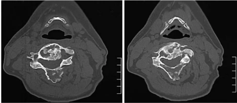
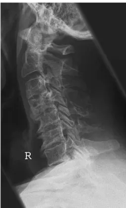
Radiography of the spine showed a lytic and cystic destruction with diVuse expansion of the vertebral body and the spinous process of the fourth cervical ver-tebra (Fig.1). CT scans and MR imaging conWrmed the
above-mentioned Wndings with no cortical destruction D. Proschek · R. Orler · P. Heini (&)
Department of Orthopaedic Surgery, Spine Service, Inselspital, University of Bern, 3010 Bern, Switzerland e-mail: [email protected]
E. StauVer
Department of Pathology, Inselspital, University of Bern, Bern, Switzerland
(Fig.2). Radiological Wndings suggested diVerential
diagnosis including bone cysts, Wbrous dysplasia, osteo-blastoma, histiocytic Wbroma, low-grade Wbrosarcoma, enchondroma, metastases and chronic infection (Figs.1, 2, 3).
Based on the radiological Wndings a biopsy of the spinous process was performed and conWrmed histo-pathologically as Wbrous dysplasia. Details showed a stroma, being rich in plump Wbroblasts with formations of osteoid substance not assuming the aspects of osteo-blasts. Bone trabeculae presented in a uniform and woven structure with no atypic cells or mitotic hyperac-tivity (Fig.4).
logical Wndings were those of Wbrous dysplasia. Six months later, clinical examination again showed no abnormal status. The patient reported about only minor pain and radiological Wndings revealed a consol-idation without progression of the Wbrous dysplasia (Figs.5, 6).
Due to minor symptoms no surgical treatment was performed or is planned unless in case of increasing pain, an acute instability or neurological symptoms.
Fig. 1 Lateral view of the cervical spine, taken xy weeks after on-set of clinical symptoms. It shows an expansion and cystic degen-eration of the fourth cervical vertebra. The severe degenerative changes only provoke minor symptoms
Fig. 2 Axial computed tomography of the fourth cer-vical vertebra. A diVuse expansion as well as lytic and cystic degeneration with no cortical destruction is seen in the vertebral body, the pedi-cles, the lamina and the spi-nous process. No extraosseus/ soft-tissue transformations are spotted
Fig. 3 Skeletal scintigraphy. Note the hot spots in the cervical part of the spine
Discussion
Fibrous dysplasia of the skeleton, congenital and simi-lar to hamartomas, is an intraooseus neoformation of a Wbrous tissue, which may be monostotic or polyostotic [3, 6, 9, 10]. Medullary components are replaced by Wbroplastic tissue. Skeletal lesions may be associated with skin pigmentation spots. Together with early skel-etal maturity and (in females) precocious puberty, this association is known as Albright’s syndrome [1, 11]. Other associations may be hyperthyroidism, diabetes mellitus, renal and cardiovascular anomalies. In agree-ment with its congenital nature, Wbrous dysplasia begins during early childhood and is therefore rela-tively often recognised in younger age [3, 9, 10]. It accounts for about 5–7% of benign bone tumours. No
bone is immune to Wbrous dysplasia. There is a predi-lection for the femur, tibia, maxillary bone, humerus, ribs and iliac bone [3]. After cessation of bone growth, Wbrous dysplasia usually tends to be exhausted. A pro-gression after having achieved adult age is a rare occur-rence. However, passive progress is possible due to continuing degenerative changes in bone. The insur-gence of a sarcoma on an area of Wbrous dysplasia is a rare occurrence, and seems to be less than 1% of all cases [3, 8, 18]. Vertebral involvement in Wbrous
dys-plasia is extremely rare, especially in monostotic vari-ants of the cervical spine [2–4, 6, 14]. Table1 gives a summary of the cases of Wbrous dysplasia of the cervi-cal spine found in literature. The 11 cases being reported separate into seven monostotic and four poly-ostotic variants.
The vertebral body is aVected most frequently. However, the vertebral body has, in comparison to the other parts of the vertebra, the biggest amount of can-cellous bone. Due to their close relationship to the ver-tebral body, involvement of the pedicles has been described in nearly all cases of body aVection [5, 7, 12,
13, 15–17, 19–22]. It could therefore be a possibility that Wbrous dysplasia in the spine starts in the vertebral Fig. 4 Histological imaging of the biopsy taken from the fourth
cervical vertebra. Bone trabeculae presented in a uniform and woven structure with many plump Wbroblasts, few mitoses and no osteoblasts
Fig. 5 Lateral view of the cervical region after 6 months still showing expansion and cystic degeneration of the fourth cervical vertebra. In comparison to the earlier radiological Wndings (Fig.1) an organisation and consolidation of the Wbrous dysplasia can be seen. The degenerative changes still provoke only minor symptoms
body and then continuously expands, via the pedicles, into the vertebral lamina and spinous process.
In the reported cases of Wbrous dysplasia of the spine there is an expansion in nearly all parts of the
when only conventional X-rays were available for diag-nosis. Therefore, it seems that Wbrous dysplasia of the cervical spine does not aVect only one part but the whole vertebra.
Constellation in the case presented here is the same with an aVection of the vertebral body, the pedicles, the lamina and the spinous process of the fourth cervi-cal vertebra.
Fibrous dysplasia usually starts in early childhood. After puberty, dysplastic areas rarely expand. In gen-eral, the progression of the skeletal lesions tends to be exhausted once adult age has been achieved. There-fore, cases with older patients getting symptomatic, especially those involving the spine, are rare. Only few cases of new foci of Wbrous dysplasia in adult have been described [3]. It still remains unclear whether those new foci are de novo lesions or based upon secondary, degenerative changes in bones. However, adult patients usually are not getting symptomatic due to the Wbrous dysplasia but the degenerative changes of the aVected motion segment. Respecting the age of our patient, he is the oldest reported with having Wbrous dysplasia of the cervical spine.
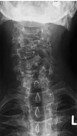
Diagnosis is diYcult in adult patients. Main diVeren-tial diagnosis includes metastatic lesions, bone cysts, enchondroma, histiocytic Wbroma and malignant pri-mary bone tumours. Clinical and radiological Wndings are often not speciWc for Wbrous dysplasia. Tomogra-phy is important but cannot replace histopathology. The histological examination is essential. Stroma shows Wbrous tissue with plump Wbroblasts and forma-Fig. 6 An a.p. view of the cervical region after 6 months. C4 has
collapsed on the left-hand side to a certain extend. (*) Compare Figs.1 and 5
Table 1 Reported cases of Wbrous dysplasia of the cervical spine
Cases Age Location Symptoms Treatment Outcome
Resnik et al. [17] 27 C6 (compl. vertebra) Acute pain Biopsy NA GarWn et al. [22] NA C7 (body + pedicle) Pain for years Arthrodesis
(C6–Th1)
Full recovery
Wright et al. [21] NA C2 + C5 (body and spinous process)
NA NA NA
Stirrat et al. [20] 25 C2 (compl. vertebra) Pain Iliac bone craft (arthrodesis)
Asymptomatic for 2 years Smith et al. [19] 47 C4–C6 (ant. + post. elements) Pain for months Partial corpectomy
C4–C6
Died postop. (bronchopneumonia) Hu et al. [7] 41 C2 (compl. vertebra) Mild pain Arthrodesis C1–C3 Asymptomatic
for 2.5 years Ohki et al. [15] 20 C2 (spinous process) No pain, local expansion
of the tumour mass
Local excisions Asymptomatic for 5 years Nishiura et al. [13] 37 C1–C3 (compl. vertebra) Pain, atlanto-axial
dislocation
Ventral + dorsal stabilisation
Asymptomatic for 1 year
Ehara et al. [5] 19 C1 (lateral mass) Asymptomatic NA NA
Mezzadri et al. [12] 35 C5 (body + pedicle) Spontaneous pain C5-corpectomy Asymptomatic for 3 years Perlick et al. [16] 55 C1 + C2 (spinous process,
body, pedicle)
Chronic pain Dorsal stabilisation C1 + C2
Asymptomatic for months
tions of osteoid substance not assuming the aspects of osteoblasts. Bone trabeculae are presented in a uni-form and woven structure with no atypic cells or mitotic hyperactivity. These Wndings are typical and together with the examination allow Wnding the exact diagnosis. DiYculties sometimes arise in diVerentiating Wbrous dysplasia from low-grade Wbrosarcoma and low-grade osteosarcoma [3, 18]. Histopathologically, these low-grade tumours show more and especially atypic mitoses, and furthermore atypic Wbro- and osteoblasts. Biopsy is unabdicable to isolate the above-mentioned diVerential diagnosis and clear the way for exact diagnosis of Wbrous dysplasia. Meanwhile genetic assessment allows for a deWnitive diagnosis by checking for the GS-alpha mutation.
Whether treatment of Wbrous dysplasia in cervical spine is necessary is controversial [5, 7, 12, 13, 15–17,
19–22]. If there is a mechanical reason for the pain or even a compromise of neurological structures then a surgical intervention is indicated. DiVerent ways are described, such as arthrodesis, laminectomy, corpec-tomy and local excisions. Due to diVerent expansion of Wbrous dysplasia there is no gold standard in operative treatment existent up to now. Each treatment should be oriented towards symptoms. A non-operative treat-ment can be followed as long as there are only minor complaints and no compromise of neurological struc-tures.
The initial symptoms of our patient showed signs of instability. After 3 months, the vertebral body col-lapsed to a certain extent and regained stability (Fig.6). After 9 months, the patient showed a consoli-dation in the clinical and radiological Wndings of the cervical vertebra. With having only minor complaints and no compromise of neurological structures opera-tive treatment is not planned and not indicated at the moment.
References
1. Albright F, Butler AM, Hampton AO (1937) Syndrome char-acterized by osteitis Wbrosa disseminata, areas of pigmenta-tion and endocrine dysfuncpigmenta-tion. N Engl J Med 216:727–746
2. Avimadje AM, Goupille P, Zerback D, Begnard D (2000) Monostotic Wbrous dysplasia of the lumbar spine. Joint Bone Spine 67(1):65–70
3. Campanacci M Bone and soft tissue tumors. Aulo Gaggi Edi-tore Bologna and Springer, Berlin Heidelberg New York 4. Chow LTC, GriYth J, Chow WH (2000) Monostotic Wbrous
dysplasia of the spine: report of a case involving the lumbar transverse process and review of literature. Arch Orthop Trauma Surg 120:460–464
5. Ehara S, Kattapuram SV, Rosenberg AE (1992) Fibrous dys-plasia of the spine. Spine 17:977–979
6. Henry A (1969) Monostotic Wbrous dysplasia. J Bone Joint Surg 51B:300–306
7. Hu SS, Healey JH, Huvos AG (1990) Fibrous dysplasia of the second cervical vertebra. J Bone Joint Surg 72-A(5):781–783 8. Huvos AG, Higinbotham NL, Miller TR (1972) Bone
sarco-mas arising in Wbrous dysplasia. J Bone Joint Surg Am 54:1047–1056
9. Lichtenstein L (1938) Polyostotic Wbrous dysplasia. Arch Surg 36:874–898
10. Lichtenstein L, JaVe HL (1942) Fibrous dysplasia of the bone: a condition aVecting one, several or many bones, the graver cases of which may present abnormal pigmentation of skin, hyperthyroidism or still other skeletal abnormalities. Arch Pathol 33:777–816
11. McCune DJ, Bruch H (1937) Osteodystrophia Wbrosa. Am J Dis Child 54:806–848
12. Mezzadri JJ, Acotto CG, Mautalen C (1999) Surgical treat-ment of cervical spine Wbrous dysplasia: technical case report and review. Neurosurgery 44(6):1342–1349
13. Nishiura I, Koyama T, Takayama S (1992) Fibrous dysplasia of the cervical spine with atlanto-axial dislocation. Neuro-chirurgia 35(4):123–126
14. Oba M, Nakagami W, Maeda M, Kobayashi K (1998) Symp-tomatic monostotic Wbrous dysplasia of the thoracic spine. Spine 23(6):741–743
15. Ohki I (1990) Monostotic Wbrous dysplasia in the spine. J West Pacif Orthop Assoc 27:107–110
16. Perlick L, Rolf V, Wallny T, Schmitt O (2000) Die atlantoax-iale Instabilität als seltene Komplikation der Wbrösen Dyspla-sie. Unfallchirurg 103:73–75
17. Resnik CS, Liniger JR (1984) Monostotic Wbrous dysplasia of the cervical spine: case report. Radiology 151(1):49–50 18. Schajowicz F (1994) Tumor like lesions of the bone. Springer,
Berlin Heidelberg New York, pp 567–581
19. Smith MD, Bohlman HH, Gideonse N (1990) Fibrous dyspla-sia of the cervical spine: a fatal complication of treatment. J Bone Joint Surg 72-A(8):1254–1258
20. Stirrat AN, Fisher CJ (1989) Fibrous dysplasia of the axis. Spine 14(2):243–245
21. Wright JF, Stoker DJ (1988) Fibrous dysplasia of the spine. Clin Radiol 39:523–527
22. GarWn WR, Rothman RH (1986) Case report 346. Skeletal Radiol 15:72–76