Christian W. A. Pfirrmann Bernhard Jost Christof Pirkl Gernot Aitzetmüller Georg Lajtai Received: 23 September 2007 Revised: 4 January 2008 Accepted: 7 February 2008 Published online: 2 April 2008
# European Society of Radiology 2008
Quadriceps tendinosis and patellar tendinosis
in professional beach volleyball players:
sonographic findings in correlation with clinical
symptoms
Abstract The purpose was to assess quadriceps and patellar tendinosis in professional beach volleyball players and to correlate ultrasound findings with clinical symptoms. During a grand-slam beach volleyball tourna-ments all 202 athletes (100 men and 102 women) were invited to partici-pate at this study. Sixty-one athletes (38 male, mean age 29.6, 23 female, mean age 27.1) were included. The dominant leg was right in 51 (84%) and left in ten athletes (16%). Lysholm knee score and pain during the game was assessed using a visual analogue scale. Sonography of the quadriceps tendon and the patellar tendon was performed by a blinded sonographer. Sonographic findings were compared between both legs and correlated to clinical findings using a regression analysis. Quadriceps tendinosis was diagnosed in 13 (21%, dominant leg)/ 21 (34%, non-dominant leg), patellar tendinosis in 13(21%)/18(30%). Only sonographic findings at the quadriceps tendon were significantly associated with pain: thickness of the quadriceps
tendon (mean diameter
6.9 mm/7.1 mm, significant for both legs P=0.011/P=0.030), abnormal echo texture (11/16; P=0.001/ P=0.228), areas with positive power Doppler signals (mean number 0.3/0.4; P=0.049/0.346), calcifica-tions (mean number: 0.9/1.1; P=0.021/0.864). A relationship be-tween findings at patellar tendon was not found. Quadriceps tendinosis is as common as patellar tendinosis in pro-fessional beach volleyball players. Thickening and structure alteration of the quadriceps tendon is associated with anterior knee pain during beach volleyball.
Keywords Knee . Sports injury . Overuse injury . Ligament . Tendon . Tendinosis
Introduction
Beach volleyball is an increasingly popular sport and has been recognized as an Olympic sport since 1996. Overuse injuries in professional beach volleyball players represent an important source of disability and impaired performance [1]. The most affected body regions are the lower back, knee, and shoulder [1]. The knee is among the most
frequent sites of overuse injuries in volleyball players, affecting more than 40% of high level volleyball players [2]. Repetitive jumping is typical in beach volleyball and the extensor apparatus of the knee is subject to continuous repetitive stress. The jumper’s knee or patellar tendinosis is a well-known condition in athletes with high stress on the leg extensors [3,4]. However, not only the patellar tendon but also the quadriceps tendon is subject to the same forces. C. W. A. Pfirrmann . B. Jost . C. Pirkl .
G. Aitzetmüller . G. Lajtai Humanomed Center Althofen, Altis Center for Sports Surgery, Moorweg 30,
9330 Althofen, Austria
C. W. A. Pfirrmann (*) . B. Jost University Hospital Balgrist, Forchstrasse 340, 8008 Zurich, Switzerland e-mail: Christian@pfirrmann.ch Tel.: +41-44-3861111 Fax: +41-44-3863319 C. Pirkl Klinikum Wels, Grieskirchnerstraβe 42, 4600 Wels, Austria G. Aitzetmüller Diakonissen-Krankenhaus Linz, Weissenwolffstraβe 15, 4020 Linz, Austria
In our own experience, volleyball players often reported pain not only at the distal pole of the patella but also at the proximal pole of the patella and at the distal quadriceps tendon. There is only limited knowledge about overuse injuries of the quadriceps tendon in beach volleyball athletes.
The purpose of the study was to assess the frequency of quadriceps tendinosis and patellar tendinosis in profes-sional beach volleyball players, comparing the dominant leg and the contralateral leg, and to correlate ultrasound findings with clinical symptoms.
Materials and methods
Data acquisition was performed during one of the grand-slam beach volleyball tournaments of the FIVB (Federation Internationale de Volleyball) world tour in Klagenfurt. All players were informed about the study during the technical meeting, which all teams were required to attend. They were invited to participate voluntarily at the study during the tournament. All examinations were performed on-site during the tournament in the medical treatment area. All players gave their informed consent. The study was approved by the local ethics committee. Only professional beach volleyball players participating actively at the tournament were included. The main draw of the tourna-ment consisted of 32 women’s teams and 32 men’s teams. Additionally, 19 women’s teams and 18 men’s teams participated at the qualifications. This results in a total of 202 athletes (100 men and 102 women). Sixty-one athletes (30%) were included in the study, no athlete was excluded.
Clinical assessment
All players were examined by one of four experienced orthopedic surgeons. A standardized knee score (Lysholm knee score) [5] was assessed for both knees. The maximum value of the Lysholm score is 100 points: 100–96 is an excellent score, 95–84 is a good score, 83–65 is s a fair score and <65 is a poor score. Peripatellar pain while playing beach volleyball was rated by the player on a visual analogue scale (from “0” indicating no pain to “10” indicating worst pain imaginable) separately for both legs.
Sonographic assessment
Each athlete underwent a standardized examination for both knees. The sonographer was blinded to the symptoms and to which side is the dominant leg. To further reduce a possible bias of the sonographer 15 volunteers were randomly sent to the sonographic assessment additionally. These volunteers were recruited from the staff of the tournament but were not active beach volleyball players.
The outfit of the volunteers was similar to that of the player group. The purpose of these additional examinations was to reduce a possible bias of the sonographer only. These additional examinations were not included in the data analysis.
Sonographic assessment was performed with a linear transducer (Toshiba Nemio 35, 8–14 MHz linear transduc-er, set to 12 MHz): length of field of view: 3 cm; depth: 4 cm; two foci were used. The examination was performed in a standardized fashion for both knees. The knee was positioned in a moderate flexion of about 45 degrees [6]. For the quadriceps tendon three sagittal images (patellar insertion, mid-tendon and musculo-tendinous junction), transverse images, as well as a power Doppler images in the same planes were obtained. For the patellar tendon three sagittal images (patellar attachment, mid-tendon, tibial attachment) transverse images and power Doppler images in the same planes were obtained.
Following criteria were assessed for the quadriceps and the patellar tendon in both knees: echo texture of the fibre structure was rated as “normal”, defined by well visible fibrillar architecture, or as “abnormal” with loss the visibility of fibre texture. Hypoechoic foci within the tendon substance were recorded as“present” or “absent”. A tendinosis was diagnosed based on the loss of the fibrillar architecture and/or the presence of hypoechoic foci within the tendon. The number of calcifications was counted. A calcification was defined as a hyperechoic area within the tendon substance with an associated acoustic shadowing. The number of independent areas with positive power-Doppler signals within the tendon was counted. The antero-posterior diameter of the quadriceps tendon and the patellar tendon was measured at the attachment of the patella.
Statistics
All data analysis was performed using statistical software (SPSS, version 11; SPSS, Chicago, Ill.). P values less than 0.05 were considered statistically significant.
The Wilcoxon signed-ranks test was used to test if continuous and ordinal variables have the same distribution between the dominant and the non-dominant leg. For nominal variables the Fisher’s exact test was used.
Multiple linear regression was performed to estimate which of the sonographic variables (independent variables) best predict the amount of pain during the game (dependent variable). The multiple regression analysis was preformed for both the dominant and the non-dominant leg separately. The threshold of significance, which was the criterion for entry into the model, was set at .05.
The Mann-Whitney U-test was used to compare contin-uous data between tendons with tendinosis and tendons without tendinosis.
Results
Demographic data of athletes
Sixty-one athletes (38 male athletes, mean age 29.6 years, range 20–36 years; 23 female athletes, mean age 27.1 years, range 20–39) were included in the study. The dominant leg was right in 51 athletes (84%) and left in ten athletes (16%).
Clinical and sonographic findings in the dominant leg and the non-dominant leg
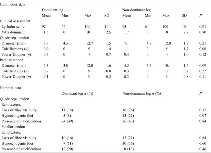
Clinical and sonographic findings of the dominant leg and the non-dominant leg are listed in Table 1. Examples of sonographic findings are shown in Figs. 1and 2. Quad-riceps tendinosis was diagnosed in 13 athletes (21%) at the
dominant leg and in 21 athletes (34%) at the non-dominant leg. Patellar tendinosis was diagnosed in 13 athletes (21%) at the dominant leg and in 18 athletes (30%) at the non-dominant leg. Differences between the non-dominant and the non-dominant leg were not significant (Table1).
Multiple regression analysis
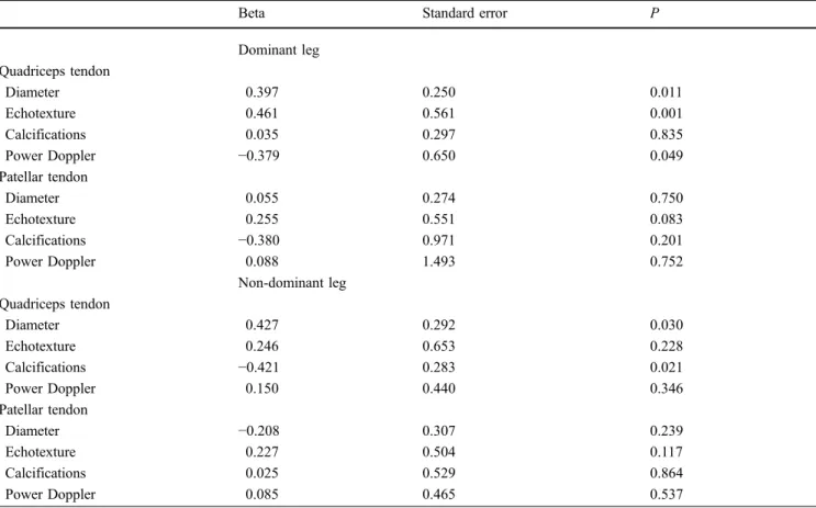
Only sonographic findings of the quadriceps tendon significantly predicted peripatellar pain during sports: thickness of the quadriceps tendon (significant for both legs, P=0.011/P=0.030), abnormal echo texture (domi-nant leg P=0.001), power-Doppler signal (domi(domi-nant leg: P=0.049), calcifications (non-dominant leg, P= 0.021) (Table2). We were not able to demonstrate a relationship between findings at the patellar tendon and symptoms.
Table 1 Clinical and sonographic findings of the dominant leg and the non-dominant leg Continuous data
Dominant leg Non-dominant leg
Mean Min Max SD Mean Min Max SD Pa
Clinical assessment Lysholm score 92 64 100 11 93 64 100 10 0.91 VAS dominant 1.5 0 10 2.5 1.7 0 10 2.7 0.80 Quadriceps tendon Diameter (mm) 6.9 4.5 12.7 1.5 7.1 4.7 12.8 1.8 0.21 Calcifications (n) 0.9 0 5 1.4 1.1 0 5 1.7 0.06 Power Doppler (n) 0.3 0 4 0.7 0.4 0 4 1.0 0.12 Patellar tendon Diameter (mm) 5.3 3.0 12.0 1.6 5.5 3.2 10.1 1.5 0.09 Calcifications (n) 0.2 0 5 0.8 0.3 0 3 0.7 0.22 Power Doppler (n) 0.1 0 3 0.5 0.3 0 3 0.8 0.11 Nominal data
Dominant leg n (%) Non-dominant leg n (%) Pb
Quadriceps tendon Echotexture:
Loss of fibre visibility 11 (18) 16 (26) 0.12
Hypoechogenic foci 5 (8) 13 (21) 0.07
Presence of calcifications 24 (39) 26 (43) 0.84
Patellar tendon Echotexture:
Loss of fibre visibility 10 (16) 13 (21) 0.64
Hypoechogenic foci 7 (11) 10 (16) 0.60
Presence of calcifications 12 (20) 8 (13) 0.46
a
Wilcoxon signed-rank test
Tendon diameter
Mean quadriceps tendon thickness with tendinosis was 8.4 mm/8.5 mm (dominant leg/non-dominant leg), in normal tendons 6.4 mm/6.4 mm (P<0.001/P<0.001). Mean patellar tendon thickness with tendinosis was 7.6 mm/7.0 mm, in normal tendons 4.7 mm/4.8 mm (P< 0.001/P=0.001) (Table3).
Discussion
Early report about jumper’s knee described involvement of the quadriceps and patellar tendon [7, 8]. In most subsequent investigations the term jumper’s knee is used synonymously for the overuse syndrome of the patellar tendon [9–12]. Our data suggest that, unlike in many other jumping sports [13], beach volleyball affects both the patellar and the quadriceps tendon. In our study population, the frequency of patellar tendon and quadriceps tendon involvement was comparable with other previous reports [4, 13–15]. Quadriceps tendinosis and tears have rarely been reported as an overuse syndrome in high-performance athletes [16,17]. This condition seems to be rather typical for beach volleyball athletes.
Calcifications were surprisingly frequent within the quadriceps tendon of our high performance volleyball players. Frequency of calcifications within the patellar tendon was considerably lower. The number of calcifica-tions within the quadriceps tendon was also one of the significant factors predicting the amount of peripatellar pain during the game in the non-dominant legs. Calcifica-tion is not a common reacCalcifica-tion of a tendon to chronic repetitive stress. Calcification is also not a common phenomenon of tendon degeneration or tendon tearing. Calcifications within the quadriceps tendons have been reported in renal failure and secondary hyperparathyroid-ism [18] and occasionally within a soft-tissue mass with calcification representing the retracted quadriceps tendon in cases with complete quadriceps tendon tears [19]. Calcifications may be the results of a chronic inflammatory process as reaction to chronic repetitive tendon injury. Calcifications are a remarkable feature of quadriceps tendon overuse in beach volleyball athletes.
Although expected, dominant leg versus non-dominant leg did not have an influence on the frequency of tendon overuse syndromes. Both the clinical assessment and the sonographic evaluation revealed symmetric distribution. One possible reason that training is focused on both sides equally.
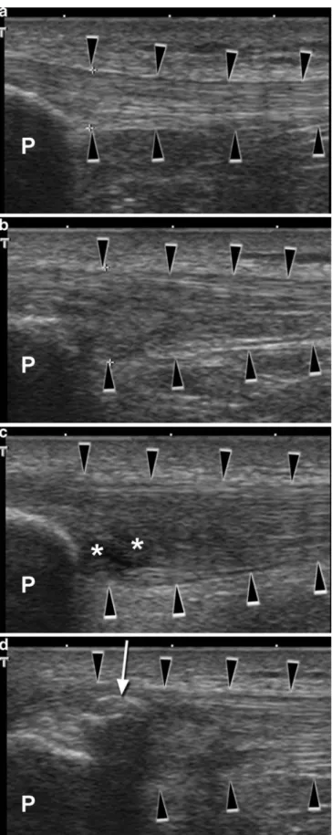
Fig. 1 Sagittal sonograms of quadriceps tendons (black arrow-heads): (a) normal, (b) thickening of the tendon and loss of fibre structure, (c) hypoechogenic foci (asterisk), (a) calcification with acoustic shadowing. (P patella)
Aagaard and Jorgensen [20] reported an overall inci-dence of 3.8 injuries per player per 1,000 volleyball hours played. In their comprehensive assessment, Bahr and Reeser [1] were able to assess 95% of all beach volleyball players participating at five consecutive tournaments similar to the tournament of our study. They reported 54 acute injuries. Injuries to the knee (30%), ankle (17%), and finger injuries (17%) were most common. Sixty-seven players reported 79 overuse injuries. The three most common affected regions were the low back (19%), the knee (12%), and the shoulder (10%). In our study population, the frequency of quadriceps tendinosis (21– 34%), the frequency of patellar tendinosis was (21–30%) considerably higher. In our study we were able to examined about one-third of the players of the tournament. Symp-tomatic players were probably more likely to participate at our study; therefore, some selection bias has probably been introduced, explaining the higher frequency in our data.
The normal quadriceps tendon has a hyperechoic, multi-laminar, structure at sonography [6]. The multi-laminar sonographic appearance is explained by the presence of two to four tendon layers. Most commonly three layers are present. The superficial layer originates from the posterior fascia of the rectus femoris muscle. The deep layer originated from the anterior fascia of the vastus intermedius muscle. The middle layer arises from the fascia between the vastus intermedius and the vastus lateralis and vastus medialis [21]. Sonography has been reported as a sensitive and specific tool in the evaluation of quadriceps tendon rupture [6, 22]. In a study using MR arthrography, the thickness of a normal quadriceps tendon measured between 7 and 8 mm [23], In our study using ultrasound, the mean thickness of normal tendons was lower (6.4 mm).
Ultrasound is recommended as the initial investigation in the assessment of patients with overuse syndrome of the patellar tendon [24]. The normal sonographic appearance shows a hyperechoic regular band with well-defined borders and homogeneous internal fibrillar echo texture. Contrarily to the quadriceps tendon, no internal tendon laminae are present [25]. The normal patellar tendon should not exceed 7 mm [26]. The mean diameter of the tendons with tendinosis was between 7 and 7.6 mm in our study.
Normal tendons do not show power-Doppler signals within the tendon substance. Positive power-Doppler signals indicate the presence of neovascularization in abnormal patellar tendons, which is associated with greater tendon pain compared with abnormal tendons without neovascularization [27, 28]. Positive power-Doppler sig-nals were also significant predictors for pain in the dominant leg in our data.
Fig. 2 Sagittal sonograms of patellar tendons (black arrowheads): (a) normal, (b) loss of fibre structure, (c) hypoechogenic foci (asterisk), (d) calcification with acoustic shadowing. (P patella) 3
In a study assessing the relationship between symp-toms of jumper’s knee and the ultrasound characteristics of the patellar tendon among high-level male volleyball players, Lian and co-workers did not find specific ultrasound findings correlating significantly with the degree or the duration of symptoms [29]. This is in line with the results of our analysis. Ultrasound findings about the patellar tendon did not significantly predict peripatellar pain during the beach volleyball game. However, findings in the quadriceps tendon, especially
thickening of the tendon, did significantly predict symptoms.
Several study limitations have to be considered. There is a possible selection bias as symptomatic players were probably more likely to participate in our study. We were not able to find an association between findings at the patellar tendon and symptoms, which may be because of the relatively low sample size. We tried to minimize any possible bias of the sonographer. The sonographer was blinded to the symptoms of the athletes and volunteers Table 2 Multiple regression to estimate which of the sonographic variables (independent variables) best predict the amount of peripatellar pain during the game (dependent variable)
Beta Standard error P
Dominant leg Quadriceps tendon Diameter 0.397 0.250 0.011 Echotexture 0.461 0.561 0.001 Calcifications 0.035 0.297 0.835 Power Doppler −0.379 0.650 0.049 Patellar tendon Diameter 0.055 0.274 0.750 Echotexture 0.255 0.551 0.083 Calcifications −0.380 0.971 0.201 Power Doppler 0.088 1.493 0.752 Non-dominant leg Quadriceps tendon Diameter 0.427 0.292 0.030 Echotexture 0.246 0.653 0.228 Calcifications −0.421 0.283 0.021 Power Doppler 0.150 0.440 0.346 Patellar tendon Diameter −0.208 0.307 0.239 Echotexture 0.227 0.504 0.117 Calcifications 0.025 0.529 0.864 Power Doppler 0.085 0.465 0.537
Table 3 Comparison of the tendon diameter between normal tendons and tendons with tendinosis
Normal Tendinosis n Mean SD n Mean SD Pa (mm) (mm) (mm) (mm) Quadriceps tendon Dominant leg 48 6.4 0.9 13 8.4 2.1 <0.001 Non-dominant leg 40 6.4 1.0 21 8.5 2.0 <0.001 Patellar tendon Dominant leg 48 4.7 0.8 13 7.6 1.7 <0.001 Non-dominant leg 43 4.8 0.8 18 7.0 1.7 0.001 a Mann-Whitney U-test
were randomly mingled to the study population. Never-theless there might be some bias introduced by the direct interaction of the sonographer and the athlete during the examination. In our study, the knee was positioned in a moderate flexion of about 45 degrees for the sonographic evaluation. The flexed position of the knee may lead to tension within the tendons and therefore to a lower sensitivity to detect neovascularity using power-Doppler sonography.
In conclusion, quadriceps tendinosis is as common as patellar tendinosis in professional beach volleyball players. Quadriceps tendinosis appears to be clinically more
relevant than patellar tendinosis. Thickening and structure alteration of the quadriceps tendon is associated with anterior knee pain during elite beach volleyball.
Acknowledgements This work was made possible by the support of the Humanomed Center Althofen, Altis Center for Sports Surgery, Althofen, Austria. The authors would also like to thank Sabina Vogel Mag, Sylvia Schöffmann MD, Julia Sostaric MD, Daniel Leiner, Peter Hoffmann - Bachinger Hoffmann, Reinhard Lischka and Hannes Jagerhofer– Acts, for their help and commitment to make this work possible.
References
1. Bahr R, Reeser JC (2003) Injuries among world-class professional beach volleyball players. The Federation In-ternationale de Volleyball beach vol-leyball injury study. Am J Sports Med 31:119–125
2. Ferretti A, Papandrea P, Conteduca F (1990) Knee injuries in volleyball. Sports Med 10:132–138
3. Khan KM, Bonar F, Desmond PM et al (1996) Patellar tendinosis (jumper’s knee): findings at histopathologic ex-amination, US, and MR imaging. Victorian Institute of Sport Tendon Study Group. Radiology 200:821–827 4. Lian OB, Engebretsen L, Bahr R
(2005) Prevalence of jumper’s knee among elite athletes from different sports: a cross-sectional study. Am J Sports Med 33:561–567
5. Lysholm J, Gillquist J (1982) Evalua-tion of knee ligament surgery results with special emphasis on use of a scoring scale. Am J Sports Med 10:150–154
6. Bianchi S, Zwass A, Abdelwahab IF, Banderali A (1994) Diagnosis of tears of the quadriceps tendon of the knee: value of sonography. AJR Am J Roentgenol 162:1137–1140 7. Blazina ME, Kerlan RK, Jobe FW,
Carter VS, Carlson GJ (1973) Jumper’s knee. Orthop Clin North Am 4:665– 678
8. Ferretti A, Ippolito E, Mariani P, Puddu G (1983) Jumper’s knee. Am J Sports Med 11:58–62
9. Kalebo P, Sward L, Karlsson J, Peterson L (1991) Ultrasonography in the detection of partial patellar ligament ruptures (jumper’s knee). Skeletal Radiol 20:285–289
10. Myllymaki T, Bondestam S, Suramo I, Cederberg A, Peltokallio P (1990) Ultrasonography of jumper’s knee. Acta Radiol 31:147–149
11. Schmid MR, Hodler J, Cathrein P, Duewell S, Jacob HA, Romero J (2002) Is impingement the cause of jumper’s knee? Dynamic and static magnetic resonance imaging of patellar tendinitis in an open-configuration system. Am J Sports Med 30:388–395
12. Yu JS, Popp JE, Kaeding CC, Lucas J (1995) Correlation of MR imaging and pathologic findings in athletes under-going surgery for chronic patellar ten-dinitis. AJR Am J Roentgenol 165:115–118
13. Ferretti A (1986) Epidemiology of jumper’s knee. Sports Med 3:289–295 14. Kujala UM, Kvist M, Osterman K
(1986) Knee injuries in athletes. Re-view of exertion injuries and retro-spective study of outpatient sports clinic material. Sports Med 3:447–460 15. Malliaras P, Cook J, Ptasznik R,
Thomas S (2006) Prospective study of change in patellar tendon abnormality on imaging and pain over a volleyball season. Br J Sports Med 40:272–274 16. Kelly DW, Carter VS, Jobe FW, Kerlan
RK (1984) Patellar and quadriceps tendon ruptures–jumper’s knee. Am J Sports Med 12:375–380
17. Martens M, Wouters P, Burssens A, Mulier JC (1982) Patellar tendinitis: pathology and results of treatment. Acta Orthop Scand 53:445–450 18. Bhole R, Flynn JC, Marbury TC (1985)
Quadriceps tendon ruptures in uremia. Clin Orthop Relat Res 200–206 19. Newberg A, Wales L (1977)
Radio-graphic diagnosis of quadriceps tendon rupture. Radiology 125:367–371 20. Aagaard H, Jorgensen U (1996)
Inju-ries in elite volleyball. Scand J Med Sci Sports 6:228–232
21. Zeiss J, Saddemi SR, Ebraheim NA (1992) MR imaging of the quadriceps tendon: normal layered configuration and its importance in cases of tendon rupture. AJR Am J Roentgenol 159:1031–1034
22. La S, Fessell DP, Femino JE, Jacobson JA, Jamadar D, Hayes C (2003) Sonography of partial-thickness quad-riceps tendon tears with surgical correlation. J Ultrasound Med 22:1323–1329 quiz 1330–1321 23. Staeubli HU, Bollmann C, Kreutz R,
Becker W, Rauschning W (1999) Quantification of intact quadriceps tendon, quadriceps tendon insertion, and suprapatellar fat pad: MR arthro-graphy, anatomy, and cryosections in the sagittal plane. AJR Am J Roent-genol 173:691–698
24. Davies SG, Baudouin CJ, King JB, Perry JD (1991) Ultrasound, computed tomography and magnetic resonance imaging in patellar tendinitis. Clin Radiol 43:52–56
25. Bianchi S, Poletti PA, Martinoli C, Abdelwahab IF (2006) Ultrasound ap-pearance of tendon tears. Part 2: lower extremity and myotendinous tears. Skeletal Radiol 35:63–77
26. El-Khoury GY, Wira RL, Berbaum KS, Pope TL, Monu JU (1992) MR imaging of patellar tendinitis. Radiology 184:849–854
27. Cook JL, Malliaras P, De Luca J, Ptasznik R, Morris ME, Goldie P (2004) Neovascularization and pain in abnormal patellar tendons of active jumping athletes. Clin J Sport Med 14:296–299
28. Zanetti M, Metzdorf A, Kundert HP et al (2003) Achilles tendons: clinical relevance of neovascularization diag-nosed with power Doppler US. Radiology 227:556–560
29. Lian O, Holen KJ, Engebretsen L, Bahr R (1996) Relationship between symp-toms of jumper’s knee and the ultra-sound characteristics of the patellar tendon among high level male volley-ball players. Scand J Med Sci Sports 6:291–296