HAL Id: hal-02388221
https://hal.univ-angers.fr/hal-02388221
Submitted on 2 Dec 2019
HAL is a multi-disciplinary open access
archive for the deposit and dissemination of
sci-entific research documents, whether they are
pub-lished or not. The documents may come from
teaching and research institutions in France or
abroad, or from public or private research centers.
L’archive ouverte pluridisciplinaire HAL, est
destinée au dépôt et à la diffusion de documents
scientifiques de niveau recherche, publiés ou non,
émanant des établissements d’enseignement et de
recherche français ou étrangers, des laboratoires
publics ou privés.
Pathogenicity of Mitochondrial DNA Variants in the
Field of Next Generation Sequencing
Céline Bris, David Goudenège, Valérie Desquiret-Dumas, Majida Charif,
Estelle Colin, Dominique Bonneau, Patrizia Amati-Bonneau, Guy Lenaers,
Pascal Reynier, Vincent Procaccio
To cite this version:
Céline Bris, David Goudenège, Valérie Desquiret-Dumas, Majida Charif, Estelle Colin, et al..
Bioinformatics Tools and Databases to Assess the Pathogenicity of Mitochondrial DNA Variants
in the Field of Next Generation Sequencing.
Frontiers in Genetics, Frontiers, 2018, 9, pp.632.
�10.3389/fgene.2018.00632�. �hal-02388221�
Edited by: Ivana Kurelac, Università degli Studi di Bologna, Italy Reviewed by: Caterina Garone, University of Cambridge, United Kingdom Roberto Preste, Università degli Studi di Bari, Italy *Correspondence: Vincent Procaccio viprocaccio@chu-angers.fr
Specialty section: This article was submitted to Genetic Disorders, a section of the journal Frontiers in Genetics Received: 05 September 2018 Accepted: 27 November 2018 Published: 11 December 2018 Citation: Bris C, Goudenege D, Desquiret-Dumas V, Charif M, Colin E, Bonneau D, Amati-Bonneau P, Lenaers G, Reynier P and Procaccio V (2018) Bioinformatics Tools and Databases to Assess the Pathogenicity of Mitochondrial DNA Variants in the Field of Next Generation Sequencing. Front. Genet. 9:632. doi: 10.3389/fgene.2018.00632
Bioinformatics Tools and Databases
to Assess the Pathogenicity of
Mitochondrial DNA Variants in the
Field of Next Generation Sequencing
Céline Bris
1,2, David Goudenege
1,2, Valérie Desquiret-Dumas
1,2, Majida Charif
1,
Estelle Colin
1,2, Dominique Bonneau
1,2, Patrizia Amati-Bonneau
1,2, Guy Lenaers
1,
Pascal Reynier
1,2and Vincent Procaccio
1,2*
1UMR CNRS 6015-INSERM U1083, MitoVasc Institute, Angers University, Angers, France,2Biochemistry and Genetics
Department, Angers Hospital, Angers, France
The development of next generation sequencing (NGS) has greatly enhanced the
diagnosis of mitochondrial disorders, with a systematic analysis of the whole
mitochondrial DNA (mtDNA) sequence and better detection sensitivity. However,
the exponential growth of sequencing data renders complex the interpretation of
the identified variants, thereby posing new challenges for the molecular diagnosis
of mitochondrial diseases. Indeed, mtDNA sequencing by NGS requires specific
bioinformatics tools and the adaptation of those developed for nuclear DNA, for
the detection and quantification of mtDNA variants from sequence alignment to the
calling steps, in order to manage the specific features of the mitochondrial genome
including heteroplasmy, i.e., coexistence of mutant and wildtype mtDNA copies. The
prioritization of mtDNA variants remains difficult, relying on a limited number of specific
resources: population and clinical databases, and in silico tools providing a prediction
of the variant pathogenicity. An evaluation of the most prominent bioinformatics tools
showed that their ability to predict the pathogenicity was highly variable indicating that
special efforts should be directed at developing new bioinformatics tools dedicated
to the mitochondrial genome. In addition, massive parallel sequencing raised several
issues related to the interpretation of very low mtDNA mutational loads, discovery of
variants of unknown significance, and mutations unrelated to patient phenotype or the
co-occurrence of mtDNA variants. This review provides an overview of the current
strategies and bioinformatics tools for accurate annotation, prioritization and reporting
of mtDNA variations from NGS data, in order to carry out accurate genetic counseling in
individuals with primary mitochondrial diseases.
Keywords: mitochondria, mitochondrial diseases, mitochondrial DNA, next generation sequencing, bioinformatics, mtDNA variant interpretation
KEY POINTS FOR IN SILICO
PRIORITIZATION AND INTERPRETATION
OF mtDNA VARIANTS
• Query dedicated mtDNA databases which are regularly
updated (e.g., Mitomap, HmtDB).
• Consider the variant’s frequency both within the general
population and specific haplogroup.
• Detection and interpretation of low heteroplasmy levels
should be carefully evaluated.
• Integrate
additional information that might modulate
the clinical penetrance (e.g., mitochondrial haplogroup,
synergistic or helper mtDNA variants, nuclear variants).
• Evaluate inter-species and primates amino-acid or nucleotide
conservation.
• Favor in silico prediction tools dedicated to mtDNA (e.g.,
APOGEE, Mitotip, Mtool Box).
INTRODUCTION
The
prevalence
of
mitochondrial
diseases
primarily
affecting oxidative phosphorylation (OXPHOS) is estimated
at about 1 in 4,300 (
Gorman et al., 2015
). As mitochondrial
proteins are encoded both by the nuclear genome and their
own genome (mtDNA), their clinical presentation are highly
heterogeneous. Human mtDNA is a 16,569 bp circular
double-stranded molecule, encoding for 13 polypeptides involved in
the oxidative phosphorylation (OXPHOS), together with 2
ribosomal RNAs and 22 tRNAs supporting the translational
machinery (
Wallace et al., 2010
). Pathogenic variants of the
mitochondrial genome can affect either the protein coding genes
(
Wallace et al., 2013
), tRNAs (
Tang et al., 2013; Gorman et al.,
2015
) and rRNA genes (
Smith et al., 2014; Elson et al., 2015
).
Genetic defects in the mitochondrial genome can be identified in
a variable proportion of patients with mitochondrial respiratory
disorders, reaching up to 20% of patients (
Thorburn et al.,
2004
). Hundreds of pathogenic mtDNA variants implicated in
a variety of human diseases (
Lott et al., 2013
) have now been
reported in the continuously updated Human Mitochondrial
Genome Database—the Mitomap (
Ruiz-Pesini et al., 2007; Lott
et al., 2013
) but as of today (July 2018) only 84 variants have a
confirmed status, whereas a total of 595 other variants classified
as reported, awaiting a final confirmation of pathogenicity
(Figure 1). These mtDNA variants lead to a broad spectrum
of maternally-inherited diseases, ranging from lethal neonatal
syndromes to multisystemic disorders, with high variable clinical
phenotypes and penetrance, mainly resulting from shifts and
differences in the mutant load (
Wallace et al., 2010
). Indeed,
due to stochastic segregation of mtDNA, the percentage of
mutant and normal mtDNAs may drift during cellular divisions,
and the percentage of the mutation load may vary drastically
among the different tissues and organs, from 100% mutant
load, defining homoplasmy, to the coexistence of mutant and
wildtype copies, defining heteroplasmy. As the percentage of
heteroplasmy increases, the energy production declines until
the energy output falls below the minimum necessary for the
physiological maintenance of cellular functions, causing the
appearance of symptoms (
Rossignol et al., 2003
).
Until the development of next generation sequencing (NGS),
molecular diagnosis of mitochondrial disorders was based
on a combination of several techniques, including targeted
Sanger sequencing for the detection of mutations, long-range
polymerase chain reaction (PCR) and Southern blotting, for the
detection of mtDNA rearrangements and depletions, whereas
fluorescent PCR restriction fragment length polymorphism
(RFLP) and pyrosequencing were used for the quantification
of mtDNA variants and rearrangements (
Moraes et al., 2003;
Bannwarth et al., 2005; Wong and Boles, 2005
). These techniques
are still useful as confirmatory and independent tools to ascertain
the presence of a given mtDNA variant identified by NGS.
The development of massive parallel sequencing techniques now
allows the systematic screening of the whole mitochondrial
genome thus increasing the efficacy of the workflow with
increased sample throughput and greater sensitivity in the
detection of mtDNA variants (
Vancampenhout et al., 2014; Ye
et al., 2014; Seneca et al., 2015
). However, massive parallel
sequencing of the whole mitochondrial genome, with the
increasing quantity and complexity of mtDNA data, led to
difficulties to appreciate the variants identified, thereby posing
new challenges in the molecular diagnosis of mitochondrial
diseases. Indeed, several issues related to the interpretation of
very low mutational loads (
Guo et al., 2013
), the discovery of
variants of unknown significance (
van der Walt et al., 2012
),
and mutations unrelated to the patient phenotype, generate
difficulties in prioritizing the variants, and as a consequence
different interpretations of mtDNA variants in the diagnostic
process. This review provides an overview of the current
strategies, databases and bioinformatics tools for an accurate
annotation, prioritization and report of mtDNA variations
coming from NGS, in order to carry out fast and accurate genetic
counseling in patients with primary mitochondrial disease.
mtDNA VARIANT ANNOTATION
Careful mtDNA annotation of mtDNA variants is a prerequisite
for accurate prioritization, addressed by several pipelines and
online tools (Table 1A), like MSeqDR mvTool (
Shen et al., 2018
),
Mitomaster (
Lott et al., 2013
), mtDNA-Server (
Weissensteiner
et al., 2016a
), MitoTool (
Fan and Yao, 2011
), or SG-ADVISER
(
Rueda and Torkamani, 2017
). Then, the general workflow for
mtDNA variants prioritization could parallel that of the nuclear
genome and rely on an accurate standardized annotation, based
on consensus databases and in silico prediction tools.
Population and Clinical Databases
Whereas mtDNA data are available from exome and genome
sequencing data (
Griffin et al., 2014; Patowary et al., 2017
), the
frequency of mtDNA variants in the general population is not
reported in the databases such as GnomAD (
Lek et al., 2016
)
or MARRVEL (
Wang et al., 2017
). The number of dedicated
databases focusing on mtDNA with an active curation is limited
(Table 1B), only three being available online: Mitomap (
Kogelnik
et al., 1996; Lott et al., 2013
), HmtDB (
Clima et al., 2017
) and
FIGURE 1 | Graphical representation of human mitochondrial DNA variations. The outer circle depicts the mitochondrial genome with annotated tRNAs (gray), rRNAs (purple), protein-coding genes (Bentley et al., 2008), and non-coding regions (white). In the inner circles each point represent an mtDNA variant reported in GenBank sequences collected from Mitomap according to the variant status reported in Mitomap (polymorphisms in green, reported pathogenic variants in orange, confirmed pathogenic variants in red) and variant frequency in GenBank (<0.2%, light color; 0.2–2.0%, medium color; >2.0%, dark color).
HmtVar (
Preste et al., 2018
). Together these resources gathered
for instance more than 45,000 whole mtDNA sequences and
over 70,000 mtDNA control region sequences for the Mitomap
database. However, the interpretation of the variant frequency
in the general population is difficult given that databases
include patient data and because of the peculiarities of mtDNA
genetics (incomplete penetrance, heteroplasmy level, influence
of mitochondrial haplogroup background). For example, the
m.3460G>A primary LHON mutation is reported 20 times in
GenBank (
Bentley et al., 2008
) as of July 10, 2018 (Mitomap
database), the pathogenic and unquestionable variant being
reported both in patients, and also from phylogenetic studies.
There is currently no consensus threshold to consider, often
mtDNA variant is frequent in the population, thresholds between
0.2 and 0.5% being arbitrarily chosen in several studies (
Wang
et al., 2012; Lieber, 2013
). However, they do not consider the
variant frequency within a peculiar haplogroup which can lead
to misinterpretation as some haplogroups are underrepresented
in databases. For instance, Asian and African lineages represent
only 21 and 13% of the Mitomap GB dataset. To overcome this
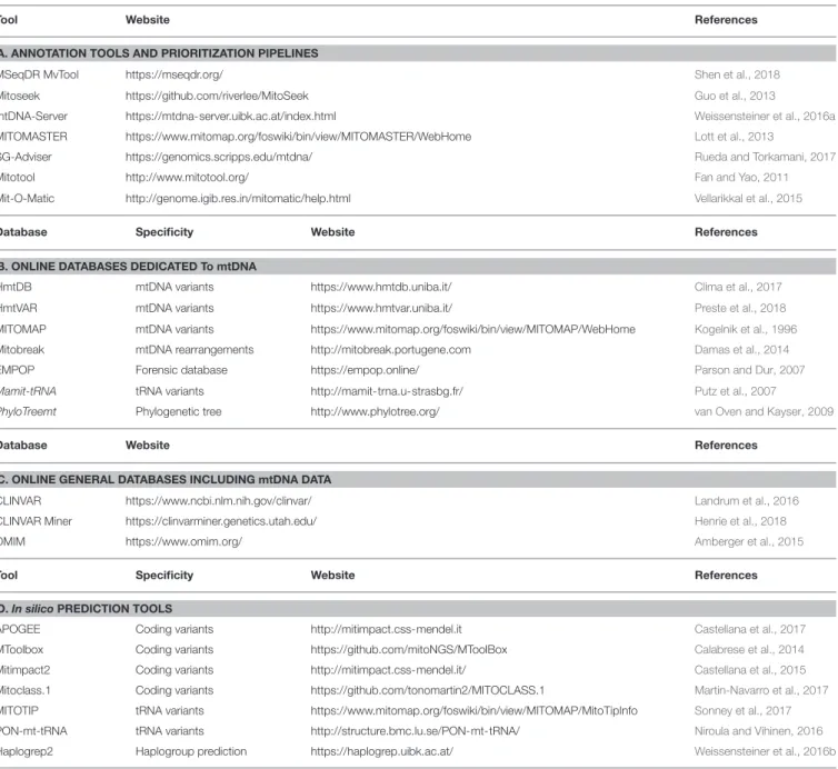
TABLE 1 | Online resources for annotation and prioritization of mtDNA variants.
Tool Website References
A. ANNOTATION TOOLS AND PRIORITIZATION PIPELINES
MSeqDR MvTool https://mseqdr.org/ Shen et al., 2018 Mitoseek https://github.com/riverlee/MitoSeek Guo et al., 2013
mtDNA-Server https://mtdna-server.uibk.ac.at/index.html Weissensteiner et al., 2016a MITOMASTER https://www.mitomap.org/foswiki/bin/view/MITOMASTER/WebHome Lott et al., 2013
SG-Adviser https://genomics.scripps.edu/mtdna/ Rueda and Torkamani, 2017 Mitotool http://www.mitotool.org/ Fan and Yao, 2011 Mit-O-Matic http://genome.igib.res.in/mitomatic/help.html Vellarikkal et al., 2015
Database Specificity Website References
B. ONLINE DATABASES DEDICATED To mtDNA
HmtDB mtDNA variants https://www.hmtdb.uniba.it/ Clima et al., 2017 HmtVAR mtDNA variants https://www.hmtvar.uniba.it/ Preste et al., 2018 MITOMAP mtDNA variants https://www.mitomap.org/foswiki/bin/view/MITOMAP/WebHome Kogelnik et al., 1996 Mitobreak mtDNA rearrangements http://mitobreak.portugene.com Damas et al., 2014 EMPOP Forensic database https://empop.online/ Parson and Dur, 2007 Mamit-tRNA tRNA variants http://mamit-trna.u-strasbg.fr/ Putz et al., 2007
PhyloTreemt Phylogenetic tree http://www.phylotree.org/ van Oven and Kayser, 2009
Database Website References
C. ONLINE GENERAL DATABASES INCLUDING mtDNA DATA
CLINVAR https://www.ncbi.nlm.nih.gov/clinvar/ Landrum et al., 2016 CLINVAR Miner https://clinvarminer.genetics.utah.edu/ Henrie et al., 2018 OMIM https://www.omim.org/ Amberger et al., 2015
Tool Specificity Website References
D. In silico PREDICTION TOOLS
APOGEE Coding variants http://mitimpact.css-mendel.it Castellana et al., 2017 MToolbox Coding variants https://github.com/mitoNGS/MToolBox Calabrese et al., 2014 Mitimpact2 Coding variants http://mitimpact.css-mendel.it/ Castellana et al., 2015 Mitoclass.1 Coding variants https://github.com/tonomartin2/MITOCLASS.1 Martin-Navarro et al., 2017 MITOTIP tRNA variants https://www.mitomap.org/foswiki/bin/view/MITOMAP/MitoTipInfo Sonney et al., 2017 PON-mt-tRNA tRNA variants http://structure.bmc.lu.se/PON-mt-tRNA/ Niroula and Vihinen, 2016 Haplogrep2 Haplogroup prediction https://haplogrep.uibk.ac.at/ Weissensteiner et al., 2016b
problem, the Mitomap database warns if a variant is identified
at >1% in at least one of the macro-lineages or over 10% in the
major haplogroups for tRNA variants (
Sonney et al., 2017
). For
the first step of prioritization, forensic databases such as EMPOP
(
Parson and Dur, 2007
) are useful.
Clinical databases combining mtDNA variants and clinical
files are available (Table 1C). General databases such as
CLINVAR (
Landrum et al., 2016
), CLINVAR Miner (
Henrie
et al., 2018
), or OMIM (
Amberger et al., 2015
) carrying
information on mtDNA in addition to nDNA variants, can be
distinguished from databases specifically dedicated to mtDNA
such as Mitomap, HmtVar or HmtDB which are more exhaustive.
In the latter, more than 10,000 variants are gathered throughout
the whole mtDNA (Figure 1). For example in MT-ND1 gene only
42 variations are reported in CLINVAR, whereas 693 variants
are available in Mitomap (
Lott et al., 2013
), and among them,
62 are reported associated with a clinical phenotype.
MtDNA-specific information, such as heteroplasmy and haplogroup
frequency are not systematically reported in each database, hence
questioning the value of pipelines collecting information from
multiple resources in order to provide a complete description
of the variant. Other specialized databases are also helpful for
variant prioritization: Mamit-tRNA database which compiles
information on mammalian mitochondrial tRNA genes (
Putz
et al., 2007
) or the Mitobreak database focusing on mtDNA
rearrangements (
Damas et al., 2014
).
Even if the prevalence of mitochondrial diseases is in the
order of 1/4,300 (
Gorman et al., 2015
), it was shown that
at least 1 in 200 newborn cord bloods carry one of the
ten most common pathogenic mtDNA mutations, which is
much more frequent than expected (
Elliott et al., 2008
). Many
of these mutations will probably be lost through stochastic
segregation, but there is still a chance that these mutations will
be transmitted to the offspring and causing mtDNA diseases in
next generations. As mtDNA variations can be obtained from
WES (
Griffin et al., 2014
) or WGS data, the identification of
pathogenic variants unrelated to the patient’s phenotype poses
a challenge for the interpretation and report of the variant.
Selected mtDNA confirmed variants, such as LHON (e.g.,
m.11778G>A, m.14484T>C, m.3460T>C or other rare LHON
mutations as classified in the top 19 mutations, see mitomap)
or deafness pathogenic variants such as the m.1555G>A, should
be considered as secondary and actionable findings. As defined
by the American College of Medical Genetics and Genomics
which claimed that “the results of a deliberate search for
pathogenic alterations in genes that are not apparently relevant
to a diagnostic indication for which the sequencing test was
ordered but which may nonetheless be of medical value or
utility to the ordering physician and the patient” (
Green et al.,
2013; Kalia et al., 2017
). As an example, we discovered in
our clinical center the m.1555G>A variant in the MT-RNR1,
responsible for the amino-glycoside-induced and non-syndromic
hearing loss (
Bitner-Glindzicz et al., 2009; Vandebona et al.,
2009
) in a family with isolated optic atrophy but without
hearing impairment. The discovery of a pathogenic variant
should be considered as “actionable,” i.e., pathogenic variants
whose penetrance would result in medical recommendations and
health care prevention. Indeed, the presence of the m.1555G>A
may modify patient management, audiometric follow-up and
hearing protection, prompting appropriate genetic counseling
and recommendations with the avoidance of the use of specific
drugs such as aminoglycosides (
Estivill et al., 1998
). Other
mtDNA variants with unrelated or unclear relation to the clinical
phenotype have been identified in the general population, such as
primary LHON mutations found in patients, but unaccompanied
by any ophthalmological sign (
Inagaki et al., 2006; Yang et al.,
2016
). Incomplete penetrance is a hallmark of optic neuropathies,
starting with LHON, and several parameters such as modifier
genes or environmental factors such as tobacco smoking or the
consumption of alcohol or administration of ethambutol, have
been identified as risk factors for asymptomatic carriers of LHON
mutations (
Yu-Wai-Man et al., 2016; Caporali et al., 2017
).
Thus, some of the mtDNA pathogenic variants, such as LHON
or deafness pathogenic variants should be carefully treated as
“actionable mutations” and call for genetic counseling, especially
to avoid exposure to risk factors and for a clinical follow up.
In silico
Prediction Tools and Pathogenicity
Scores
The systematic mtDNA screening by NGS revealed a large
number of novel variants of unknown significance (
Lieber, 2013;
McCormick et al., 2013
), as exemplified by the identification
of 11 patients among 71 from a pediatric cohort, harboring a
novel variant of unknown significance (VUS) (
van der Walt
et al., 2012
). In our diagnostic laboratory setting, about 6.5%
of the patients carried a VUS. The interpretation of the clinical
significance of mtDNA VUS is more complicated than for the
nuclear VUS, in part due to the mtDNA characteristics, as
heteroplasmy and high mutation rate (
Marcelino and Thilly,
1999
), which are not considered in the classical prioritization
algorithms, and because of the limited guidelines for the mtDNA
compared to those provided by the American College of Medical
Genetics for nuclear VUS (
Richards et al., 2015
). Special efforts
are currently underway with an initiative from an international
group of mitochondrial researchers and clinicians to revise the
ACMG guidelines specifically for mtDNA variants (Procaccio,
personal communication).
In silico prediction tools, which evaluate the functional impact
of variations using approaches based on interspecies sequence
conservation and/or structure analysis, are currently the last step
of variant prioritization. As prediction tools are specific for a
type of variations, we will distinguish those dedicated to coding
regions to those dedicated to other mtDNA regions.
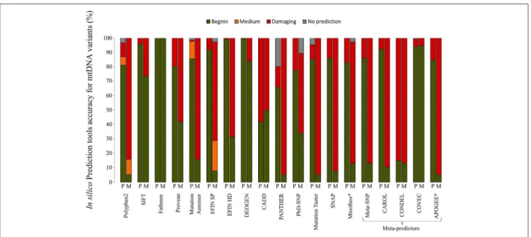
A plethora of in silico bioinformatics prediction tools exists for
the prioritization of nuclear DNA coding variants. A thorough
analysis of the main tools commonly used to evaluate the
pathogenicity of variants demonstrated that the performances
vary drastically when variants of the mtDNA-encoded proteins
were tested. A set of 38 confirmed pathogenic variants (M) and
224 variants considered to be polymorphisms (P) according to
Mitomap, were assessed with a set of 19 different prediction
tools gathered in MitImpact2 (
Castellana et al., 2015
; Figure 2).
Variants of the mitochondrial genome were subdivided into four
categories based on the prediction of pathogenicity, i.e., benign,
medium, damaging, and no prediction. The performances of the
prediction of pathogenic mtDNA variants differed significantly
between the different bioinformatics tools. For instance, more
than 70% of the confirmed pathogenic mutations were predicted
to be benign with SIFT, whereas about 15% of the pathogenic
variants were not predicted as such by Polyphen2, highlighting
that the tools developed for nDNA are barely suitable for
mtDNA. Conversely, recent tools developed for mtDNA using
machine learning based approaches (Table 1D) show better
performances (Figure 2), as MToolBox (
Calabrese et al., 2014
),
the meta-predictor APOGEE (
Castellana et al., 2017
), or
Mitoclass.1 (
Martin-Navarro et al., 2017
), confirming the need
to pursue the development of tools dedicated to mitochondrial
genetics.
Few tools are dedicated to mitochondrial tRNAs, accounting
for nearly 50% of the mtDNA alterations identified in patients
(
Schaefer et al., 2008; Gorman et al., 2015
). PON-mt-RNA
is a multifactorial score associating 12 features including
evolutionary conservation, primary to tertiary structures, and
functional assays including biochemistry and histochemistry
(
Niroula and Vihinen, 2016
). All the precomputed data
are downloadable at http://structure.bmc.lu.se/PON-mt-tRNA/
datasets.html/. MITOTIP, the most recent tool available through
Mitomap combines conservation data, structural analogies with
other tRNA variants and secondary structure information
(
Sonney et al., 2017
), giving the best prediction performances in
terms of sensitivity and specificity. However, specific tools for the
interpretation of VUS in MT-RNR1 and MT-RNR2 combining
FIGURE 2 | Performances of in silico prediction tools for non-synonymous mtDNA variants. A set of 38 confirmed pathogenic variants (M) and 224 non-synonymous variants classified as mtDNA polymorphisms (P) according to Mitomap, were assessed with 19 different prediction tools. Information about the different in silico tools is available at MitImpact2 website (http://mitimpact.css-mendel.it/). Variants were classified into 4 categories: benign (green), medium (orange), damaging (red) and no prediction (hatched) according to the tool prediction. Results are expressed as percentages. *In silico tools developed for mtDNA.
conservational information with functional and structural data
are helpful to better interpretation of ribosomal variants (
Smith
et al., 2014; Elson et al., 2015
).
Thus, many bioinformatics tools are useful for predicting
the functional impact of mtDNA variants, nevertheless results
should still be considered with caution, due to the high rate
of false negative and false positive predictions as demonstrated
in Figure 2. To overcome this problem and improve the
prioritization of mtDNA VUS, several teams have developed
scoring approaches (
McFarland et al., 2004; Mitchell et al., 2006;
Wong, 2007
). These scores which combined algorithms similar
to those of in silico prediction tools (i.e., structure, conservation)
and functional in vivo and in vitro evaluation show better
performances, but their use is still limited, because functional
studies are time-consuming and tissues such as fibroblasts,
cybrids, or muscle samples, are not always available for assessing
the consequences of the variants on mitochondrial physiology.
Given the increasing number of variants of unknown significance
identified by NGS, it would be interesting to regularly re-evaluate
these pathogenicity scores based on new information.
Heteroplasmy Level Interpretation
The
development
of
NGS
techniques
and
dedicated
bioinformatics pipelines (
Calabrese et al., 2014; Weissensteiner
et al., 2016a; Marquis et al., 2017
) has widely improved the
detection of low-level mtDNA variations. While the major
drawback of Sanger sequencing is its lack of sensitivity for
detecting DNA mutant loads lower than 20% (
Procaccio et al.,
2006; Wong, 2010
), the limit of detection (LOD) of NGS
strategies is considerably lower for the major NGS technologies
with an LOD close to 5% for pyrosequencing methods (
Zaragoza
et al., 2010; Sosa et al., 2012
) and semiconductor technology
(
Huang, 2011; McElhoe et al., 2014; Vancampenhout et al., 2014;
Seneca et al., 2015
), and close to 1% for reversible terminated
chemistry (
Huang, 2011; Zhang et al., 2012
). Recently, the
development of duplex sequencing further improved the power
to detect low-level of heteroplasmy down to 0.01% (
Schmitt
et al., 2012; Ahn et al., 2015
).
One of the first pitfalls to this gain of sensitivity is the
difficulty to confirm very low heteroplasmy mtDNA variations,
eliminating possible sequencing artifacts, which could impair
the diagnosis accuracy and prevent sound genetic counseling.
Several sensitive technics have been developed such as fluorescent
PCR-RFLP (
Procaccio et al., 2006; Bannwarth et al., 2013
),
PNA clamp PCR (
Urata et al., 2004
), digital or real-time PCR
(
He et al., 2002; Grady et al., 2014
). Thus nowadays, although
no consensus threshold has yet been defined, previous studies
establishing the detection limit from 1 to 10% according to
the technology used (
Cui et al., 2013; Wong, 2013; Seneca
et al., 2015
). The detection of low mutation loads improves the
diagnosis of mitochondrial diseases and the quality of genetic
counseling, particularly for mutation carriers. However, the
clinical relevance is sometimes difficult to interpret in probands,
with the risk of false conclusion of the implication of mtDNA
in the disease, instead of a nuclear gene variant. For example,
a report described a patient presenting the Alper’s syndrome,
carrying both nuclear and mtDNA mutations: two pathogenic
variants in POLG and also the m.3243A>G at 8% mutation load
in blood (
Tang et al., 2013
). The mtDNA mutation was probably
not responsible for the phenotype, considered as secondary
finding, but may potentially modulate the clinical phenotype as
described (
Tang et al., 2013
). Indeed, it is commonly accepted
that mtDNA mutations have clinical consequences only over a
certain heteroplasmy level, also called threshold effect (
Rossignol
et al., 2003
). It was recently shown that clinical phenotypes are
associated with low heteroplasmic mtDNA pathogenic variants
(
Ng et al., 2018
) or deletions (
Leung et al., 2018
), and additional
low-level heteroplasmic variants can explain the phenotypic
variability of mtDNA homoplasmic mutations (
Ballana et al.,
2008
). Unfortunately, mtDNA databases do not specify the
level of heteroplasmy leading to clinical phenotype in patient,
with the exception of Mitomap (
Kogelnik et al., 1996; Lott
et al., 2013
), which provides partial information mentioning the
homoplasmic or heteroplasmic nature of pathogenic variants.
So, there are different situations to consider depending on
the mtDNA variation and the tissue analyzed in the proband.
When the mutation has been identified from a blood sample,
or from the analysis of more relevant tissues, such as muscle
or uroepithelial cells (
McDonnell et al., 2004; Blackwood et al.,
2010; Liu et al., 2013; Fayssoil et al., 2017; Grady et al., 2018
),
the presence of the heteroplasmic mutation can be linked to
the phenotype of the patient. Indeed, due to the stochastic
segregation of mtDNA, mutation loads can drastically vary
in-between and within tissues, and several mutations may
undergo selection in blood cells, as for example the m.3243A>G,
for which heteroplasmy decreases by 1.4% per year in blood
(
Rahman et al., 2001
). Recently, a new algorithm was developed
to estimate the m.3243A>G mutation heteroplasmy in muscle
based on the quantification in blood or uroepithelial cells
(
Grady et al., 2018
). This tool available through an online
webserver (https://newcastle-mito-apps.shinyapps.io/m3243ag_
heteroplasmy_tool/) is then helpful for the clinicians for
the interpretation of low-level of the m.3243A>G mutation
identified in peripheral tissues. Coupling the mtDNA copy
number with mutant load quantification is another argument
to assess the clinical variability (
Frey et al., 2017; Emperador
et al., 2018
). Indeed, an increase of the mtDNA copy number
in a heteroplasmic situation will modify the absolute value of
the wild type mtDNA copies, even if the mutant load remains
unchanged and therefore may explain the variability of the
clinical phenotypes of mtDNA-related disorders, as shown for
LHON (
Giordano et al., 2014
) or MELAS syndrome (
Liu et al.,
2013; Grady et al., 2018
). The gain of sensitivity enabled by
massive parallel sequencing also allows identifying high level
of heteroplasmy, in samples that were initially considered as
homoplasmic (
Genasetti et al., 2007; Ballana et al., 2008; Carrasco
Salas et al., 2016
). The detection of heteroplasmy has always
been considered as strong argument for the variant scoring
pathogenicity (
McFarland et al., 2004; Mitchell et al., 2006
).
PERSPECTIVES: TOWARD AN
INTEGRATIVE ANALYSIS OF THE
MITOCHONDRIAL GENOME
With the development of NGS, we have now access to the
entire mtDNA sequencing information. Therefore, additional
information such as mitochondrial haplogroups, identification of
helper or synergistic mutations and co-occurrences of variants
should be incorporated in clinical diagnostic settings, as they
are thought to modulate the phenotypic expression of mtDNA
pathogenic variants.
Influence of Mitochondrial Haplogroups
Mitochondrial
haplogroups,
i.e.,
clusters
of
nucleotide
polymorphisms accumulated in mtDNA during human
evolution and transmitted through maternal lineage, play a
role in modulating the penetrance of mitochondrial diseases
(
Ghelli et al., 2009; Gomez-Duran et al., 2012
), or in age-related
disorders (
van der Walt et al., 2004; Wolf et al., 2010; Hudson
et al., 2013
). Haplogroups are defined by ancient sequence
polymorphisms that occur at the base of a particular branch of
the mtDNA phylogenetic tree (
Ingman et al., 2000
). For example,
the higher prevalence of specific subclades of haplogroup J have
been shown to modify the pathogenicity and penetrance of
LHON (
Brown et al., 2002; Ghelli et al., 2009; Caporali et al.,
2017
). Computing mitochondrial haplogroups from NGS data
is relatively easy, as many bioinformatics tools (Tables 1C,D)
have been developed based on the PhyloTree data (
van Oven
and Kayser, 2009
) such as HaploGrep2 (
Weissensteiner et al.,
2016b
), Mitomaster (
Lott et al., 2013
), or HmtDB (
Clima
et al., 2017
) available on a web-server, or integrated into
an all-in-one pipeline as in MToolBox bioinformatics suite
(
Calabrese et al., 2014
), MseqDR mvTool (
Shen et al., 2018
),
mit-o-matic (
Vellarikkal et al., 2015
). Conversely, the Phy-Mer
software allows the classification of haplogroups from the
FASTQ files, i.e., without prior alignment, avoiding mistakes
caused by artifactual sequencing variants (
Navarro-Gomez
et al., 2015
). However, few databases provide information
about the prevalence of a variant in a specific haplogroup.
Mitomaster allows a quick overview of the variant distribution
in the different haplogroups, while HmtDB through multiple
queries gives the frequency of a pathogenic variant within the
haplogroup.
Co-occurrence of mtDNA Variants
Even with well-characterized mitochondrial phenotypes such
as LHON or MELAS, shown to be associated with confirmed
mtDNA variants, such as the m.11778G>A or m.3243A>G,
respectively, mitochondrial whole genome screening may
provide additional information with the co-occurrences of
mtDNA variants that may modulate the phenotype (
El Meziane
et al., 1998; Khan et al., 2013
). For example, the presence of the
heteroplasmic m.12300G>A variant in MT-TL2, with a mutant
load of about 10%, was shown to suppress the mitochondrial
dysfunction in transmitochondrial cybrid cells carrying the
m.3243A>G mutation with 99% mutated mtDNA, emphasizing
the need for a complete mtDNA screening (
El Meziane et al.,
1998
). A large study analyzing the distribution of known
disease-causing mutations in a set of more than 30,000 mtDNA
sequences has recently suggested that the mtDNA background
influences the development of mtDNA mutagenesis with the
acquisition of recurrent mtDNA variants (
Wei et al., 2017
). In
addition, it was recently shown that, apart from any pathogenic
mtDNA variants, the combination of rare non-synonymous
polymorphisms could lead to LHON, as exemplified by both
combinations of variants m.14258G>A in the MT-ND6 gene
(p.Pro139Leu) and m.14582A> G (p.Val31Ala); m.14258G> A,
m.10680G> A in the MT-ND4L gene and m.12033A> G in
the MT-ND4 gene (
Caporali et al., 2018
). Functional studies of
cybrid cells carrying both variant combinations revealed that
the biochemical deficiency was transferred to mutant cybrids.
Unfortunately, currently databases and bioinformatic pipelines
do not allow identifying rare co-occurrences of variants, and
further developments of these databases are needed to implement
a searchable function of possible combinations of mtDNA
variants.
Influence of the Nuclear Genome
As mitochondria are driven by two genomes, several studies
have demonstrated that nuclear variants may modulate the
phenotypic expression of mtDNA pathogenic variants (
Davidson
et al., 2009; Jiang et al., 2016
). For example, it has recently
been suggested that the c.572G> T variant (p.Gly191Val) in
YARS2, a gene coding for mitochondrial tyrosyl-tRNA synthetase
was associated with a mitochondrial protein translation defect,
worsening mitochondrial respiratory chain deficiency in patients
carrying the m.11778G>A LHON mutation (
Jiang et al., 2016
).
Thus, YARS2 appeared as a nuclear modifier, capable of
triggering optic atrophy in individuals carrying the m.11778G>A
mutation, and would explain the incomplete penetrance of
LHON, in addition to other parameters or environmental
factors (
Dimitriadis et al., 2014; Giordano et al., 2015
).
Unfortunately, major databases such as Mitomap, HmtDB,
and HmtVar do not currently allow the search of the
co-occurrence of mtDNA variants or in combination with nuclear
variants. As the mtDNA data can be extracted from exome
or genome sequencing data (
Griffin et al., 2014
), these
information could be integrated in general databases such as
GnomAD.
The integration of additional information in mitochondrial
databases or in the filtering and prioritization process of
bioinformatics pipelines, such as haplogroups, co-occurrences of
mtDNA or nuclear variants shown to modulate the phenotype
should be very helpful to assess the pathogenicity of a given
variant for a better interpretation and as a possible explanation
for incomplete penetrance or phenotypic variability. Special
efforts should be directed at developing bioinformatics
tools dedicated to the mitochondrial genome such as
MseqDR.
CONCLUSION
Due to NGS technologies the amount of mtDNA is now
constantly increasing and special efforts from the mitochondrial
and scientific community have to be made to collect and
organize the large quantity of generated information. In addition,
the complexity of mtDNA interpretation is increasing in an
exponential manner, requiring better and specific prediction
tools to assess mtDNA variant pathogenicity or to assess the
co-occurrence of variants.
AUTHOR CONTRIBUTIONS
CB and VP decided on the content and structure of the first
version of the manuscript. CB, DG, VD-D, EC, PA-B, and MC
collected the information. VP and CB drafted the first and final
version of the manuscript while DG, GL, DB, and PR revised the
manuscript.
FUNDING
This work was founded by grants from: Institut National de la
Santé et de la Recherche Médicale (INSERM), Centre National
de la Recherche Scientifique (CNRS), University Hospital of
Angers. This work was also supported by the following patients’
foundations: Association Contre les Maladies Mitochondriales
and Association Française Myopathies-Telethon, Fondation
VISIO, Ouvrir les Yeux, Union Nationale des Aveugles et
Déficients Visuels.
REFERENCES
Ahn, E. H., Hirohata, K., Kohrn, B. F., Fox, E. J., Chang, C. C., and Loeb, L. A. (2015). Detection of ultra-rare mitochondrial mutations in breast stem cells by duplex sequencing. PLoS ONE 10:e0136216. doi: 10.1371/journal.pone.0136216 Amberger, J. S., Bocchini, C. A., Schiettecatte, F., Scott, A. F., and Hamosh, A. (2015). OMIM.org: Online Mendelian Inheritance in Man [OMIM(R)], an online catalog of human genes and genetic disorders. Nucleic Acids Res. 43, D789–D798. doi: 10.1093/nar/gku1205
Ballana, E., Govea, N., de Cid, R., Garcia, C., Arribas, C., Rosell, J., et al. (2008). Detection of unrecognized low-level mtDNA heteroplasmy may explain the variable phenotypic expressivity of apparently homoplasmic mtDNA mutations. Hum. Mutat. 29, 248–257. doi: 10.1002/humu.20639
Bannwarth, S., Procaccio, V., Lebre, A. S., Jardel, C., Chaussenot, A., Hoarau, C., et al. (2013). Prevalence of rare mitochondrial DNA mutations in mitochondrial disorders. J. Med. Genet. 50, 704–714. doi: 10.1136/jmedgenet-2013-101604
Bannwarth, S., Procaccio, V., and Paquis-Flucklinger, V. (2005). Surveyor nuclease: a new strategy for a rapid identification of heteroplasmic mitochondrial DNA
mutations in patients with respiratory chain defects. Hum. Mutat. 25, 575–582. doi: 10.1002/humu.20177
Bentley, D. R., Balasubramanian, S., Swerdlow, H. P., Smith, G. P., Milton, J., Brown, C. G., et al. (2008). Accurate whole human genome sequencing using reversible terminator chemistry. Nature 456, 53–59. doi: 10.1038/nature07517 Bitner-Glindzicz, M., Pembrey, M., Duncan, A., Heron, J., Ring, S. M., Hall, A.,
et al. (2009). Prevalence of mitochondrial 1555A–>G mutation in European children. N. Engl. J. Med. 360, 640–642. doi: 10.1056/NEJMc0806396 Blackwood, J. K., Whittaker, R. G., Blakely, E. L., Alston, C. L., Turnbull, D.
M., and Taylor, R. W. (2010). The investigation and diagnosis of pathogenic mitochondrial DNA mutations in human urothelial cells. Biochem. Biophys. Res. Commun. 393, 740–745. doi: 10.1016/j.bbrc.2010.02.072
Brown, M. D., Starikovskaya, E., Derbeneva, O., Hosseini, S., Allen, J. C., Mikhailovskaya, I. E., et al. (2002). The role of mtDNA background in disease expression: a new primary LHON mutation associated with Western Eurasian haplogroup. J. Hum. Genet. 110, 130–138. doi: 10.1007/s00439-001-0660-8 Calabrese, C., Simone, D., Diroma, M. A., Santorsola, M., Gutta, C.,
Gasparre, G., et al. (2014). MToolBox: a highly automated pipeline for heteroplasmy annotation and prioritization analysis of human mitochondrial
variants in high-throughput sequencing. Bioinformatics 30, 3115–3117. doi: 10.1093/bioinformatics/btu483
Caporali, L., Iommarini, L., La Morgia, C., Olivieri, A., Achilli, A., Maresca, A., et al. (2018). Peculiar combinations of individually non-pathogenic missense mitochondrial DNA variants cause low penetrance Leber’s hereditary optic neuropathy. PLoS Genet. 14:e1007210. doi: 10.1371/journal.pgen.1007210 Caporali, L., Maresca, A., Capristo, M., Del Dotto, V., Tagliavini, F., Valentino, M.
L., et al. (2017). Incomplete penetrance in mitochondrial optic neuropathies. Mitochondrion 36, 130–137. doi: 10.1016/j.mito.2017.07.004
Carrasco Salas, P., Palma Milla, C., Lopez Montiel, J., Benito, C., Franco Freire, S., and Lopez Siles, J. (2016). Leber hereditary optic neuropathy: usefulness of next generation sequencing to study mitochondrial mutations on apparent homoplasmy. Med. Clin. 146, 163–166. doi: 10.1016/j.medcli.2015.10.015 Castellana, S., Fusilli, C., Mazzoccoli, G., Biagini, T., Capocefalo, D., Carella,
M., et al. (2017). High-confidence assessment of functional impact of human mitochondrial non-synonymous genome variations by APOGEE. PLoS Comput. Biol. 13:e1005628. doi: 10.1371/journal.pcbi.1005628
Castellana, S., Ronai, J., and Mazza, T. (2015). MitImpact: an exhaustive
collection of pre-computed pathogenicity predictions of human
mitochondrial non-synonymous variants. Hum. Mutat. 36, E2413–E2422. doi: 10.1002/humu.22720
Clima, R., Preste, R., Calabrese, C., Diroma, M. A., Santorsola, M., Scioscia, G., et al. (2017). HmtDB 2016: data update, a better performing query system and human mitochondrial DNA haplogroup predictor. Nucleic Acids Res. 45, D698–D706. doi: 10.1093/nar/gkw1066
Cui, H., Li, F., Chen, D., Wang, G., Truong, C. K., Enns, G. M., et al. (2013). Comprehensive next-generation sequence analyses of the entire mitochondrial genome reveal new insights into the molecular diagnosis of mitochondrial DNA disorders. Genet. Med. 15, 388–394. doi: 10.1038/gim.2012.144
Damas, J., Carneiro, J., Amorim, A., and Pereira, F. (2014). MitoBreak: the mitochondrial DNA breakpoints database. Nucleic Acids Res. 42, D1261– D1268. doi: 10.1093/nar/gkt982
Davidson, M. M., Walker, W. F., Hernandez-Rosa, E., and Nesti, C. (2009). Evidence for nuclear modifier gene in mitochondrial cardiomyopathy. J. Mol. Cell. Cardiol. 46, 936–942. doi: 10.1016/j.yjmcc.2009.02.011
Dimitriadis, K., Leonhardt, M., Yu-Wai-Man, P., Kirkman, M. A., Korsten, A., De Coo, I. F., et al. (2014). Leber’s hereditary optic neuropathy with late disease onset: clinical and molecular characteristics of 20 patients. J. Rare Dis. 9:158. doi: 10.1186/s13023-014-0158-9
El Meziane, A., Lehtinen, S. K., Hance, N., Nijtmans, L. G., Dunbar, D., Holt, I. J., et al. (1998). A tRNA suppressor mutation in human mitochondria. Nat. Genet. 18, 350–353. doi: 10.1038/ng0498-350
Elliott, H. R., Samuels, D. C., Eden, J. A., Relton, C. L., and Chinnery, P. F. (2008). Pathogenic mitochondrial DNA mutations are common in the general population. Am. J. Hum. Genet. 83, 254–260. doi: 10.1016/j.ajhg.2008.07.004 Elson, J. L., Smith, P. M., Greaves, L. C., Lightowlers, R. N.,
Chrzanowska-Lightowlers, Z. M., Taylor, R. W., et al. (2015). The presence of highly disruptive 16S rRNA mutations in clinical samples indicates a wider role for mutations of the mitochondrial ribosome in human disease. Mitochondrion 25, 17–27. doi: 10.1016/j.mito.2015.08.004
Emperador, S., Vidal, M., Hernández-Ainsa, C., Ruiz-Ruiz, C., Woods, D., Morales-Becerra, A., et al. (2018). The decrease in mitochondrial DNA mutation load parallels visual recovery in a leber hereditary optic neuropathy patient. Front. Neurosci. 12:61. doi: 10.3389/fnins.2018.00061
Estivill, X., Govea, N., Barcelo, E., Badenas, C., Romero, E., Moral, L., et al. (1998). Familial progressive sensorineural deafness is mainly due to the mtDNA A1555G mutation and is enhanced by treatment of aminoglycosides. Am. J. Hum. Genet. 62, 27–35. doi: 10.1086/301676
Fan, L., and Yao, Y. G. (2011). MitoTool: a web server for the analysis and retrieval of human mitochondrial DNA sequence variations. Mitochondrion 11, 351–356. doi: 10.1016/j.mito.2010.09.013
Fayssoil, A., Laforet, P., Bougouin, W., Jardel, C., Lombes, A., Becane, H. M., et al. (2017). Prediction of long-term prognosis by heteroplasmy levels of the m.3243A>G mutation in patients with the mitochondrial encephalomyopathy, lactic acidosis and stroke-like episodes syndrome. Eur. J. Neurol. 24, 255–261. doi: 10.1111/ene.13176
Frey, S., Geffroy, G., Desquiret-Dumas, V., Gueguen, N., Bris, C., Belal, S., et al. (2017). The addition of ketone bodies alleviates mitochondrial dysfunction by
restoring complex I assembly in a MELAS cellular model. Biochim. Biophys. Acta 1863, 284–291. doi: 10.1016/j.bbadis.2016.10.028
Genasetti, A., Valentino, M. L., Carelli, V., Vigetti, D., Viola, M., Karousou, E. G., et al. (2007). Assessing heteroplasmic load in Leber’s hereditary optic neuropathy mutation 3460G->A/MT-ND1 with a real-time PCR quantitative approach. J. Mol. Diagn. 9, 538–545. doi: 10.2353/jmoldx.2007.060183 Ghelli, A., Porcelli, A. M., Zanna, C., Vidoni, S., Mattioli, S., Barbieri, A., et al.
(2009). The background of mitochondrial DNA haplogroup J increases the sensitivity of Leber’s hereditary optic neuropathy cells to 2,5-hexanedione toxicity. PLoS ONE 4:e7922. doi: 10.1371/journal.pone.0007922
Giordano, C., Iommarini, L., Giordano, L., Maresca, A., Pisano, A., Valentino, M. L., et al. (2014). Efficient mitochondrial biogenesis drives incomplete penetrance in Leber’s hereditary optic neuropathy. Brain 137(Pt 2), 335–353. doi: 10.1093/brain/awt343
Giordano, L., Deceglie, S., d’Adamo, P., Valentino, M. L., La Morgia, C., Fracasso, F., et al. (2015). Cigarette toxicity triggers Leber’s hereditary optic neuropathy by affecting mtDNA copy number, oxidative phosphorylation and ROS detoxification pathways. Cell Death Dis. 6:e2021. doi: 10.1038/cddis. 2015.364
Gomez-Duran, A., Pacheu-Grau, D., Martinez-Romero, I., Lopez-Gallardo, E., Lopez-Perez, M. J., Montoya, J., et al. (2012). Oxidative phosphorylation differences between mitochondrial DNA haplogroups modify the risk of Leber’s hereditary optic neuropathy. Biochim. Biophys. Acta 1822, 1216–1222. doi: 10.1016/j.bbadis.2012.04.014
Gorman, G. S., Schaefer, A. M., Ng, Y., Gomez, N., Blakely, E. L., Alston, C. L., et al. (2015). Prevalence of nuclear and mitochondrial DNA mutations related to adult mitochondrial disease. Ann. Neurol. 77, 753–759. doi: 10.1002/ana.24362 Grady, J. P., Murphy, J. L., Blakely, E. L., Haller, R. G., Taylor, R. W., Turnbull, D. M., et al. (2014). Accurate measurement of mitochondrial DNA deletion level and copy number differences in human skeletal muscle. PLoS ONE 9:e114462. doi: 10.1371/journal.pone.0114462
Grady, J. P., Pickett, S. J., Ng, Y. S., Alston, C. L., Blakely, E. L., Hardy, S. A., et al. (2018). mtDNA heteroplasmy level and copy number indicate disease burden in m.3243A>G mitochondrial disease. EMBO Mol. Med. 10:e8262. doi: 10.15252/emmm.201708262
Green, R. C., Berg, J. S., Grody, W. W., Kalia, S. S., Korf, B. R., Martin, C. L., et al. (2013). ACMG recommendations for reporting of incidental findings in clinical exome and genome sequencing. Genet. Med. 15, 565–574. doi: 10.1038/gim.2013.73
Griffin, H. R., Pyle, A., Blakely, E. L., Alston, C. L., Duff, J., Hudson, G., et al. (2014). Accurate mitochondrial DNA sequencing using off-target reads provides a single test to identify pathogenic point mutations. Genet. Med. 16, 962–971. doi: 10.1038/gim.2014.66
Guo, Y., Li, C.-I., Sheng, Q., Winther, J. F., Cai, Q., Boice, J. D., et al. (2013). Very low-level heteroplasmy mtDNA variations are inherited in humans. J. Genet. Genomics 40, 607–615. doi: 10.1016/j.jgg.2013.10.003
He, L., Chinnery, P. F., Durham, S. E., Blakely, E. L., Wardell, T. M., Borthwick, G. M., et al. (2002). Detection and quantification of mitochondrial DNA deletions in individual cells by real-time PCR. Nucleic Acids Res. 30:e68. doi: 10.1093/nar/gnf067
Henrie, A. H., Hemphill, S. E., Ruiz-Schultz, N., Cushman, B., DiStefano, M. T., Azzariti, D., et al. (2018). ClinVar Miner: demonstrating utility of a web-based tool for viewing and filtering ClinVar data. Hum. Mutat. 39, 1051–1060. doi: 10.1002/humu.23555
Huang, T. (2011). Next generation sequencing to characterize mitochondrial genomic DNA heteroplasmy. Curr. Protoc. Hum. Genet. Chapter 19:Unit19.8. doi: 10.1002/0471142905.hg1908s71
Hudson, G., Nalls, M., Evans, J. R., Breen, D. P., Winder-Rhodes, S., Morrison, K. E., et al. (2013). Two-stage association study and meta-analysis of mitochondrial DNA variants in Parkinson disease. Neurology 80, 2042–2048. doi: 10.1212/WNL.0b013e318294b434
Inagaki, Y., Mashima, Y., Fuse, N., Ohtake, Y., Fujimaki, T., and Fukuchi, T. (2006). Mitochondrial DNA mutations with Leber’s hereditary optic neuropathy in Japanese patients with open-angle glaucoma. Jpn. J. Ophthalmol. 50, 128–134. doi: 10.1007/s10384-005-0290-0
Ingman, M., Kaessmann, H., Paabo, S., and Gyllensten, U. (2000). Mitochondrial genome variation and the origin of modern humans. Nature 408, 708–713. doi: 10.1038/35047064
Jiang, P., Jin, X., Peng, Y., Wang, M., Liu, H., and Liu, X., et al (2016). The exome sequencing identified the mutation in YARS2 encoding the mitochondrial tyrosyl-tRNA synthetase as a nuclear modifier for the phenotypic manifestation of Leber’s hereditary optic neuropathy-associated mitochondrial DNA mutation. Hum. Mol. Genet. 25, 584–596. doi: 10.1093/hmg/ddv498 Kalia, S. S., Adelman, K., Bale, S. J., Chung, W. K., Eng, C., Evans, J. P., et al. (2017).
Recommendations for reporting of secondary findings in clinical exome and genome sequencing, 2016 update (ACMG SF v2.0): a policy statement of the American College of Medical Genetics and Genomics. Genet. Med. 19, 249–255. doi: 10.1038/gim.2016.190
Khan, N. A., Govindaraj, P., Jyothi, V., Meena, A. K., and Thangaraj, K. (2013). Co-occurrence of m.1555A>G and m.11778G>A mitochondrial DNA mutations in two Indian families with strikingly different clinical penetrance of Leber hereditary optic neuropathy. Mol. Vis. 19, 1282–1289.
Kogelnik, A. M., Lott, M. T., Brown, M. D., Navathe, S. B., and Wallace, D. C. (1996). MITOMAP: a human mitochondrial genome database. Nucleic Acids Res. 24, 177–179. doi: 10.1093/nar/24.1.177
Landrum, M. J., Lee, J. M., Benson, M., Brown, G., Chao, C., Chitipiralla, S., et al. (2016). ClinVar: public archive of interpretations of clinically relevant variants. Nucleic Acids Res. 44, D862–D868. doi: 10.1093/nar/gkv1222
Lek, M., Karczewski, K. J., Minikel, E. V., Samocha, K. E., Banks, E., Fennell, T., et al. (2016). Analysis of protein-coding genetic variation in 60,706 humans. Nature 536, 285–291. doi: 10.1038/nature19057
Leung, D. G., Cohen, J. S., Michelle, E. H., Bai, R., Mammen, A. L., and Christopher-Stine, L. (2018). Mitochondrial DNA deletions with low-level heteroplasmy in adult-onset myopathy. J. Clin. Neuromuscul. Dis. 19, 117–123. doi: 10.1097/CND.0000000000000200
Lieber, D. S. (2013). Computational and Experimental Approaches for Evaluating the Genetic Basis of Mitochondrial Disorders. Harvard University.
Liu, H., Ma, Y., Fang, F., Zhang, Y., Zou, L., Yang, Y., et al. (2013). Wild-type mitochondrial DNA copy number in urinary cells as a useful marker for diagnosing severity of the mitochondrial diseases. PLoS ONE 8:e67146. doi: 10.1371/journal.pone.0067146
Lott, M. T., Leipzig, J. N., Derbeneva, O., Xie, H. M., Chalkia, D., Sarmady, M., et al. (2013). mtDNA variation and analysis using mitomap and mitomaster. Curr. Protoc. Bioinformatics 44, 1 23.1–26. doi: 10.1002/0471250953.bi0123s44
Marcelino, L. A., and Thilly, W. G. (1999). Mitochondrial
mutagenesis in human cells and tissues. Mutat. Res. 434, 177–203. doi: 10.1016/S0921-8777(99)00028-2
Marquis, J., Lefebvre, G., Kourmpetis, Y. A. I., Kassam, M., Ronga, F., De Marchi, U., et al. (2017). MitoRS, a method for high throughput, sensitive, and accurate detection of mitochondrial DNA heteroplasmy. BMC Genomics 18:326. doi: 10.1186/s12864-017-3695-5
Martin-Navarro, A., Gaudioso-Simon, A., Alvarez-Jarreta, J., Montoya, J., Mayordomo, E., and Ruiz-Pesini, E. (2017). Machine learning classifier for identification of damaging missense mutations exclusive to human mitochondrial DNA-encoded polypeptides. BMC Bioinformatics. 18:158. doi: 10.1186/s12859-017-1562-7
McCormick, E., Place, E., and Falk, M. J. (2013). Molecular genetic testing for mitochondrial disease: from one generation to the next. Neurotherapeutics 10, 251–261. doi: 10.1007/s13311-012-0174-1
McDonnell, M. T., Schaefer, A. M., Blakely, E. L., McFarland, R., Chinnery, P. F., Turnbull, D. M., et al. (2004). Noninvasive diagnosis of the 3243A > G mitochondrial DNA mutation using urinary epithelial cells. Eur. J. Hum. Genet. 12, 778–781. doi: 10.1038/sj.ejhg.5201216
McElhoe, J. A., Holland, M. M., Makova, K. D., Su, M. S., Paul, I. M., Baker, C. H., et al. (2014). Development and assessment of an optimized next-generation DNA sequencing approach for the mtgenome using the Illumina MiSeq. Forensic Sci. Int. Genet. 13:20–29. doi: 10.1016/j.fsigen.2014.05.007 McFarland, R., Elson, J. L., Taylor, R. W., Howell, N., and Turnbull, D.
M. (2004). Assigning pathogenicity to mitochondrial tRNA mutations: when “definitely maybe” is not good enough. Trends Genet. 20, 591–596. doi: 10.1016/j.tig.2004.09.014
Mitchell, A. L., Elson, J. L., Howell, N., Taylor, R. W., and Turnbull, D. M. (2006). Sequence variation in mitochondrial complex I genes: mutation or polymorphism? J. Med. Genet. 43, 175–179. doi: 10.1136/jmg.2005.0 32474
Moraes, C. T., Atencio, D. P., Oca-Cossio, J., and Diaz, F. (2003). Techniques and pitfalls in the detection of pathogenic mitochondrial DNA mutations. J. Mol. Diagn. 5, 197–208. doi: 10.1016/S1525-1578(10)60474-6
Navarro-Gomez, D., Leipzig, J., Shen, L., Lott, M., Stassen, A. P., Wallace, D. C., et al. (2015). Phy-Mer: a novel alignment-free and reference-independent
mitochondrial haplogroup classifier. Bioinformatics 31, 1310–1312.
doi: 10.1093/bioinformatics/btu825
Ng, Y. S., Lax, N. Z., Maddison, P., Alston, C. L., Blakely, E. L., Hepplewhite, P. D., et al. (2018). MT-ND5 mutation exhibits highly variable neurological manifestations at low mutant load. EBioMedicine 30, 86–93. doi: 10.1016/j.ebiom.2018.02.010
Niroula, A., and Vihinen, M. (2016). PON-mt-tRNA: a multifactorial probability-based method for classification of mitochondrial tRNA variations. Nucleic Acids Res. 44, 2020–2027. doi: 10.1093/nar/gkw046
Parson, W., and Dur, A. (2007). EMPOP–a forensic mtDNA database. Forensic Sci. Int. Genet. 1, 88–92. doi: 10.1016/j.fsigen.2007.01.018
Patowary, A., Nesbitt, R., Archer, M., Bernier, R., and Brkanac, Z. (2017). Next generation sequencing mitochondrial DNA analysis in autism spectrum disorder. Autism Res. 10, 1338–1343. doi: 10.1002/aur.1792
Preste, R., Vitale, O., Clima, R., Gasparre, G., and Attimonelli, M. (2018). HmtVar: a new resource for human mitochondrial variations and pathogenicity data. Nucleic Acids Res. gky1024. doi: 10.1093/nar/gky1024
Procaccio, V., Neckelmann, N., Paquis-Flucklinger, V., Bannwarth, S., Jimenez, R., Davila, A., et al. (2006). Detection of low levels of the mitochondrial tRNALeu(UUR) 3243A>G mutation in blood derived from patients with diabetes. Mol. Diagn. Ther. 10, 381–389. doi: 10.1007/BF03256215
Putz, J., Dupuis B Fau - Sissler, M., Sissler M Fau - Florentz, C., and Florentz, C. (2007). Mamit-tRNA, a database of mammalian mitochondrial tRNA primary and secondary structures. RNA 13, 1184–1190.doi: 10.1261/rna.588407 Rahman, S., Poulton, J., Marchington, D., and Suomalainen, A. (2001). Decrease of
3243 A–>G mtDNA mutation from blood in MELAS syndrome: a longitudinal study. Am. J. Hum. Genet. 68, 238–240. doi: 10.1086/316930
Richards, S., Aziz, N., Bale, S., Bick, D., Das, S., Gastier-Foster, J., et al. (2015). Standards and guidelines for the interpretation of sequence variants: a joint consensus recommendation of the American College of Medical Genetics and Genomics and the Association for Molecular Pathology. Genet. Med. 17, 405–424. doi: 10.1038/gim.2015.30
Rossignol, R., Faustin, B., Rocher, C., Malgat, M., Mazat, J. P., and Letellier, T. (2003). Mitochondrial threshold effects. Biochem, J. 370(Pt 3), 751–762. doi: 10.1042/BJ20021594
Rueda, M., and Torkamani, A. (2017). SG-ADVISER mtDNA: a web server for mitochondrial DNA annotation with data from 200 samples of a healthy aging cohort. BMC Bioinformatics 18:373. doi: 10.1186/s12859-017-1778-6 Ruiz-Pesini, E., Lott, M. T., Procaccio, V., Poole, J. C., Brandon, M. C., Mishmar,
D., et al. (2007). An enhanced MITOMAP with a global mtDNA mutational phylogeny. Nucleic Acids Res. 35, D823–D828. doi: 10.1093/nar/gkl927 Schaefer, A. M., McFarland, R., Blakely, E. L., He, L., Whittaker, R. G., Taylor, R. W.,
et al. (2008). Prevalence of mitochondrial DNA disease in adults. Ann. Neurol. 63, 35–39. doi: 10.1002/ana.21217
Schmitt, M. W., Kennedy, S. R., Salk, J. J., Fox, E. J., Hiatt, J. B., and Loeb, L. A. (2012). Detection of ultra-rare mutations by next-generation sequencing. Proc. Natl. Acad. Sci. U.S.A. 109, 14508–14513. doi: 10.1073/pnas.1208715109 Seneca, S., Vancampenhout, K., Van Coster, R., Smet, J., Lissens, W., Vanlander, A.,
et al. (2015). Analysis of the whole mitochondrial genome: translation of the Ion Torrent Personal Genome Machine system to the diagnostic bench? Eur. J. Hum. Genet. 23, 41–48. doi: 10.1038/ejhg.2014.49
Shen, L., Attimonelli, M., Bai, R., Lott, M. T., Wallace, D. C., Falk, M. J., et al. (2018). MSeqDR mvTool: a mitochondrial DNA Web and API resource for comprehensive variant annotation, universal nomenclature collation, and reference genome conversion. Hum. Mutat. 39, 806–810. doi: 10.1002/humu.23422
Smith, P. M., Elson, J. L., Greaves, L. C., Wortmann, S. B., Rodenburg, R. J., Lightowlers, R. N., et al. (2014). The role of the mitochondrial ribosome in human disease: searching for mutations in 12S mitochondrial rRNA with high disruptive potential. Hum. Mol. Genet. 23, 949–967. doi: 10.1093/hmg/ddt490 Sonney, S., Leipzig, J., Lott, M. T., Zhang, S., Procaccio, V., Wallace,
in mitochondrial tRNA with MitoTIP. PLoS Comput. Biol. 13:e1005867. doi: 10.1371/journal.pcbi.1005867
Sosa, M. X., Sivakumar, I. K., Maragh, S., Veeramachaneni, V., Hariharan, R., Parulekar, M., et al. (2012). Next-generation sequencing of human mitochondrial reference genomes uncovers high heteroplasmy frequency. PLoS Comput. Biol. 8:e1002737. doi: 10.1371/journal.pcbi.1002737
Tang, S., Wang, J., Zhang, V. W., Li, F. Y., Landsverk, M., Cui, H., et al. (2013). Transition to next generation analysis of the whole mitochondrial genome: a summary of molecular defects. Hum. Mutat. 34, 882–893. doi: 10.1002/humu.22307
Thorburn, D. R., Sugiana, C., Salemi, R., Kirby, D. M., Worgan, L., and Ohtake, A., et al. (2004). Biochemical and molecular diagnosis of mitochondrial respiratory chain disorders. Biochim. Biophys. Acta 1659, 121–128. doi: 10.1016/j.bbabio.2004.08.006
Urata, M., Wada, Y., Kim, S. H., Chumpia, W., Kayamori, Y., Hamasaki, N., et al. (2004). High-sensitivity detection of the A3243G mutation of mitochondrial DNA by a combination of allele-specific PCR and peptide nucleic acid-directed PCR clamping. Clin. Chem. 50, 2045–2051. doi: 10.1373/clinchem.2004.033761 van der Walt, E. M., Smuts, I., Taylor, R. W., Elson, J. L., Turnbull, D. M., Louw, R., et al. (2012). Characterization of mtDNA variation in a cohort of South African paediatric patients with mitochondrial disease. Eur. J. Hum. Genet. 20, 650–656. doi: 10.1038/ejhg.2011.262
van der Walt, J. M., Dementieva, Y. A., Martin, E. R., Scott, W. K., Nicodemus, K. K., Kroner, C. C., et al. (2004). Analysis of European mitochondrial haplogroups with Alzheimer disease risk. Neurosci. Lett. 365, 28–32. doi: 10.1016/j.neulet.2004.04.051
van Oven, M., and Kayser, M. (2009). Updated comprehensive phylogenetic tree of global human mitochondrial DNA variation. Hum. Mutat. 30, E386–E394. doi: 10.1002/humu.20921
Vancampenhout, K., Caljon, B., Spits, C., Stouffs, K., Jonckheere, A., De Meirleir, L., et al. (2014). A bumpy ride on the diagnostic bench of massive parallel sequencing, the case of the mitochondrial genome. PLoS ONE 9:e112950. doi: 10.1371/journal.pone.0112950
Vandebona, H., Mitchell, P., Manwaring, N., Griffiths, K., Gopinath, B., Wang, J. J., et al. (2009). Prevalence of mitochondrial 1555A–>G mutation in adults of European descent. N. Engl. J. Med. 360, 642–644. doi: 10.1056/NEJMc0806397 Vellarikkal, S. K., Dhiman, H., Joshi, K., Hasija, Y., Sivasubbu, S., and Scaria, V. (2015). mit-o-matic: a comprehensive computational pipeline for clinical evaluation of mitochondrial variations from next-generation sequencing datasets. Hum. Mutat. 36, 419–424. doi: 10.1002/humu.22767
Wallace, D. C., Fan, W., and Procaccio, V. (2010). Mitochondrial
energetics and therapeutics. Annu. Rev. Pathol. 5, 297–348.
doi: 10.1146/annurev.pathol.4.110807.092314
Wallace, D. C., Lott, M. T., and Procaccio, V. (2013). “Mitochondrial medicine: the mitochondrial biology and genetics of metabolic and degenerative diseases, cancer, and aging,” in Emery and Rimoin’s Principle and Practice of Medical Genetics, 6th Edn. (Waltham, MA: Elsevier), 1–153.
Wang, J., Al-Ouran, R., Hu, Y., Kim, S. Y., Wan, Y. W., Wangler, M. F., et al. (2017). MARRVEL: integration of human and model organism genetic resources to facilitate functional annotation of the human genome. Am. J. Hum. Genet. 100, 843–853. doi: 10.1016/j.ajhg.2017.04.010
Wang, J., Schmitt, E. S., Landsverk, M. L., Zhang, V. W., Li, F. Y., Graham, B. H., et al. (2012). An integrated approach for classifying mitochondrial DNA variants: one clinical diagnostic laboratory’s experience. Genet. Med. 14, 620–626. doi: 10.1038/gim.2012.4
Wei, W., Gomez-Duran, A., Hudson, G., and Chinnery, P. F. (2017).
Background sequence characteristics influence the occurrence and
severity of disease-causing mtDNA mutations. PLoS Genet. 13:e1007126. doi: 10.1371/journal.pgen.1007126
Weissensteiner, H., Forer, L., Fuchsberger, C., Schopf, B., Kloss-Brandstatter, A., Specht, G., et al. (2016a). mtDNA-Server: next-generation sequencing data analysis of human mitochondrial DNA in the cloud. Nucleic Acids Res. 44, W64–W69. doi: 10.1093/nar/gkw247
Weissensteiner, H., Pacher, D., Kloss-Brandstatter, A., Forer, L., Specht, G., Bandelt, H. J., et al. (2016b). HaploGrep 2: mitochondrial haplogroup classification in the era of high-throughput sequencing. Nucleic Acids Res. 44, W58–W63. doi: 10.1093/nar/gkw233
Wolf, C., Gramer, E., Muller-Myhsok, B., Pasutto, F., Wissinger, B., and Weisschuh, N. (2010). Mitochondrial haplogroup U is associated with a reduced risk to develop exfoliation glaucoma in the German population. BMC Genet. 11:8. doi: 10.1186/1471-2156-11-8
Wong, L. J. (2007). Pathogenic mitochondrial DNA mutations in protein-coding genes. Muscle Nerve. 36, 279–293. doi: 10.1002/mus.20807
Wong, L. J. (2010). Molecular genetics of mitochondrial disorders. Dev. Disabil. Res. Rev. 16, 154–162. doi: 10.1002/ddrr.104
Wong, L. J. (2013). Challenges of bringing next generation sequencing technologies to clinical molecular diagnostic laboratories. Neurotherapeutics 10, 262–272. doi: 10.1007/s13311-012-0170-5
Wong, L. J., and Boles, R. G. (2005). Mitochondrial DNA analysis
in clinical laboratory diagnostics. Clin. Chim. Acta 354, 1–20.
doi: 10.1016/j.cccn.2004.11.003
Yang, H., Liu, R., and Wang, C. C. (2016). Searching the co-occurrence of pathogenic mutations for Leber’s hereditary optic neuropathy and hearing loss in more than 26,000 whole mitochondrial genomes. Mitochondrial DNA A DNA Mapp. Seq. Anal. 27, 3399–3402. doi: 10.3109/19401736.2015.10 18239
Ye, F., Samuels, D. C., Clark, T., and Guo, Y. (2014). High-throughput sequencing in mitochondrial DNA research. Mitochondrion. 17, 157–163. doi: 10.1016/j.mito.2014.05.004
Yu-Wai-Man, P., Hudson, G., Klopstock, T., and Chinnery, P. F. (2016). Reply: parsing the differences in affected with LHON: genetic versus environmental triggers of disease conversion. Brain 139(Pt 3):e18. doi: 10.1093/brain/ awv340
Zaragoza, M. V., Fass, J., Diegoli, M., Lin, D., and Arbustini, E. (2010).
Mitochondrial DNA variant discovery and evaluation in human
Cardiomyopathies through next-generation sequencing. PLoS ONE 5:e12295. doi: 10.1371/journal.pone.0012295
Zhang, W., Cui, H., and Wong, L. J. (2012). Comprehensive one-step molecular analyses of mitochondrial genome by massively parallel sequencing. Clin. Chem. 58, 1322–1331. doi: 10.1373/clinchem.2011. 181438
Conflict of Interest Statement: The authors declare that the research was conducted in the absence of any commercial or financial relationships that could be construed as a potential conflict of interest.
Copyright © 2018 Bris, Goudenege, Desquiret-Dumas, Charif, Colin, Bonneau, Amati-Bonneau, Lenaers, Reynier and Procaccio. This is an open-access article distributed under the terms of the Creative Commons Attribution License (CC BY). The use, distribution or reproduction in other forums is permitted, provided the original author(s) and the copyright owner(s) are credited and that the original publication in this journal is cited, in accordance with accepted academic practice. No use, distribution or reproduction is permitted which does not comply with these terms.