CRYSTAL STRUCTURE OF THE ANTICANCER DRUG
CISPLATIN BOUND TO DUPLEX DNA
by
Patricia Michele Takahara
B.S., Chemistry
University of California at Berkeley
(December, 1990)
Submitted to the Department of Chemistry
in Partial Fulfillment of
the Requirements for the Degree of
Doctor of Philosophy
at the
Massachusetts Institute of Technology
June, 1996
© 1996 Massachusetts Institute of Technology
All rights reserved
Signature of A uthor ... ... ...
Department of Chemistry
r
May 16, 1996
Certified by
...
.
...
..
Q
...
...
f ...
...
Stephen J. Lippard
Arthur Amos Noyes Professor of Chemistry
S
;,
Thesis Supervisor
Accepted by...
...
Dietmar Seyferth
Chairman, Departmental Committee on Graduate Students
Science
MASSACHUSETTS INSTiEJ ½ OF TECHNOLOGY
JUN
1
21996
This doctoral thesis has been examined by a Committee of the Department of
Chemistry as follows:
Alan Davison
Professor of Chemistry
Committee Chairman
S.
0r
Stephen J. Lippard
Arthur Amos Noyes Chair and Professor of Chemistry
Thesis Supervisor
Carl O. Pabo
Professor of Biology
Howard Hughes Medical Institute
Department of Biology
5
CRYSTAL STRUCTURE OF THE ANTICANCER DRUG CISPLATIN BOUND TO DUPLEX DNA
by
Patricia Michele Takahara
Submitted to the Department of Chemistry on May 16, 1996 in partial
fulfillment of the requirements for the Degree of
Doctor of Philosophy in Chemistry
ABSTRACT
Cisplatin is a simple coordination compound used in chemotherapeutic
regimens and is considered to be a cure for testicular cancer. The major
targets of cisplatin in the cell are the N7 atoms of adjacent guanine residues
on DNA. The structure of cisplatin bound to a single-stranded dinucleotide
was solved in 1985. Subsequent gel electrophoresis studies of cisplatin bound
to duplex DNA revealed that platinum coordination induces a bend in the
double helix, but molecular details of the structure were unavailable until
now.
The X-ray crystal structure of the major (GpG) adduct of cisplatin on a duplex
DNA dodecamer has been solved to a resolution of 2.6
A
(R = 0.203, R-free =
0.245). The crystals are triclinic, spacegroup P1, with unit cell constants a =
31.3 A, b = 35.5
A,
c = 47.0
A,
a = 79.80
,1
= 84.80
,y = 82.80
,and Z = 2. The two
molecules in the asymmetric unit are related by a non-crystallographic
two-fold axis, but no symmetry constraints were used during the refinement. The
crystal structure reported here affords two independent views of a
cisplatin-modified DNA duplex.
The DNA duplex in this crystal structure is bent by a 260 roll toward the major
groove at the site of platinum coordination. The platinum atom binds to the
N7 atoms of adjacent guanine residues, compacts the major groove, and
widens and flattens the minor groove. The crystal structure shows that the
platinum atom sits out of the planes of the guanine bases by ~1
A
and is
considerably strained.
The overall structure of the cisplatin-modified duplex is a unique junction of
A-like and B-like helices with an overall bend of 38°-55'. This bent structureis accommodated by an interesting and novel packing arrangement in the
crystal. One end of each duplex packs end-to-end like crystals of B-DNA while
the other end of each duplex packs into the minor groove of an adjacent
6
molecule much like A-DNA crystal packing. The opened minor groove and the bend caused by platinum binding probably facilitates protein recognition of the adduct and potentiates the antitumor activity of the drug.
Certain cellular proteins containing a basic domain of about 80 amino acids, known as the high mobility group (HMG) domain, have been found to bind cisplatin-DNA adducts in a structure specific manner and affect cell survival. The details of the cisplatin/DNA structure will facilitate the rational design of new platinum antitumor drug candidates. The DNA adducts of the candidates should bind more strongly to HMG domains.
Thesis Supervisor: Stephen
J.
LippardACKNOWLEDGMENTS
I'd like to start by thanking Stephen J. Lippard for allowing me to join his lab and work on the cisplatin/DNA crystallography project. It has not always been easy but I have learned quite a lot from him and from his group. Many thanks also to the people who got me started on the cisplatin/DNA project: Steve Bellon, Dan Bancroft, Chris Frederick. Steve Bellon worked very hard on the project before I arrived and crystallized two sequences of cisplatin-modified DNA. He laid the groundwork for my project and made things much easier for me when I started. Dan helped me mount my first crystals and allowed me to use his cold room and equipment when I first started collecting data. Chris, a professor at the Dana-Farber Cancer Institute at Harvard Medical School, allowed me to work in her lab, use her equipment, and encouraged me along during the whole project. She essentially allowed me to become a member of her research group and provided me with valuable crystallography experience.
Others who also have been very supportive throughout the project are Carl Pabo (MIT, Department of Biology) and Greg Petsko (Brandeis University). Whenever I needed help, they took time out of their busy schedules to help me understand what I was doing. I'd also like to thank Pir Nordlund for talking over my data problems when we got together on various MMO synchrotron trips. Things definitely start to make more sense after a few cups of sake.
Many thanks also to Mark Rould for his patience and help with many aspects of the structure solution. He met with me many times, looked at my maps, looked at my statistics, helped me understand what I was doing during my refinements, and answered at least a million questions about X-PLOR.
Amy Rosenzweig also answered a million questions about O, CCP4, computers, and Lippard lab life during my time in graduate school. We struggled with UNIX, FORTRAN, and SSRL ants together. Those synchrotron trips were fun at first, but I think we've had enough. Amy, I hope you get lots of students so you never have to go again.
Ken Comess also helped with my project by providing cisplatin expertise, reading drafts of my papers, and providing some distraction for my husband. Ken, thanks for keeping Mark occupied once in a while by playing tennis, seeing violent movies, and drinking beer with him. Thank you also for allowing me the honor of riding in your Taurus.
Thanks also to Amy Anderson for being my nucleic acid crystallography buddy. It's hard to do a DNA or RNA structure when people all around you always seem to get crystals that look wonderful and diffract no
matter what you do. Good luck with your structures
-
stick with it and things
will work out eventually.
There have been many people in the Lippard Lab who have helped me
out and supported me during my time at MIT. First, thanks to Chad Davis for
being a wonderful UROP. All that HPLC and all those setups took a lot of
patience and must have been really boring (is that why you went to medical
school?) and I appreciate the help you gave me. Thank you for Thursday@3!
Maybe you should drop out of medical school and make cannolis at Veniero's
for a living. Thanks to Joanne Yun for introducing me to golden crowns and
for talking basketball with Mark. Jon Wilker and Linda Doerrer, my
classmates, have been very supportive during our whole time in the group
and made my first year at MIT less boring than I thought it would be. Special
thanks to Dave Coufal for taking such good care of the computers, spending
so much time learning about the system, and keeping everything updated.
Thank you also to all the members of the Manchanda family (Rajesh,
Stephanie, Karen, Betsy, Andy, and Shari) for making me an honorary
member, letting me hang a stocking by your fireplace, and introducing me to
Stella.
My decision to go to graduate school dates back to my undergraduate
days at Berkeley and the inorganic and small molecule crystallography classes
I took there. Caroline, thanks for helping me get through those classes and
put up with Ginny. I still laugh every time I open up Stout and Jensen.
Caroline, I admire your free spirit, your sense of humor, and your hair-styling
technique. I hope you never change.
My family also deserves many thanks for all their encouragement and
help during my time in graduate school. Thanks Mom, Dad, and Tako23,
your e-mail always made me laugh. Thanks Deb-the-celeb for all the
newspaper clippings, pictures, tapes, and Arkansas stories. I still want to meet
Peter, though, so keep going with all that tee-vee-in'.
And finally, I'd like to thank Mark Hasegawa, my husband. Thank you
Mark for all your help and for helping me stick with grad school when it got
really frustrating. Thank you for taking a chance with your career to be with
me. I could not have gotten through all this without you, and this thesis is
dedicated to you.
TABLE OF CONTENTS
Abstract ... ... 5
A cknow ledgm ents...7
Table of Contents ... ... 9
List of Tables...10
List of Figures... ... 11
Abbreviations and Physical Constants... . ... 15
D edication ... ... ... 17
Chapter 1. Introduction ... ... 19
Chapter 2. Chapter 3. Chapter 4. Materials and Methods ... ... 35
M aterials ... ... 36
Deoxyoligonucleotide Synthesis and Purification...36
Crystallization...37
Analysis of Platinum/DNA Ratios in the Crystals ... 42
X-ray Data Collection...43
Data Reduction and Structure Determination ... 54
R esults ... 63
Canonical DNA Structures ... 64...
The Unit Cell ... 70
Packing ... ... ... ... 80
The Platinated DNA Duplex ... 94
Structure of Molecule A... 104
Structure of Molecule B ... ... 126
Bending ... ... 128
Details of the Platinum Binding Site...129
D iscussion ... 135
Crystal Structure of Cisplatin-modified DNA ... 136
Comparison with the NMR Solution Structure... 138
Stabilization of the Cisplatin Lesion by Further Bending...145
HMG Domain Protein Binding to Cisplatin-modified DNA...148
Other DNA-binding Proteins and Bent DNA ... 149
Interstrand Adducts... ... 151
Concluding Remarks ... 154
References... ... 155
LIST OF TABLES
Table 1 Sequences previously used for crystallization trials ... 32 Table 2 DNA sequences used for crystallization trials ... 34
Table 3 Experimental details of the X-ray diffraction study of
cisplatin-m odified D N A ... 50
Table 4 Summary of crystallographic data ... 61 Table 5 Comparison of selected A-DNA and B-DNA structural
p aram eters ... 65 Table 6 Selected atomic positions related by noncrystallographic
sy m m etry ... 73 Table 7 Backbone-backbone packing contacts between molecules
A an d B ... 90
Table 8 Water contacts within the crystal structure of cisplatin-modified
D N A ... ... ... ... ... 93
Table 9 Base pair parameters calculated by using the program
C U R V ES... 98 Table 10 Base step parameters calculated by using the program
C U R V ES...100 Table 11 Pseudorotation angles, sugar puckers, and torsion angles for
molecules A and B...108
Table 12 Comparison of hSRY/DNA and LEF-1/DNA NMR structures
LIST OF FIGURES
Figure 1 Platinum compounds that have been tested for anticancer
activity ... ... 21
Figure 2 Biological cross-links caused by
cis-diamminedichloro-platinum (II) ... ... 23 Figure 3 Structure of the dodecamer d(CGCGAATTCGCG) after pre-formed
crystals were soaked with cisplatin ... 25
Figure 4 X-ray crystal structure of cis-{Pt(NH3)2}2+ bound to d(pGpG)...26
Figure 5 A representative molecular mechanics model of cis-{Pt(NH3)2}2 + bound to duplex DNA ... 28
Figure 6 NMR model of cis-{Pt(NH3)212+ bound to duplex DNA...30 Figure 7 HPLC purification of a platinated deoxyoligonucleotide... 38
Figure 8 HPLC purification an unplatinated, complementary
deoxyoligonucleotide strand ... 40 Figure 9 A sitting drop crystallization and conditions for
cisplatin-m odified DN A ... 44
Figure 10 Photomicrograph of a cluster of cisplatin-modified DNA
crystals ... ... 45
Figure 11 Digestion analysis of the platinated oligonucleotide,
d(CCTCTG*G*TCTCC) where the -G*G*- site is coordinated to
platinum, from crystals of TT12d ... 46
Figure 12 Photomicrograph of a crystal of cisplatin-modified DNA in a glass
capillary ... ... 48 Figure 13 CCD detector image collected on a frozen crystal of platinated
DNA at NSLS-X8C ... 52 Figure 14 Fluorescence intensity scan of a crystal of platinated DNA ... 53 Figure 15 Patterson maps (010 projections) calculated with native data...55 Figure 16 Difference Patterson maps (010 projections) calculated with
Figure 17
Figure 18
Figure 19
12
Maps of molecule A, residues C19-A20-G21-A22, generated
during refinement of the structure ... 59
aa plot for the estimated coordinate error for the final
m odel .. ... ... ... ... 60
DNA is composed of bases connected by a sugar phosphate
backbone ... 66 The Watson-Crick base pairs of DNA ... 67
Labeling of the torsion angles and deoxyribose ring along the sugar-phosphate backbone of a segment of DNA...68 The structures of B-DNA and A-DNA...69 Space filling diagram of the structure of cisplatin-modified
duplex D N A ... ... 71 Ball and stick models of
d(CCTCTG*G*TCTCC)-d(GGAGACCAGAGG) where -G*G*- is modified by
cis-{Pt(N H 3)2}2 ..... . . ..... .. . . . 74 The numbering schemes for individual bases, base pairs, and base steps are shown schematically ... 75 Rotation axis relating the two molecules in each unit cell ... 76 Rotation axis relating molecules A and B...77 Rotation axis relating the two molecules in each unit cell shown with neighboring molecules ... 78 Superposition of molecules A and B...79 Packing diagram of cisplatin-modified DNA ... 81 Packing diagrams showing the large solvent channels which run through the crystal of cisplatin-modified DNA...82 End-to-end packing interaction of molecule A stacking on
m olecule B... 83 End-to-groove packing interaction: the end of molecule A
packing into the minor groove of molecule B...85
Figure Figure Figure 22 Figure 23 Figure 24 Figure 25 Figure Figure Figure Figure Figure Figure Figure 32 Figure 33
Figure 34 Figure 35 Figure 36 Figure 37 Figure 38 Figure 39 Figure 40 Figure 41 Figure 42 Figure 43 Figure 44 Figure 45 Figure 46 Figure 47 Figure 48 Figure 49 Figure 50 13
End-to-groove packing interaction: the end of molecule B
packing against the minor groove of molecule A...86
The C1-G24 base pair of molecules A and B packing against the sugar-phosphate backbone of molecules B and A, respectively... ... 87
The hydrogen bonding interactions between the C1-G24 base pair of molecule A and the G6-C19 base pair of molecule B...88
Backbone-to-backbone packing interaction between molecule A and m olecule B... ... 89
Schematic diagram of the water positions in the crystal structure of cisplatin-modified DNA ... 92
Views of the B-like and A-like ends of cisplatin-modified D N A ... 95
Base pair parameters for nucleic acids... .... 96
Base step parameters for nucleic acids... .... 97
Graphical representations of the base pair parameters for m olecules A and B... ... 102
Graphical representations of the base step parameters for molecules A and B... ... 103
Schematic diagrams of phosphorus atom distances in the structure of cisplatin-modified DNA ... 106
Graphs of the phosphorus atoms distances in the structure of cisplatin-modified DNA...110
Base stacking patterns for the different types of steps of A-DNA and B-D N A ... 113
Stacking arrangement in base step 1, C1-G24/C2-G23 ... 114
Stacking arrangement in base step 2, C2-G23/UBr3-A22 ...115
Stacking arrangement in base step 3, UBr3-A22/C4-G21 ... 116
Figure Figure Figure Figure Figure Figure Figure Figure Figure Figure Figure 14
Stacking arrangement in base step 5, T5-A20/G6-C19...118
Stacking arrangement in base step 6, G*6-C19/G*7-C18 ... 119
Stacking arrangement in base step 7, G7-C18/T8-A17...120
Stacking arrangement in base step 8, T8-A17/C9-G16... 121
Stacking arrangement in base step 9, C9-G16/T10-A15...122
Stacking arrangement in base step 10, T10-A15/C11-G14...123
Stacking arrangement in base step 11, C11-G14/C12-G13...124
Hydrogen bonding in base step 7, G7-C18/T8-A17...125
Helical axes calculated with the program CURVES ... 131
The -G*G*- platination site. ... 132
Electron density map and stereo image of cis-{Pt(NH3)2}2 + bound to a d(GpG) site on duplex DNA... ... 133
The platination sites of cisplatin-modified DNA from the crystal structure and the NMR solution structure ... 140
The structures of cisplatin-modified DNA determined by X-ray crystallography and by NMR... ... 141
Space-filling picture of cisplatin-modified DNA, an octahedral metal complex docked in the major groove at the site of platinum binding, and a platinum-lysine complex docked in place of cis-{Pt(N H 3)2}2+ ... ... .. ... ....... ... . . . ..... ... . . ... .. . . . .. 144
-TG*G*- segment from the crystal structure of cisplatin-modified DNA and model with -TG*G*- replaced by -CG*G*-, -AG*G*-, and -G G *G - ... ... ... ... 147
NMR solution structure of SRY bound to its DNA sequence ... ... ... 150
NMR solution structure of an interstrand cross-link of cis-{Pt(NH3)2)2+ on duplex DNA ... 153 Figure 62 Figure 63 Figure 64 Figure 65 Figure 66 Figure 67
ABBREVIATIONS
CCD charge coupled device
DDP diamminedichloroplatinum(II) DNA deoxyribonucleic acid
HMG high mobility group
HPLC high pressure liquid chromatography MIR multiple isomorphous replacement NMR nuclear magnetic resonance
NSLS National Synchrotron Light Source (Brookhaven) rmsd root-mean-square deviation
RNA ribonucleic acid
SIR single isomorphous replacement
SSRL Stanford Synchrotron Radiation Laboratory
PHYSICAL CONSTANTS
1 eV = 1.6021892 x 10⁻¹⁹ J
h = 6.626176 x 10⁻³⁴ J.s c = 2.9979258 x 10⁸ m/s
17
19
CHAPTER 1
20
Cisplatin, or cis-diamminedichloroplatinum(II) (cis-DDP), is an anticancer drug used against bladder, ovarian, head, and neck cancers, and its use in chemotherapeutic regimens has been important in achieving a cure rate of approximately 95% for testicular cancer (Comess & Lippard, 1993). Because cisplatin is effectively a cure for testicular cancer, much research has gone into understanding the molecular basis for its mechanism of anticancer activity so that drugs effective against a wider variety of tumors might be developed. Some basic information about how cisplatin works as an antitumor agent came from tests of the biological activity of other platinum compounds (Figure 1) (McA'Nulty & Lippard, 1995). The geometric isomer of cisplatin, trans-DDP, is not an effective antitumor agent. Also ineffective are square-planar platinum(II) complexes with only one labile ligand such as a platinum(II) diethylenetriamine complex, [Pt(dien)Cl]+. Carboplatin, an analog of cisplatin, has good activity as well as reduced toxicity and has been in clinical use since 1990. Also in clinical trials is an oral analog of cisplatin which contains an octahedral platinum(IV) atom. This compound loses two axial ligands and is reduced to a square-planar platinum(II) complex in cells. It was concluded that a platinum(II) atom with two labile cis ligands as well as two stable cis ammine moieties is necessary for the anticancer activity of platinum compounds, and that the effect of the drug is related to specific bifunctional adducts formed with biological targets in vivo.
Cisplatin, carboplatin, and the Pt(IV) oral analog have the ability to react with many biomolecules in vivo (Bruhn et al., 1990). In chemotherapy, cisplatin and carboplatin are administered intravenously and the Pt(IV) complex is administered orally. The drugs enter the bloodstream in their neutral states, and in this environment, the chloride ion concentration is about 100 mM. The neutral complexes enter cells by passive diffusion and are
Figure 1. Platinum compounds that have been tested for anticancer activity.
Active
H
3N,
Cl
0
2CR
cisplatin
carboplatin
oral analog
Inactive
H
3N
Cl
C1
NH
3H2N yNH
S/NH
Cl
N
trans-DDP
[Pt(dien)C1]+
H
3
N
C1
H
3
N
Cl1
22
immersed in a medium where the chloride ion concentration is 20 mM to 55 mM (Jennerwein & Andrews, 1995). The chloride ligands in the case of cisplatin and its oral analog and the carboxy ligands in the case of carboplatin are displaced by water and cis-[Pt(NH3)2(H20)2]2+ forms. This Pt(II) diaqua species has the ability to react with many biomolecules within the cell. The water ligands can readily be displaced by sulfur, nitrogen, or oxygen atoms on the side chains of peptides or proteins or by nitrogen or oxygen atoms on the nucleobases of deoxyribonucleic acid (DNA) or ribonucleic acid (RNA). Studies have shown that the most persistent adducts formed in vivo and those likely to be responsible for the antitumor activity of cisplatin are the ones in which platinum binds to the N7 atoms of the purine bases on DNA. Studies in vitro have shown that when cisplatin is allowed to react with DNA, 65% of adducts formed are intrastrand cross-links where platinum is coordinated to two adjacent guanine residues, 25% are intrastrand adducts with the platinum atom coordinated to an adjacent adenine and guanine, and the remaining 10% are other intrastrand cross-links, interstrand cross-links, monofunctional adducts, or protein-DNA cross-links (Figure 2) (Eastman, 1986; Fichtinger-Schepman et al., 1985).
Recent studies have shown that certain cellular proteins which contain a region of about 80 amino acids known as the high mobility group (HMG) domain bind to DNA modified by cisplatin (Pil & Lippard, 1992; Whitehead & Lippard, 1995). HMG proteins bind to intrastrand Pt-GpG and Pt-ApG adducts in a structure specific manner and the proteins may potentiate the antitumor activity of the drug by interfering with cellular repair processes (Huang et al., 1994). It is therefore important to understand the detailed molecular structures of cisplatin-DNA adducts and how these structures correlate with HMG protein binding.
.4-C 0 UE So . ! N "00 0) 0Ocl~ -3 sm Q o 3~C Ui UqT o Cl) 4-0-1 Cl) Cl) 0
UI
2
0) O C" II!V.
t=l r: CWhen cis-[Pt(NH3)2(H20) 2]2+ interacts with duplex DNA, significant disruptions in base stacking must occur in order to accommodate the square-planar coordination requirements of the platinum(H) atom. In an attempt to understand the structural perturbations caused by platinum binding to DNA, many structural studies have been undertaken (Sherman & Lippard, 1987). The first crystallographic studies were attempted by soaking cisplatin into crystals of the self-complementary B-DNA dodecamer sequence
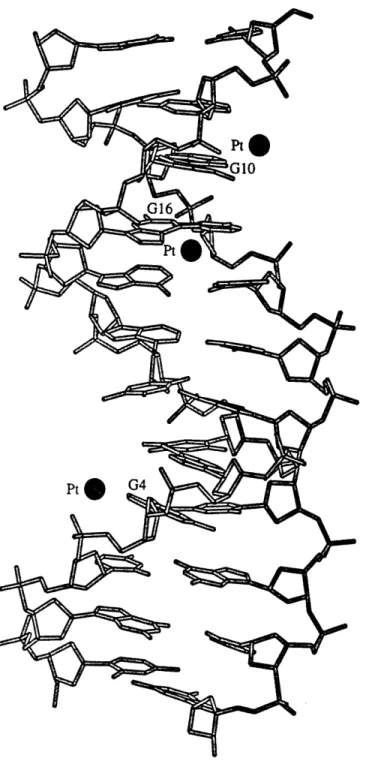
d(CGCGAATTCGCG) (Wing et al., 1984). In this work, three of the eight
guanine residues, G4, G10, and G16, appeared to have affinity for cisplatin but all the platinum sites in the crystal had only partial occupancy (Figure 3). Crystal structures with three different levels of cisplatin substitution were solved, and the most occupied platinum site, near the N7 atom of residue
G16, was compared. The crystals had platinum-G16(N7) bond lengths of 2.51
A,
2.43 A, and 2.16A
with occupancies of 20%, 38%, and 61%, respectively. Attempts to obtain more highly substituted platinum sites resulted in degradation of the crystals. The authors reasoned, from a linear plot of bond length versus percent substitution, that the platinum-N7 bond length would be 1.8A
at 100% occupancy. This conclusion was only for a monofunctional cisplatin-DNA adduct and yielded no information about the bifunctional adducts thought to be responsible for the antitumor activity of the drug.The structure of the major adduct formed,
cis-[Pt(NH3)2{d(pGpG)-N7(G1),-N7(G2)}], has been probed on short segments of single-stranded DNA
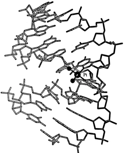
by X-ray crystallography. These studies showed coordination at the N7 atoms
of adjacent guanine bases causes a head-to-head orientation of the bases and a dihedral angle of about 800 between the planes of the guanine rings (Figure 4) (Admiraal et al., 1987; Sherman et al., 1985; Sherman et al., 1988). On a segment of short, single-stranded DNA, the sugar pucker of the 5' nucleotide
25
Figure 3. Structure of the dodecamer d(CGCGAATTCGCG) after pre-formed crystals were soaked with cisplatin.
26
Figure 4. X-ray crystal structure of cis-tPt(NH3)2}2 + bound to d(pGpG). Cisplatin binds to the N7 atoms on each guanine base and causes a roll of
-80' between the guanine ring planes. A hydrogen bond forms between
27
adopts a C3'-endo conformation whereas the 3' ribose ring is C2'-endo. Another interesting aspect of the structure is the hydrogen bond formed between an ammine on the platinum atom and a phosphate oxygen 5' to the platinum lesion. It was hypothesized that this hydrogen bond plays a role in stabilizing the adduct and may therefore be important for the anticancer activity of the drug. Because single-stranded DNA molecules lack the constraints of base stacking and Watson-Crick hydrogen bonding, further studies of specific platinum adducts were carried out on segments of duplex
DNA.
Nuclear magnetic resonance (NMR) studies of a specific adduct of cis-{Pt(NH 3)2}2+ on duplex DNA suggested a 40-70' bend as a possible deformation of the double helix and showed a C3'-endo sugar pucker to the 5' side of the platinum lesion and a C2'-endo sugar pucker on the 3' side (den Hartog et al., 1985). Later, NMR data were combined with geometric parameters from the crystal structure of the
cis-[Pt(NH3)2{d(pGpG)-N7(Gl),-N7(G2)}] adduct and used in molecular mechanics studies (Kozelka et al., 1987;
Kozelka & Chottard, 1990; Kozelka et al., 1985). These studies confirmed the head-to-head orientation of the bases, the presence of a bend, the sugar pucker alternation across the platinum lesion, and the hydrogen bond between a phosphate oxygen and an ammine on the platinum atom. Because DNA is such a large and complex molecule, it was not possible to derive a single best structure for the adduct. Instead, several models resulted from these studies, all energetically feasible and all with a bend of about 600 toward the major groove and unwound by 12-190 (Figure 5).
The bend angle of duplex DNA with an intrastrand cis-{Pt(NH3)2}2+ cross-link seen in molecular mechanics models was much larger than the
28
Figure 5. A representative molecular mechanics model of cis-{Pt(NH3)2}2 + bound to duplex DNA. The overall bend in the duplex was estimated to be
bend observed by gel electrophoresis studies on site-specifically modified
platinum-DNA adducts.
Multimers of a duplex containing a specific
intrastrand cis-[Pt(NH
3)
2{d(GpG)-N7(G),-N7(G)) }] site showed anomalous
electrophoretic mobility and a bend of ~40' toward the major groove wascalculated (Rice et al., 1988). Further gel electrophoresis studies on intrastrand
site-specific cisplatin/DNA adducts showed that the major intrastrand
cross-links cis-[Pt(NH
3)
21d(GpG)-N7(G),-N7(G)}] and cis-[Pt(NH
3)
2{d(ApG)-N7(A),-N7(G)}] bend the double helix by -35' and unwind it by -13' (Bellon et al.,
1991; Bellon & Lippard, 1990).
Recently, an NMR structure of a double-stranded DNA octamer,
d(CCTG*G*TCC).d(GGACCAGG) with an intrastrand cis-[Pt(NH
3)
2{d(GpG)-N7(G
4),-N7(Gs)}] cross-link at the -G*G*- site was reported (Yang et al., 1995).
The duplex exhibits a bend of -58
°, an unwinding angle of ~-21'
,a C3'-endo
sugar pucker on the 5' side of the platinum lesion, and a C2'-endo sugar
conformation on the 3' side of the platinum cross-link (Figure 6). This study
afforded a significantly more detailed description of the cisplatin adduct than
previous NMR studies. In particular, it showed that the minor groove of the
octamer opposite the platinum binding site had widened to about 8
A,
as
compared to the width of 5.7
A
for canonical B-DNA. The platinated octamer
used in the NMR analysis was metastable, however. The intrastrand
cross-link rearranged to an interstrand cross-cross-link under the experimental
conditions employed, but the significance of such a rearrangement is not yet
clear.
Although the experimental NMR and gel electrophoresis studies
provided important information about the distortions caused by
cisplatin-DNA cross-links, structural details of bifunctional platinum adducts were
limited to short single-stranded DNA oligonucleotides.
The one
30
Figure 6. NMR model of cis-(Pt(NH
3)
2)
2 +bound to duplex DNA. The
31
crystallographic attempt to study duplex DNA crystals soaked with cisplatin
did not result in cisplatin-DNA cross-links and provided no structural
information about biologically relevant adducts.
It was therefore an
extremely important objective to obtain the X-ray crystal structure of a specific
and biologically relevant intrastrand cisplatin cross-link on duplex DNA.
Crystallographic studies of such a site, a specific cisplatin cross-link on
duplex DNA were first undertaken in the laboratory of Stephen J. Lippard by
Steven F. Bellon (Bellon, 1992). His work involved the self-complementary
dodecamer DNA oligonucleotide d(GCTG*G*TTAACCA) with an intrastrand
cis-{Pt(NH
3)
2}
2+ cross-link at the -G*G*- site. The platinated dodecamer was
synthesized, purified, and annealed to afford a duplex with two cisplatin
cross-links per double helical turn. The cisplatin adducts were strategically
placed such that the bends in each helical turn would be 1800 apart. The
annealed duplex DNA also had a 5' -GC- overhang on each end for base
pairing with the 5' ends of neighboring helices. The bend placements and the
overhangs on the ends of the helices were designed to create a structure
which was effectively a long, continuous, albeit curved helix that might pack
well in crystals. Crystals with a flat plate-like morphology were obtained but
diffracted poorly. An analogous sequence, d(ATTG*G*TTAACCA), was also
synthesized, purified, annealed, and crystallized. Again, the crystals diffracted
poorly and were not suitable for study by X-ray crystallography. The unit cell
dimensions and space groups for the crystals of the sequences described above
are listed in Table 1.
Further attempts at obtaining diffraction quality crystals of an
intrastrand cisplatin cross-link on duplex DNA involved a more
conventional approach.
Oligonucleotides with a single -GG- site for
platination were synthesized and, in order to increase the yield of purified
Table 1. Platinated deoxyoligonucleotides duplexes crystallized by Steven F.
Bellon (Bellon, 1992). The -GG- sites were coordinated to cis-{Pt(NH3)2}2 +.
Sequence Unit cell parameters
GCTGGTTAACCA Trigonal: ACCAATTGGTCG a = 30
A
a = 900b = 30 A
b = 90
0 c = 83A
g = 1200 ATTGGTTAACCA P21: ACCAATTGGTTA a = 27 A a = 900 b = 87 A b = 1110c = 36
A
g = 90
platinated product, the oligonucleotide contained no purines other than the
-GG- platination site. The pure platinated oligonucleotide was then annealed
to its purified, complementary strand to form duplex DNA with an intrastrand cisplatin cross-link. Many cisplatin-modified DNA duplexes were synthesized and all were screened for crystallization (Table 2), but only one sequence yielded diffraction quality crystals. Crystals of the sequence
d(CCTCTG*G*TCTCC)-d(GGAGACCAGAGG) were obtained and the X-ray
crystal structure was solved by multiple isomorphous replacement. One of the brominated derivatives diffracted to 2.6
A
and data from this crystal were used to obtain the structure described in this thesis.The structure of the platinated duplex dodecamer,
d(CCUBrCTG*G*TCTCC).d(GGAGACCAGAGG) where the -G*G*- site was
multiple isomorphous replacement (MIR) methods with brominated
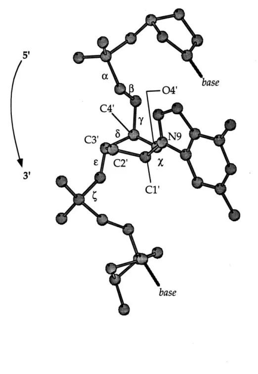
derivatives. The numbering scheme for the oligonucleotide is as follows:
5'-Ci -C2 -U*3-C4 -T5 -G*6-G*7-T8 -C9 -T10-C11-C12-3' 3'-G24-G23-A22-G21-A20-C19--C18-A17-G6-A15-GI4-GI3-5'
where U*3 is 5-bromouridine and platinum binds to the N7 atoms of G*6 and
G*7. In this thesis, the ends of the double helix will be referred to as the "3'
end" and "5' end" with respect to the platinated deoxyoligonucleotide.
Crystals of this cisplatin-modified duplex diffracted to 2.6
A
and these data
provide the basis for the structure discussed. A preliminary report of this
crystal structure has been published (Takahara et al., 1995). The structure
shows how a segment of platinated DNA flexes to accommodate a platinum
lesion and packing interactions in the crystal illustrate how a
cisplatin-modified duplex might come in close contact with other biomolecules.
Table 2. Deoxyoligonucleotides used for crystallization trials. Name Sequence TT8d 5'-CTCGGTTC-3 ' 3'-GAGCCAAG-5' TT6d 5'-CCGGTC-3 ' 3'-GGCCAG-5' TT12d 5'-CCTCTGGTCTCC-3' 3'-GGAGACCAGAGG-5' TT12-3d 5 -CCCCTGGTTTCC-3' 3'-GGGGACCAAAGG-5' TT12-4d 5'-CTCTTGGCCTAC-3' 3'-GAGAACCGGATG-5' TT12-5d 5 -CCCCCGGTCCCC-3' 3' -GGGGGCCAGGGG-5' TT12-2A 5'-CTGGC-3' TT12-2B 5'-CGGCCAG-3 ' TT32A 5' -CCTCTCTGGTTCTTC-3 ' TT32B 5'-CGGAAGAACCAGAGAGG-3' TT12Br-a 5'-CCUBrCTGGTCTCC- 3' TT12Br-b 5'-CCBrTCTGGTCTCC-3' TT12Br-c 5 -CCTCTGGTCBrTCC-3' TT12Br-d 5'-CCTCTGGUBrCTCC-3' TT12Br-e 5'-CCTCUBrGGTCTCC-3' TT12Br-f 5 '-CCTCTGGTCUBrCC-3' CD13-1d 5'-CTCTTGTGTCCTC-3' 3'-GAGAACACAGGAG-5' CD13-2d 5'-CTCCTGTGTTCTC-3' 3'-GAGGACACAAGAG-5' CD13-3d 5'-CCCTTGTGTCCCC-3' 3'-GGGAACACAGGGG-5' CD12d 5'-CCTCTGTGTCTC-3' 3'-GGAGACACAGAG-5' CD20 5'-TCTCCTTCTGGTCTCTTCTC-3' 3'-AGAGGAAGACCAGAGAAGAG-5'
*Strands with GG sites or GTG sites for platination were reacted with cisplatin. Top and bottom strands of TT6, TT8, TT12, TT12-3, CD13-1, CD13-2, CD13-3, and CD12 were annealed to form duplexes. TT12Br-a, TT12Br-b, TT12Br-c, TT12Br-d, TT12Br-e, and TT12Br-f were mixed in a 1:1 molar ratio with TT12b. TT12A andTT12B were mixed in a 1:1 molar ratio and annealed to form a 12-mer. TT32A and TT32B were mixed in a 1:1 molar ratio and annealed to form a 32-mer. TT12A/B and TT32A/B were designed with the help of Professor Carl Pabo.
35
CHAPTER 2
Materials. Phosphoramidites and DNA synthesis reagents were
purchased from Cruachem and Glen Research. Crystallization reagents were obtained from Fluka or Aldrich. cis-Diamminedichloroplatinum(II) was a gift from the Engelhard Corporation. DNase 1 and alkaline phosphatase were obtained from Boehringer Mannheim and P1 Nuclease was purchased from Gibco BRL. Reverse phase C4 and C18 high pressure liquid chromatography (HPLC) columns were purchased from Vydac, and ion exchange HPLC columns were purchased from Dionex.
Large scale HPLC was performed on a Waters 600E pumping system with either a Waters 486 or Waters 484 ultraviolet detector set at 260 nm. Analytical HPLC was done by using a Perkin-Elmer Series 4 Liquid Chromatograph with an LC-95 UV/vis detector set at 260 nm. Atomic absorption was done by using a Varian AA1475 instrument. X-ray diffraction data were collected on a Marresearch imaging plate system equipped with a Rigaku Cu Ka rotating anode radiation source.
Deoxyoligonucleotide Synthesis and Purification.
Deoxyoligonucleotides were prepared on a Cruachem synthesizer by using standard solid phase phosphoramidite methods. Deoxyoligonucleotides were deprotected by using concentrated NH40H at 55 °C for 12 hours. Protecting groups and excess trityl groups were removed by G25 Sephadex size exclusion chromatography, and the deoxyoligonucleotides were then lyophilized to dryness. Deoxyoligonucleotides were then converted to their sodium salts by using a Dowex cation exchange column and quantitated by optical spectroscopy with calculated extinction coefficients (A2 60) (Borer, 1975).
The diaqua complex of cisplatin was prepared by allowing 1.97 equivalents of AgNO3 to react with cis-[Pt(NH3)2C12] in water at room
37
temperature in the dark. AgC1, a white precipitate, formed after about 30 min. After 12 h, the reaction mixture was centrifuged at 13000 rpm for ten min. The aqueous layer was drawn away from the AgCl pellet by using a pipet and centrifuged for another ten min. This process was repeated twice, and the final platinum solution was allowed to react with deoxyoligonucleotides with a -GpG- site for platination at 37 TC in the dark for 6-8 h.
Platinated deoxyoligonucleotides and their complementary strands were initially purified by an NaCl gradient on an ion exchange HPLC column. HPLC buffer A was composed of 25 mM NH4OAc, 10% CH3CN, and distilled,
deionized H20 (ddH20). Buffer B was composed of 25 mM NH40Ac, 10%
CH 3CN, 1 M NaC1, and ddH20. Oligonucleotides were eluted from the
column by using a gradient of 90% A and 10% B to 50% A and 50% B over 30 min. Oligonucleotide fractions were collected and desalted by dialysis against a solution of 0.1 M NH4OAc in ddH20. Oligonucleotides rich in adenine or
guanine residues were sometimes difficult to purify because they tended to aggregate on the ion exchange columns. In these cases, 50% formamide was used as a denaturing agent in the ion exchange HPLC solutions.
The DNA strands were then purified by an acetonitrile gradient on a C4 or C18 reverse phase HPLC column. Reverse phase HPLC buffer A was composed of 0.1 M NH40Ac in ddH20 and buffer B was 50% buffer A and 50%
HPLC grade CH3CN. The typical gradient used was 95% A and 5% B to 60% A
and 40% B over 30 minutes. DNA sequences used in this study are listed in
Table 2 and example purification schemes are shown in Figures 7 and 8.
Crystallization. Purified complementary strands of each
38
Figure 7. HPLC purification of a platinated deoxyoligonucleotide.
(a)
Dodecamer oligonucleotide after reaction with cisplatin. Ion exchange
gradient B: 10-40% over 30 min. (b) Platinated oligonucleotide after ion
exchange purification. Reverse phase gradient B: 5-40% over 30 min. (c)
Platinated oligonucleotide after ion exchange and reverse phase purification.
Reverse phase B: 5-40% over 30 min.
(a) A2 60 Elution time (b) A2 60 Elution time (c) A260 Elution time
I
E on.Figure 8.
HPLC purification an unplatinated, complementary
deoxyoligonucleotide strand. (a) Crude oligonucleotide after deprotection.
Ion exchange B: 20-80% over 30 min. (b) Crude oligonucleotide after
deprotection. Ion exchange B with 50% formamide: 10-50% over 30 min. (c)
Oligonucleotide after ion exchange purification. Reverse phase B: 5-40%
over 30 min. (d) Oligonucleotide after ion exchange and reverse phase
purification. Ion exchange B with 50% formamide: 10-50% over 30 min.
A260 Elution time Elution time Elution time Elution time
(b)
(c)
A260 A2 60(d)
A260 (a)i
Elution timeconcentration of 2.5 mM. Crystals were grown by using sitting drops (Figure 9) (Drenth, 1994). Each crystallization trial drop contained deoxyoligonucleotide, sodium cacodylate, magnesium chloride, and 2-methyl-2,4-pentanediol (MPD). Drops also contained a polyamine; spermine hydrochloride or [Co(NH3)6]C13 were most often used. Crystallization drops were mixed at room temperature and equilibrated against a 5% MPD reservoir at 4 'C. In successful trials, clusters of crystals appeared after 3-30 days and were allowed to grow for 9-12 months.
Successful crystallizations of TT12d and its derivatives resulted in clusters of thin rods. The small dimensions of the rods are usually 0.01 - 0.07 mm x 0.01 - 0.10 mm, too thin to be studied by X-ray diffraction. In order to get crystals with dimensions of about 0.05 x 0.10 x 1.0 mm3, a slow cooling technique was employed. Crystallization drops were constructed as described in the previous section, the crystallization boxes were sealed, and the boxes were then covered with three layers of bubble wrap. Wrapped boxes were then packed into a styrofoam box at room temperature. The styrofoam box was sealed and placed in the cold room and allowed to equilibrate to 4 OC.
After 9-12 months, the drops produced diffraction quality crystals.
Clusters of TT12d and its brominated derivatives TT12-Brl, TT12-Br2, and TT12-Br3 (Table 3) were grown in drops containing 0.2 mM duplex DNA (TT12), 52 mM cacodylic acid (sodium salt, pH 6.0), 15 mM magnesium chloride, 6 mM [Co(NH3)6]C13 and 3% 2-methyl-2,4-pentanediol (MPD) (Figure 10). Diffraction quality crystals of the other sequences listed in Table 2 were not obtained.
Analysis of Platinum/DNA Ratios in the Crystals. Ten TT12d crystals
and dissolved in water. The platinated oligonucleotide was separated from its complementary strand by reverse phase HPLC. The
cis-[Pt(NH3)2{d(GpG)-N7(G6),-N7(G7)}] adduct was confirmed by enzymatic digestion analysis of the
platinated oligonucleotide. 1 nmol of single-stranded platinated
oligonucleotide from TT12d crystals was dissolved in 50 mM NaOAc and 10 mM MgC12 at pH 5.6 and digested with DNase I (40 units) and P1 Nuclease (2 units) at 37 °C for 24 h. An aliquot of this digestion solution was diluted ten-fold and digested further with alkaline phosphatase (5 units) in a 100 mM
EDTA and 50 mM Tris-HCI buffer at 37 oC for 24 h. The final digestion solution was then analyzed by reverse phase HPLC. The presence of cytosine and thymine were confirmed by comparison with standards purchased from Aldrich, and the ratio of 6 cytosine residues to 4 thymine residues was confirmed by peak integration. The cisplatin intrastrand cross-link was confirmed by coinjection with authentic cis-[Pt(NH 3)2{d(GpG)-N7(G 1 ),-N7(G2)}] and unplatinated d(GpG). The digestion analysis of the platinated
oligonucleotide isolated from TT12d crystals is shown in Figure 11.
The platinated oligonucleotide isolated from TT12d crystals by using HPLC was also analyzed twice by using flameless atomic absorption spectroscopy and yielded platinum per single-stranded oligonucleotide ratios of 0.98 and 0.96.
X-ray Data Collection. Crystals cut from clusters were mounted in
sealed glass capillaries containing a drop of mother liquor (Figure 12) and sealed with melted wax. The capillaries were attached to pre-cooled brass pins and secured with epoxy resin. The brass pins were then attached to pre-cooled goniometer heads and stored in a styrofoam box packed with ice.
Figure 9. A sitting drop crystallization and conditions for
cisplatin-modified DNA.
0.2 mM DNA
52 mM cacodylic acid (Na
+salt, pH 6.0)
15 mM MgC12
6 mM [Co(NH
3)
6]C1
33% 2-methyl-2,4-pentanediol (MPD)
5% MPD reservoir
I
sitting drop
4 OC
III)Figure 10.
Photomicrograph of a cluster of cisplatin-modified DNA
crystals. The picture was taken under polarized light and the crystals are
actually colorless.
T ~~ ;NW,""7-V7
ft-AC ~l·-ir~ L~~
Figure 11. Digestion analysis of the platinated oligonucleotide,
d(CCTCTG*G*TCTCC) where the -G*G* site is coordinated to cis-{Pt(NH3)2}2 +, from crystals of TT12d. (a) Digestion products from the platinated oligonucleotide. (b) Coinjection of d(GpG) and cis-[Pt(NH3)2{d(GpG)}]+ with the digestion products. (c) d(GpG) and cis-[Pt(NH 3)2{d(GpG)}]+ standards.
Peaks are as follows: (1) digestion buffer, (2) dC, (3) dT, (4)
cis-[Pt(NH3)2{d(GpG)}]+, (5) undigested platinated oligonucleotide, (6) d(GpG), (7)
(a) A26 0 (b) A2 60 Elution time 1 2 Elution time (c) A260 Elution time 6 4
7
t ~ 1___2__48
Figure 12. Photomicrograph of a crystal of cisplatin-modified DNA in a
glass capillary. The capillary contains the crystal and a plug of mother
liquor and is sealed on both ends by wax.
49
The cold stream on the X-ray instrument to be used was adjusted to 4
°C and allowed to equilibrate for 30-60 min. The temperature was monitored
with a thermocouple. After the cold stream was stabilized at 4 OC, the styrofoam box containing the goniometer head was removed from the cold room, transported to the machine and mounted as quickly as possible. Crystals were optically centered by using a videomicroscope.
Data sets for TT12d and its derivatives were collected on a Marresearch image plate with CuKa (k = 1.5418
A)
radiation. Unit cell parameters were determined by autoindexing several images in each data set separately with the program DENZO (Z. Otwinowski, University of Texas, Southwestern Medical Center). The unit cell volume was determined to be 50,770A3
and indicated the presence of two DNA duplexes in each asymmetric unit. For each data set, rotation images were collected in 30 increments with a total rotation of 3600 about phi. Unit cell parameters, sequences used, and additional X-ray information are summarized in Table 3.Crystals of TT12d, TT12Br2, and TT12Br3, in the original drops from which they were grown, were packed in a styrofoam box fitted with several cold packs and foam and were transported to the Stanford Synchrotron Radiation Laboratory (SSRL). At SSRL, the crystallization plates were immediately transferred to a cold room (4 °C) and removed from the styrofoam box. The crystals were visually inspected by using a microscope, and no damage was seen. Several crystals of TT12d, TT12Br2, and TT12Br3 were mounted in a loops made from strands of dental floss and fastened to the end of brass pins with epoxy. Crystals mounted in loops were flash frozen in a nitrogen cold stream (-170 'C). No diffraction was seen for the frozen crystals. The remaining crystals were mounted in sealed capillaries and tested
Table 3. Experimental details of the
modified DNA.
X-ray diffraction study of
cisplatin-Unit cell parameters: a = 31.3 A a = 79.80
b = 35.5 A P = 84.80
c = 47.0 A
y = 82.8
0Unit cell volume: 50,770 A3
Space group: P1
Molecules per asymmetric unit: 2
Instrument: Mar Research Imaging Plate
Radiation: Cu Ka
Diffraction limit: 2.6
A
for Brl
Structure solution method: multiple isomorphous replacement (MIR)
Sequences used
Native
Brl
Br2
Br3
for MIR:
d(CCTCTG*G*TCTCC).d(GGAGACCAGAGG)
d(CCUBrCTG*G*TCTCC).d(GGAGACCAGAGG)
d(CCTCTG*G*UBrCTCC).d(GGAGACCAGAGG)
d(CCTCTG*G*TCBrTCC)-d(GGAGACCAGAGG)
51
for diffraction. Many of the crystals failed to diffract at all, and the ones that
did diffract had split spots throughout the diffraction pattern indicating that
the crystal had cracked during the trip or during mounting. The best crystals,
TT12d, were split badly but diffracted to about 2.8
A.
Crystals were packed and transported to the Brookhaven National
Synchrotron Light Source Beamline X-8C in the manner described for the
Stanford experiments. The crystals were inspected under a microscope after
they had been transferred to the cold room. Several were cracked and a few
had disintegrated, but there were also many crystals which showed no visible
signs of damage. Two TT12d crystals were mounted in loops and frozen by
immersion in liquid propane. The frozen crystals were then transferred into
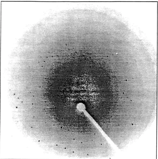
a nitrogen cold stream set at -165 'C. The first frozen crystal diffracted to about
2.7 angstroms but had many split spots indicating that the crystals were
damaged either by the transportation or freezing procedure (Figure 13). No
data were collected on the first crystal but it was used to tune the wavelength
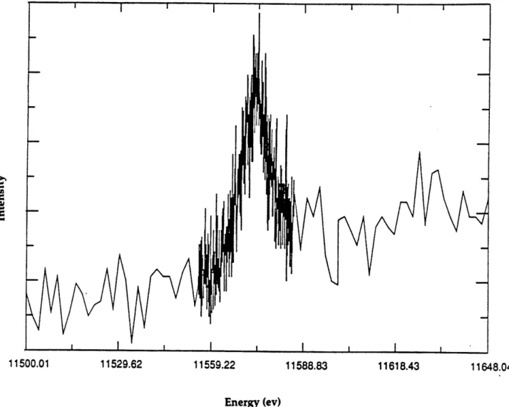
of the radiation to the platinum edge. The edge was found by monitoring
fluorescence while stepping the energy of the X-ray beam. The maximum
fluorescence for platinum foil was found to be 11550 eV. The maximum
fluorescence for a TT12d crystal was found to be 11574 eV and corresponded to
a wavelength of 1.072 A (Figure 14). This experiment provided further
confirmation that crystals contained platinum.
A partial data set was collected on a second crystal that had been frozen
in liquid propane. The data were collected on a charge coupled device (CCD)
imaging plate with rotation increments of 0.20. 492 pictures corresponding to
a total rotation of 98.40 were collected. The data could not be indexed.
52
Figure 13. CCD detector image collected on a frozen crystal of platinated
DNA at NSLS-X8C. The outer spots on the image correspond to 2.7 A
diffraction. Many spots are split and indicate crystal damage.
Figure 14. Fluorescence intensity scan of a crystal of platinated DNA. The
maximum energy corresponds to an energy of 11574 eV and a wavelength
of
1.072A.
11500.01 11529.62 11559.22 11588.83 11618.43 11648.04
54
Data Reduction and Structure Determination. Data sets collected on a
Marresearch imaging plate were used to solve the structure of the
cisplatin-modified dodecamer, d(CCTCTG*G*TCTCC).d(GGAGACCAGAGG), where
the -G*G*- site has been modified with cis-{Pt(NH3)2}2+. Data were processed and merged by using DENZO and SCALEPACK (Z. Otwinowski) and further processed by using the CCP4 program suite (CCP4, 1994). An anomalous difference Patterson map was calculated from the native data and used to determine the relative positions of the platinum atoms and this map confirmed the presence of two platinated duplexes in each unit cell with the platinum atoms 15.1A
apart (Figure 15 a). A conventional Patterson map was calculated by using the data from 3.2A to 3.5
A
and showed a slight bend in the stacking pattern of the bases (Figure 15 b).Difference Patterson maps between the native and derivative data were calculated for each brominated duplex for which data were collected. Each derivative DNA duplex was synthesized with one bromine placed specifically in the sequence (Table 3). Difference Patterson maps clearly showed one peak corresponding to a bromine-bromine vector, in accord with the presence of two DNA duplexes in the asymmetric unit (Figures 16 a-c).
The bromine positions were used to calculate single isomorphous replacement (SIR) phases for each heavy atom derivative. Fourier maps were then calculated by using the SIR phases and the native structure factor amplitudes. Since Patterson maps are centrosymmetric, the Fourier map derived was superimposed on its inverse. From each map, two pairs of possible platinum atoms positions were obtained. Shifting one platinum atom to the origin of the unit cell with concomitant shifting of the bromine atoms afforded two possible pairs of bromine positions for each derivative.
Figure 15. (a) Anomalous difference Patterson map (010 projection) calculated with native data from 12.0 to 3.0 A. The peak is at fractional
coordinates (0.18, 0.24, 0.19) and corresponds to a platinum-platinum vector of 15.1 A. (b) Patterson map (010 projection) calculated with native data from 3.2 to 3.5 A. w (fractional coordinates) 0.0( I. - 0.5 s-i 'a (a) w (fractional coordinates) 0. 8 (00 ,O ca5-' (U 0 (b) I I r~vv
Figure 16. Difference Patterson maps (010 projections) calculated with
native and derivative data from crystals of (a) Brl, (b) Br2, and (c) Br3. The
fractional coordinates for the main peaks are (a) (0.48, 0.50, 0.06), (b) (0.16,
0.19, 0.35), and (c) (0.20, 0.20, 0.53).
(a)
(b)
The possible pairs of heavy atom positions afforded eight possible
combinations of heavy atom positions, each of which was used to calculate
trial phases. The correct bromine coordinates for each derivative were found
by choosing the best multiple isomorphous replacement (MIR) electron
density map. Bromine positions were confirmed by calculating electron
density maps with MIR phases and I Fnat
-
Fder I structure factor amplitudes.
The platinum atom positions were confirmed by calculating maps with MIR
phases and the anomalous differences in the native data as the structure
factor amplitudes.
The MIR maps calculated at this stage of the refinement clearly showed
the platinum atoms, spherical electron density for the phosphate groups, and
elongated electron density for a few of the bases. An initial model of B-DNA
modified with cis-{Pt(NH
3)
2}
2+ was built with the program INSIGHT II
(Biosym). Model manipulation was done by using the program O (Jones et
al., 1989). No symmetry restraints between the two molecules in the unit cell
were used during refinement. The initial model was fit to the MIR maps
calculated with native structure factor amplitudes to 3.0
A.
After positional
refinement in X-PLOR (Briinger, 1992b; Briinger et al., 1987), the phases
obtained were applied to Brl derivative data with I Fobs I to 2.6
A.
10% of the
reflections were set aside for the free-R factor calculation prior to model
building and refinement (Briinger, 1992a; Briinger, 1993). Seventeen cycles of
model building, positional refinement, and phase combination, yielded a
model for which R
= I(I
Fobs I -I Fcaic I )/I
I Fobs I =0.25. Another round of
positional refinement in which all restraints on the platinum geometry were
removed, followed by temperature factor (B) refinement resulted in R
=
0.225.
Finally, 31 water molecules were added to the model and gave a final
structure with a free-R
=
0.249 and R
=
0.203. The final structure was checked
58
by using a series of simulated annealing omit maps in which one base step at
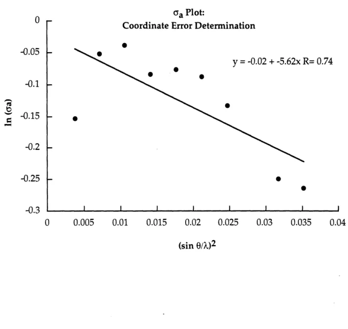
a time was left out of the calculation. Refinement statistics are given in Table 4 and examples of maps obtained and models built during the refinement are shown in Figure 17. The overall estimated coordinate error for this structure is 0.46
A
and was determined by using a plot of ln(GA) vs. resolution (Figure18) (Drenth, 1994; Read, 1986) which is based on equation (1):
In GA = 1/2 [ln(Ip/1N)] -R3( I Ar I)2(sine/X)2 (1)
where CYA is D(yp/1N)1/2, Ip and IN are the summations of the squares of
atomic structure factors for all atoms in the full and partial structures, respectively, I Dr I is the average coordinate error for the model, and D is the Fourier transform of the probability distribution of I Ar I.
Figure 17. Maps of molecule A, residues C19-A20-G21-A22, generated during
refinement of the structure. (a) is the MIR map, (b) is a 2Fo-Fc map calculated
from a partial structure after five rounds of model building, (c) is a 2Fo-Fc
map calculated after ten rounds of model building, and (d) is the final 2Fo-Fc
map with the final model superimposed.
iCi:i iii? i. ii~ i ;;; C· ii ·- · · .. I : I ~r~cf*- ii~i ~·:"i·? ir z: r i `·; ~;··s, ~ \.. · i irr i)C' TC 'i :~ ; ri ;; ·i ,·: .*: i\ ~i~ll (a) (b) (d)
60
Figure 18. ca plot for the estimated coordinate error for the final model.
Ga Plot:
Coordinate Error Determination
R= 0.74 (sin O/X)2 I -0.05 -0.1 -0.15 -0.2 -0.25 -0.3 0 0.005 0.01 0.015 0.02 0.025 0.03 0.035 0.04 · · · ·
n a ao o ; Nd oN O O 00 00 c n C 0O ~Co Lfo co C C e C F c~l LOo II -1 0 k o -4-0).. 0 0L
ccj t<
r-4 .. 0) 0 0 1 T6 .4-0 0iz
UO
0 ui rJt o o_ +, , , (. I -! .=ii rm o *o -4-0) I cFj 0) '.4-'.4-I '.4-'.4-I63
CHAPTER 3