HAL Id: hal-02450494
https://hal.univ-reims.fr/hal-02450494
Submitted on 2 Jun 2020
HAL is a multi-disciplinary open access
archive for the deposit and dissemination of
sci-entific research documents, whether they are
pub-lished or not. The documents may come from
teaching and research institutions in France or
abroad, or from public or private research centers.
L’archive ouverte pluridisciplinaire HAL, est
destinée au dépôt et à la diffusion de documents
scientifiques de niveau recherche, publiés ou non,
émanant des établissements d’enseignement et de
recherche français ou étrangers, des laboratoires
publics ou privés.
Hereditary multiple exostoses of the ribs as an
uncommon cause of pneumothorax
Antoine Dumazet, Claire Launois, Sandra Dury, Frédéric Sailhan, Marco
Alifano, Maxime Dewolf, François Lebargy, Gaetan Deslée, Jeanne-Marie
Perotin
To cite this version:
Antoine Dumazet, Claire Launois, Sandra Dury, Frédéric Sailhan, Marco Alifano, et al.. Hereditary
multiple exostoses of the ribs as an uncommon cause of pneumothorax. Medicine, Lippincott, Williams
& Wilkins, 2018, 97 (35), pp.e11894. �10.1097/MD.0000000000011894�. �hal-02450494�
Downloaded from https://journals.lww.com/md-journal by BhDMf5ePHKav1zEoum1tQfN4a+kJLhEZgbsIHo4XMi0hCywCX1AWnYQp/IlQrHD36ls6+OKM7O1o0e2K2SF5Xo/CGpuLFDLL6OVW52vIsXs= on 06/02/2020 Downloadedfrom https://journals.lww.com/md-journalby BhDMf5ePHKav1zEoum1tQfN4a+kJLhEZgbsIHo4XMi0hCywCX1AWnYQp/IlQrHD36ls6+OKM7O1o0e2K2SF5Xo/CGpuLFDLL6OVW52vIsXs=on 06/02/2020
Hereditary multiple exostoses of the ribs as an
uncommon cause of pneumothorax
A case report
Antoine Dumazet, MD
a,∗, Claire Launois, MD
a, Sandra Dury, MD
a,b, Frédéric Sailhan, MD
c,
Marco Alifano, MD, PhD
d, Maxime Dewolf, MD
a, François Lebargy, MD, PhD
a,b,
Gaëtan Deslee, MD, PhD
a,e, Jeanne-Marie Perotin, MD, PhD
a,eAbstract
Rationale:Hereditary multiple exostoses (HME) is a genetic musculoskeletal condition causing multiple exostoses. Rib location of exostosis can be complicated by thoracic injuries.
Patient concerns and diagnoses: We report a case of pneumothorax in a 32-year-old man with a partial left-sided pneumothorax caused by an exostosis of the fourth andfifth ribs.
Interventions and outcomes:Clinical and radiological presentations allowed a conservative management. A video-assisted thoracoscopic surgery was performed a few weeks later to avoid any recurrence.
Lessons:Rib exostosis represents an unusual cause of pneumothorax. Any local modification of symptoms or size of the exostosis should lead to investigations in regard to chondrosarcoma transformation.
Abbreviations: FEV1= forced expiratory volume in the first second, HME = hereditary multiple exostoses. Keywords:costal exostosis, hereditary multiple exostoses, pneumothorax, VATS
1. Introduction
Hereditary multiple exostoses (HME) is a genetic musculoskele-tal condition with an autosomal dominant inheritance and a variable penetrance, involvingEXT1 and EXT2 genes.[1]HME is
defined by the presence of at least two exostoses (or osteochondromas) of the juxta epiphyseal region of long bones. HME incidence is approximately 1:50000 in general popula-tion.[2]The most frequent localizations of exostoses are around the knees and proximal humerus.[3]Ribs exostoses are usually
asymptomatic but can occasionally be associated with pleural, diaphragm or pericardial injuries. [4–6] We report a case of pneumothorax caused by costal exostosis.
2. Case report
A 32-year-old man was admitted for a spontaneous oppressive left side chest pain with a left arm irradiation for 2 days. He had a
history of HME diagnosed in the childhood, with multiple leg exostosis resections and a leg-length inequalities correction. No genetic testing was available. He was a tobacco and cannabis smoker (13-pack-years). At admission, clinical exam did not reveal any sign of acute respiratory failure but a slight decrease in breath sounds in the left lung. Blood pressure was 130/80 mmHg, cardiac rate: 62 per minute, Sa02: 98%. Standard blood analysis and ECG were normal. A chest X-ray identified a left pneumothorax extending on axillary line and 2 dense opacities, 1 is located near the leftfifth rib and the other being located near the right sixth rib (Fig. 1A). A chest computed tomography (CT) was performed and confirmed the left side pneumothorax and multiple costal exostoses (Fig. 1B–D). One exostosis was developed from the anterior arch of the left fifth rib with an intra-thoracic involvement and had a contact with the pneumothorax. Furthermore, CT-scan revealed bilateral paraseptal emphysema with an apical predominance.
Given clinical and radiological presentations, a conservative management wasfirst proposed, resulting in a progressive and spontaneous improvement. The patient was discharged from hospital after 2 days management. Chest X-ray performed 2 weeks later exhibited complete resolution of the pneumothorax. Pulmonary function tests identified: forced expiratory volume in thefirst second (FEV1) 93% of predicted value, FEV1/forced vital
capacity (FVC) 92%, RV 179% pred. The Alpha-1-antitrypsin level was normal.
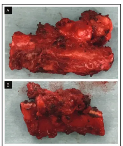
Several weeks after this event, a surgical management of rib exostoses was proposed in order to prevent any pneumothorax recurrence. Surgery was performed by left-sided video-assisted thoracoscopy (VATS) and revealed exostoses of the left-sided fourth andfifth ribs with tight pulmonary adherences. A partial resection of the left-sided fourth and fifth ribs exhibiting intrathoracic exostosis lesions as well as a resection of 2 small emphysematous bullae were performed (Fig. 2). Due to double exostoses withdrawal, an early pulmonary hernia occurs and was taken care with a Vicryl plate tofilling the anterior parietal defect.
Editor: N/A.
The authors have no conflicts of interest to disclose. a
Department of Respiratory Diseases, University Hospital, Reims, France,bEA 4683, Laboratoire D’immunologie et de Biotechnologies, UFR de Pharmacie, Reims,cDepartment of Orthopedic Surgery, Cochin Hospital, APHP, Paris Descartes University,d
Department of Thoracic Surgery, Cochin hospital, APHP, Paris Descartes University, Paris,e
INSERM UMRS 1250, University Hospital, Reims, France.
∗
Correspondence: Antoine Dumazet, Department of Respiratory Diseases, University Hospital, Reims, France (e-mail: adumazet@chu-reims.fr). Copyright© 2018 the Author(s). Published by Wolters Kluwer Health, Inc. This is an open access article distributed under the Creative Commons Attribution License 4.0 (CCBY), which permits unrestricted use, distribution, and reproduction in any medium, provided the original work is properly cited. Medicine (2018) 97:35(e11894)
Received: 16 April 2018 / Accepted: 17 July 2018 http://dx.doi.org/10.1097/MD.0000000000011894
Clinical Case Report
Medicine
®
OPEN
Histological examination demonstrated emphysematous bul-lae and exostosis of the fourth and fifth ribs, with no sign of malignant transformation.
Written informed consent was obtained from the patient for publication of this case report.
3. Discussion
HME is a rare genetic musculoskeletal disease characterized by exostoses of long bones usually appearing and extending in the first decade of life with no extension after puberty. The number of exostoses is variable and more than 20 exostoses can occur in a patient.[1] Exostoses are mostly located around the knees and proximal humerus, usually sparing facial bones. Exostoses are usually asymptomatic but can induce different symptoms depending on exostoses localization, including pain, neuro-vascular compression, fractures or inequality in limb-length, as occurred in our case. Exostoses can also evolve with a chondrosarcoma transformation (0.5%– 5% of patients),[7]
which can be revealed by an increase in pain or size of the exostoses. Such symptoms should be evaluated by magnetic
resonance imaging (MRI) and a removal of exostosis should be discussed.
Ribs exostoses are described between 35% and 44% of cases, depending on genotype (EXT2 or EXT1 respectively), and are usually asymptomatic.[3]However, rib exostosis can occasionally
be associated with hemothorax,[4]pneumothorax,[6]diaphragm or pericardial injuries. All 7 previously reported cases of pneumothorax[5,6,8–12]occurred in young patients (12–36 years) and required surgical removal of the affected rib. We report here the second case of pneumothorax associated with a rib exostosis with a spontaneous improvement. The first was described by Assefa et al in a 15-year-old boy with a mild left apical pneumothorax. The surgical procedure in our case was performed several months after pneumothorax recovery in order to avoid any recurrence. Local recurrence rate is very low, less than 2%, especially after a complete excision and puberty.[13]
In our patient, the pneumothorax may have been induced by the close contact between thefifth rib exostosis and the pleura, as well as by paraseptal emphysema. No association between HME and emphysema has been previously described. In our case, emphysema could be related to tobacco and cannabis use. Alpha-1-antitrypsin level was normal.
Figure 1. A, Chest X-ray showing left pneumothorax. B–D, Chest CT scan showing left pneumothorax, peripheral emphysema and rib exostosis.
Dumazet et al. Medicine (2018) 97:35 Medicine
HME physiopathology involves EXT1 and EXT2 genes, located respectively on chromosome 8 and 11.[1]EXT1 mutation and male gender are associated to a more severe HME phenotype with a greater degree of functional limitation and deformity.[3]
EXT1 and EXT2 genes are tumor suppressor genes coding for exostosins 1 and 2, two glycosyl-transferases required for the biosynthesis of heparan sulfate. Exostosins 1 and 2 are ubiquitous[14]predominantly present in the lung and are thought to be involved in vascular development and angiogenesis in endothelial cells of adult lung.[15]A role of EXT1 in asthma has also been suggested by Nonaka et al in anEXT1 knockout mouse model of asthma.[16]However, the role ofEXT genes and
exostosins in lung development and pathology remains to be elucidated.
In conclusion, rib exostosis represents an unusual cause of pneumothorax. Although a spontaneous improvement of pneumothorax can occur, rib exostosis removal can be performed to avoid any recurrence. Any local modification of
symptoms or size of the exostosis should lead to investigations in regard to chondrosarcoma transformation
Author contributions
Conceptualization: Antoine Dumazet.
Writing– original draft: Antoine Dumazet, Jeanne-Marie Perotin.
Writing– review & editing: Antoine Dumazet, Claire Launois, Sandra Dury, Frédéric Sailhan, Marco Alifano, Maxime Dewolf, François Lebargy, Gaëtan Deslee.
References
[1] Bovée JVMG. Multiple osteochondromas. Orphanet J Rare Dis 2008;3:3.
[2] Schmale GA, Conrad EU, Raskind WH. The natural history of hereditary multiple exostoses. J Bone Joint Surg Am 1994;76:986–92.
[3] Clement ND, Porter DE. Hereditary multiple exostoses: anatomical distribution and burden of exostoses is dependent upon genotype and gender. Scott Med J 2014;59:35–44.
[4] Asmat A, Tam J. Spontaneous haemothorax from an osteochondroma. Eur J Cardiothorac Surg 2009;36:394.
[5] Chawla JK, Jackson M, Munro FD. Spontaneous pneumothoraces in hereditary multiple exostoses. Arch Dis Child 2013;98:495–6. [6] Imai K, Suga Y, Nagatsuka Y, et al. Pneumothorax caused by costal
exostosis. Ann Thorac Cardiovasc Surg 2014;20:161–4.
[7] Czajka CM, DiCaprio MR. What is the proportion of patients with multiple hereditary exostoses who undergo malignant degeneration? Clin Orthop Relat Res 2015;473:2355–61.
[8] Suzuki T, Hori G, Yoshimatsu T, et al. A case of pneumothorax in a patient with costal exostosis. Nihon Kyobu Geka Gakkai Zasshi 1991;39:1077–80.
[9] Assefa D, Murphy RC, Bergman K, et al. Three faces of costal exostoses: case series and review of literature. Pediatr Emerg Care 2011;27: 1188–91.
[10] Pollitzer RC, Harrell GT, Postlethwait RW. Recurrent pneumothorax associated with hereditary deforming chondrodysplasia; report of a case apparently due to puncture of the lung by an exostosis of a rib. N C Med J 1952;13:668–73.
[11] Bini A, Grazia M, Stella F, et al. Acute massive haemopneumothorax due to solitary costal exostosis. Interact Cardiovasc Thorac Surg 2003; 2:614–5.
[12] Khosla A, Parry RL. Costal osteochondroma causing pneumothorax in an adolescent: a case report and review of the literature. J Pediatr Surg 2010;45:2250–3.
[13] Florez B, Mönckeberg J, Castillo G, et al. Solitary osteochondroma long-term follow-up. J Pediatr Orthop B 2008;17:91–4.
[14] Lüdecke HJ, Ahn J, Lin X, et al. Genomic organization and promoter structure of the human EXT1 gene. Genomics 1997;40:351–4. [15] Favre CJ, Mancuso M, Maas K, et al. Expression of genes involved in
vascular development and angiogenesis in endothelial cells of adult lung. Am J Physiol Heart Circ Physiol 2003;285:H1917–38.
[16] Nonaka M, Bao X, Matsumura F, et al. Synthetic di-sulfated iduronic acid attenuates asthmatic response by blocking T-cell recruitment to inflammatory sites. Proc Natl Acad Sci USA 2014;111:8173–8. Figure 2. A, B, Surgical resection of rib exostoses.
Dumazet et al. Medicine (2018) 97:35 www.md-journal.com