LIFE-HISTORY EVOLUTION AND THE POLYPHENIC REGULATION
OF SOMATIC MAINTENANCE AND SURVIVAL
Thomas Flatt*
Institut fu¨r Populationsgenetik, Vetmeduni Vienna, A-1210 Vienna, Austria and Wissenschaftskolleg zu Berlin, 14193 Berlin, Germany
e-mail: [email protected]
Gro V. Amdam
School of Life Sciences, Arizona State University, Tempe, Arizona 85287-4501 USA and Department of Chemistry, Biotechnology and Food Science, Norwegian University of Life Sciences, N-1432 Ås, Norway
e-mail: [email protected]
Thomas B. L. Kirkwood
Institute for Ageing and Health, Newcastle University, Newcastle upon Tyne NE4 5PL United Kingdom e-mail: [email protected]
Stig W. Omholt
Centre for Ecological and Evolutionary Synthesis, Department of Biology, University of Oslo, N-0316 Oslo, Norway and Department of Biology, Centre for Biodiversity Dynamics, NTNU Norwegian University of
Science and Technology, NO-7491 Trondheim, Norway e-mail: [email protected]
keywords
life history, polyphenism, somatic maintenance, survival, evolution of aging, disposable soma
abstract
Here we discuss life-history evolution from the perspective of adaptive phenotypic plasticity, with a focus on polyphenisms for somatic maintenance and survival. Polyphenisms are adaptive discrete alternative phenotypes that develop in response to changes in the environment. We suggest that dauer larval diapause and its associated adult phenotypes in the nematode (Caenorhabditis elegans), reproductive dormancy in the fruit fly (Drosophila melanogaster) and other insects, and the worker
*Present Address: Department of Ecology and Evolution, University of Lausanne, CH-1015 Lausanne, Switzerland The Quarterly Review of Biology, September 2013, Vol. 88, No. 3
Copyright © 2013 by The University of Chicago Press. All rights reserved. 0033-5770/2013/8803-0002$15.00
castes of the honey bee (Apis mellifera) are examples of what may be viewed as the polyphenic regulation of somatic maintenance and survival. In these and other cases, the same genotype can— depending upon its environment— express either of two alternative sets of life-history phe-notypes that differ markedly with respect to somatic maintenance, survival ability, and thus life span. This plastic modulation of somatic maintenance and survival has traditionally been underappreci-ated by researchers working on aging and life history. We review the current evidence for such adaptive life-history switches and their molecular regulation and suggest that they are caused by temporally and/or spatially varying, stressful environments that impose diversifying selection, thereby favoring the evolution of plasticity of somatic maintenance and survival under strong regulatory control. By considering somatic maintenance and survivorship from the perspective of adaptive life-history switches, we may gain novel insights into the mechanisms and evolution of aging.
Adaptive Regulatory Plasticity of Somatic Maintenance and Survival
R
ECENT progress in unraveling the molecular basis of aging suggests that somatic maintenance and life span are strongly influenced by evolutionarily conserved signal-ing pathways that respond to changes in the environment, for example, the nutrient sens-ing insulin/insulin-like growth factor signalsens-ing (IIS) and target of rapamycin (TOR) path-ways (e.g., Tatar et al. 2003; Kenyon 2005; Partridge et al. 2005; Fielenbach and Antebi 2008; Kenyon 2010; Flatt and Heyland 2011). This raises the intriguing but rarely discussed possibility that such pathways have evolved to respond to environmental con-ditions in a way that allows organisms to plastically match their life history, and in particular their somatic maintenance and survival ability, with the prevailing environmen-tal conditions (e.g., Shanley and Kirkwood 2000; Tatar and Yin 2001; Ackermann and Pletcher 2007; Fielenbach and Antebi 2008; Flatt and Schmidt 2009; Gerisch and Antebi 2011; Schaedel et al. 2012).Somatic maintenance and survivorship matter evolutionarily only insofar as they enable or promote fitness (i.e., reproductive success, a function of both survival and repro-ductive effort). Under favorable conditions, in-dividuals might opt to invest in reproduction at the cost of somatic maintenance and survival, while under suboptimal or stressful conditions they might plastically switch to a state of improved stress resistance, somatic mainte-nance, and survival until conditions for re-production have become favorable again (e.g., Kirkwood 1977; Kirkwood and Rose 1991; Flatt 2011). Environmental
heteroge-neity, and stressful conditions associated with such environmental variability, might thus se-lect for the ability of organisms to sense the state of their environment and to plastically adjust their life history in response to these changes, thereby maintaining and optimizing fitness (e.g., Shanley and Kirkwood 2000; Ackermann and Pletcher 2007; Fielenbach and Antebi 2008; Flatt and Schmidt 2009; Gerisch and Antebi 2011). Traditionally, how-ever, this adaptive plastic modulation of so-matic maintenance and survival in response to the environment, and in particular its im-portance for making biologically sound in-ferences from experimental data, has not received much attention from biologists working on aging and longevity (but see Finch 1990; Finch and Rose 1995; Tatar and Yin 2001; Bateson et al. 2004; Flatt and Schmidt 2009). Here we discuss environmen-tally induced, alternative life-history pheno-types that differ markedly with respect to somatic maintenance, stress resistance, and survival ability and argue that they represent polyphenisms (also see Finch 1990; Finch and Rose 1995; Tatar and Yin 2001).
The term polyphenism, a form of phenotypic plasticity, denotes the ability of a genotype to produce two or more discrete alternative phe-notypes in response to environmental stimuli or cues (e.g., Stearns 1989); it was introduced by Mayr (1963) to distinguish it from genetic polymorphism. Although polyphenism (some-times also called “developmental switch,” “adaptive switch,” or “discrete plasticity”) was originally meant to describe all kinds of phe-notypic plasticity (Mayr 1963; also see Canfield and Greene 2009; Simpson et al. 2011), most current definitions are restricted to environ-mentally induced, discrete alternative
pheno-types (e.g., Levins 1968; Shapiro 1976, 1984; Stearns 1989; West-Eberhard 1989, 2003; Mo-ran 1992; Nijhout 2003; Beldade et al. 2011; but see discussion below). Well-known exam-ples of polyphenisms include: the castes of ants, termites, bees, and wasps; the alternative seasonal color pattern and life-history morphs of some moths and butterflies; density-induced “phase polyphenisms” in armyworm moths, locusts, and grasshoppers; predator-induced helmet formation in water fleas; horn-length polyphenisms in male dung beetles; dispersal-related wing formation polyphenisms in crickets, ants, aphids, and other insects; and heterophylly in some aquatic plants (e.g., Cook 1968; Nijhout and Wheeler 1982; Wheeler 1986; Nijhout 1994, 2003; Dingle and Winchell 1997; Tollrian and Harvell 1999; Emlen and Nijhout 2000; West-Eberhard 2003; Hunt and Amdam 2005; Braendle et al. 2006; Hunt et al. 2007; Simpson et al. 2011).
Traditionally, polyphenisms are best known for conspicuous morphological traits, but whether and how they affect life-history traits is in many cases unknown. Notable exceptions are, for example, the caste polyphenism in ants where queens are known to live sub-stantially longer than workers (reviewed in Keller and Genoud 1997; Keller and Je-mielity 2006); the seasonal polyphenism in the African squinting brown butterfly (Bicyclus anynana), where the dry- and wet-season forms not only differ in their wing patterns (uniform brown wings versus wings patterned with con-spicuous eyespots) but also in several impor-tant life-history traits, including survival ability and stress resistance (e.g., reviewed in Brake-field and Zwaan 2011); and the wing poly-phenism of aphids where the morphological differences between winged and wingless phe-notypes are often correlated with differences in developmental time, length of the reproduc-tive period, offspring production, and life span (reviewed in Braendle et al. 2006).
Here we focus on three specific cases of adaptive life-history switches that have dramatic qualitative and quantitative effects on survival ability and thus life span (e.g., Finch 1990; Finch and Rose 1995; Tatar and Yin 2001). The first and probably best-known example is dauer formation in Caenorhabditis elegans and other nematode worms. C. elegans and related
species exhibit a facultative larval diapause elic-ited by stressful environmental conditions, the so-called dauer larva, a long-lived and stress-resistant dispersal morph (e.g., Cassada and Russell 1975; Riddle and Albert 1997; Braendle et al. 2007; Fe´lix and Braendle 2010). Impor-tantly, many of the genes involved in C. elegans dauer formation are also implicated in the reg-ulation of adult stress resistance and adult life span (e.g., Vanfleteren and Braeckman 1999; Rottiers and Antebi 2006; Fielenbach and Antebi 2008; Gerisch and Antebi 2011). A second, similar example, albeit restricted to the adult stage, is found in fruit flies of the genus Drosophila and other insects. In D. melanogaster, for example, low tempera-ture and short day length trigger a state of reproductive dormancy characterized by ovarian arrest, increased stress resistance, and greatly improved survival (e.g., Saun-ders et al. 1989; Tatar and Yin 2001; Tatar et al. 2001a; Schmidt et al. 2005a,b; Schmidt and Paaby 2008). Nematode diapause and insect adult reproductive dormancy are cases of what has been called senescence plasticity, i.e., pheno-typic plasticity affecting the rate of aging (e.g., Tatar and Yin 2001). A third striking example is provided by honey bees (Apis mellifera), where functionally sterile females that be-long to the worker “helper” caste develop a stress-resistant and long-lived phenotype in the absence of colony reproduction, i.e., the production of new individuals and col-onies from eggs laid by the queen (e.g., Amdam and Omholt 2002, 2003; Mu¨ nch et al. 2008; Mu¨ nch and Amdam 2010; Am-dam 2011). Using these three cases as ex-amples, we aim to illustrate how viewing somatic maintenance and survival from the perspective of adaptive life-history switches can inform our understanding of the mechanisms and evolution of aging.
Many other examples of life-history poly-phenisms (and polymorphisms) with dra-matic effects on adult survival and sodra-matic maintenance could be given, for instance, from ants where—as mentioned above— genetically identical individuals can differ in life span by several orders of magnitude (e.g., reviewed in Keller and Jemielty 2006), but discussing these cases is beyond the scope of this paper. Similarly, we do not
discuss alternative reproductive strategies, even though these might be illuminating for our understanding of survival and mainte-nance polyphenisms; interesting examples in-clude semelparous and iteroparous (albeit genetically determined, yet environmentally sensitive) life-history morphs in the ascidian Botryllus schlosseri, which differ dramatically in reproductive effort and life span (Grosberg 1988). We refer interested readers to the excellent monograph by Finch (1990) and the review by Taborsky and Brockmann (2010).
We begin by showing that the environmen-tally induced somatic maintenance and sur-vival phenotypes in worms, flies, and bees meet the defining criteria of polyphenism. Second, we argue that such adaptive life-history switches may have been shaped by the same selection principle: the evolution of adaptive regulatory plasticity in somatic maintenance and survival is likely caused by temporally and/or spatially stressful environments that impose diversifying selection. Third, we ask whether this likely shared ultimate (evolu-tionary) basis of polyphenisms has resulted in similar proximate (mechanistic) solutions and find that the molecular regulatory prin-ciples that govern the expression of somatic maintenance and survival polyphenisms share many commonalities, especially at the endocrine level. Fourth, we discuss how ac-counting for plastic aspects of somatic main-tenance and survival might affect the design and interpretation of experiments in molec-ular biogerontology. Finally, we address how polyphenisms in somatic maintenance and survival not only challenge aspects of the principal evolutionary theories of aging, but also how they can highlight important differ-ences between them.
Somatic Maintenance and Survival are Polyphenic in Worms, Flies, and
Bees
Polyphenisms are usually characterized by the following combination of features (e.g., see Levins 1968; Stearns 1976, 1982; Nijhout 2003): they are adaptive, discrete (but see below), environmentally induced, alternative phenotypes; they are produced in response to reliable and predictable token
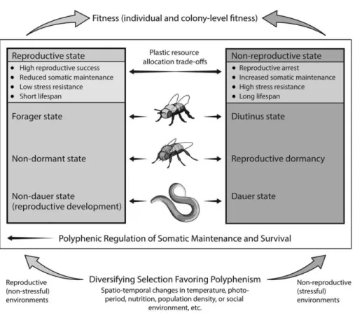
stimuli or cues from the environment; and their discrete alternative nature is either due to environmental discontinuity (e.g., discrete generations experiencing different seasons) or a discrete developmental switch in an envi-ronmentally sensitive threshold trait (e.g., trig-gered by critical photoperiod). Moreover, although perhaps not being a defining feature per se, polyphenic switches are typically cen-trally regulated by the neuroendocrine sys-tem, at least as far as is known (Nijhout 2003). In the following, we show that the plastic life histories of nematode worms, flies, and honey bees match these defining criteria and that they provide striking ex-amples of adaptive somatic maintenance and survival polyphenisms (Figure 1).
Note, however, that the criterion that poly-phenisms only refer to discrete (discontinu-ous) plastic traits is somewhat restrictive and artificial; typically this requirement can be re-laxed without loss of generality (see Mayr 1963; Canfield and Greene 2009; Simpson et al. 2011). This is because the boundaries between cases of continuous phenotypic plasticity and discrete alternative morphs are often very blu-rry: for example, as discussed by Nijhout (2003), the butterfly Araschnia levana occurs in nature in two seasonal polyphenic morphs, a spring and a summer form. However, a whole range of intermediate forms can be produced by timed injections of the steroid hormone ecdysone, or by imposing intermediate envi-ronmental conditions in the laboratory (also see discussion in Canfield and Greene 2009). Also note that the original and more inclusive definition of polyphenism by Mayr (1963) en-compasses cases of both discrete and continu-ous plasticity; thus, although the survival and maintenance polyphenism we discuss here can be said to involve qualitatively discrete pheno-typic states, we follow Canfield and Greene (2009) and Simpson et al. (2011) by applying Mayr’s more inclusive definition of polyphen-ism. Moreover, as pointed out by Simpson et al. (2011), it is very common that the same devel-opmental hormones (or other regulatory mechanisms) are involved in different cases of discrete or continuous plasticity. Our current mechanistic knowledge therefore does not support the dichotomy between discrete versus continuous plasticity (Simpson et al. 2011).
dauer diapause in C.ELEGANS
Under normal environmental conditions, nematodes such as C. elegans develop from an embryo through four larval stages (L1 to L4) into reproductively mature adults in about 3.5–4 days (at 20°C; reproductive develop-ment). In contrast, under stressful conditions (e.g., crowding, starvation, high tempera-ture), larvae arrest their development and alter their metabolism at the second molt (Figure 2). They bypass the normal L3 lar-val stage by forming a “dauer” larva (from the German word for “enduring”), an al-ternative stage that represents a facultative larval diapause, also called L3d (e.g., Cas-sada and Russell 1975; Riddle and Albert 1997; Braendle et al. 2007; Hu 2007; Fielen-bach and Antebi 2008; Fe´lix and Braendle
2010). The dauer, like other forms of dia-pause, represents a programmed state of ar-rested development and altered physiology that ensures somatic persistence and survival (e.g., Tatar and Yin 2001). In addition to dauer, C. elegans can also exhibit other less well-understood types of diapause or developmen-tal and physiological arrest (e.g., Ruaud and Bessereau 2006; Padilla and Ladage 2012). For example, under starvation conditions at hatch-ing, larvae undergo L1 diapause (Baugh and Sternberg 2006). These nondauer cases of de-velopmental plasticity in C. elegans are, how-ever, beyond the scope of our review.
It is easy to see how dauer larvae are somat-ically highly persistent: they have a hardened cuticle; do not feed (since they lack pharyngeal pumping) but use their fat reserves; are highly Figure 1. Maintenance and Survival Polyphenisms in Nematode Worms, Flies, and Bees
Dauer larval diapause and its associated adult phenotypes in the nematode (C. elegans), reproductive dormancy in the fruit fly (D. melanogaster) and other insects, and the worker castes of the honey bee (Apis mellifera) represent examples of adaptive life-history polyphenisms for somatic maintenance and survival in response to stressful (nonreproductive) environments.
resistant to multiple stresses, including heat, oxygen deprivation, starvation, and oxidation; and can survive without food for three to six months (e.g., Cassada and Russell 1975; Klass and Hirsh 1976; Larsen 1993; Lithgow et al. 1995; Riddle and Albert 1997; Hu 2007; Fielen-bach and Antebi 2008), or occasionally even up to 12 months (C. Braendle, pers. comm.). Whether dauer formation also protects against somatic DNA damage remains, to our knowl-edge, largely unknown; however, while dauer larvae are not more resistant than nondauer larvae to the life span-shortening effects of ion-izing radiation (Johnson and Hartman 1988), the microRNA mir-34, which is strongly upreg-ulated in the dauer stage, is known to be re-quired for a robust DNA damage response (see Karp et al. 2011 and references therein).
The changes leading to dauer formation are accompanied by substantial alterations in
metabolism, for example, involving a switch to glycolysis and fermentative metabolism (e.g., Vanfleteren and De Vreese 1995, 1996; Riddle and Albert 1997; Holt and Riddle 2003; Bur-nell et al. 2005; Fuchs et al. 2010). Once favorable conditions have returned, dauer larvae rapidly resume reproductive devel-opment, developing into postdauer L3/L4 larvae that, in turn, develop into reproduc-tively mature adults with normal adult life span (Riddle and Albert 1997; Fielenbach and Antebi 2008).
The determinants of entry to the dauer state act at the L1 stage and depend on the interplay of three environmental cues: population den-sity of conspecific individuals (i.e., crowding), food scarcity, and temperature (e.g., Golden and Riddle 1982, 1984a,b,c; Riddle and Albert 1997; Hu 2007). Crowding appears to be the primary cue triggering dauer formation and is Figure 2. Dauer Diapause in Nematode Worms
In response to adverse environmental conditions (starvation, crowding, high temperature), nematode worms (C. elegans) can enter a stress-resistant and long-lived larval diapause stage called “dauer”; the “decision” to enter the dauer state is mediated by several molecular pathways, in particular, by different hormonal signaling pathways.
mediated by several different small molecules that act as pheromones, called “ascarosides,” which can induce dauer arrest while at the same time preventing dauer recovery (e.g., Golden and Riddle 1984c; Jeong et al. 2005; Butcher et al. 2007, 2008, 2009; Srinivasan et al. 2008; Gallo and Riddle 2009; Kim et al. 2009; Braendle 2012; Ludewig and Schroeder 2013). The effects of these prodauer pheromones are opposed by a “food cue,” a heat-stable lipo-philic molecule produced by bacteria, the food source of the nematodes, which inhibits dauer arrest and promotes dauer recovery (Golden and Riddle 1984b). The option to enter/exit the dauer depends on the ratio of these two cues and is further modulated by temperature (Golden and Riddle 1982, 1984a,b). At growth temperatures between 15°C and 25°C, induc-tion of dauer formainduc-tion is moderate, whereas at 27°C induction is very strong and indepen-dent of the presence of prodauer pheromones (Ailion and Thomas 2000).
The phenomenon of dauer diapause is not only relevant for larval survival but also for survival and life span in the adult worm: many of the genes involved in C. elegans dauer formation (so-called daf genes; Rid-dle et al. 1981; Albert and RidRid-dle 1988) are also implicated in the regulation of adult stress resistance and longevity (e.g., Van-fleteren and Braeckman 1999; Tatar and Yin 2001; Rottiers and Antebi 2006; Fielen-bach and Antebi 2008; Gerisch and An-tebi 2011). Different mutations in these genes have been classified as “dauer for-mation-defective” (daf-d), i.e., mutants that always bypass “dauer” irrespective of envi-ronmental conditions, or as “dauer forma-tion-constitutive” (daf-c), i.e., mutants that always enter dauer diapause (e.g., Riddle et al. 1981; Albert and Riddle 1988; Rot-tiers and Antebi 2006; Fielenbach and An-tebi 2008). Interestingly, although dauer is a larval trait, certain weak daf-c mutant alleles of genes in the insulin/insulin-like growth factor signaling (IIS) or steroid hormone pathways (see below) allow the animals to bypass the larval dauer stage and to become stress-resistant and long-lived adults (e.g., Kenyon et al. 1993; Larsen 1993; Lithgow et al. 1994; Dor-man et al. 1995; Larsen et al. 1995; Gems et al. 1998; Rottiers and Antebi 2006; Fielenbach
and Antebi 2008; Gerisch and Antebi 2011). Furthermore, dauer pheromone can extend adult life span in C. elegans (Kawano et al. 2005). The somatic persistence and slow aging that is characteristic of dauer diapause larvae can thus apparently also apply to the reproduc-tive adult phase (Tatar and Yin 2001).
Dauer diapause clearly matches the above definition of an adaptive life-history poly-phenism (also see Braendle et al. 2007). It is induced by defined environmental token cues and involves a switch between two adap-tive, qualitatively discrete life-history “modes” (Figures 1 and 2). Moreover, the associated somatic maintenance and survival “program” can also extend to the adult stage. In addi-tion, the dauer syndrome is under strong hormonal control (see below). Dauer dia-pause thus represents a clear example of the kind of adaptive somatic maintenance and survival polyphenisms that we have in mind (also see Tatar and Yin 2001). This being said, we are not aware of any study that has directly quantified the demographic fitness benefits and costs of the wild-type dauer ver-sus nondauer life-history “strategy” under dif-ferent environmental conditions.
reproductive dormancy in
D. MELANOGASTER
Many insects, including Drosophila, can exhibit a state of “dormancy.” Dormancy is de-fined as an environmentally induced arrest of growth, development, and activity, accompa-nied by a downregulation of metabolic func-tion, which may or may not be adaptive, but which enables somatic persistence over time (e.g., Danilevskii 1965; Tauber et al. 1986; Danks 1987; Tatar et al. 2001a; Košta´l 2006; Schmidt 2011). Two basic types of dormancy can be distinguished, quiescence and dia-pause. In quiescence, the dormant state is a direct and immediate response to unfavorable aseasonal and/or unpredictable environmen-tal conditions. Such a response may be faculta-tive or inevitable and can be adapfaculta-tive or not. In diapause, by contrast, the dormant state: is a profound physiological response to anticipa-tory token cues (e.g., temperature, photope-riod) in unfavorable (seasonally) predictable environments; includes defined physiological phases (prediapause, diapause, postdiapause);
is under central neuroendocrine control; and clearly represents an adaptation to (typically seasonally) unfavorable environments (e.g., Danilevskii 1965; Tauber et al. 1986; Danks 1987; Tatar et al. 2001a; Košta´l 2006; Schmidt 2011).
In response to low temperatures and short day length, several temperate-zone species of Drosophila enter a state of adult reproductive dormancy, characterized by arrested ovarian development, increased stress resistance, and improved survival ability (e.g., Carson and Stalker 1948; Kambysellis and Heed 1974; Lumme et al. 1974; Lumme 1978; Lumme and Lakovaara 1983; Kimura 1988a,b; Saunders et al. 1989; Tatar and Yin 2001; Tatar et al. 2001a; Tatar 2004; Schmidt et al. 2005a,b; Schmidt and Paaby 2008; Schmidt 2011; Figure 3). Al-though most authors refer to this phenome-non as reproductive or ovarian “diapause,” at least in D. melanogaster this state of
reproduc-tive dormancy is quite weak, lacking both a preparatory prediapause and a postdiapause phase, and with dormancy induction being rapid, taking place within a few days after eclosion (e.g., Saunders et al. 1989; Saunders and Bertossa 2011). This dormant state might thus not qualify as a proper diapause but might rather represent quiescence, although there is still some debate about this issue (e.g., Tatar et al. 2001a; Emerson et al. 2009b; Schmidt 2011); for simplicity here we use the neutral term “reproductive dormancy.”
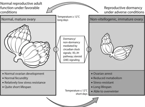
The biology of reproductive dormancy is best understood in D. melanogaster (e.g., Saunders et al. 1989, 1990; Saunders and Gilbert 1990; Tatar and Yin 2001; Tatar et al. 2001a; Schmidt 2011). In this species, flies enter dormancy at temperatures ⱕ 12–13°C and short-day photoperiod (ⱕ 12 hours light). In response to these conditions, flies exhibit ovarian arrest (blocked vitello-Figure 3. Adult Reproductive Dormancy in Fruit Flies
In response to short day length and cool temperatures, fruit flies (D. melanogaster and related species) can undergo a state of reproductive dormancy, which is associated with ovarian arrest, increased stress resistance, and greatly improved adult survival. Although the details are not yet fully understood, this dormant state appears to be mediated by changes in the circadian clock, the insulin/insulin-like growth factor (IIS), ecdysone (20E), and juvenile hormone (JH) signaling pathways.
genesis), improved resistance to oxidative and heat stress, and strongly reduced rates of senescence during diapause (e.g., Saun-ders et al. 1989, 1990; SaunSaun-ders and Gil-bert 1990; Tatar et al. 2001a; Schmidt et al. 2005a,b; Schmidt and Paaby 2008). This state of dormancy is reversible and can be “bro-ken” by higher temperature and longer pho-toperiod (e.g., Saunders and Gilbert 1990). Yet, whether flies enter dormancy or not, and to what extent dormancy is triggered by tem-perature and/or photoperiod, depends on their genotype and geographic origin (lati-tude; e.g., Saunders et al. 1989; Williams and Sokolowski 1993; Mitrovski and Hoffmann 2001; Tatar et al. 2001a; Schmidt et al. 2005a,b; Emerson et al. 2009b; Schmidt 2011). In par-ticular, while certain genotypes can readily enter reproductive dormancy under dorman-cy-inducing conditions, others cannot. This suggests the existence of substantial amounts of genetic variation and genotype-by-en-vironment (GxE) interactions for dormancy expression (e.g., Schmidt et al. 2005b, 2008). As compared to “low-dormancy” genotypes (i.e., those that are unable to undergo dor-mancy under dordor-mancy-inducing conditions), “high dormancy” genotypes (i.e., those that always undergo dormancy under dormancy-inducing conditions) are characterized by con-stitutively longer life span (i.e., median female life span is about 50 days as compared to 35 days in nondormancy genotypes), reduced age-specific mortality, lower early fecundity, im-proved resistance to starvation and cold stress, increased lipid sequestration, and a suite of other phenotypes, even when measured under nondormancy-inducing conditions (Schmidt et al. 2005b). Reproductive dormancy is thus a pleiotropic life-history syndrome, with multiple fitness traits being coordinately expressed in response to specific environmental cues (Flatt et al. 2005; Schmidt 2011). Similar kinds of reproductive dormancy have been described for grasshoppers and butterflies as well as other insects (e.g., Pener 1972; Nijhout 1994; Her-man and Tatar 2001; Tatar and Yin 2001; Flatt et al. 2005).
The available evidence indicates that re-productive dormancy in D. melanogaster (and presumably other species) is a life-history adap-tation that is most likely associated with the
ability of flies to overwinter (e.g., Izquierdo 1991; Mitrovski and Hoffmann 2001; Boule´-treau-Merle and Fouillet 2002; Hoffmann et al. 2003). This notion is consistent with the obser-vation from population genetic studies that some genotypes in local fly populations of tem-perate regions persist over long time periods, implying that the flies overwinter (e.g., Ives 1945, 1970). Moreover, the ability to undergo dormancy varies predictably with latitude, with flies in temperate northern populations hav-ing a much greater propensity to become dormant than flies from southern popula-tions, suggesting that the ability to express dormancy is a seasonal adaptation that has evolved along latitudinal gradients (Schmidt et al. 2005a). Flies from ancestral African populations seem unable to undergo mancy, consistent with the idea that dor-mancy is a recent evolutionary adaptation to temperate climates of an ancestrally tropical insect that has become cosmopolitan (Schmidt 2011). Additional evidence for the adaptive nature of reproductive dormancy comes from population cage experiments in the laboratory where, under stressful environ-mental conditions (i.e., bouts of starvation and cold stress), the frequency of genotypes able to express dormancy increased over time relative to the frequency of nondormant genotypes, whereas under favorable control conditions the reverse was observed (Schmidt and Conde 2006).
Like dauer diapause in nematodes, re-productive dormancy in Drosophila matches the defining criteria of polyphenism (e.g., Nijhout 2003). First, reproductive dormancy is clearly environmentally induced; seems to be adaptive; and involves a switch between two qualitatively discrete suites of life-history traits. Nondormant flies express a life-history “pro-gram” that favors reproduction at the expense of somatic maintenance and survival, whereas dormant flies express a “program” that favors somatic maintenance, stress resistance, and survival at the expense of reproduction (e.g., Flatt and Schmidt 2009; Paaby and Schmidt 2009; Schmidt 2011; Figures 1 and 3). Second, reproductive dormancy appears to be an en-vironmentally sensitive threshold trait whose expression is triggered by reliable and pre-dictable environmental token cues, namely
seasonally predictable changes in temperature and/or photoperiod. Moreover, reproductive dormancy is under strong neuroendocrine control (see below).
worker polyphenism inA.MELLIFERA
In contrast to C. elegans and D. melano-gaster, individuals of the A. mellifera worker caste usually do not reproduce directly. Honey bee reproduction is defined as the production of male individuals (drones) and of daughter colonies that “bud off ” from the mother colony by a process of fission called swarming (Winston 1987). To reproduce, a colony must build up to the critical size that enables fission in par-allel with the production of drones and new queens (Lee and Winston 1987). Col-ony size is defined by the number of workers. Workers, queens, and drones are produced during favorable ambient (sum-mer) conditions with the majority of re-sources allocated to worker rearing, since swarming requires 10,000 workers or more, but only one new queen (Winston 1987). Worker numbers are critical to colony func-tion since the workers (with the excepfunc-tion of egg laying) are responsible for all colony be-haviors, such as hygiene, caregiving, con-struction, foraging, and defense. Workers divide labor based on a system of temporal or “age” polyethism in summer in which individuals perform different tasks in se-quence as they age—i.e., a sort of sequen-tial behavioral polyphenism (Seeley 1982; Figure 4). Within-nest activities, such as nest hygiene, caregiving (often called nurs-ing), and construction, are typically per-formed prior to outside-nest activities like foraging. Summer workers make this transition to outside-nest activities around their third or fourth week of life and only survive another seven to 18 days as foragers (Visscher and Du-kas 1997; DuDu-kas 2008). Colony reproduction and foraging activities cease in the fall, and workers enter an alternative phenotypic state with a mean life span of greater than 100 days, with reported maxima between 212 and 304 days (Maurizio 1950; Fukuda and Sekiguchi 1966; Sakagami and Fukuda 1968; Mattila et al. 2001 and references therein); in contrast, “spring” and “summer”
bees have a mean life span of approximately only 30–40 days and 25–30 days, respectively (Fukuda and Sekiguchi 1966).
Although seasonal in principle, the long-lived worker phenotype of A. mellifera is not triggered by ambient cues but rather tied to the dynamic demography of the colony (Mau-rizio 1950; Amdam and Omholt 2002). The phenotype is called “diutinus” worker (Latin for long-lived) or winter bee, but is also trig-gered in summer if the brood (eggs, larvae, and pupae) is removed from the colony (Maurizio 1950; Amdam et al. 2004; Figure 4). This removal simulates the social envi-ronmental conditions of winter, when very little brood is produced, but also condi-tions that may occur short-term during swarming, while the colony waits for its new queen to mature, mate, and begin egg lay-ing. The winter bee state is “broken” when brood rearing commences as it does natu-rally in spring, and workers develop sum-mer bee characteristics with division of labor and short life spans (Sakagami and Fukuda 1968; Terada et al. 1975).
Honey bees originated in Africa (Whit-field et al. 2006), and the long-lived worker bee is likely an adaptation to life in temperate zones (Amdam et al. 2005). In accordance with this idea, the ability to produce “diutinus” or winter bees is not equally present among A. mellifera subspecies. For example, it is absent from African A. mellifera scutellata as well as from the scutellata hybrids called African-ized bees in America, which have conse-quently been unable yet to colonize areas north of California (Terada et al. 1975; Amdam et al. 2005). Winter bees are resis-tant to the oxidative stress-inducing agent paraquat (Seehuus et al. 2006) and to star-vation, and they show reduced accumula-tion of lipofuscin (cellular waste) in the brain as well as intact brain function (i.e., cognitive function as measured by a olfactory learning performance assay) for more than 200 days (D. Muench, G. V. Amdam, unpublished data). In comparison, summer bees are sensitive to par-aquat, with foragers being more susceptible than nest bees (Seehuus et al. 2006). Foragers also show rapid lipofuscin accumulation in the brain and glands (D. Muench, G. V. Amdam, unpublished data), and reduced brain
func-tion after 14 days (Behrends et al. 2007; Schei-ner and Amdam 2009).
Like dauer diapause in nematodes and reproductive dormancy in Drosophila, the long-lived worker phenotype of A. mellifera ful-fills the criteria of polyphenism: it is induced by changes in the (social) environment and seems to involve a switch between two qualita-tively discrete sets of life-history traits: sum-mer bees express a suite of life-history traits that favor colony reproduction at the ex-pense of worker somatic maintenance and survival, whereas winter bees express a life-history “program” that favors mainte-nance, stress resistance, and survival of the
colony until the next favorable season (Fig-ures 1 and 4). The winter bee state is trig-gered by a reliable environmental cue, since the absence of brood implies a halt in worker production that requires average worker life spans to increase if colony size and functions are to be maintained (Om-holt 1988; Smedal et al. 2009). Similar to worms and flies, this polyphenic switch in bees is also centrally regulated by the en-docrine system.
Thus, taken together, the plastic somatic maintenance and survival phenotypes of worms, flies, and bees clearly represent bona fide polyphenisms, and similar prin-Figure 4. Worker Polyphenism in Honey Bees
In the honey bee (A. mellifera), workers divide labor that is important for colony function (e.g., hygiene, caregiving, construction, foraging, and defense) based on temporal “age polyethism,” i.e., a sort of sequential polyphenism. In summer, nurse bees perform within-nest activities (e.g., nest hygiene and cargiving) prior to outside-nest activities such as foraging. In their third or fourth week of life, the nurses transition to become foragers. In fall, colony reproduction and foraging activities cease, and the workers enter a very long-lived phenotypic state called the “diutinus” worker or winter bee. Interestingly, the same state can be elicited in summer if the brood is removed from the colony.
ciples might apply to other organisms as well (“senescence plasticity”; Tatar and Yin 2001; Figure 1). However, we would also like to point out that the ephemeral poly-phenic nature of dauer/reproductive dia-pause in worms and flies is qualitatively distinct, at least to some extent, from more permanent developmental switches, such as, for example, wing morphs seen in crickets or aphids. Similarly, we note that there is also a certain disjunction between the polyphenic na-ture of dauer/diapause in worms and flies and that of worker polyphenisms in bees. Nonethe-less, all three cases provide clear examples of the plastic regulation of suites of correlated life-history traits, involving profound, environ-mentally induced changes in somatic mainte-nance and survival in response to conditions that are unfavorable to reproduction. We now turn to addressing how these adaptive life-history switches might have evolved.
Somatic Maintenance and Survival Polyphenisms Evolve By Diversifying
Selection
Mather (1955) laid the foundation for our current understanding of how poly-phenic patterns evolve. He suggested that in addition to segregational (genetic) poly-morphisms, disruptive (also called diversi-fying) selection may select for a genotype that is developmentally reactive, by means of a switching mechanism, to some environmental factor such that the “morphic types continue to share a common gene pool” (Mather 1955: 52). Mather thus anticipated that disruptive se-lection could convert a continuously variable trait into a discontinuous one governed by an environmentally sensitive switch. This was con-firmed, for example, by Scharloo (1970) who, by using a negative assortative mating system and artificial disruptive selection targeting cu-bitus interruptus mutant phenotype expres-sion in Drosophila, was able to obtain a bimodal distribution of the relative length of the fourth wing vein. Genetic analyses of these lines showed that this bimodal distribution is likely caused by a temperature-sensitive threshold mechanism.
The idea that temporally and/or spatially varying environments impose disruptive or diversifying selection that causes the evolution
of phenotypic plasticity and polyphenisms (“developmental switches”) rather than ge-netic diversification has been substantially elab-orated since (e.g., Levins 1968; Thoday 1972; Mather 1973; Moran 1992; Nijhout 2003; West-Eberhard 2003; Berrigan and Scheiner 2004; Leimar et al. 2006; Rueffler et al. 2006). Al-though plasticity can evolve under both spatial and temporal environmental heterogeneity (e.g., Levins 1968; Berrigan and Scheiner 2004; Leimar 2005; Leimar et al. 2006), theory suggests that it most readily evolves when envi-ronments vary temporally, for example, when nonoverlapping generations experience differ-ent environmdiffer-ents that alternate regularly and that are associated with reliable environmental cues (e.g., Levins 1968; Leimar 2005; Rueffler et al. 2006). This is indeed how seasonal poly-phenisms, such as the wet and dry season mor-phs in tropical butterflies, are thought to have evolved (e.g., Shapiro 1976; Brakefield and Zwaan 2011, and references therein).
The above explanatory scheme is fully con-sistent with how we believe that the observed somatic maintenance and survival polyphen-isms in worms, flies, and bees might have evolved (Figure 1). First, there exists ample genetic variation for the expression of poly-phenic life histories in all three systems for selection to act upon, i.e., genotype by envi-ronment interactions (G⫻E), and the ob-served life-history polyphenisms are stably maintained in natural populations (e.g., Viney et al. 2003; Rueppell et al. 2004; Schmidt 2005a,b; Schmidt and Conde 2006; Page and Amdam 2007; Harvey et al. 2008, 2009; Schmidt et al. 2008). Second, diversi-fying selection, which has likely shaped the evolution of these adaptive switches, appears to be caused in all three species by temporal and/or spatial environmental variation, such that reproductive possibilities are reduced or lacking because of unfavorable conditions in one environment as compared to the other. Third, as we have seen above, in each case the phenotypic switch is associated with reliable environmental cues, a major prereq-uisite for the evolution of polyphenisms. Finally, in each species, exposure to the un-favorable environment results in stored en-ergy reserves being plastically allocated to somatic maintenance, which allows
individu-als to survive until conditions have improved. We therefore consider all three polyphen-isms to be adaptive. Their existence suggests that variation in the underlying regulatory mechanisms must have been readily avail-able for selection to act upon and thus to shape life-history plasticity. Given that the somatic maintenance and survival polyphen-isms of worms, flies, and bees likely share a common selection principle that underlies their evolution, it is interesting to ask what precisely these mechanisms are and to see whether evolution has perhaps led to similar proximate solutions in all three species.
Molecular Basis of Polyphenic Regulation of Somatic Maintenance
and Survival
The plastic life histories of worms, flies, and bees all require a regulatory machin-ery that makes switch-like developmental and physiological transitions in response to changes in the environment. These species thus exhibit what we may call polyphenic regula-tion of somatic maintenance and survival. Over the past few decades, a great deal has been learned about the mechanistic underpinnings of these adaptive life-history switches (see Tatar and Yin 2001; Flatt et al. 2005; Rottiers and Antebi 2006; Braendle et al. 2007; Fielenbach and An-tebi 2008; Emerson et al. 2009a; Flatt and Hey-land 2011; Ga´likova´ et al. 2011; Gerisch and Antebi 2011; Schiesari et al. 2011; Schmidt 2011; Lee and Schroeder 2012; Schaedel et al. 2012). Based on the currently available evi-dence, we argue that there are many profound similarities in the proximate regulation of life-history polyphenisms among worms, flies, and bees, particularly at the hormonal level (Figure 5).
mechanisms of dauer diapause in
C.ELEGANS
The polyphenic regulation of somatic main-tenance and survival is mechanistically best un-derstood in C. elegans, a powerful genetic model system. These mechanisms have been reviewed in depth elsewhere; here we give a pre´cis of the molecular regulation of dauer formation as reviewed and discussed by Hu (2007), Fielenbach and Antebi (2008), and Gerisch and Antebi (2011).
Genetic studies have discovered more than 30 daf genes, mutations which are known to misregulate dauer formation (e.g., Riddle et al. 1981; Albert and Riddle 1988). These loci belong to at least six dif-ferent functional pathways: sensory neuronal function and perception, including G-protein-coupled receptor (GPCR)/G-protein signal-ing; neurosensory transduction via cyclic guanosine monophosphate (cGMP) signaling; serotonergic neurotransmission; paracrine and endocrine signaling via the transforming growth factor beta (TGF-) pathway; insulin/ insulin-like growth factor signaling (IIS); and steroid hormone signaling (see Hu 2007; Fielenbach and Antebi 2008; Gerisch and An-tebi 2011; Lee and Schroeder 2012, and refer-ences therein; Figures 2 and 5).
Although the regulatory connections that lead to dauer formation versus repro-ductive development (i.e., normal develop-ment bypassing the dauer) are not strictly hierarchical but should rather be thought of as comprising a highly complex net-work, including parallel and independent signaling outputs as well as feedback and feedforward loops, a number of major reg-ulatory steps or levels can be recognized (Hu 2007; Fielenbach and Antebi 2008; Gerisch and Antebi 2011).
The first step in the regulation of dauer versus nondauer is the detection, process-ing, and integration of environmental stimuli and cues, such as nutrition, tem-perature, and dauer pheromone, by the nervous system; for example, GPCRs that reside in the ciliated endings of neurons situated in the two major head sensory organs, the amphids, detect dauer phero-mone and food signals. In turn, these neu-rosensory signals are transduced via G-proteins and cGMP signaling, including signaling through a transmembrane guanylyl cyclase encoded by daf-11, which converts guanosine triphosphate (GTP) to cGMP, and two subunits of a cGMP-gated ion chan-nel encoded by tax-2 and tax-4, which trans-late cGMP levels into ion flux. The second step is the integration and transduction of neurosensory signals by the hormonal sys-tem: cGMP signaling acts upstream of two major endocrine signaling pathways, TGF-
signaling and IIS, and—under conditions of reproductive development—high levels of cGMP stimulate the production of hormone ligands, i.e., TGF- and insulin-like peptides (ilps). In various target tissues, binding of the ilp ligands to the insulin-like receptor encoded by daf-2 sets off the IIS cascade, which involves a series of phosphorylation steps that leads to the cytoplasmic retention and inactivation of the forkhead box-O transcription factor Foxo encoded by daf-16. Similarly, the TGF- ligand encoded by daf-7 activates TGF- signaling in target tissues by binding to two TGF- recep-tors encoded by daf-1 and daf-4. This in turn leads to the phosphorylation and nuclear local-ization of two nuclear effectors of the pathway,
so-called SMADs (the term is a merger of the names of the Mad—mothers against decapentaple-gic—gene in D. melanogaster and the Sma genes in C. elegans), encoded by daf-8 and daf-14. Notably, TGF- signaling also regulates the production of ilps, and there exists extensive cross-talk between TGF- signaling and IIS. In the third step, the IIS and TGF-  signal-ing pathways converge on the regulation of steroid hormone signaling; in particular, both pathways regulate the production of steroid hormones called dafachronic acids (DA) produced in the neuroendocrine XXX cells, which are ligands of the nuclear/steroid hormone receptor encoded by daf-12. Finally, DA-liganded DAF-12 exerts transcriptional ef-Figure 5. Molecular Pathways Underlying Life-History Polyphenisms in Worms, Flies, and Bees
Comparative models showing the molecular pathways leading to dauer diapause in C. elegans (left panel), reproductive dormancy in D. melanogaster (middle panel), and the winter bee (diutinus) state in A. mellifera (right panel). Many of the mechanistic details underlying the regulation of these adaptive life-history switches still remain unknown (as indicated by question marks or dashed lines). However, some general regulatory features are beginning to emerge: for example, a central (maybe event dominant) role in all three systems is played by insulin/insulin-like growth factor signaling (IIS). Similarly, in all three cases, IIS seems to regulate the production of lipophilic hormones downstream of IIS, such as the steroid hormones (dafachronic acid or 20-hydroxyecdysone) and/or the sesquiterpenoid juvenile hormone. These hormones in turn mediate the switch between two alternative states: a “program” expressed under normal environmental conditions that promotes reproduction at the expense of somatic maintenance and survival and an alternative “program,” expressed under suboptimal or stressful conditions, which promotes somatic maintenance, stress resistance, and survival at the expense of reproduction.
fects that promote reproductive development and inhibit dauer formation. In contrast, when IIS or TGF- signaling are downregulated, the expression of steroid hormone biosynthetic en-zymes is inhibited; under such conditions, the unliganded DAF-12 receptor promotes the dauer (nonreproductive) program and inhib-its reproductive development (also see Rottiers and Antebi 2006; Fielenbach and Antebi 2008; Gerisch and Antebi 2011; Lee and Schroeder 2012; Schaedel et al. 2012; Wollam et al. 2012). As already mentioned above, many of the mechanisms involved in specifying dauer versus nondauer (including GPCR, serotonin, TGF- signaling, IIS, and ste-roid hormone signaling) also play a major role in regulating C. elegans adult life his-tory, especially somatic maintenance (e.g., energy storage, stress resistance) and sur-vival (e.g., Kenyon et al. 1993; Larsen 1993; Lithgow et al. 1994; Dorman et al. 1995; Larsen et al. 1995; Gems et al. 1998; Apfeld and Kenyon 1999; Alcedo and Kenyon 2004; Rottiers and Antebi 2006; Shaw et al. 2007; Fielenbach and Antebi 2008; Gerisch and Antebi 2011; Lee and Schroeder 2012). The probably most famous example of a gene involved in dauer regulation with known effects on the adult phenotype is the insulin-like receptor gene, daf-2. Mod-est impairment of daf-2 causes increased resistance of adults to a variety of stresses and a doubling of adult life span (e.g., Kenyon et al. 1993). Depending on the specific mutant allele, the strength of its phe-notypic effects, as well as the maintenance tem-perature in the laboratory, daf-2 mutants are highly pleiotropic, also affecting traits such as developmental rate, mobility, fat storage, brood size, and the length of the reproductive period; however, many of these traits can be decoupled from longevity (Gems et al. 1998; Dillin et al. 2002; Fielenbach and Antebi 2008; Gerisch and Antebi 2011).
Interestingly, many of the mechanisms that modulate C. elegans life history also regulate Drosophila life history (e.g., Tatar and Yin 2001; Flatt et al. 2005; Fielenbach and Antebi 2008; Ga´likova´ et al. 2011; Gerisch and Antebi 2011), as we will dis-cuss next. In fact, it can be argued that the regulation of the dauer stage and its
asso-ciated adult phenotypes in C. elegans is functionally homologous to the regulation of adult reproductive dormancy in insects such as Drosophila (cf. Tatar and Yin 2001).
mechanisms of reproductive dormancy inD.MELANOGASTER
Compared to C. elegans, much less is known about the molecular regulation of reproductive dormancy in D. melanogaster. In contrast to the over 30 daf genes affect-ing C. elegans dauer formation (e.g., Riddle et al. 1981; Albert and Riddle 1988; Fielen-bach and Antebi 2008; Gerisch and Antebi 2011), only three loci have so far been impli-cated in controlling the propensity of D. mela-nogaster to undergo dormancy (for reviews see Emerson et al. 2009a; Schiesari et al. 2011; Schmidt 2011; see below). Nonetheless, the endocrine regulation of reproductive dor-mancy in flies and other insects is reason-ably well understood, and there seem to exist several intriguing parallels between the hormonal mechanisms involved in dauer diapause in worms and adult repro-ductive dormancy in insects (e.g., Tatar and Yin 2001; Flatt et al. 2005; Tu et al. 2006; Ga´likova´ et al. 2011; Gerisch and An-tebi 2011; Schmidt 2011). The mecha-nisms known to underlie dormancy in D. melanogaster can be grouped—somewhat artificially—into two categories: endocrine and circadian/photoperiodic regulation (Figures 3 and 5).
Similar to its role in C. elegans dauer formation, insulin/insulin-like growth fac-tor signaling (IIS) also seems to mediate reproductive dormancy in fruit flies and other insects (e.g., Tatar and Yin 2001; Flatt et al. 2005; Tu et al. 2006; Emerson et al. 2009a; Gerisch and Antebi 2011; Schiesari et al. 2011; Schmidt 2011, and references therein). By applying deletion mapping to two natural dormancy variants of D. melanogaster, one line with high dia-pause expression and the other with low expression, Williams et al. (2006) found a strong association between the propensity of flies to undergo dormancy and natural varia-tion at PI3K (synonyms: Pi3K92E, Dp110), the locus encoding phosphoinositide 3-kinase, an enzyme intimately involved in IIS. Further
complementation and transgenic analyses showed that a reduction in PI3K dosage and function increases dormancy expression, sug-gesting that reduced IIS might induce a state of adult reproductive dormancy (Williams et al. 2006). Interestingly, the Drosophila PI3K locus is the homolog of age-1 in C. elegans, which is known to regulate dauer formation (e.g., Wolkow et al. 2000), thus lending further sup-port to the homology between dauer diapause and insect reproductive dormancy (Tatar and Yin 2001; Gerisch and Antebi 2011).
Other observations also suggest that IIS might be a major endocrine determinant of dormancy in insects. Various D. melano-gaster mutants in the IIS pathway, for ex-ample, mutants of the insulin receptor InR (the homolog of C. elegans daf-2) or the insulin receptor substrate chico, phenocopy (at least partially) suites of phenotypes ob-served in the dormancy state, including arrest of vitellogenic egg development, in-creased stress resistance, and extended life span (e.g., Clancy et al. 2001; Tatar and Yin 2001; Tatar et al. 2001b, 2003; Flatt et al. 2005; Tu et al. 2006). Moreover, in the mosquito Culex pipiens, RNA interference (RNAi) di-rected against InR in nondormant mosquitos reared under long day conditions phenocopies aspects of reproductive diapause, whereas RNAi silencing of the forkhead transcription dfoxo (Drosophila foxo, the homolog of C. elegans daf-16) downstream of IIS in mosquitos pro-grammed to undergo reproductive diapause has the opposite effect (Sim and Denlinger 2008). Taken together, these observations strongly indicate that IIS in insects plays a role in regulating dormancy/diapause that is func-tionally homologous to that in C. elegans, as previously postulated (Tatar and Yin 2001; Gerisch and Antebi 2011; also see below). In addition, it is noteworthy that major compo-nents of IIS not only profoundly affect life span in C. elegans and D. melanogaster, but also seem to influence mammalian and in particular also human longevity. For example, variants of FOXO3A and FOXO1, two human orthologs of dFOXO/DAF-16, have been associated with longevity in humans (for a recent review see Kenyon 2010).
Adult reproductive dormancy (and other forms of dormancy/diapause) is also
modu-lated by two other major insect hormones, the sesquiterpenoid juvenile hormone (JH) and the steroid hormone ecdysone (20-hy-droxyecdysone, 20E), two lipophilic molecules whose production is regulated, at least in part, by IIS (e.g., Nijhout 1994; Tatar and Yin 2001; Flatt et al. 2005; Tu et al. 2006). In larval in-sects, both hormones are produced in the so-called ring gland, a composite gland complex situated behind the brain: the precursors of 20E (the final conversion into the active hor-mone 20E occurs at the target tissues) are pro-duced in a part of the ring gland called the prothoracic gland, whereas JH and its precur-sors are produced in the corpora allata glands or, in Dipterans such as D. melanogaster, in their single corpus allatum (e.g., Nijhout 1994; Gäde et al. 1997; Gilbert et al. 2002; Flatt et al. 2005; Jones and Jones 2007). Although the protho-racic gland degenerates at metamorphosis and the gonads become the major ecdysteroido-genic tissue, the corpus allatum persists through metamorphic development and con-tinues to be the adult source of JH. Under dormancy-inducing conditions, the titers of JH and 20E are decreased, whereas ectopic treat-ment of dormant flies with natural or synthetic JH and 20E terminates their dormancy and restores vitellogenesis (e.g., Saunders et al. 1989, 1990; Richard et al. 1998, 2001a,b; Tatar and Yin 2001; Tatar et al. 2001a; Flatt et al. 2005). In support of the notion that JH plays a major role in regulating dormancy, surgical re-moval of the corpora allata results in reduced fecundity or sterility and extends life span in grasshoppers, butterflies, bugs, and D. melano-gaster, suggesting that JH is a positive regulator of fecundity, but a negative regulator of life span (e.g., Herman and Tatar 2001; Tatar and Yin 2001; Flatt et al. 2005; Flatt and Kawecki 2007; Hodkova 2008; Tatar et al. 2010; Hod-kova and Tatar 2011). However, evidence sug-gests that the effects of JH on life span and reproduction may be separable since they can be experimentally uncoupled (Hodkova 2008; Tatar et al. 2010; Hodkova and Tatar 2011). Consistent with the idea that 20E is an impor-tant mediator of dormancy, ecdysteroid titers differ between North American wild-type fly lines that differ in dormancy propensity, with low dormancy lines showing a higher titer un-der dormancy-inducing conditions than high
dormancy lines (K. J. Min, T. Flatt, and P. S. Schmidt, unpublished data). Moreover, cur-rent evidence suggests that there exist strong genetic and physiological similarities between 20E/ecdysone receptor (EcR) signaling in Drosoph-ila and dafachronic acid (DA)/daf-12 signaling in C. elegans aging and life history, thus under-scoring the importance of steroid hormone signaling in regulating life-history plasticity (Ga´likova´ et al. 2011). Although the mechanis-tic details await further investigation, it is clear that both JH and 20E play important endo-crine roles in affecting dormancy propensity.
Several lines of evidence suggest that the production of both JH and 20E is regu-lated by IIS. Hypomorphic mutations at both InR and chico reduce JH biosynthesis levels, and ectopic treatment of sterile long-lived and JH-deficient InR mutant fe-males with synthetic JH (methoprene) partly restores vitellogenesis and reduces life span to wild-type level (Tatar et al. 2001b; Tu et al. 2005; but also see Richard et al. 2005). Similarly, ovarian ecdysone synthesis is impaired in InR mutant females (Tu et al. 2002), and genetically up- or downregulating IIS in the prothoracic gland increases and decreases circulating ecdysone titers, respectively (Colombani et al. 2005).
The second major candidate gene known to affect dormancy propensity in natural popula-tions of D. melanogaster, couch potato (cpo), also appears to be part of this IIS/ecdysone signal-ing network. The cpo locus encodes a RNA-binding protein expressed in the peripheral nervous system, in glia cells, the midgut, sali-vary glands, and—notably—the ring gland (Bellen et al. 1992; Harvie et al. 1998). Using a combination of quantitative trait (QTL) map-ping, genetic complementation mapmap-ping, and linkage association analysis, Schmidt et al. (2008) found that a single amino acid substi-tution at cpo, which varies clinally along the North American east coast, determines whether flies under dormancy-inducing condi-tions enter dormancy or not. Interestingly, cpo interacts genetically with PI3K in regulating dormancy expression, thus establishing a link between cpo and IIS (Schmidt 2011). Moover, cpo contains a number of ecdysone re-sponse elements, suggesting that cpo might be
targeted by or involved in ecdysone signaling (Schmidt et al. 2008). In summary, these ob-servations clearly implicate the neuroendo-crine-ovarian IIS/20E signaling axis in the regulation of reproductive dormancy in D. melanogaster (e.g., Tatar and Yin 2001; Flatt et al. 2005; Tu et al. 2006; Emerson et al. 2009a; Schiesari et al. 2011; Schmidt 2011). This is consistent with the fact that dormancy expres-sion varies clinally (see above) and with the observation that several major genes in the IIS and 20E signaling pathways show major ge-netic differentiation along the Australian and North American latitudinal clines (Kolacz-kowski et al. 2011; Fabian et al. 2012).
In addition to the regulation of 20E and JH by IIS, recent studies have found that JH and 20E production are regulated by transforming growth factor  (TGF-)/ bone morphogenetic protein (BMP) sig-naling. The TGF- ligand activin regulates the competence of the prothoracic gland to receive and respond to prothoracico-tropic hormone (PTTH) and insulin sig-nals that in turn control the expression of 20E biosynthesis enzymes (Gibbens et al. 2011; also see McBrayer et al. 2007; Rewitz et al. 2009). Similarly, Huang et al. (2011) found that decapentaplegic (DPP)-mediated TGF-/BMP signaling regulates JH biosynthe-sis by activating the expression of JH acid meth-yltransferase (JHAMT). Thus, although it remains unclear whether TGF-/BMP signal-ing affects reproductive dormancy in Drosoph-ila, these findings suggest that there may be parallels between worms and flies in the TGF-/BMP regulation of lipophilic hormones known to be involved in modulating diapause/ dormancy.
The third candidate gene found to affect dormancy in natural populations, timeless (tim), does not seem to be directly involved in the endocrine control of dormancy, but probably plays a role in its photoperiodic regulation (Sandrelli et al. 2007; Tauber et al. 2007; Emerson et al. 2009a; Schiesari et al. 2011; Schmidt 2011). Since dormancy is elicited by a shortening of the photope-riod, its expression is dependent on light and day length. Consequently, the circa-dian clock, which regulates different kinds of daily rhythms, and the so-called
“circa-dian” or “clock genes” may be involved in the regulation of dormancy expression, al-though the link between photoperiodism and circadian rhythms is somewhat contro-versial (e.g., Saunders 2002; Danks 2005; Emerson et al. 2009a; Saunders and Ber-tossa 2011; Schiesari et al. 2011; Schmidt 2011). Sandrelli et al. (2007) and Tauber et al. (2007) found that the derived ls-tim mutation at the tim locus has spread by selection over the past 10,000 years in Eu-rope. The ls-tim allele attenuates the pho-tosensitivity of the circadian clock and increases the incidence of ovarian dor-mancy in response to changes in light and temperature in different genetic backgrounds (Sandrelli et al. 2007; Tauber et al. 2007). Moreover, since dormancy incidence varies cli-nally, it is interesting to note that some of the clock genes and the cryptochrome (cry) gene, which affects circadian resetting and photosen-sitivity, are genetically differentiated along the Australian and North American clines (Kolac-zkowski et al. 2011; Fabian et al. 2012).
The tim locus then, with its role in photo-periodic regulation of dormancy, represents an example of a mechanism that mediates the sensory perception of an environmental signal. These signals are then integrated and medi-ated by the endocrine system (e.g., IIS/ ecdysone signaling), and the hormonal signals are in turn translated into the physiological responses underlying dormancy. So what is the connection between circadian rhythms/ photoperiod and the endocrine system? Not much is known, but a few observations suggest that both systems interact quite intimately. In D. melanogaster, neurosecretory cells in the pars intercerebralis (the insulin-producing cells, IPCs) and corpus allatum cells exhibit rhyth-mic daily changes in nuclear size that might be related to their secretory activity (Rensing 1964), and in several other insects ecdysone and JH titers undergo circadian/diurnal fluc-tuations (e.g., Zhao et al. 2004a,b; Steel and Vafopoulou 2006; Polanska et al. 2009; P. Klep-satel, C. Dauphin-Villemant, and T. Flatt, un-published data). Furthermore, clock genes seem to be strongly expressed in the protho-racic part of the ring gland (Plautz et al. 1997), and Itoh et al. (2011) have recently found that
ecdysone signaling is involved in the regulation of circadian oscillations in D. melanogaster.
mechanisms of worker polyphenism in
A.MELLIFERA
The correlation between brood and worker bee life spans led researchers to suspect that brood pheromone, a blend of 10 fatty acid methyl and ethyl esters produced by the larval salivary glands, is the environmental cue that controls the alternative programs of worker longevity (Smedal et al. 2009). Brood phero-mone has complex effects on worker gene ex-pression, physiology, and behavior (Pankiw et al. 1998, 2008; Pankiw and Page 2001; Alaux et al. 2009). Importantly, it inhibits adult bees from sequestering a lipoprotein called vi-tellogenin (Vg) into the abdominal fat body (Smedal et al. 2009), a tissue func-tionally homologous to mammalian liver and adipose tissue. Vitellogenins are phy-logenetically widespread in oviparous ani-mals, where they serve as egg-yolk proteins. Vitellogenin was identified in honey bees about 40 years ago and its reproductive role in queens was instantly recognized (Engels 1974). But, curiously, vitellogenin was also found in considerable amounts in the wor-kers, which are normally sterile. The workers’ expression of vitellogenin was first deemed evolutionary baggage, an unavoidable conse-quence of selection for extreme vitellogenin production rates in queens. Later studies, how-ever, revealed pleiotropic functions: vitelloge-nin coordinates social behaviors in workers and enhances stress resistance, immunity, and survival in both workers and queens (see Seehuus et al. 2006; Nelson et al. 2007, and references therein). At least some of these functions result from interplay with JH (Fig-ures 5 and 6).
In summer, vitellogenin and JH act to-gether in a feedback loop to control forag-ing onset in worker bees (Amdam and Omholt 2003; Figure 6). High vitellogenin levels suppress JH and foraging behavior, while high JH levels suppress vitellogenin and nest activities. RNAi experiments con-firm that JH increases when vitellogenin is suppressed (Guidugli et al. 2005), and that vitellogenin knockdown workers forage preco-ciously (Nelson et al. 2007; Marco Antonio et
al. 2008). The vitellogenin knockdowns, moreover, are more sensitive to the oxida-tive stress-inducing agent paraquat and are characterized by reduced longevity inde-pendent of when they begin foraging (See-huus et al. 2006; Nelson et al. 2007). In temperate regions, workers’ circulating blood levels of vitellogenin appear to be very high when the brood is absent from colonies (almost 100 g/l blood) com-pared to when the brood is present (up to 25g/l blood; Amdam et al. 2004, 2005). The very high vitellogenin levels are paral-leled by low JH levels (Fluri et al. 1977). The accumulation of vitellogenin in the absence of brood was first explained simply by reduced consumption rates of the pro-tein, as workers expend vitellogenin in the production of proteinaceous food secre-tions that are fed to brood, queen, and other adult colony members (Amdam and Omholt 2002). However, it was shown later that brood pheromone is sufficient to
in-hibit vitellogenin accumulation, suggesting a different level of regulatory control than simple feeding rates, since the actual amount of brood to feed was taken out of the equation (Smedal et al. 2009).
It is currently unknown how honey bee brood pheromones interact with worker phys-iology to regulate vitellogenin and also how vitellogenin acts, in a molecular sense, to exert its different functions on worker behav-ior and longevity. Recently, the honey bee vitellogenin gene was found to have been subject to recurrent positive selection in Eu-ropean but not African populations (Kent et al. 2011). We believe this new finding sup-ports the hypothesis that changes in vitel-logenin occurred to accommodate colony survival in colder climates during and after the prehistoric migrations of A. mellifera from Africa to Europe (Amdam et al. 2005; See-huus et al. 2006). Kent et al. (2011) discuss 64 single nucleotide polymorphisms (SNPs) that are unequally distributed in the vitel-Figure 6. The Vitellogenin (Vg)-Juvenile Hormone (JH) Double Repressor Model
Behavioral and life-history maturation in workers of the honey bee (A. mellifera) is regulated by a negative feedback loop between vitellogenin (Vg) and juvenile hormone (JH). In nurse bees, which are characterized by high pollen intake, corpulent bodies, and high levels of stress resistance, Vg titers are high which causes a reduction in JH levels. When workers transition to become foragers, characterized by high nectar intake, lean bodies, and low stress resistance, JH titers increase which causes a drop in Vg levels.
logenin sequence. Roughly dividing the pro-tein into two parts, the N-terminal domain (N-sheet) is resistant to change, while the major lipid-binding cavity is sprinkled with SNP hot-spots. The N-sheet contains the phylogeneti-cally conserved, putative receptor-binding do-main of vitellogenin that presumably is important for uptake into the ovary (Li et al. 2003). The remaining part of vitellogenin is dominated by a lipid-binding cavity where poly-morphisms may alter ligand-binding proper-ties. JH is a proposed ligand of vitellogenin, as is brood pheromone (Smedal et al. 2009; Nilsen et al. 2011). Alternatively, the general lipid load of vitellogenin might vary in size or composition based on structural features of the cavity and influence traits associated with be-havior and survival (Havukainen et al. 2011).
Other studies have connected vitellogenin to IIS. Honey bees have two insulin-like pep-tides (ilp1 and ilp2), both of which are ex-pressed in the fat body (Nilsen et al. 2011). Expression of ilp1 is correlated with vitelloge-nin expression, but not affected by vitellogevitelloge-nin knockdown, suggesting that both genes are sensitive to (the same) nutrients. In contrast, ilp2 and vitellogenin are associated indirectly: there is a correlation between ilp2 expression and JH, which is positive when vitellogenin is expressed and negative when vitellogenin is silenced. A model in which ilp1 and ilp2 is the agonist versus antagonist of honey bee insulin receptors, respectively, has been put forward to explain these results, similar to the INS-7 versus INS-1 (insulin-like peptide) agonist/antagonist system of C. elegans (Nilsen et al. 2011). It is, however, unknown whether and how IIS affects honey bee longevity. Correlations be-tween the expression of JH, ilp1, ilp2, and (life-shortening) foraging activities have been pointed out, but recent data from insulin re-ceptor substrate (IRS) knockdown do not sug-gest that worker life spans are extended by reduced IIS (K. Ihle, G. V. Amdam, unpub-lished data). In other words, vitellogenin is the only gene that is functionally validated as a life span regulator in this animal.
The Relevance of Polyphenisms for Molecular Biogerontology “Context and interaction are of the es-sence” (Lewontin 1974:318).
What is the relevance of polyphenisms for somatic maintenance and survival, or more generally of “senescence plasticity” (i.e., phenotypic plasticity in life span or the rate of aging), for molecular biogeron-tology, which focuses on discovering and understanding genetic effects on life span in model organisms?
The most important point is that life-history traits such as life span are charac-terized by a very high degree of phenotypic plasticity, i.e., the expression of specific life-history phenotypes is highly contingent upon the environment (e.g., Finch 1990; Stearns 1992; Chippindale et al. 1993, 1997; Nylin and Gotthard 1998; Roff 2002; Fielen-bach and Antebi 2008; Mu¨nch et al. 2008; Flatt and Schmidt 2009; Mu¨nch and Amdam 2010; Flatt and Heyland 2011, and references therein). For example, Drosophila life span is highly sensitive to changes in diet (e.g., Mair et al. 2003; Tatar 2007) and temperature (e.g., Maynard Smith 1958; Mair et al. 2003).
An experimentalist who studies, say, the effects of a well-defined gene mutation upon life span, for example, in response to changes in nutrition, temperature, or pho-toperiod, can view the problem of “senes-cence plasticity” from two different angles. One angle would be to deliberately ignore the environmental dimension of the prob-lem by treating environmentally engendered variation in life span as a confounding nui-sance and thus to strictly control all aspects of the environment so that the experiment is carried out in a single, well-defined, and con-stant setting. The other angle would be to accept the possibility that a given genotype might exhibit different life spans when ex-posed to, say, different food levels, and thus to ask whether and how this genotype’s life span varies as a function of systematic, con-trolled changes in food levels. When adopting this point of view, researchers might further anticipate that different genotypes, say the mu-tant allele versus the “wild-type” allele (the “control”), might differ in their life span re-sponse to different environments due to a genotype by environment interaction (G⫻E interaction; Stearns 1992). These approaches are clearly very different, so what are the