*Corresponding author: Andrea R. Genazzani, MD, PhD, HcD, FRCOG, Division of Gynecology and Obstetrics, University of Pisa, Pisa, Italy, Phone: +39 050 503985, Fax: +39 050 220 7028, Mobile: +39 335 7211224, E-mail: argenazzani@gmail.com Nicola Pluchino: Division of Gynecology and Obstetrics, University Hospital of Geneva, Geneva, Switzerland
Marinella Russo: Division of Gynecology and Obstetrics, University of Pisa, Pisa, Italy
Nicola Pluchino, Marinella Russo and Andrea R. Genazzani*
The fetal brain: role of progesterone and
allopregnanolone
DOI 10.1515/hmbci-2016-0020
Received March 30, 2016; accepted May 29, 2016; previously published online July 21, 2016
Abstract: Progesterone and allopregnanolone have crucial
and different roles in brain development, function and recovery after injury. Pregnancy is characterized by an increased synthesis of progesterone and its neuro-active metabolites by the placenta, maternal and fetal brain. This supports the critical role of these steroids in mater-nal brain adaptation during pregnancy and development of the fetal brain. Moreover, allopregnanolone may play a brain-protective role during complications of pregnancy, complications of pregnancy, such as preterm delivery or intrauterine growth restriction (IUGR), by reducing the impact of hypoxia and excitotoxic brain damage or impair-ment myelination. Behavioral consequences of altered progesterone/allopregnanolone fetal brain programming have also been hypothesized, although further evidence is needed. New potential applications of allopregnanolone as a treatment strategy have also been proposed, address-ing unmet clinical needs in perinatal care.
Keywords: allopregnanolone; fetal brain; progesterone.
Introduction
The brain is a target and a source of progesterone synthesis and action. The synthesis of progesterone in the nervous system has been demonstrated in several species and the enzymes required for progesterone and allopregnanolone synthesis are widely distributed throughout the brain and spinal cord. The behavioral and neuroprotective effects of progesterone and allopregnanolone have been widely recognized in traumatic brain injury, ischemic stroke and neurodegenerative disease, at least in in vitro and in vivo studies [1].
Multiple receptors or associated proteins may contrib-ute to the progesterone effects: classical nuclear recep-tors (PRs), membrane progesterone receptor component 1 (PGRMC1), membrane progesterone receptors (mPRs), and γ-aminobutyric acid type A (GABAA) receptors after conversion to allopregnanolone.
Progesterone and allopregnanolone are the most important neuroactive steroids during pregnancy, as they are found in remarkably high concentrations in the fetal and maternal circulation and brain. Increased local biosynthesis of pregnenolone, progesterone and 5α-dihydroprogesterone may be a part of an endogenous adaptive mechanism in maternal and fetal brain during pregnancy [2].
Here, we describe the potential mechanisms involved in the action of progesterone (and its metabolite, allopreg-nanolone) in the brain during pregnancy.
Mechanism of actions of
progesterone in the brain
A comprehensive account of the molecular and cellular activities of progesterone on the central nervous system (CNS) is beyond the scope of this article, and several recent reviews of the subject are available [1, 3]. However, in an attempt to describe the biological plausibility of the hypotheses that progesterone and progestins greatly affect brain function, the mechanisms that seem most rel-evant will be briefly described here.
The different physiological effects of progesterone can be mediated by both PRs and membrane receptors [4].
The most common isoforms of PRs are PR-A and PR-B, which are responsible for the transcriptional effects of progesterone. Although both PRs are generated from a single gene [5], PR-B differs from PR-A by an additional 164 amino acid sequence in the N-terminal region [6]; thus the different structure gives them diverse transactivational properties observed both in vitro [7, 8] and in vivo [9, 10]. Interestingly, a third isoform, PR-C, has also been iden-tified, which is thought to modulate the transcriptional activity of PR-A and PR-B [11, 12]. The differential struc-ture of the PR isoforms confers distinct tissue-specific
responses to progesterone, through post-translational modifications, dimerization and recruitment of cofactor proteins contributing to the differential transactivation properties of each isoform. Consequently, these events lead to the regulation of distinct substrates of progester-one-dependent target genes [3].
In the CNS, PR-A and PR-B were identified, although their biological properties are not yet completely defined [13–15]; instead, there is no evidence for the existence of PR-C isoform to date [3]. Reverse transcription-polymerase chain reaction (RT-PCR) analyses revealed the expression of both the PR-A and PR-B mRNA transcripts in all regions of the brain where the neural PRs are known to be present [3]. Their co-localization in several brain districts such as amygdala, hippocampus, cortex, basal forebrain, cerebel-lum, locus coeruleus, midbrain rafe nuclei, glial cells and gray matter could confirm the involvement of progester-one in the control of well-being, cognitive functions and memory processes in physiological as well as pathologi-cal conditions [16]. Furthermore, in the adult female rat brain, estradiol (E2) and progesterone differentially regu-late the isoforms in distinct regions of the brain [17]. In the hypothalamus, estrogen can up-regulate PR-A and PR-B expression as PRs are expressed in the same areas of the estrogen receptors, while progesterone itself down-regu-lates them. In the hippocampus, only PR-A expression is up-regulated by E2, while progesterone has no influence in both PR expressions [17]. However, in the cerebellum and the frontal cortex, neither E2 nor progesterone has any effect on PR isoforms’ mRNA expression. Moreover, the transcription of PR isoforms varies with the estrous cycle in a region-specific manner; for example, studies on E2-treated rhesus macaques indicate a region-specific regulation of the PR isoforms, with PR-B expression being predominant in the hypothalamus and PR-A in the pitui-tary [18, 19].
The increasing in vitro and in vivo evidence of dif-ferential transcriptional activities and co-regulator inter-actions between PR-A and PR-B predict that these two isoforms could have distinct roles in mediating additional and/or alternate signaling pathways within steroid- sensitive neurons [3]. In addition, genetic variations of the common progesterone receptor gene described in the endometrium and the breast tissue might be associated with functional differences inside the brain. Similarly, an association between receptor transcriptional silenc-ing and the methylation status of PR-A and PR-B promoter regions is well documented, and hypermethylation of PR-B induces a down-regulation of the receptor [20]. However, no data is currently available for the methylation status of brain PR-A and PR-B. In ongoing clinical trials, premature
infants have been treated with a continuous infusion of P and estradiol for the initial few weeks of life: prema-ture infants treated with hormones achieved normal psy-chomotor development earlier than untreated premature infants [21].
Concerning non-classical pathways, several studies have demonstrated that progesterone is able to interact with membrane receptors such as the PGRMC1, s1 recep-tor and GABAA receprecep-tor, through allopregnanolone; PGRMC1 is localized on the membrane of hypothalamic and spinal neurons [22, 23] and its expression was shown to be induced by E2 treatment, thus suggesting a role in the activation of female sex behavior [22]. Moreover, a role of PGRMC1 in mediating protective effects of progesterone in the nervous system is also supported by the observa-tion that its mRNA and protein were up-regulated by progesterone treatment in dorsal horn neurons of spinal cord-injured male rats [23]. Another membrane receptor of progesterone is the s1 receptor that is involved in the neuronal aging processes [24, 25]. The receptor is involved in the potentiation of the N-methyl-D-aspartate (NMDA) response of hippocampal neurons and the NMDA-evoked norepinephrine release but the presence of progesterone leads to a reduction of s1 activity [26, 27].
Furthermore, progesterone plays a role in the control of other transmission systems like opioidergic, serotonin-ergic and cholinserotonin-ergic. The nicotinic receptor of acetylcho-line is a target of progesterone as well, and progesterone inhibits the activity of the receptor independently of the membrane potential [28, 29].
Studies in vitro have shown that PR, like other steroid receptors, can be modulated by compounds other than steroids in a “ligand-independent manner”. These mole-cules include cyclic nucleotides that increase intracellular kinase activity [30], as well as extracellular compounds that interact with membrane receptors and stimulate intracellular phosphorylation pathways, including growth factors and neurotransmitters like dopamine.
Thus, the “ligand-dependent” (genomical/ classical and non-genomical/non-classical) and the ligand- independent mechanisms of PRs activation and sensi-tization allow steroids to widely affect the regulation of cerebral activities.
Allopregnanolone synthesis
Neurons and glia cells possess all the enzymes neces-sary for progesterone, testosterone and estradiol metabo-lism [aromatase, 5α-reductase (5α-R) mainly in neurons, (3α-hydroxysteroid dehydrogenase, 3α-HSD) mainly
in type 1 astocytes]. Allopregnanolone (3-hydroxy-5- pregnan-20-1), is a 3,5-reduced metabolite of progesterone produced by the enzyme 5α-R and 3α-HSD. Allopregna-nolone is a neurosteroid produced by the CNS, adrenals and ovaries [11]. Allopregnanolone is a potent endogenous steroid that rapidly affects the excitability of neurons and glial cells through direct modulation of GABAA receptor activity [12, 13]. In addition, allopregnanolone exhib-its neurotrophic/neuroprotective actions, reducing cell death, gliosis, and functional deficits after traumatic brain injury in rats and in experimental models of Alzhei-mer’s disease [14]. Experimental data suggested a direct functional association between allopregnanolone brain content, neurosteroids and sex steroid concentration in experimental models of ovarian function withdrawal.
Effects of progesterone and allopreg
nanolone in the developmental brain
The critical role of progesterone in brain activity has been postulated as evidence that Purkinje cells (a typical cer-ebellar neuron) have been shown to express P450scc and 3β-HSD during postnatal development and in adulthood, and were demonstrated to be a source of progesterone and allopregnanolone particularly during the neonatal period when enzymatic activities increase [31].
Progesterone may also affect oligodendrocyte ferentiation. Oligodendrocytes and their precursors dif-ferentially express enzymes needed for progesterone and other neurosteroid production, suggesting that these com-pounds may be involved in oligodendrocyte progenitor proliferation and differentiation during development [32]. Oligodendrocyte pre-progenitors, precursors, and fully differentiated oligodendrocytes differentially express 3β-HSD, 5α reductase and 3α-HSD. Pre-progenitors have highest expression of 3β-HSD and 3α-HSD, and can convert pregnenolone to progesterone. 3α-HSD activity is highest in oligodendrocyte pre-progenitors, but is also found in oligodendrocyte precursors and mature oligo-dendrocytes. In contrast, mature oligodendrocytes have the highest levels of expression of 5α-reductase but are unable to convert pregnenolone to progesterone, suggest-ing a lack of 3β-HSD expression [32, 33].
Progesterone also stimulates myelination in the CNS [33, 34]. In slice cultures of 7-day-old rat and mouse cer-ebella, high concentrations of progesterone (20–50 μM) increased expression of myelin basic protein about four-fold. This effect may be mediated through progesterone receptors as the selective progesterone receptor agonist R5020 also increased expression of myelin basic protein
while the progesterone receptor antagonist RU-486 abol-ished the effect of progesterone. The involvement of the progesterone receptor was confirmed using cerebellar slice cultures from progesterone receptor knockout mice. In those animals, progesterone had no significant effect on myelin basic protein expression. In addition to direct effects of progesterone on its nuclear receptor, some effects on expression of myelin basic protein were likely mediated through neurosteroid metabolites of progester-one (allopregnanolprogester-one). A 5α-reductase inhibitor partially inhibited the effect of progesterone, and allopregnanolone significantly increased expression of myelin basic protein, although this stimulation was less than that found with progesterone treatment. In addition, the GABAA receptor antagonist bicuculline inhibited the effect of allopregna-nolone on increasing expression of myelin basic protein. Thus, progesterone affects myelination not only in the peripheral nervous system but also in the CNS as well through mechanisms that involve both the progesterone receptor and the GABAA receptor [35].
The role of progesterone and
allopregnanolone in fetal brain
Progesterone and allopregnanolone are the most impor-tant neuroactive steroids during pregnancy, as they are found in remarkably high concentrations in the fetal circulation and brain [36, 37]. Besides contributing to the maintenance of pregnancy, these hormones are also important to facilitate adaptations of maternal brain needed for timely parturition, lactation and for expression of appropriate maternal behavior postpartum [36, 37]. During gestation, increased levels of allopregnanolone are observed both in maternal and fetal circulation [38]. This could probably be explained by the fact that the maternal brain has an increased capacity to generate neu-rosteroids during pregnancy [38]. Neurosteroid levels drop quickly after birth and this event may reduce neuroprotec-tion and cause some problems for preterm newborns [39]. Studies carried out on sheep have shown that allopregna-nolone levels in the fetal brain are increased during gesta-tion, particularly near term [36, 39]. Moreover, it has also been shown that the GABAA receptor is expressed in fetal sheep brain and its expression rises with advancing ges-tation [2, 40]. Several research works indicate that some interactions between placenta and brain, due to increas-ing expression of 5α-reductase in these two organs, may regulate concentrations of allopregnanolone in the fetal and maternal brain [40]. 5α-Reductase activity may be the
major determinant of the levels of neuroactive steroids found locally within regions of the brain. Total activity may be a product of the activities of the two 5α-reductase isoforms, type-1 and type-2. Both isoforms are expressed in fetal sheep and guinea pig brains throughout late gestation.
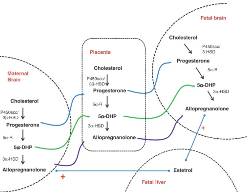
We previously demonstrated that estetrol, E4, a nat-urally occurring estrogen only produced by the human fetal liver, increased allopregnanolone levels in the brain and in the circulation, underlying the crucial role of allo-pregnanolone during human fetal brain development [41] (Figure 1).
Recent findings suggest that the neurosteroid milieu deeply influences behavioral state and exerts a tonic suppression of CNS excitability in the fetus [42]. This is supported by findings that the inhibition of neuroactive steroid synthesis during pregnancy, either by lowering placental progesterone synthesis, using a 3β-HSD inhibi-tor, or blocking the metabolism of progesterone to allo-pregnanolone, markedly increases arousal-like behavior and excitation in the ovine fetus. Furthermore, this indi-cates that allopregnanolone levels markedly influence behavioral states during fetal life and may have a major impact on brain development. The hypothesis according to which the fetal brain itself plays a key role in regulating levels of allopregnanolone is supported by the evidence that its concentrations in the fetal brain rise further after hypoxic stress [40, 42].
There is now increasing evidence that neurosteroids improve outcomes following hypoxic/ischemic brain injury in adults by aiding tissue repair. These processes involve increased production of allopregnanolone and its interaction with GABAA receptors. Suppression of allopregnanolone production alone increases apoptotic cell numbers in the fetal brain in the absence of any inju-rious process. Reduced allopregnanolone levels nega-tively affect the number of brain cells. These effects of inhibiting neuroactive steroid synthesis were blocked by the co-infusion of alfaxalone, suggesting that allo-pregnanolone in the fetal brain is required to maintain constitutive levels of cell death and proliferation in late development.
Previous findings show that neuroactive steroids stimulate myelination, an action thought to involve neu-roactive steroid-induced stimulation of GABAA receptors, which appears to indirectly affect oligodendrocytes. This idea is supported by a recent study where recovery from acute perinatal hypoxic injury involved increased prolifer-ation of oligodendrocyte progenitor cells and their matu-ration into mature oligodendrocytes [35].
Allopregnanolone responses to chronic placental insufficiency are mediated by an increase of 5α-reductase expression in the placenta and brain, especially in the hippocampus [40]. Changes in brain allopregnanolone levels seem to be observed also after birth in lambs [36]. After exposure to hypoxia, these newborn animals show
Placenta Cholesterol Progesterone 5α-DHP 5α-DHP 5α-DHP Allopregnanolone Fetal brain Cholesterol Progesterone Allopregnanolone P450scc/ 3β-HSD P450scc/ 3β-HSD 5α-R 5α-R 3α-HSD 3α-HSD Fetal liver Estetrol + P450scc/ 3-HSD 5α-R 3α-HSD Maternal Brain Cholesterol Progesterone Allopregnanolone
a rise in brain allopregnanolone concentrations [2, 36] and plasma cortisol [2]. These observations suggest that allopregnanolone may also play a neuroprotective role in newborns, not only during gestation [2].
Conclusions
Progesterone and allopregnanolone have crucial and somewhat different roles in brain development, function and recovery after injury. Pregnancy is characterized by an increased synthesis of progesterone and its neuro-active metabolites by the placenta, and the maternal and fetal brain. This supports the critical role of these ster-oids in maternal brain adaptation during pregnancy and development of fetal brain. Moreover, allopregnanolone may play a brain-protective role during complications of pregnancy, such as preterm delivery or intrauterine growth restriction (IUGR), reducing the impact of hypoxia and excitotoxic brain damage or impairment myelination. Behavioral consequences of altered progesterone/allo-pregnanolone fetal brain programming have also been hypothesized, although further evidence is needed. New potential applications of allopregnanolone as a treatment strategy have been proposed, addressing unmet clinical needs in perinatal care.
References
1. Schumacher M, Guennoun R, Ghoumari A, Massaad C, Robert F, El-Etr M, Akwa Y, Rajkowski K, Baulieu E-E. Novel perspectives for progesterone in hormone replacement therapy, with special refer-ence to the nervous system. Endocr Rev 2007;28:387–439. 2. Hirst JJ, Kelleher MA, Walker DW, Palliser HK. Neuroactive steroids
in pregnancy: key regulatory and protective roles in the foetal brain. J Steroid Biochem Mol Biol 2014;139:144–53.
3. Mani S. Progestins receptor subtypes in the brain: the known and the unknown. Endocrinology 2008;149:2750–6.
4. Tsai M-J, O’Malley BW. Molecular mechanisms of action of steroid/thyroid receptor superfamily members. Ann Rev Biochem 1994;63:451–86.
5. Kastner P, Krust A, Turcotte B, Stropp U, Tora L, Gronemeyer H, Chambon P. Two dinstinct estrogen-regulated promoters generate transcripts encoding the two functionally different human progesterone receptors forms A and B. EMBO J 1990;9:1603–14. 6. Takimoto GS, Tung L, Abdel H, Abel MG, Sartorius CA, Richer JK, Jacobsen BM, Bain DL, Horwitz KB. Functional properties of the N-terminal region of progesterone receptors and their mecha-nistic relationship to structure. J Steroid Biochem Mol Biol 2003;85:209–19.
7. Dijkema R, Schoonen WG, Teuwen R, van der Struik E, de Ries RJ, van der Kar BA, Olijve W. Human progesterone receptor A and B
isoforms in CHO cells. I. Stable transfection of receptor and receptor-responsive reporter genes: transcription modulation by (anti)progestagens. J Steroid Biochem Mol Biol 1998;64:147–56. 8. Hovland AR, Powell RL, Takimoto GS, Tung L, Horwitz KB. An
N-terminal inhibitory function, IF, suppresses transcription by the A-isoform but not the B-isoform of human progesterone receptors. J Biol Chem 1998;273:5455–60.
9. Shyamala G, Yang X, Silberstein G, Barcellos H, Dale E. Transgenic mice carrying an imbalance in the native ratio of A to B forms of progesterone receptor exhibit developmental abnormalities in mammary glands. Proc Natl Acad Sci USA 1998;95:696–701.
10. Shyamala G, Yang X, Cardiff RD, Dale E. Impact of progesterone receptor on cell-fate decisions during mammary gland develop-ment. Proc Natl Acad Sci USA 2000;97:3044–9.
11. Wei LL, Gonzales-Aller C, Wood WM, Miller LA, Horwitz KB. 5′-heterogeneity in human progesterone receptor transcripts predicts a new amino-terminal truncated C-receptor and unique A-receptor messages. Ml Endocrinol 1990;4:1833–40.
12. Wei LL, Hawkins P, Baker C, Norris B, Sheridan PL, Quinn PG. An amino-terminal truncated progesterone receptor isoform, PRc, enhances progestin-induced transcriptional activity. Mol Endocrinol 1996;10:1379–87.
13. Camacho-Arroyo I, Perez-Palacios G, Pasapera AM, Cerbon MA. Intracellular progesterone receptors are differentially regulated by sex steroid hormones in the hypothalamus and the cerebral cortex of the rabbit. J Steroid Biochem Mol Biol 1994;50:299–303. 14. Guerra-Araiza C, Coyoy-Salgado A, Camacho-Arroyo I. Sex
dif-ferences in the regulation of progesterone receptor isoforms expression in the rat brain. Brain Res Bull 2002;59:105–9. 15. Inoue T, Akahira JI, Takeyama J, Suzuki T, Darnel AD, Kaneko C,
Kurokawa Y, Satomi S, Sasano H. Spatial and topological distri-bution of progesterone receptor A and B isoforms during human development. Mol Cell Endocrinol 2001;182:83–9.
16. Pluchino N, Luisi M, Lenzi E, Centofanti M, Begliuomini S, Freschi L, Ninni F, Genazzani AR. Progesterone and progestins: effects on brain, allopregnanolone and β-endorphin. J Steroid Biochem Mol Biol 2006;102:205–13.
17. Camacho-Arroyo I, Guerra-Araiza C, Cerbon MA. Progesterone receptor isoforms are differentially regulated by sex steroids in the rat forebrain. Neuroreport 1998;9:3993–6.
18. Guerra-Araiza C, Cerbon MA, Morimoto S, Camacho-Arroyo I. sex differences in the regulation of progesterone receptor isoforms expression in the rat brain during the estrous cycle. Life Sci 2000;66:1743–52.
19. Bethea CL, Widmann AA. Differential expression of progestins receptor isoforms in the hypothalamus, pituitary and endome-trium of rhesus macaques. Endocrinology 1998;139:677–87. 20. Wu Y, Strawn E, Basir Z, Halverson G, Guo SW. Promoter
hypermethylation of progesterone receptor isoform B (PR-B) in endometriosis. Epigenetics 2006;1:106–11.
21. Trotter A, Bokelmann B, Sorgo W, Bechinger-Kornhuber D, Heinemann H, Schmücker G, Oesterle M, Köhntop B, Brisch KH, Pohlandt F. Follow-up examination at the age of 15 months of extremely preterm infants after postnatal estradiol and proges-terone replacement. J Clin Endocrinol Metab 2001;86:601–3. 22. Krebs CJ, Jarvis ED, Chan J, Lydon JP, Ogawa S, Pfaff DW. A
membrane-associated progesterone-binding protein, 25-Dx, is regulated by progesterone in brain regions involved in female reproductive behavior. Proc Natl Acad Sci USA 2000;97:12816–21.
23. Labombarda F, Gonzalez SL, Deniselle MC, Vinson GP, Schumacher M, De Nicola AF, Guennoun R. Effects of injury and progesterone treatment on progesterone receptor and proges-terone binding protein 25-Dx expression in the rat spinal cord. J Neurochem 2003;87:902–13.
24. Phan VL, Miyamoto Y, Nabeshima T, Maurice T. Age-related expression of 1 receptors and antidepressant efficacy of a selective agonist in the senescence-accelerated (SAM) mouse. J Neurosci Res 2005;79:561–72.
25. Monnet FP, Maurice T. The 1 protein as a target for the non-genomic effects of neuro(active)steroids: molecular, physiological, and behavioral aspects. J Pharmacol Sci 2006;100:93–118.
26. Monnet FP, Mahé V, Robel P, Baulieu EE. Neurosteroids, via receptors, modulate the (3H)norepinephrine release evoked by N-methyl-D-aspartate in the rat hippocampus. Proc Natl Acad Sci USA 1995;92:3774–8.
27. Debonnel G, Bergeron R, Monnet FP, de Montigny C. Differential effects of ligands on the N-methyl-D-aspartate response in the CA1 and CA3 regions of the dorsal hippocampus: effect of mossy fiber lesioning. Neuroscience 1996;71:977–87.
28. Lena C, Changeux JP. Allosteric modulations of the nicotinic acetyl-choline receptor. Trends Neurosci 1993;16:181–6. 29. Valera S, Ballivet M, Bertrand D. Progesterone modulates a
neuronal nicotinic acetylcholine receptor Proc Natl Acad Sci USA 1992;89:9949–53.
30. Denner LA, Weigel NL, Maxwell BL, Schrader WT, O’Malley BW. Regulation of progesterone receptor-mediated transcription by phosphorylation, Science 1990;250:1740–3.
31. Ukena K, Kohchi C, Tsutsui K. Expression and activity of 3beta-hydroxysteroid dehydrogenase/delta5-delta4-isomerase in the rat Purkinje neuron during neonatal life. Endocrinology 1999;140:805–13.
32. Ghoumari AM, Baulieu EE, Schumacher M. Progesterone increases oligodendroglial cell proliferation in rat cerebellar slice cultures. Neuroscience 2005;135:47–58.
33. Mellon SH. Neurosteroid regulation of central nervous system development. Pharmacol Ther 2007;116:107–24.
34. Guennoun R, Labombarda F, Gonzalez Deniselle MC, Liere P, De Nicola AF, Schumacher M. Progesterone and allopregnanolone in the central nervous system: response to injury and implication for neuroprotection. J Steroid Biochem Mol Biol 2015;146:48–61. 35. Melcangi RC, Giatti S, Calabrese D, Pesaresi M, Cermenati G,
Mitro N, Viviani B, Garcia-Segura LM, Caruso D. Levels and actions of progesterone and its metabolites in the nervous system during physiological and pathological conditions. Prog Neurobiol 2014;113:56–69.
36. Brunton PJ, Russell JA, Hirst JJ. Allopregnanolone in the brain: protecting pregnancy and birth outcomes. Prog Neurobiol 2014;113:106–36.
37. Pluchino N, Santoro A, Casarosa E, Wenger JM, Genazzani AD, Petignat P, Genazzani AR. Advances in neurosteroids: role in clinical practice. Climacteric 2013;16(Suppl 1):8–17. 38. Hill M, Pašková A, Kančeva R, Velíková M, Kubátová J,
Kancheva L, Adamcová K, Mikešová M, Žižka Z, Koucký M, Šarapatková H, Kačer V, Matucha P, Meloun M, Pařízek A. Steroid profiling in pregnancy: a focus on the human fetus. J Steroid Biochem Mol Biol 2014;139:201–22.
39. Nguyen PN, Billiards SS, Walker DW, Hirst JJ. Changes in 5alpha-pregnane steroids and neurosteroidogenic enzyme expression in fetal sheep with umbilicoplacental embolization. Pediatr Res 2003;54:840–7.
40. Crossley KJ, Nitsos I, Walker DW, Lawrence AJ, Beart PM, Hirst JJ. Steroid-sensitive GABAA receptors in the fetal sheep brain. Neuropharmacology 2003;45:461–72.
41. Pluchino N, Santoro AN, Casarosa E, Giannini A, Genazzani A, Russo M, Russo N, Petignat P, Genazzani AR. Effect of estetrol administration on brain and serum allopregnanolone in intact and ovariectomized rats. J Steroid Biochem Mol Biol 2014;143:285–90. 42. Yawno T, Yan EB, Walker DW, Hirst JJ. Inhibition of neurosteroid
synthesis increases asphyxia-induced brain injury in the late gestation fetal sheep. Neuroscience 2007;146:1726–33.