DOI 10.1007/s00402-014-2038-0 HIP ARTHROPLASTY
Long‑term prognosis of nerve palsy after total hip arthroplasty:
results of two‑year‑follow‑ups and long‑term results after a mean
time of 8 years
B. Zappe · P. M. Glauser · M. Majewski · H. R. Stöckli · P. E. Ochsner
Received: 27 December 2013 / Published online: 6 July 2014 © Springer-Verlag Berlin Heidelberg 2014
patients showed a complete recovery after 2 years. Out of the remaining 17 patients, six out of seven patients with a final examination after a median time of 93 months achieved further improvement. The different nerves showed no significant different potential of recovery.
Conclusions In contrast to the literature, an improvement beyond the limit of 2 years is probable and independent of the nerve affected.
Keywords Nerve lesion · Total hip arthroplasty ·
Long-term prognosis · .Potential for recovery Introduction
Perioperative iatrogenic nerve damage after total hip arthroplasty (THA) is a fatal complication. Even though several risk factors like dysplasia, revision surgery, post-traumatic osteoarthritis, limb lengthening and female gender are well known the incidence persisted on a fairly constant level (0.17–3.2 %) during the last 30 years [2, 5–7, 9, 10]. However, there are just a few studies refer-encing the long-term prognosis of nerve damage consid-ering the influence of the severity of damage as well as the type of the affected nerve [4, 14]. On the other hand a prognosis about the development and possibility of reha-bilitation would be immensely helpful for patients as well as for surgeons.
Aim of the study was to observe affected patients and to answer the following questions: which prognoses can be made concerning the potential for recovering of the nerve lesion? Is the potential of recovery related to the severity of the nerve damage observed? How long can we expect further recovery? Are there differences among different nerves?
Abstract
Introduction Nerve damage is a rare but serious com-plication after THA. There exist only little data about the outcome of these patients particularly regarding the long-term results later than 2 years postoperatively. Aim of this study is to answer the following questions: Is the recovery to be expected for light nerve lesions different from the severe ones? Is there a possibility of nerve recovery more than 2 years after THA? Is the potential of nerve recovery depending on the affected nerve?
Materials and methods This study investigates 2,255 pri-mary THA as well as revision surgeries performed from 1988 to 2003 relating to iatrogenic nerve lesion. We classi-fied the nerve lesion according to the core muscle strength in severe (M0–M2) and light (M3–M4) nerve damage and differentiated between femoral, sciatic and superior gluteal nerve, according to the electromyography.
Results We found 34 cases of iatrogenic nerve dam-age representing an incidence of 1.5 %. 17 of 34 (50 %)
B. Zappe (*) · P. M. Glauser
Department of Traumatology, University Hospital, 4031 Basel, Switzerland
e-mail: bjoern.zappe@usb.ch M. Majewski
Department of Orthopedic Surgery, University Hospital, Basel, Switzerland
H. R. Stöckli
Kantonsspital Liestal, Liestal, Switzerland P. E. Ochsner
Department for Orthopaedic Surgery, Kantonsspital Liestal, Liestal, Switzerland
Patients and method
Patients
This work is based on a retrospective analysis of a prospec-tive-maintained database to detect neurological deficits imme-diately, postoperatively, after THA on all patients in our clinic from January 1988 until December 2003. Inclusion criteria were planned THA and normal preoperative neurological exam on the day before surgery. Preoperative electromyogra-phy (EMG) was not performed. We included 2,162 patients with overall 2,255 primary implanted THAs or revision sur-geries. All THA patients got clinical and radiological exami-nations 6 weeks, 4 months, 1 and 2 years after surgery. The results of all examinations were documented in the Interna-tional Documentation and Evaluation System (IDES) [13]. Surgery and implants
The majority of patients have been operated in a supine position using the transgluteal approach according to Bauer [1]. Only 51 operations in 49 patients were performed in lateral position. In rare exceptions, in which the planned elongation of one leg was more than 4 cm, the sciatic nerve was explored using a dorsal approach with a special emphasis on a careful approach and placing of surgical instruments as well as surgical hooks [11, 15]. In most of the cases, a “Muller Straight Stem“ and an uncemented cup were used. All operations were conducted by or under the supervision of a senior orthopedic surgeon (consultant). Identification and classification of nerve damage
Standardized examinations of the function of the femoral, sciatic and superior gluteal nerve were performed on all patients pre- and postoperatively. The function of the femo-ral nerve was tested by examining the active knee extension and the sciatic nerve by testing the function of the feet and the extensor muscle of the toes. The superior gluteal nerve could only be approximately estimated by active abduction in the hip against resistance caused by the examiner [11]. We classified the deficit in muscular strength of the core muscle in M0–M5 according to Daniels et al. [3] (Table 1). Hereby we defined M0–M2 as severe, M3–M4 as light nerve damage. All patients who were detected with deficit in muscular strength were examined by EMG, to objectify the nerve damage. So, we could differentiate nerve dam-ages according to the intervention from positional damage. Empirical final examination
Patients not showing full recovery after 2 years were con-tacted for a final examination. Gait pattern, muscle strength
of the core muscle were tested including a neurological sta-tus of the lower extremities and an EMG. During the whole study period the same neurologist did all the testing. Statistics
We used SPSS® version 17.0 for Windows (SPSS Inc,
Chicago, IL, USA) for the statistical analyses. Categorical data are presented as absolute values and relative frequen-cies and continuous data as median and interquartile range (IQR). Groups have been tested for difference by Fisher’s exact test or Mann–Whitney U test, as appropriate.
Results
Incidence and development of nerve lesion
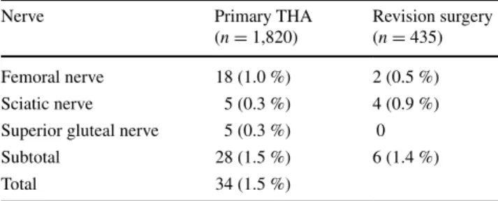
Overall, we documented 35 patients with postoperative nerve lesions. In one patient the electromyography showed a nerve lesion in a lower level (peroneal nerve) interpreted as a posi-tional damage. This case was excluded from the analyses. Therefore, 34 out of 2,255 (1.5 %) patients with a mean age of 65 years (SD 10.6) which suffered from iatrogenic nerve damage after THA were included in our study. For primary surgery the incidence was 1.5 % (28 out of 1,820), for revi-sion surgery it was 1.4 % (6 out of 435) (Table 2). Accord-ing to gender the incidence for nerve lesion after THA was 2.5 % in female and 0.6 % in male patients (27 out of 1,071 vs. 7 out of 1,184). Chi-square test shows a high statisti-cal significance for female gender as a risk factor for nerve lesion (p < 0.01). All these 34 patients with postoperative nerve lesion have been operated in a supine position using the transgluteal approach according to Bauer [1].
20 patients (59 %) suffered from damage of the femo-ral nerve, 9 patients (26 %) had a sciatic nerve lesion and 5 patients (15 %) showed damage in the superior gluteal nerve (Table 2). 23 patients (68 %) presented a severe damage, whereas 11 (32 %) showed a lighter nerve lesion (Table 3).
In all 34 patients the postoperatively performed EMG did show a neurapraxia. Neurotmesis with the need for a
Table 1 Classification of muscle function adapted from Daniels et al. [3]
Grades of muscle function
M0: no contraction or muscle movement
M1: trace of contraction, but no movement at the joint M2: movement at the joint with gravity eliminated
M3: movement against gravity, but not against added resistance M4: movement against external resistance, but less than normal M5: normal strength
revision including nerve reconstruction was not found. Evacuation of postoperative haematoma is not part of our clinical standard treatment. Therefore, all patients were treated conservatively.
Chronological development of improvement (Table 4) At the time of the 2-year-examination 17 (50 %) of 34 patients showed complete remission. For these patients the median duration up to complete remission was 19 months (IQR 8–24). Patients suffering from severe nerve damage needed a median time to recovery of 21 months (IQR 15– 24), whereas patients suffering from a light nerve damage recovered in a mean time of 13 (IQR 3–23) months.
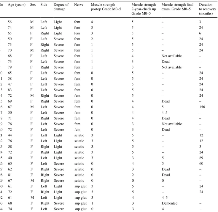
At the final empirical examination of the seven patients controlled, additional three patients reached complete recovery; three presented slight improvement whereas only one remained unchanged. Overall, 20 out of 34 (59 %) patients had achieved complete recovery. The median dura-tion of recovery for all patients was 21 months (IQR 13– 25). While patients with severe nerve damage had required 23 months (IQR 18–37), the patients with lighter nerve damage had needed 16 months (IQR 3–24) (Table 4). Femoral nerve (n = 20)
12 patients (60 %) developed complete recovery within a median of 21 months (IQR 14–25). All 3 patients with light damage did show complete remission. Of the 17 severe cases 9 took a median of 24 months (IQR 17–25) until complete remission. Of the remaining 8 cases with no com-plete recovery after 2 years only 2 patients were available
for long-term follow-up. One achieved full recovery (No. 16) and the other improved slightly (No. 17).
Sciatic nerve (n = 9)
All of the 5 light cases showed complete remission after a median of 13 months (IQR 3–63). One of these patients showed full recovery after 7 years (No. 25). One out of 4 severe lesions healed completely after 5 years. One did stay without any recovery at all. The remaining two were par-tially restored at 2 years, but died before the final control. Superior gluteal nerve (n = 5)
Following a light lesion only 2 out of 3 patients achieved complete remission after 2 years. The remaining case showed further improvement but only restored to M 4–5. Two severe cases presented partial recovery after 2 years. In the long-term follow-up one improved to M4, the other was lost to follow-up.
With Fig. 1 we could illustrate the chronological devel-opment of the nerve recovery dependent on each nerve. It shows once more that most of the recoveries appear within the first 25 months. But mainly the sciatic nerve and the femoral nerve too show later recoveries.
Potential for recovery
In 10 out of 11 patients with light nerve lesions complete recovery was achieved (91 %). Patients with severe nerve damage ended in complete remission in 10 out of 23 cases (43 %). In 12 cases at least partial remission was observed. Only one patient did not show any improvement at all.
For the long-term follow-up, all patients with no com-plete recovery after 2 years (n = 17) were contacted for a final exam. At that time 6 patients were dead, 3 patients were lost to follow-up and one in poor general conditions that did not allow an examination. 7 patients were con-trolled at a median time interval between surgery and final examination of 93 months (interquartile range (IQR) (80– 156) (Table 4). Six out of these 7 patients presented fur-ther improvement at the final examination. Three patients achieved full recovery to M5.
Discussion
Graduation in severe and light lesions
We believe that classifying iatrogenic nerve damage between (M0–2) as severe and (M3–4) as light is reason-able. Our graduation is more practical than the one of Far-rell in “complete and incomplete lesions”. The attribution
Table 2 Incidence of iatrogenic peripheral nerve injury, separated to different nerves
Nerve Primary THA
(n = 1,820)
Revision surgery (n = 435)
Femoral nerve 18 (1.0 %) 2 (0.5 %)
Sciatic nerve 5 (0.3 %) 4 (0.9 %)
Superior gluteal nerve 5 (0.3 %) 0
Subtotal 28 (1.5 %) 6 (1.4 %)
Total 34 (1.5 %)
Table 3 Degree of damage and no. of recoveries Nerve damage Light (M3–5)
No./no. of recovery
Severe (M0–2) No./no. of recovery
Femoral nerve 3/3 17/9
Sciatic nerve 5/5 4/1
Superior gluteal nerve 3/2 2/0
of M2 lesions is uncertain. We only had one such lesion amongst our cases.
Prognosis of light and severe lesions
The most positive finding is that light nerve lesions have a chance of 91 % for full recovery within a median of 13 months. In contrast only 43 % of severe lesions fully recover within a median of 21 months (Table 3). Only one of 16 M0-lesions did not show any improvement. This is in contrast to Pekkarinen et al. [14], which quoted, that it
would be impossible to predict the prognosis of a nerve injury at an early stage. We also cannot confirm the say-ing of Farrell et al. [5] that “the majority of patients with nerve palsy—whether complete or incomplete—never fully recovered to preoperative strength.” Simmons showed that in case of retractor-related injury of the femoral nerve all his patients showed full recovery [17]. But he did not dif-ferentiate between mild and severe nerve injuries. Further-more the significant higher incident in female gender indi-cates that it is not mainly a question of a retractor-related problem.
Table 4 Temporal and qualitative development of nerve damage of all patients No Age (years) Sex Side Degree of
damage
Nerve Muscle strength postop Grade M0–5
Muscle strength 2-year-check up Grade M0–5
Muscle strength final exam. Grade M0–5
Duration to recovery (months)
1 56 M Left Light fem 4 5 – 3
2 74 M Left Light fem 3 5 – 24
3 65 F Right Light fem 3 5 – 6
4 50 F Left Severe fem 2 5 – 24
5 73 F Right Severe fem 1 5 – 24
6 70 M Right Severe fem 1 5 – 24
7 68 F Left Severe fem 1 4 Not available –
8 73 F Left Severe fem 1 3 Dead –
9 79 F Right Severe fem 1 3 Not available –
10 65 F Left Severe fem 0 5 – 24
11 58 F Left Severe fem 0 5 – 24
12 47 F Left Severe fem 0 5 – 24
13 83 F Left Severe fem 0 5 – 24
14 72 M Right Severe fem 0 5 – 24
15 69 F Right Severe fem 0 4 Dead –
16 67 M Left Severe fem 0 4 5 156
17 50 F Left Severe fem 0 4 4–5 –
18 71 F Right Severe fem 0 4 Dead –
19 76 F Left Severe fem 0 3 Not available –
20 72 F Left Severe fem 0 3 Dead –
21 44 F Left Light sciatic 3 5 – 12
22 76 F Left Light sciatic 3 5 – 12
23 58 F Right Light sciatic 3 5 – 3
24 72 F Right Light sciatic 3 5 – 24
25 40 F Left Light sciatic 3 3 5 89
26 65 F Left Severe sciatic 0 4 5 60
27 62 F Right Severe sciatic 0 3 Dead –
28 81 F Right Severe sciatic 0 2 Dead –
29 67 M Right Severe sciatic 0 0 0 –
30 61 F Left Light sup glut 3 5 – 24
31 72 F Right Light sup glut 3 5 – 24
32 61 M Left Light sup glut 3 4 4–5 –
33 68 F Right Severe sup glut 1 3 Demented –
Prognosis of the severe lesions according to the damaged nerve
Femoral, sciatic and superior gluteal nerve lesions were observed in the frequency relation of 4 : 2 : 1. The femoral nerve lesions seem to carry a higher potential for recovery than sciatic or superior gluteal nerve lesions. 59 % (9/17) with severe lesions of the femoral nerve had a full recov-ery (Table 3). But only 25 % (1/4) of our severe sciatic nerve lesions and none of the two of the superior gluteal nerve fully restored. Brown et al. [2] describe a higher potential for recovery of sciatic nerves if the tibial divi-sion is affected instead of the peroneal one. This fits to our only patient without improvement with a nearly exclusive damage of the peroneal fibers of the sciatic nerve (No. 29 Table 4). On the other hand we have had another patient (patient No. 27) with severe sciatic nerve lesions with a similar EMG-result as patient No. 29, which showed no recovery but an improvement from M0 to M3 during the first 2 years postoperatively. So, affection of the peroneal fibers is maybe disadvantageous but even then, recovery is possible.
Overall, the incidence of postoperative nerve lesion is 1.5 %. This is in line with the literature. But in the majority of cases (20 of 34) the femoral nerve was affected. This is an incidence of 0.9 %. On one hand this is not higher than
reported by Brown et al. [2], who reported an incidence of femoral nerve lesion after THA of 0.01–2.3 %. On the other hand this finding is contrary to most studies including Farrell et al. [5], where femoral nerve lesions were the most uncommon one. But in this study different approaches to the hip were analyzed and the approach according to Bauer was not included. Therefore, one possible explanation for this finding could be the approach. Only one study docu-mented complications after THA with the Bauer approach [16]. In this study only 60 patients were examined and not any nerve lesion was documented. According to the very low incidence of nerve lesions after THA, this is not sur-prising. So, we cannot compare our finding of mainly fem-oral nerve lesions to the literature.
Further improvement later than 2 years after the incidence Holzapfel et al. [8] claimed that later than 18 months since lesion you cannot expect a further regeneration. To our sur-prise we are able to show the contrary. At a late control 7 patients without full remission after 2 years could be reex-amined. Six of them showed further improvement, three of them even reached full recovery. Because of death, loss to follow-up and poor general condition, 10 patients were missed for this late control. The authors are aware of this high number of missing data. But this does not change the finding that there is a high probability of further improve-ment up to full recovery after 2 years. We have to empha-size that in all our patients except one at least a slight improvement of the nerve function was documented. Male–female relation
Our data confirm a higher risk of women (79 %) suffering a nerve lesion after THA as mentioned in the literature [10, 12, 14, 15]. We have no explanation for this finding. Vas-cular changes after pregnancy [18], the higher rate of hip dysplasia and smaller bulk of muscle [19] are discussed as a possible reason.
Revision total hip surgery in our hands did not nega-tively influence the risk of nerve palsy.
Conclusion
Our classification in light (M3–4) and severe (M0–2) nerve lesions is of clinical significance. Light lesions reach full restoration in 91 %, most of them within 2 years. Severe lesions show full recovery only in 43 % with a potential for further improvement after 2 years. We cannot demonstrate a significant difference in recovery potential between the affected nerves. Females show a significant higher risk to suffer a nerve lesion after THA.
Acknowledgments We want to thank Susanna Häfliger for the data research and support.
Conflict of interest Authors have no conflicts of interest or finan-cial ties to disclose.
Ethical standard This study was approved by the local ethical committee (Ref. 192/13).
References
1. Bauer R, Kerschbaumer F, Poisel S, Oberthaler W (1979) The transgluteal approach to the hip joint. Arch Orthop Trauma Surg Archiv fur orthopadische und Unfall-Chirurgie 95(1–2):47–49 2. Brown GD, Swanson EA, Nercessian OA (2008) Neurologic
injuries after total hip arthroplasty. Am J Orthop 37(4):191–197 3. Daniels LWC (1986) Muscle testing: techniques of manual
exam-ination, 5th edn. Saunders, Philadelphia
4. DeHart MM, Riley LH Jr (1999) Nerve injuries in total hip arthroplasty. J Am Acad Orthop Surg 7(2):101–111
5. Farrell CM, Springer BD, Haidukewych GJ, Morrey BF (2005) Motor nerve palsy following primary total hip arthroplasty. J Bone Joint Surg Am 87(12):2619–2625
6. Fox AJ, Bedi A, Wanivenhaus F, Sculco TP, Fox JS (2012) Femo-ral neuropathy following total hip arthroplasty: review and man-agement guidelines. Acta Orthop Belg 78(2):145–151
7. Goetz MB, Seybold D, Gosse F, Muhr G, Roetman B (2010) The risk of nerve lesions in hip alloarthroplasty. Zeitschrift fur Ortho-padie und Unfallchirurgie 148(2):163–167
8. Holzapfel BM, Heinen F, Holzapfel DE, Reiners K, Noth U, Rudert M (2012) Nerve lesions after minimally invasive total hip arthroplasty. Der Orthopade 41(5):354–364
9. Kirschner S, Goronzy J, Storch A, Gunther KP, Hartmann A (2011) Avoidance, diagnostics and therapy of nerve lesions after total hip arthroplasty. Der Orthopade 40(6):491–499
10. Navarro RA, Schmalzried TP, Amstutz HC, Dorey FJ (1995) Sur-gical approach and nerve palsy in total hip arthroplasty. J Arthro-plasty 10(1):1–5
11. Ochsner P, Brunazzi M (2003) Die Hüfttotalprothese: Implanta-tionstechnik und lokale Komplikationen; eine Darstellung auf der Basis des Systems nach M. E. Müller unter Einbezug einer Lang-zeitkontrolle; mit 24 Tabellen. Springer, Berlin [u.a.]
12. Oldenburg M, Muller RT (1997) The frequency, prognosis and significance of nerve injuries in total hip arthroplasty. Int Orthop 21(1):1–3
13. Paterson D (1993) The International Documentation and Evalua-tion System (IDES). Orthopedics 16(1):11–14
14. Pekkarinen J, Alho A, Puusa A, Paavilainen T (1999) Recovery of sciatic nerve injuries in association with total hip arthroplasty in 27 patients. J Arthroplasty 14(3):305–311
15. Schoellner C, Schoellner D (2003) Nerve injuries in total hip arthroplasty—prophylactic strategies—quality assurance and risk management in orthopaedic and trauma surgery. Z Orthop Ihre Grenzgeb 141(3):289–295
16. Sendtner E, Borowiak K, Schuster T, Woerner M, Grifka J, Ren-kawitz T (2011) Tackling the learning curve: comparison between the anterior, minimally invasive (Micro-hip(R)) and the lateral, transgluteal (Bauer) approach for primary total hip replacement. Arch Orthop Trauma Surg. Archiv fur orthopadische und Unfall-Chirurgie 131(5):597–602
17. Simmons C Jr, Izant TH, Rothman RH, Booth RE Jr, Balderston RA (1991) Femoral neuropathy following total hip arthroplasty. Anatomic study, case reports, and literature review. J Arthroplasty 6(Suppl):S57–S66
18. Solheim LF, Hagen R (1980) Femoral and sciatic neuropathies after total hip arthroplasty. Acta Orthop Scand 51(3):531–534 19. Weber ER, Daube JR, Coventry MB (1976) Peripheral
neuropa-thies associated with total hip arthroplasty. J Bone Joint Surg Am 58(1):66–69