HAL Id: hal-01523680
https://hal.archives-ouvertes.fr/hal-01523680
Preprint submitted on 16 May 2017HAL is a multi-disciplinary open access archive for the deposit and dissemination of sci-entific research documents, whether they are pub-lished or not. The documents may come from teaching and research institutions in France or abroad, or from public or private research centers.
L’archive ouverte pluridisciplinaire HAL, est destinée au dépôt et à la diffusion de documents scientifiques de niveau recherche, publiés ou non, émanant des établissements d’enseignement et de recherche français ou étrangers, des laboratoires publics ou privés.
Homogeneous non-selective and slice-selective
parallel-transmit excitations at 7 Tesla with universal pulses: a
validation study on two commercial RF coils
Vincent Gras, Markus Boland, Alexandre Vignaud, Guillaume Ferrand, Alexis
Amadon, Franck Mauconduit, Denis Le Bihan, Tony Stöcker, Nicolas Boulant
To cite this version:
Vincent Gras, Markus Boland, Alexandre Vignaud, Guillaume Ferrand, Alexis Amadon, et al.. Homo-geneous non-selective and slice-selective parallel- transmit excitations at 7 Tesla with universal pulses: a validation study on two commercial RF coils. 2017. �hal-01523680�
Homogeneous non-selective and slice-selective
parallel-transmit excitations at 7 Tesla with universal pulses: a
validation study on two commercial RF coils
Vincent Gras1, Markus Boland2, Alexandre Vignaud1, Guillaume Ferrand3, Alexis Amadon1, Franck Mauconduit4, Denis Le Bihan1, Tony Stöcker2, Nicolas Boulant1
1CEA/DRF/IFJ/NeuroSpin/Unirs, Gif sur Yvette 91191, France 2DZNE, Bonn, Germany
3CEA/DRF/IRFU/SACM, Gif sur Yvette 91191, France 4Siemens Healthcare, Saint Denis, France
Corresponding author:
Nicolas Boulant, CEA, NeuroSpin, Gif sur Yvette, 91191 Cedex, France e-mail: nicolas.boulant@cea.fr
Abstract
Parallel transmission (pTx) technology, despite its great potential to mitigate the transmit field inhomogeneity problem in magnetic resonance imaging at ultra-high field (UHF), suffers from a cumbersome calibration procedure, thereby making the approach problematic for routine use. The purpose of this work is to demonstrate on two different 7T systems respectively equipped with 8-transmit-channel Rapid-Biomedical and Nova coils, the benefit of so-called universal pulses (UP), optimized to produce uniform excitations in the brain in a population of adults and making unnecessary the calibration procedures mentioned above. Non-selective and slice-selective UPs were designed to return homogeneous excitation profiles throughout the brain simultaneously on a group of ten subjects, which then were subsequently tested on ten additional volunteers in magnetization prepared rapid gradient echo (MPRAGE) and multi-slice gradient echo (2D GRE) protocols. The results were additionally compared experimentally with the standard non-pTx circularly-polarized (CP) mode, and in simulation with subject-specific tailored excitations.
For both pulse types and both coils, the UP mode returned a better signal and contrast homogeneity than the CP mode. Retrospective analysis of the flip angle (FA) suggests that the FA deviation from the nominal FA on average over a healthy adult population does not exceed 10.6% with the calibration-free parallel-transmit pulses whereas it goes beyond 25% with the CP mode. As a result the universal pulses designed in this work confirm their relevance in 3D and 2D protocols with commercially available equipment. Plug-and-play pTx implementations henceforth become accessible to exploit with more flexibility the potential of UHF for brain imaging.
1.
Introduction
Magnetic Resonance Imaging (MRI) of the brain has improved considerably in the last decade with the advent of ultra-high field (UHF) scanners (B0 ≥ 7T). To explore the brain non-invasively, the use of high
field allows increasing the signal-to-noise ratio (SNR), reducing the acquisition times, yielding greater image resolution, but also improving the sensitivity to several effects of great importance in the study of brain organization and function. The use of high field strengths comes nevertheless with a dramatic increase in the radiofrequency (RF) field inhomogeneity which can severely degrade imaging performance. Within this context, parallel transmission (pTx) [1] has revealed a great potential in mitigating the transmit field inhomogeneity problem in the human head at field strengths equal or larger than 7T [2–8]. The pTx approach is the transmit analogy to parallel reception, pRx, where the MR signal is acquired with multiple receivers (phased array). Parallel reception is nowadays a standard tool in clinical MRI, e.g. for parallel imaging acceleration. While pRx increases image SNR and encodes spatial information via the reception profile, pTx provides additional degrees of freedom to shape the total excitation profile and to reduce the Specific Absorption Rate (SAR), a critical limiting measure at UHF. Today, however, despite its power the pTx technology has remained marginally exploited. In addition to a significant financial investment in hardware to enable pTx (monitoring equipment, dedicated RF coils, etc.) and the problems engendered in patient safety management [9–11], it can be attributed mostly to the cumbersome calibration procedures (measurement of the transmit field sensitivities and pulse design) and to the expertise necessary to drive the parallel transmit coil adequately. Yet recently, a new concept of performing MRI using pTx was proposed, in which the user was spared the conventional calibration procedure [12]. In this approach, instead of computing RF pulses tailored to the subject’s actual transmit field sensitivities and static field offset map, RF pulses are designed offline, based on
measured fields obtained from a group of subjects representative of a population, and blindly applied on new subjects of the considered population, without any calibration. This method, implemented experimentally with non-selective kT-point RF pulses [8], allowed designing so-called universal pulses
(UP) whose excitation performance in terms of flip angle homogenization greatly exceeded the performance of the standard circularly polarized (CP) transmission mode commonly used in single channel transmission systems. Interestingly also, the reported performance exceeded as well the performance of the subject-specific RF shim mode, although the latter technique does require the measurement of the transmit field sensitivities. The penalty in performance compared to the subject-specific tailored pulse design in fact was found to be mild. Although the reported results appeared promising, they were limited to non-selective excitations and involved a specific, home-made, pTx RF coil prototype, which obviously prevents from disseminating the pulses. Given the apparent necessity to provide broadband solutions to be robust against the inter-subject variability of the static field offset [12], it remained also to be determined whether the concept could be transposed to spatially selective pulses of much longer durations.The aim of the present study thus is to extend the proof of concept of UP at 7T with two commercially available pTx head coils, namely the 8TX-8RX Rapid-Biomed (RAPID Biomedical GmbH, Rimpar, Germany) and 8TX-32RX Nova (Nova medical Inc., Wilmington, MA, USA) coils, and to investigate non-selective as well as slice-selective parallel transmit pulses using larger groups of subjects than initially reported (40 versus 12 subjects). Following the approach in Ref. [12], non-selective UPs will be determined from the kT-point framework while slice-selective UPs will
be based on multi-spoke RF pulses [4,13].
2.
Methods
The study was conducted at two different sites (site 1 and site 2) and on two different groups of subjects. Measurements at site 1 and 2 were both made with Magnetom 7T scanners (Siemens Healthcare, Erlangen, Germany) equipped with eight-channel transmit arrays (1 kW peak power per channel). Site 1 measurements made use of the Rapid-Biomed 8TX-8RX head coil and AC84 head gradient set (50-mT/m maximum amplitude and 333-T/m/s maximum slew rate), while the measurements at site 2 were carried out with the Nova 8TX-32RX head coil and SC72 whole body gradient insert (70 mT/m maximum amplitude and 200 T/m/s maximum slew rate). At both sites, measurements were performed in the local SAR supervision mode (Siemens Tim Tx Array Step 2.3), requiring for each coil detailed numerical simulations of the electromagnetic fields on generic head models and construction of Virtual Observation Points (VOPs) [9,14] with appropriate safety margins [15]. Studies were approved by the respective local ethic committees and all volunteers gave written informed consent. At each site, the respective groups consisted of 20 healthy subjects (age = 40±15 years, 10 men, and 10 women). Ten out of the 20 subjects (50 % men, 50 % women) were enrolled as “database subjects” and thus served the computation of UPs. The remaining ten subjects were included in the “test subject group” and their data was used to evaluate UP performance through the quantitative analysis of the simulated flip angle profiles and through examination of the images after correction of the reception profiles.
2.1.MRI protocols
The acquisitions performed on the database subjects aimed at measuring the subject-specific static field offset ∆B0 (T) and transmit field sensitivities B1,c+ (T/V) for each transmit channel c (1 ≤ c ≤ Nc = 8).
a three-dimensional (3D) multiple gradient recalled echo (GRE) (2.5 mm isotropic resolution, matrix size 64 × 96 × 128, TR = 25 ms, 3 echoes, TE = 5, 6.5, 8 ms, TA = 3 min). The eight complex transmit field sensitivities were subsequently measured from a multi-slice interferometric turbo-FLASH acquisition (5 mm isotropic resolution, matrix size 40 × 64 × 40, TR = 20 s, TA = 4 min 40 s) [16,17]. Knowledge of the subject-based field maps on the 10 database volunteers thereby allowed designing universal pulses for each coil/site, as described in the next section.
The same field map measurements were repeated on the test subjects for retrospective control. Otherwise the MRI protocol for these subjects consisted in two different anatomical scans each repeated with two transmit modes: first in the CP mode and subsequently in the pTx mode with the universal pulses previously optimized on the database subjects (pTx-UP mode). The first scan consisted of a non-selective 3D magnetization prepared rapid gradient echo (MPRAGE) acquisition with TI = 1100 ms, TR = 2600 ms, TE = 3 ms, nominal flip angle (FA) = 5°, readout bandwidth = 260 Hz/pixel, echo train length (ETL) = 160, matrix size = 160 × 240 × 256, 1 × 1 × 1.1 mm3 voxels and TA = 4.5 min. The
second protocol was a 40-slice 2D T2*–weighted GRE acquisition with TR = 1720 ms, TE = 18 ms,
target FA = 30°, readout bandwidth = 40 Hz/pixel, 2 mm slice thickness, 50% slice gap, 0.5 × 0.5 mm2
in-plane resolution, matrix size 512 × 384. For the two acquisitions, a GRAPPA [18] acceleration factor of 2 with 24 reference lines in the phase encoding direction was used, except for the 2D GRE acquisition at site 2 which was performed with an acceleration factor of 3 (the acquisition time for the 2D GRE scans hence was decreased from 7 min at site 1 to 5 min at site 2). An additional 3D GRE CP-mode acquisition (3 mm isotropic resolution, matrix size 64 × 64 × 60, nominal FA = 5°, TR = 50 ms, TE = 2.3 ms, readout bandwidth = 300 Hz/pixel, TA = 3 min) was performed to return the reception profile of the RF coil by post-processing.
2.2.RF pulses
For the MPRAGE acquisition in CP-mode, magnetization preparation used a hyperbolic-secant adiabatic inversion pulse of 10 ms duration with peak voltage of 140 Volts at the coil plug. The 5° (nominal FA) excitation was achieved with a rectangular hard pulse. For the 2D GRE acquisition in CP-mode, the 30° (nominal FA) excitation was performed with a standard apodized sinc pulse of time-bandwidth product 2.5. In each sequence using the CP mode, the reference voltage (Vref) was defined as
the value required for a 0.5 ms rectangular pulse to return a FA of 90° on average in the brain isocentric axial slice, which was determined from the B1+ maps acquired on the database subjects. These
measurements yielded: Vref = 130 ± 5 V (mean ± std) for site 1 (Rapid-Biomed coil) and Vref = 95 ± 6 V
for site 2 (Nova coil).
The MPRAGE acquisition in the pTx-UP mode used a 4-ms 9 kT-point pulse for the inversion pulse
and a 1 ms 7 kT-point pulse for the 5° excitation. The 2D GRE acquisitions in the pTx-UP transmission
mode used 40 slice-specific 30° 5-ms long 4-spoke bipolar pulses at site 1 and 6-ms long 3-spoke monopolar pulses at site 2. Each spoke sub-pulse consisted of an apodized sinc-type pulse of time-bandwidth product equal to 2.5, as for the CP mode.
2.3.Construction of the universal pulses
Universal pulses were designed to minimize the expectation value of the normalized root-mean-square error of the FA profile (FA-NRMSE) across the possible RF and static field distributions measured over
the database subjects. A universal pulse p designed to create a uniform FA profile FAt across a region of interest R (in this study, the whole brain region for non-selective pulses and the union of the 40 2D regions for the slice selective pulses) thus minimizes the quantity:
ϵ(p)=FA1 t〈 ∑ (FA(r)-FAt) 2 r ∈ R 1/2 〉, [1]
where Nv denotes the subject-dependent number of voxels in R, FA(r) the actual FA profile for a given
realization of B1+ and ∆B0 maps on one subject, and 〈 . 〉 the expectation value operator over the
population. Assuming that the set of B1+ and ∆B0 measurements performed on the database subjects constitutes a representative sample of the RF and static field statistics, ϵ(p) can be approximated by (1/Ns) ∑ ϵNj=1s j(p), where Ns denotes the number of subjects in the database and ϵj(p) denotes the FA-NRMSE of pulse p on the jth database subject. The latter expression was used as the objective function
to construct the 5° non-selective, the 180° non-selective and the 30° slice-selective excitations. To satisfy patient safety as well as hardware constraints, limits for the RF peak power, average power, global SAR and peak 10g SAR were defined and enforced explicitly throughout the optimization of the RF pulses [19]. From the pulse design point-of-view, the parameterization of the RF pulse p (kT-point
or spoke pulses) determines the multi-dimensional variable for the optimization. With Np denoting the
number of sub-pulses and nsl the number of slices (1 and 40 for the non-selective and selective pulses
respectively), p is composed of i) nsl × Np × Nc complex RF coefficients and ii) Np × 3 (non-selective
case) or nsl × Np × 2 (selective case) real coefficients for the sparse k-space locations. In this work, all
coefficients (complex sub-pulse coefficients and transmit k-space locations) were optimized jointly under SAR and power constraints using the active-set algorithm [19,20,21]. Pulse design was conducted by using the small tip angle approximation for the 5° non-selective and 30° slice-selective pulses while numerical integrations of the Bloch equation were conducted for the inversion pulse. To reduce computation times, the latter operation was ported on a Nvidia (Nvidia, Santa Clara, CA, USA) Tesla K40 graphics processing units card. Ultimately, final pulse performance was always verified by using full numerical Bloch integrations.
2.4.Flip angle simulations
The subject-specific B1+ and ∆B0 measurements performed on the test subjects fed Bloch simulations to yield FA profiles for the CP and universal pulses integrated in the MPRAGE and 2D GRE sequences. For comparison, additionally, subject-specific static RF-shim and tailored (kT-point or spoke) pulses
were computed by minimization of the FA-NRMSE under the same RF power and SAR constraints as for the design of the pTx-UPs. For each pulse (5° non-selective, 180° non-selective, 30° selective), the CP, pTx-UP, RF-shim modes and subject tailored pTx spoke or kT-point pulses (obtained through the
minimization of the subject-specific NRMSE) were compared in terms of their respective FA-NRMSE performance.
3.
Results
3.1.MPRAGE and 2D GRE acquisitions
For two test subjects scanned respectively at sites 1 and 2, differences between the MPRAGE in CP- and the pTx-UP modes are highlighted in selected orthogonal views of the brain in Figure 1. The results for all 10 test subjects for the same scan are also provided for both coils in Figures 2 and 3 respectively. Similar comparisons are provided for the 2D GRE protocol in Figures 4 and 5. All images shown are corrected for the reception profile. Hence, up to the precision of the receive profile correction procedure, the signal inhomogeneity displayed in Figures 1 to 5 is representative of the transmission inhomogeneity only. Incidentally, the same figures reveal to some extent the variability in head geometry, size and position. For the MPRAGE sequence, except for Figure 1, the same (scanner coordinates) coronal, sagittal and axial slices are shown for all test subjects. For the 2D GRE sequence, 3 slice positions (same scanner coordinates) were selected and displayed again for all test subjects.
Figure 1. MPRAGE image comparison in sagittal, coronal and axial planes for a) a test subject of site 1 (Rapid-Biomed coil) and b) a test subject of site 2 (Nova coil). Especially at site 2, the bright center removal by UPs is more pronounced. Moreover, one can observe at the same site a larger coverage in the neck area due to the increased broadband behavior of the UPs. In both cases, the signal and contrast homogeneity is dramatically improved in the cerebellum, temporal lobes and top of the brain when using UPs. The utilization of UPs at site 1 may cause however the occasional emergence of small susceptibility artifacts in some areas (red arrows), e.g. at the interface with air cavities at the basis of the skull.
Figure 2. Coronal (a), sagittal (b) and axial (c) views of the MPRAGE scans (receive profile corrected) obtained on the 10 test subjects at site 1 (Rapid-Biomed coil) with the CP and pTx-UP mode. In general, for the entire test group, the overall signal and contrast homogeneity is clearly improved with the use of UPs. The bright center clearly visible in the CP-mode is well reduced with the proposed excitation mode. On most test subjects, a clear improvement of the contrast between white matter and gray matter is visible in the cerebellum and the temporal lobes.
Figure 3. Coronal (a), sagittal (b) and axial (c) views of the MPRAGE scans (receive profile corrected) obtained on the 10 test subjects at site 2 (Nova coil) with the CP- and the pTx-UP modes. Compared to site 1, the bright center of the CP mode appears slightly more pronounced, but the CP contrast (mostly driven by the inversion efficiency) is generally better preserved in the lower part of the brain. Yet, similar observations as for site 1 can be made regarding the signal and contrast homogeneity improvements with the utilization of UPs.
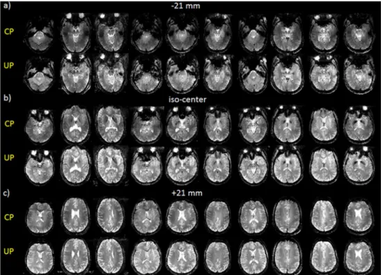
Figure 4. Comparison of the 2D GRE acquisitions (receive profile corrected) performed at site 1 (Rapid-Biomed coil) on all 10 test subjects in the CP and pTx-UP (4-spoke bipolar pulse) modes. a) -21 mm, b) iso-center and c) +21 mm positions out of the 40 acquired slices are shown. As previously mentioned, the transmit efficiency of the CP mode decaying rapidly in the lower part of the brain, the signal enhancement is important in the lowest slices. Still, in b) and c), the slight transmit fall-off in the left-right directions in the CP mode appear well corrected with the pTx UPs.
Figure 5. Comparison of the 2D GRE acquisitions (receive profile corrected) performed at site 2 (Nova coil) on all 10 test subjects with the CP and pTx-UP (3-spoke monopolar pulse) modes. The same observations as in Figure 4 can be made. For subject 7 (marked with an asterix), due to an interruption of the exam and the renewed placement of the subject in the scanner between the acquisitions in the CP- and the pTx-UP mode, the position consistency was compromised.
3.2.Retrospective control with flip angle simulations
The FA-NRMSE of the 5° pulses (CP, UP, subject specific RF-shim and subject-specific optimized kT
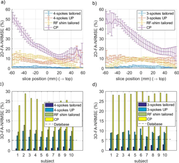
-point) is reported in Figures 6a and 6b for sites 1 and 2 respectively and for all 10 test subjects. In Figures 6c and 6d, the same comparison is provided for the non-selective 180° pulses. For the slice-selective 30° RF pulses, the slice-by-slice FA-NRMSE (2D-FA-NRMSE) is reported in Figures 7a and 7b. There, for each slice, the average FA-NRMSE and its standard deviation across the 10 test subjects (error bar) is reported for the CP, universal, subject-specific RF-shim, and subject-specific optimized multi-spoke pulses. In the bar-plots of Figure 7c and 7d, the global FA-NRMSE obtained by pooling all slices together (3D-NRMSE) is reported for each test subject, and again for both coils and sites.
Figure 6. FA-NRMSE simulations of a,b) the non-selective 5° CP, 7 kT-point universal, subject specific
RF-shim and subject specific 7 kT-point pulses and c,d) non-selective 180° adiabatic CP, adiabatic RF-shim, 9 kT
-point universal and subject specific 9 kT-point pulses designed at site 1 (a,c) and site 2 (b,d). The dashed line
represents the average UP 3D-FA-NRMSE on the database subjects. The UP NRMSE at both sites on all test subjects is noticeably lower than the 13 % threshold corresponding to the CP mode at 3T with a volume coil [22].
Figure 7. FA-2D-NRMSE (a-b) and 3D-FA-NRMSE (c-d) simulations of the 30° slice selective RF pulses designed at site 1 (a,c) and site 2 (b,d) and for all test-subjects. The CP and the 3- or 4-spoke pTx-UP were the excitation modes tested experimentally. For comparison, the subject-specific, slice-specific optimized RF-shim and 3- or 4-spoke designs were included as well. The dashed line represents the average 3D-FA-NRMSE obtained over the database subjects with the UPs. For both coils, the spoke UPs perform systematically better than (site 1), or comparably to (site 2) the subject-specific 2D RF shims. Also the 3D-FA-NRMSE is always smaller than the 13 % threshold, corresponding to the CP mode inhomogeneity at 3T in the brain with a volume coil [22].
The average 3D-FA-NRMSE values (mean ± std) obtained for the 5°, 180° and slice-selective 30° pulses on the test subjects is summarized in Table 1. Those were further exploited to attempt giving the 99% confidence interval (see Table 2) on the average performance of the pTx-UP mode over a much larger population for the two coils investigated. It can be seen in particular that the upper bound of the 99% confidence interval does not exceed 10.6% FA-NRMSE.
Site 1 (Rapid-Biomed coil) Site 2 (Nova coil) 5° ns 180° ns 30° ss 5° ns 180° ns 30° ss CP 26.1±2.4 12.1±2.6 25.1±1.7 27.4±1.0 10.5±0.8 28.8±0.9 ST RF-shim 21±1.6 10.3±1.9 10.5±1.0 17.1±0.9 4.8±0.8 8.3±0.8 UP 9.2±1.7 6.8±2.0 8.1±1.5 8.6±1.5 3.4±1.2 8.3±1.2 ST kT/spokes 3.8±0.8 4.3±1.3 1.3±0.4 4.1±1.3 2.1±0.9 3.6±1.4
Table 1. Average and standard deviation of the 3D-FA-NRMSE (in %) values obtained at sites 1 and 2 over 10 test subjects for the subject-tailored (ST) RF-shim-, universal- and subject-specific kT-point or spoke pulses
implementing the non-selective (ns) 5° and 180° and the slice-selective (ss) 30° excitations. For the 180° pulse, the CP and ST RF-shim modes are played with a hyperbolic secant (HS) shape.
Site 1 (Rapid-Biomed coil) Site 2 (Nova coil)
5° ns 9.2±1.4 8.6±1.2
180° ns 6.8±1.7 3.4±1.0
30° ss 8.1±1.2 8.3±1.0
Table 2. 99% confidence interval for the average whole brain 3D-FA-NRMSE of the pTx-UP transmission mode over the entire healthy adult population for both coils. The reported values are based on the assumption that the size of the test subject group (n = 10) is large. The confidence interval thus is given by x" ±z0.01
%
σx √n,
where x" and σx are respectively the mean value and standard deviation of the FA-NRMSE over the test subjects, and where z0.01 = 2.58 satisfies Prob(|-| > /0.0 ) = 0.01 for the centered and normalized Gaussian random variable Z.
4.
Discussion
The image comparisons provided in Figures 1 to 5 experimentally demonstrate for both RF coils and both protocols a systematic improvement of the excitation uniformity with pTx-UP compared to the CP transmission mode. This result is fully consistent with the flip angle performance simulations reported in Figures 6 and 7, where the whole brain FA-NRMSE of the CP-mode typically exceeded 25% whereas that of the UPs was often below 10%. Overall, the FA-NRMSE confidence intervals given in Table 2 indicate that the UPs designed in this study returned on average in a healthy adult population (age 40±15 years) whole-brain FA-NRMSEs below 10.6% with 99% confidence (the worst case being the 5° non selective pulse for site 1). Comparing this number with the inhomogeneity of the CP-mode B1+ field
within the brain at 3T [22], namely 13%, the proposed pTx-UP mode allows to recover a uniformity of excitation better than that of a volume coil at 3T, yet without a tedious calibration procedure. Direct flip angle measurements in this study were not performed in vivo due to incompressible exam durations. Such types of measurements, however, have been reported in many other works in both small and large
flip angle regimes and across several groups, field strengths and vendors [8,20,21,23-26], thus confirming proper hardware implementation with state of the art equipment.
Interestingly also, at both sites and for all test subjects, the 180° pTx-UP returned better whole brain FA-NRMSEs than the adiabatic hyperbolic secant pulses (see Figures 6c-d). Indeed it was observed (loss of contrast between white matter and gray matter) that the adiabatic inversion pulse often failed to fully invert the magnetization in a large part of the cerebellum and occasionally in temporal regions. In those regions, the inversion efficiency was significantly improved with the utilization of the 180° pTx-UP, which helped restoring proper white matter – gray matter contrast. The FA-NRMSE comparisons summarized in Table 1 moreover show that the UPs clearly outperform the CP and RF shim modes in 3D. For the slice-selective 30° pulses, the overall performance (3D-FA-NRMSE) of the subject and slice specific RF-shims is comparable to the one of the pTx-UP mode. Especially for the slice-selective pulses, a penalty in performance with the UPs compared to subject-specific more sophisticated approaches yet naturally remains. The inhomogeneity with the UPs, however, remained for both non-selective and selective pulses below the 13% threshold of a volume coil at 3T in the human brain [22]. For further optimality, partitioning a bigger database according to relevant criteria, such as for instance head size or position in the z direction, with corresponding pulses could increase the performance of the UPs [27].
Although the construction of the proposed pTx universal pulses was mostly driven by the RF coil properties, the gradient slew-rate limits and the eddy-current characteristics of the gradient coils also came into play for the selective case. Thus, at site 1 with the Rapid-Biomed coil, the 30° slice-selective UPs were composed of 4 bipolar spokes, i.e. played out with alternating gradient polarity with appropriate RF phase corrections to compensate for gradient delays [28]. At site 2 with the Nova coil, despite the employed correction scheme [28], the implementation of the bipolar design appeared more challenging. Eddy current effects on the SC72 gradient system indeed were found to be more severe than on the AC84 head gradient system, thus degrading more severely the excitation performance when using bipolar shapes [5,28-30]. As a result, the more robust monopolar design (all spokes played out with the same gradient polarity) was retained for site 2. Due to the time penalty engendered by this fly-back trajectory, the number of spoke sub-pulses in this case was reduced to 3 (versus 4 at site 1), partly explaining the loss of performance in simulation when comparing the two sites.
The examination of the receive profile-corrected MPRAGE and 2D GRE images indicate that the pTx-UP mode allows recovering a homogeneous signal and a contrast throughout the whole brain although some specific imperfection such as occasional susceptibility (i.e., ∆B0-induced) artefacts could
occur at site 1 with the Rapid-Biomed coil. At site 2 with the Nova coil, the MPRAGE images obtained with the UPs looked artefact-free. An incidental consequence of the broadband behavior of the UPs [12] is a larger coverage, yielding greater signal in the neck area (Figures 1 and 3), thereby making structures in this region visible (e.g. muscles, tendons). The problem of remaining susceptibility artefacts observed with the Rapid-Biomed coil most probably arises from the lower transmit efficiency (Vref = 130 V vs 95
V for Nova) vis-a-vis the broadband requirements necessary to tackle inter-subject B1 and ∆B0 map
variability [12]. This raises the importance of good static field homogenization and coil efficiency for application of UPs. The problem on the other hand could be addressed from the pulse design perspective, where a tradeoff between spatial and spectral uniformity of the UP could be potentially enforced in the objective function (Equation 1). Finally, we chose in our objective function to minimize the average (NRMSE) performance over the database subjects. Worst-case NRMSE optimization could be performed likewise [12]. Although the latter appears appealing conceptually, we found that a worst-case approach could, perhaps unsurprisingly, penalize the average performance non-negligibly and even not perform the desired task on the test subjects. Table 3 indeed reports the simulated NRMSEs over the
test and database subjects for the two optimization strategies, i.e. minimization of the average and worst-case NRMSEs respectively, and for the two different coils. The NRMSEs over the database subjects perform as expected, i.e. according to the cost-function used in the optimization. On the other hand, for the Nova coil minimizing the worst-case NRMSE over the database subjects does not guarantee a lower worst-case over the test subjects than the one obtained with the other optimization method. This illustrates the greater sensitivity of the results to subject-specific details in the RF and static field distributions when worst-case metrics are employed. Yet with careful management of the database, e.g. with segmentation [27], we do not rule out the possibility of using other optimization metrics to target more specifically radiologists’ needs.
RF coil UP construction method <NRMSE> (%) Max(NRMSE) (%)
RapidBiomed min(<NRMSE>) 9.6 (7.7) 14.2 (9.9) min(max(NRMSE)) 10.6 (8.8) 13.1 (9.1) Nova min(<NRMSE>) 8.6 (6.2) 11.7 (8.1) min(max(NRMSE)) 9.6 (7.5) 12.4 (7.6)
Table 3. NRMSE results over the test subjects of the two optimization metrics and for the two different coils. The second column indicates the optimization strategy (minimization of the average NRMSE versus worst-case NRMSE over the database subjects). Results for the database subjects are indicated between parentheses. For the Nova coil, the rightmost column (numbers in bold) illustrates the sensitivity of the results to subject-specific details in the RF transmit field and static field offset maps. In this case, the minimization of the worst-case NRMSE over the database subjects indeed does not guaranty a better worst-case NRMSE than the proposed design (minimization of the average NRMSE) on the test-subjects.
For the CP-mode, the reception profile of the phased array is known to partly compensate for the transmit inhomogeneity [31]. Hence from a global signal homogeneity perspective, it is not necessarily desirable to apply any receive profile correction in this transmit configuration. In contrast with this, since universal pulses are designed to produce homogeneous excitation patterns, correction of the receive profile in this case is perfectly adequate. As a result, the image comparisons provided in Figures 1 to 5 thus may penalize the CP mode performance in favor of the UPs. Yet, from the SNR point-of-view the proposed comparison illustrates very objectively the gain that is brought by UPs. Moreover, the "accidental" cancelation of the CP-mode transmit inhomogeneity with the reception profile of the head coil array cannot compensate for spatially varying contrast and thus does not guaranty the optimality of the contrast-to-noise ratio.
5.
Conclusion
In this work, we have designed non-selective and slice-selective parallel-transmit universal pulses for two commercially available head coils, able to mitigate the RF inhomogeneity problem in the human brain at 7T without prior calibration of the transmit RF field. Our pulse performance analysis, based on two groups of 10 test subjects (one group per site) indicates with 99% confidence that the FA-NRMSE of the designed UPs does not exceed 10.6% on average in healthy adults (age 40±15 years) for non-selective and slice-non-selective pulses. The coil-specific UPs, tested at both sites, on all test subjects in standard MPRAGE and 2D-GRE protocols, demonstrated a clear image quality improvement in terms
of flip angle and contrast uniformity in comparison with the CP-mode, confirming the robustness of the approach and the possibility of using and distributing such calibration-free solutions for clinical routine applications.
6.
Acknowledgments
The authors thank Dr. Pierre Brugières (Centre Hospitalier Henri Mondor) and Dr. Elke Hattingen (University Clinics of Bonn) for valuable discussions. The research leading to these results has received funding from the European Research Council under the European Union’s Seventh Framework Program (FP7/2016-2017), ERC Proof Of Concept Grant Agreement n. 700812.
7.
References
[1] U. Katscher, P. Börnert, C. Leussler, J. S. van den Brink. Transmit SENSE. Magn Reson Med, vol. 49, no. 1, pp. 144–150, 2003.
[2] S. Saekho, F. E. Boada, D. C. Noll, V. A. Stenger. Small tip angle three-dimensional tailored radiofrequency slab-select pulse for reduced B1 inhomogeneity at 3 T. Magn. Reson. Med., vol. 53, no. 2, pp. 479–484, 2005.
[3] W. Grissom, C. Yip, Z. Zhang, V. A. Stenger, J. A. Fessler, D. C. Noll. Spatial domain method for the design of RF pulses in multicoil parallel excitation. Magn Reson Med, vol. 56, no. 3, pp. 620–629, 2006.
[4] K. Setsompop et al. Slice-selective RF pulses for in vivo B1+ inhomogeneity mitigation at 7 tesla using parallel RF excitation with a 16-element coil. Magn. Reson. Med., vol. 60, no. 6, pp. 1422– 1432, 2008.
[5] M. Jankiewicz et al. Practical considerations for the design of sparse-spokes pulses. J. Magn.
Reson., vol. 203, no. 2, pp. 294–304, 2010.
[6] D. O. Brunner and K. P. Pruessmann. Optimal design of multiple-channel RF pulses under strict power and SAR constraints. Magn. Reson. Med., vol. 63, no. 5, pp. 1280–1291, 2010.
[7] A. T. Curtis, K. M. Gilbert, L. M. Klassen, J. S. Gati, R. S. Menon. Slice-by-slice B1+ shimming at 7 T. Magn. Reson. Med., vol. 68, no. 4, pp. 1109–1116, 2012.
[8] M. A. Cloos et al. kT-points: Short three-dimensional tailored RF pulses for flip-angle homogenization over an extended volume. Magn Reson Med, vol. 67, no. 1, pp. 72–80, 2012. [9] G. Eichfelder and M. Gebhardt. Local specific absorption rate control for parallel transmission by
virtual observation points. Magn Reson Med, vol. 66, no. 5, pp. 1468–1476, 2011.
[10] M. de Greef, O. Ipek, A. J. E. Raaijmakers, J. Crezee, C. A. T. van den Berg. Specific absorption rate intersubject variability in 7T parallel transmit MRI of the head. Magn Reson Med, vol. 69, no. 5, pp. 1476–1485, 2013.
[11] I. Graesslin et al. Comprehensive RF safety concept for parallel transmission MR. Magn Reson
Med, 2014.
[12] V. Gras, A. Vignaud, A. Amadon, D. Le Bihan, N. Boulant. Universal pulses: A new concept for calibration-free parallel transmission. Magn. Reson. Med., DOI 10.1002/mrm.26148, 2016. [13] S. Saekho, C. Yip, D. C. Noll, F. E. Boada, V. A. Stenger. Fast-kz three-dimensional tailored
radiofrequency pulse for reduced B1 inhomogeneity. Magn. Reson. Med., vol. 55, no. 4, pp. 719– 724, 2006.
[14] J. Lee, M. Gebhardt, L. L. Wald, E. Adalsteinsson. Local SAR in parallel transmission pulse design. Magn Reson Med, vol. 67, no. 6, pp. 1566–1578, 2012.
[15] V. Gras et al. Signal-domain optimization metrics for MPRAGE RF pulse design in parallel transmission at 7 tesla: Signal-Domain Optimization Metrics for MPRAGE RF Pulse Design.
Magn. Reson. Med., DOI 10.1002/mrm.26043, 2015.
[16] D. O. Brunner and K. P. Pruessmann. B1+ interferometry for the calibration of RF transmitter arrays. Magn Reson Med, vol. 61, no. 6, pp. 1480–1488, 2009.
[17] A. Amadon, M. A. Cloos, N. Boulant, M.-F. Hang, C. J. Wiggins, H.-P. Fautz. Validation of a very fast B1-mapping sequence for parallel transmission on a human brain at 7T. In Proceedings
of the 20th Annual Meeting of ISMRM, p. 3358, 2012.
[18] M. A. Griswold et al. Generalized autocalibrating partially parallel acquisitions (GRAPPA).
Magn. Reson. Med., vol. 47, no. 6, pp. 1202–1210, 2002.
[19] A. Hoyos-Idrobo, P. Weiss, A. Massire, A. Amadon, N. Boulant. On Variant Strategies to Solve the Magnitude Least Squares Optimization Problem in Parallel Transmission Pulse Design and Under Strict SAR and Power Constraints. IEEE Trans. Med. Imaging, vol. 33, no. 3, pp. 739–748, 2014.
[20] V. Gras, M. Luong, A. Amadon, N. Boulant. Joint design of kT-points trajectories and RF pulses under explicit SAR and power constraints in the large flip angle regime. J. Magn. Reson., vol. 261, pp. 181 – 189, 2015.
[21] V. Gras, A. Vignaud, A. Amadon, F. Mauconduit, D. Le Bihan, N. Boulant. In vivo demonstration of whole-brain multislice multispoke parallel transmit radiofrequency pulse design in the small and large flip angle regimes at 7 tesla: Joint Multislice Multispoke Pulse Design. Magn. Reson.
Med., DOI 10.1002/mrm.26491, 2016.
[22] N. Boulant, D. Le Bihan, A. Amadon. Strongly modulating pulses: a new method for tackling RF inhomogeneity problems at high fields. Magn. Reson. Med., vol. 68, pp. 701–708, 2008.
[23] K. Setsompop, V. Alagappan, A. C. Zelinski, A. Potthast, U. Fontius, F. Hebrank, F. Schmitt, L. L. Wald, E. Adalsteinsson. High-flip-angle slice-selective parallel RF transmission with 8 channels at 7T. J. Magn. Reson., vol. 195, pp. 76-84, 2008.
[24] Z. Cao, M. Donahue, J. Ma, W. A. Grissom. Joint design of large-tip-angle parallel RF pulses and blipped gradient trajectories. Magn. Reson. Med., vol. 75, pp. 1198-1208, 2016.
[25] S. Schmitter, L. DelaBarre, X. Wu, A. Greiser, D. Wang, E. J. Auerbach, J. T. Vaughan, K. Ugurbil, P-F. Van de Moortele. Cardiac imaging at 7 tesla: single and two-spoke radiofrequency pulse design with 16-channel parallel excitation. Magn. Reson. Med., vol. 70, pp. 1210-1213, 2013.
[26] D. H. Y. Tse, C. J. Wiggins, B. A. Poser. High-resolution gradient-recalled echo imaging at 9.4T using 16-channel parallel transmit simultaneous multislice spokes excitations with slice-by-slice flip angle homogenization. Magn. Reson. Med., DOI 10.1002/mrm.26501, 2016.
[27] M. Le Garrec, V. Gras, M. Luong, N. Boulant. B1+ maps intersubject variability study for universal pulses applications in parallel transmission MRI. In Proceedings of the 25th Annual
Meeting of ISMRM, Honolulu, USA.
[28] V. Gras, A. Vignaud, A. Amadon, F. Mauconduit, D. Le Bihan, N. Boulant. New method to characterize and correct with sub-µs precision gradient delays in bipolar multispoke RF pulses.
Magn. Reson. Med., DOI 10.1002/mrm.26614, 2017.
[29] B. Aldefeld and P. Börnert. Effects of gradient anisotropy in MRI. Magn. Reson. Med., vol. 39, no. 4, pp. 606–614, 1998.
[30] S. Rieseberg, J. Frahm, J. Finsterbusch. Two-dimensional spatially-selective RF excitation pulses in echo-planar imaging. Magn. Reson. Med., vol. 47, no. 6, pp. 1186–1193, 2002.
[31] D. Brenner, R. Stirnberg, E. Pracht, T. Stöcker. Rapid MRI System Calibration using 3DREAM.