Description and evaluation of a ventriculo-coronary artery
bypass device that provides bi-directional coronary flow
Robert W. Emery
a,*, Thierry Carrel
b, Randall K. Wolf
c,
Marvin J. Slepian
d, Katherine S. Tweden
eaMinnesota Cardiovascular and Thoracic Surgeons, LLC, 2356 University Avenue West, #258, St. Paul, MN 55114, USA b
University Hospital, Clinic for Thoracic and Cardiovascular Surgery, CH-3010 Bern, Switzerland
c
Ohio State University Medical Center, Division of Cardiothoracic Surgery, North Doan Hall 8th Floor, 410 West 10th Avenue, Columbus, OH 43210, USA
dSarver Heart Center, University of Arizona, Life Sciences North, Room 355, 1501 North Campbell Avenue, Tucson, AZ 85724-5037, USA e
HeartStent Corporation, 7145 Boone Avenue North, Minneapolis, MN 55428, USA Received 5 November 2002; received in revised form 27 May 2003; accepted 30 July 2003
Abstract
Objective: The objective of this study was to assess acute patency of a new myocardial revascularization device that connects the left ventricular cavity to a coronary artery (termed ventriculo-coronary artery bypass, VCAB) thereby providing proximal and distal blood flow from the site of the anastomosis. Methods: A device made of expanded polytetrafluoroethylene and low density polyethylene was implanted from the base of the left ventricle to the mid left anterior descending coronary artery (LAD) in 11 juvenile domestic pigs using a beating heart approach. Flow rates were measured in the distal LAD before and after implant using ultrasonic flow techniques, and patency was assessed at explant at either 2 or 4 weeks post-implantation. Myocardial perfusion using positron emission tomography (PET) was assessed in a separate set of pigs (n ¼ 2) revascularized by VCAB 2 weeks post-implant. Results: Net forward flow distal to the implanted device was 73 ^ 15% of native LAD flow. PET demonstrated that the target myocardium was perfused at 85% of that seen in the remote, control myocardium. Device patency rate was 80% (4/5) at 2 weeks in one set of pigs and 83% (5/6) at 4 weeks in a second set of pigs. Histologic analysis showed formation of neointima along the extraventricular segment of the device. Conclusions: This study demonstrates the promise of perfusing ischemic myocardium using a VCAB approach with a device that provides blood flow both proximal and distal to the anastomosis. Patency of the transmyocardial device was encouraging at 2 and 4 weeks and warrants further investigation.
q2003 Published by Elsevier B.V.
Keywords: Blood vessel prosthesis; Coronary artery bypass grafting; Myocardial revascularization; Vascular patency
1. Introduction
The viability of myocardial revascularization using devices to connect the left ventricular cavity to a coronary artery (termed ventriculo-coronary artery bypass (VCAB)) distal to a coronary occlusion has been documented in the recent literature using animal models[1 – 3]. Suehiro et al. reported acute canine study data which demonstrated that myocardial flow and function are directly related [3]. Specifically, they reported that a post-implant coronary flow rate of 70% of baseline native left anterior descending coronary artery (LAD) flow corresponded to regional wall
motion that was 70% of baseline wall motion. The authors concluded that directing blood to flow between the left ventricle and a coronary artery with an occlusion proximal to the arterial connection could significantly restore myocardial function to ischemic tissues.
We previously reported promising short-duration patency of permanently implantable VCAB devices using the porcine model [1,2]. In these studies performed off-pump, we demonstrated 91 – 100% patency at two weeks and 75% patency at 4 weeks using two different designs that directed coronary flow exclusively to the distal bed (unidirectional flow). The unidirectional flow design is ideal for chronic total occlusions in coronary vessels located epicardially with access immediately distal to the lesion. However, for many diseased vessels, a clear need exists for
1010-7940/$ - see front matter q 2003 Published by Elsevier B.V. doi:10.1016/j.ejcts.2003.07.011
www.elsevier.com/locate/ejcts
* Corresponding author. Tel.: þ1-651-917-6160; fax: þ1-651-917-6166. E-mail address: dremery@csa-heart.com (R.W. Emery).
a VCAB approach using an implanted device that provides coronary blood flow both proximal and distal to the anastomotic site (bi-directional flow). In addition, ideally a bi-directional device configuration is more flexible and versatile affording variable length to accommodate most coronary arteries for revascularization.
Of the few flexible materials historically used as vascular conduits, expanded polytetrafluoroethylene (ePTFE) was reported to provide satisfactory results in several limited clinical trials as a coronary arterial bypass conduit[4 – 9]. The advantages of ePTFE are that it is easy to handle, thin walled, pliable, resistant to infection, and not subject to atherosclerotic degeneration. It also is successful in non-coronary vascular applications, and can provide for tissue ingrowth and development of neointima, which may be important to the achievement of long-term patency [10]. However, thrombosis has been cited as the major reason for occlusion of synthetic vascular grafts in general[9,10]and the reason synthetic coronary grafts have not been widely adopted. There are some potential advantages that bi-directional VCAB offers that eliminate the risk of thrombotic occlusion. Specifically, unique hemodynamics of the investigated device provide peak forward and retrograde flows that are four times greater than those seen in the native coronary artery[2]. These high flow rates, affording transcyclic device washing, and a device design that minimizes areas for blood stasis provide theoretical advantages compared with other non-conventional prosthe-tic aorto-coronary bypass grafts. In addition, since the transmyocardial insertion site for the designs reported [2] closely approximates the diseased coronary arterial segment, only a short length of prosthetic material (approximately 4 cm) is necessary to accommodate revascularization. These advantages may result in superior patency performance. This report documents early patency results of a newly developed ePTFE and low density polyethylene (LDPE) bi-directional composite conduit for performing coronary artery bypass grafting using a VCAB procedure developed by our group.
2. Materials and methods 2.1. Device design
The bi-directional direct revascularization device (Bi-DRD) consists of ePTFE reinforced with LDPE through the transmyocardial segment. External reinforcement rings are placed along the extraventricular segment of the device. Device dimensions for animal trials were 3.0 mm outer diameter, 25 mm long transmyocardial segment, and 40 mm long extraventricular segment (Fig. 1). Expanded PTFE (Zeus Inc., Orlando, FL) was chosen as the substrate for the device based on its long history of use in the cardiovascular environment, i.e. peripheral vascular grafts and cardiac patches. A portion of the outer surface of
the transmyocardial segment is wrapped in polyethylene terephthalate (polyester velour, C.R. Bard, Inc., Billerica, MA) fabric to provide site hemostasis and to encourage integration of the device within the myocardium. The device was sterilized using ethylene oxide and degassed for 24 h before use. Tools used in the implantation were either steam or ethylene oxide sterilized using standard techniques.
2.2. Surgical technique
All animals were treated according to the ‘Guide for the Care and Use of Laboratory Animals’ prepared by the Institute of Laboratory Animal Resources, National Research Council (National Academy Press, revised 1996). Bi-DRDs were implanted in 13 normal, healthy domestic Yorkshire-cross pigs less than 1 year of age weighing 65 – 75 kg. Pigs were treated with aspirin (325 mg p.o., Q.D.) and clopidogrel (75 mg p.o., Q.D., Plavixw
, registered trademark of Sanofi~Synthelabo Pharmaceuticals Inc. licensed to Bristol-Myers Squibb Company, New York, NY) beginning 3 days prior to the procedure and continuing until sacrifice. Diltiazem (60 mg p.o.) was administered 1 day prior to and on the morning of surgery. Anesthesia was induced with a combination of Telazol (tiletamine and zolazepam, 5 mg/kg, A.H. Robbins Co., Richmond, VA) and xylazine (5 mg/kg). After intubation, anesthesia was maintained with 1 – 2% isofluorane and lidocaine (2 mg/kg) was used to minimize arterial spasms. A left thoracotomy and pericardial cradle were performed to expose the left anterior wall of the heart. Systemic arterial pressure was monitored using a fluid-filled line inserted into the internal mammary artery. The animals were systemically hepar-inized as in a human ‘off-pump’ model (125 U/kg heparin). A beating heart approach was used during the entire implant. A 1 cm segment of distal LAD located approxi-mately 3 cm distal to the anastomotic site was mobilized circumferentially to place an ultrasonic flow probe (Transo-nics, model 2.5 or 3.0 SB, Ithaca, NY) for monitoring blood flow rate using standard techniques. Flow probes were
Fig. 1. Representation of the bi-directional Direct Revascularization Device (Bi-DRD). Graft consists of ePTFE (a); and rings consist of low density polyethylene (b). Cuff consists of polyester (c).
calibrated electronically to zero before each experiment. Baseline electrocardiogram (ECG), systemic arterial press-ure, and LAD blood flow rate were recorded for approxi-mately 4 min (Gould 6600 Smart Case, Gould Instrument Systems, Inc., Valley View, OH). The heart was then pre-conditioned using the following sequence: LAD occlusion – 30 s, recovery – 5 min, LAD occlusion – 2 min, and recovery – to baseline blood flow rate.
The Bi-DRD was primed with heparinized saline (2 units/ml heparin) before implantation. The transmyocar-dial segment of the device was first seated into the left ventricle (LV) using the method and tools as described. A purse string suture is placed in the epicardial area where the device is introduced into the LV to facilitate hemostasis. Areas with significant coronary branching were avoided. A metallic inverted L needle guide is superimposed on the epicardium in the center of the purse string to measure myocardial wall thickness to choose a device of adequate length to insure protrusion of the device into the LV (Fig. 2a). A single port 16 £ 3 1/200cone ventricular needle connected to a pressure monitor via a fluid-filled catheter is inserted through the lumen and advanced through the LV free wall into the ventricular chamber (Fig. 2a). Penetration into the LV chamber is confirmed by observing the pressure tracing on the monitor. The ventricular needle is then withdrawn until the distal port just penetrates the LV chamber. A guide wire is advanced through the lumen of the needle into the LV chamber and the needle is removed (not shown). LV wall thickness is obtained by measuring the distance on the needle between the needle guide and the port at the distal end of the needle.
A 16 F cone-shaped introducing sheath with dilator is advanced over the wire and through the LV wall (Fig. 2b). The wire is removed and the ventricular arm of the device is placed in the sheath by quickly exchanging it for the dilator (Figs. 2c,d). The sheath, followed by the device handle, are removed.
The extraventricular segment of the device is trimmed to length to perform a traditional end-to-side anastomosis. An off-pump anastomotic stabilizer (Ultima OPCAB System, Guidant Corp, Indianapolis, IN) is placed around the target site of the coronary artery for immobilization during the anastomosis. The proximal LAD is then ligated to simulate a totally occluded artery. A longitudinal incision (5 mm) is made in the target site of the LAD and the anastomosis is performed using 7-O polypropylene suture (Deklenew II, XT-6, Genzyme Surgical Products Corp., Fall River, MA). The average implant ischemic time was 10.6 ^ 3.1 min (n ¼ 11). The implanted device is shown inFig. 2e.
The flow probe was replaced around the LAD distal to the implanted Bi-DRD. ECG, arterial pressure, and LAD blood flow rate were measured, as described, within 10 min post-implant. A paired t-test was used to compare baseline flow rates with device flow rates. At the conclusion of the procedure, the pericardial sac was loosely approximated,
the thoracotomy closed, and the animals recovered. Animals were sacrificed at 2 or 4 weeks.
2.3. Assessment of myocardial perfusion
In a separate group of pigs (n ¼ 2), DRDs were implanted in the LAD for assessment of perfusion by positron emission tomography. Positron emission tomogra-phy (PET) was performed with an ECAT 953B/31 (CT/Sie-mens Inc. Knoxville, TN) capable of rapid dynamic imaging for the generation of time activity curves as described in McFalls et al. [11]. Briefly, the camera consisted of 16 contiguous rings of bismuth germanate detectors. Cross sectional images of the heart were recorded with an effective transaxial resolution of 10 mm.13N-ammonia images were acquired at baseline and during a dobutamine stress at 20 mg/kg per min. Using a 3-compartment model, blood flows were calculated in the LAD and remote posterior wall regions.
2.4. Animal sacrifice procedure
Pigs were sacrificed using Buthanasiae (Schering-Plough, Kenilworth, NJ) either 2 or 4 weeks after implantation. Fifteen minutes before sacrifice the animals were given 250 units/kg heparin. Using a right thoracotomy, hearts were quickly exposed and excised. The LAD was rinsed via perfusion with lactated Ringer’s solution at 100 – 120 mmHg until the artery ran clear. McDowell-Trumps fixative was introduced subsequently and infused for 15 min at 100 mmHg. Hearts were then placed in 1 l of fresh fixative and fixed for at least 24 h before handling.
2.5. Light and scanning microscopy
After fixation, the DRD/arterial interface was exposed by carefully cutting longitudinally, starting 2 cm distal to the device anastomosis toe and ending 5 mm proximal to the anastomosis heel. The extraventricular segment was opened longitudinally. The internal surface was photographed via photo-stereomicroscopy (Nikon SMZ-U, Nikon, Inc., Mel-ville, NY). The device was removed from the myocardium by cutting the polyester cuff longitudinally along the transmyocardial segment of the device. The tissue blocks, except for the cuff block, were paraffin embedded, sectioned and stained with hematoxylin and eosin (H&E). The cuff block was embedded in plastic and stained with H&E. The remaining half of the extraventricular segment was dehydrated through graded ethanol, critical point dried with CO2 (Autosamdri-814, Tousimis, Inc., Rockville, MD), coated with 200 A˚ of gold (DV-502A Vacuum Evaporator, Denton Vacuum, Moorestown, NJ) and exam-ined via scanning electron microscopy (Hitachi S-450, Hitachi, Inc., Mountain View, CA) to assess tissue reaction and neointima formation.
3. Results
3.1. Coronary flows
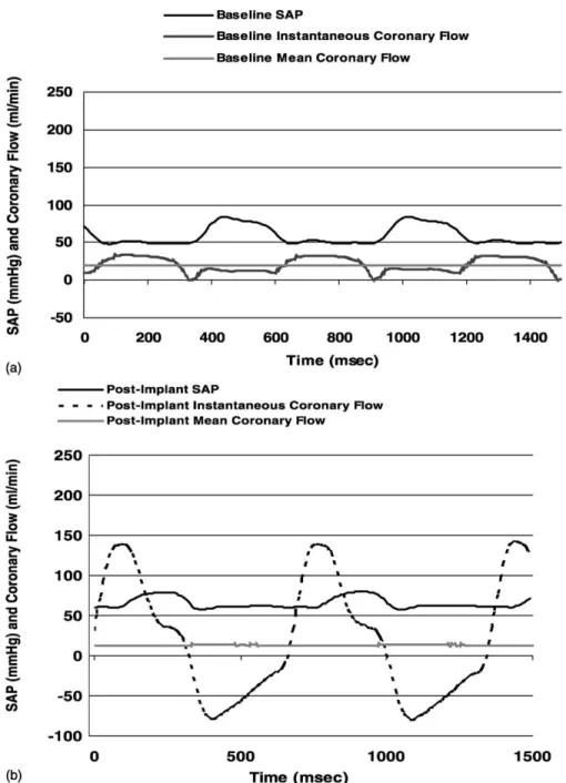
Following implantation of the Bi-DRD, the instan-taneous flow pattern seen distal to the device consisted of
forward flow during systole and retrograde flow during diastole with no areas of stasis as previously reported [2] and seen inFig. 3b.Fig. 3shows two cardiac cycles of mean and instantaneous distal LAD flow waveforms before (Fig. 3a) and after (Fig. 3b) implantation of the device in one representative animal. Superimposed on the flow
Fig. 2. Illustration of the implant technique. (a) placement of the purse string and measurement of the ventricular wall thickness with a ventricular needle and needle guide; (b) introduction of the dilator and sheath into the ventricular cavity along the wire; (c, d) placement of the device into the left ventricle and exchange of the tools; and (e) device fully implanted.
waveforms are the corresponding systemic arterial pressure waveforms. This representative animal shown inFig. 3had a net forward flow of 18 ml/min before implantation and 14 ml/min immediately post-implant (Fig. 3b) resulting in device flow that was 78% of native baseline flow. Analysis of the net forward flow through the device immediately after implantation for all animals studied revealed a mean flow equal to 73 ^ 15% of baseline native LAD coronary flow. Average peak forward flow measured 144 ^ 59 ml/min, and average peak retrograde flow measured 2 69 ^ 31 ml/min through the device compared with 37 ^ 13 ml/min average peak forward and 2 8 ^ 10 ml/min average peak retrograde flow in the native coronary artery (P ¼ 0:00003 for peak forward flow and P ¼ 0:00001 for peak retrograde flow).
3.2. Survival, patency and tissue reaction
Ten pigs survived to scheduled sacrifice with one animal in the 4 week group dying 2 days after implantation with a thrombosed device. This animal required electrical conver-sion ten times from ventricular fibrillation at implant resulting in 44 cumulative minutes of ischemia compared to an average ischemic implant time of 10.6 min for the study. Five animals each were sacrificed at both 2 and 4 weeks post-implantation. Two weeks after implantation, four of five devices were patent, and 4 weeks after implantation five of six devices were patent (including the early death). The graft that was occluded at 2 weeks failed due to thrombosis. This graft, which was damaged during
suturing to the artery, had caused a severe inflammatory reaction.
The gross analysis of the DRD/arterial interface of the four patent devices at 2 weeks showed neointimal proliferation along approximately 10% of the DRD
originating from the distal anastomosis. Scanning electron microscopic evaluation revealed minimal accumulation of platelet/fibrin coagulum on the internal lumen of the DRD. Insignificant thrombotic deposits were observed on the aspect of the DRD that protruded into the left ventricle (Fig. 4). DRDs explanted at 4 weeks showed varying levels of neointimal proliferation originating from the anastomo-sis. The entire length of the extraventricular segment was covered with neointima in one case (Fig. 5). Scanning electron microscopic evaluation demonstrated an intimal surface with bumpy or wavy appearing cells consistent with endothelial cells as the outer-most feature of the neointima (Fig. 6). The remaining 4-week explants had between 25 and 50% of the length of the extraventricular segment covered with neointima. The device cuff was found to be infiltrated with fibroblasts, as reported previously, with a mild to moderate chronic inflammatory cell response[1].
3.3. Myocardial perfusion
PET assessment of the perfusion in the region of myocardium subtended by the DRD revealed a mild decline in perfusion, though with preservation of perfusion at 85% of baseline (Table 1). With stress, a physiologic increase in perfusion for the DRD-subtended segment was noted consistent with controls.
Fig. 4. Gross photograph of endocardial/Bi-DRD interface 4 weeks post-implant. Note device protrudes into the lumen with no gross biological deposit.
Fig. 5. LAD/Bi-DRD interface 4 weeks after implantation. LAD in cross-section. (a) Gross photograph; and (b) Photomicrograph, H&E stain, original magnification 20 £ . Note no excessive neointimal proliferation at the vascular interface.
Fig. 6. Scanning electron micrograph of neointimal formation on a 4-week explanted Bi-DRD, original magnification 500 £ . Note the formation of non-obstructive, smooth endothelial-like growth into the extraventricular segment of the device.
4. Discussion
These data show encouraging results with a bi-directional DRD for achieving ventriculo-coronary artery bypass grafting in the porcine model. The results obtained in this study are comparable with those reported previously with a rigid titanium conduit having an intracoronary segment[1,2]. Those studies demonstrated 76 and 74% of baseline flow, respectively, compared with 73% of baseline flow seen in this study. This allows perfusion adequate to maintain contractibility at rest and with dobutamine stress [1,2]. Suehiro et al. [3] recently reported blood flows through an unvalved LV-LAD conduit similar in concept to be only 45% of baseline flow in an acute dog model. The differences in flows reported between this study and Suehiro et al. may be related to device design. Suehiro et al. [3] investigated improving net forward flow by inhibiting retrograde flow during diastole using a Starling resistor valve. Our studies have consistently shown that similar or even superior flows can be obtained with an unvalved conduit compared to those obtained with a valved conduit (70%). These studies demonstrate that device design dramatically affects flow. Incorporating a valve in a 3 mm or smaller diameter conduit is not practical in light of the fact that no valve smaller than 15 mm internal diameter has been hemodynamically effective or durable. Thrombosis due to turbulence created by prosthetic valve mechanisms is well documented for small caliber devices in the prosthetic heart valve field [12]. In addition, the antegrade/retrograde washing of blood, reducing stasis and deposition, is believed to be a critical component contributing to long-term patency of a VCAB device as indicated by these preliminary results.
This study also demonstrates that significant levels of perfusion both at rest and under stress occur in myocar-dium subtended by the VCAB device (85% of remote) compared to myocardium subtended by native coronary flow. Further, utilizing the present Bi-DRD device, augmentable flow is preserved and recruitable under stress conditions similar to control myocardium. It has been shown that only 20 – 30% of normal subendocardial flow is necessary to support systolic function [13]. Importantly, these devices provide significantly greater flow than that seen in patients with severe coronary artery disease. Of clinical relevance are the Hufnagel valve recipients from the 1950s whose coronaries were perfused during systole because of valve placement in the descending thoracic
aorta. Significantly, several of these patients lived asymp-tomatically for up to 18 years [14]. This study and previously published studies [1 – 3] support our strategy that flow delivered to the myocardium during systole with minimal antegrade coronary flow during diastole, while less than that which would be predicted for standard coronary artery bypass grafting, provides adequate levels of nutritive flow to sustain cardiac function.
In conduits smaller than 4 mm, particularly in coronary vascular applications, ePTFE has not gained wide accep-tance [4 – 9]. Inferior clinical outcomes reported with this material in coronary vascular applications have been postulated to be related to a number of factors. The anastomosis of large diameter ePTFE to coronary arteries that are only 1.5 – 2.5 mm outer diameter with resultant poor hemodynamics due to abrupt diameter change is felt to contribute to reduced chronic patency. Limited distal arterial runoff due to the relatively poor condition of the recipient artery and distal bed predisposes grafts to failure by thrombosis. Lower trans-conduit flow allowing stasis of blood contributes to thrombosis of smaller diameter grafts. Further, the excessive length of synthetic conduit required for a typical aorto-coronary graft also accelerates and contributes to a thrombotic response.
Despite the above limitations, encouraging data have been reported on the use of ePTFE as an aorto-coronary arterial bypass conduit. Specifically, Emery et al. reported on the early North American experience with the use of the Perma-Flow graft, an ePTFE conduit, in patients to 1 year follow-up[4]. While the numbers were low, they reported 90% early patency and 77% patency at 1 year for 73 coronary side-to-side anastomoses evaluated at 1, 3 – 8 and 12 month intervals. They concluded that the graft was very promising for completion therapy. The Perma-Flow graft has a unique design that results in high flow rates of 250 ml/ min at the aortic proximal end of the graft [4] due to the creation of a controlled left to right shunt providing continuous flow through the ePTFE graft. The Bi-DRD evaluated in the current study also sees high instantaneous flow rates and no areas of stagnation throughout the cardiac cycle, similar to the Perma-Flow graft. These data suggest that ePTFE conduits configured with an appropriate design allowing for high flows and optimal hemodynamics fares well in the coronary application.
The Bi-DRD examined in this study addresses several of the disadvantages observed in the prior use of ePTFE for coronary surgery in that the diameter of the device approaches that of the coronary artery (3 mm) and the hemodynamics of systolic flow results in peak flows four times greater than native flows with no areas of blood stasis. In addition, length of the conduit is significantly shorter in the present Bi-DRD design described (approxi-mately 4 cm) compared to previous ePTFE conduits, thereby minimizing adverse blood-material interactions. Healing of the conduit at 4 weeks in the porcine model was encouraging with the formation of non-obstructive
Table 1
Myocardial perfusion in LAD territory compared to remote territory (posterior wall) LAD (ml/min per g) Remote (ml/min per g) Percentage of remote (%) Baseline (n ¼ 2) 0.91 ^ 0.22 1.07 ^ 0.16 85 Stress (n ¼ 2) 1.21 ^ 0.06 1.46 ^ 0.08 83
neointima that originated from the host artery on the lumen of as much as half of the conduit. Finally, thrombus deposition was not significant on devices that did not experience technical issues such as infection or severely compromised flow during implantation associated with peri-operative ventricular fibrillation.
In conclusion, the novel bi-directional DRD and VCAB procedure described afford several unique and desirable revascularization characteristics. This approach allows for creation of a permanent channel between the left ventricle and coronary artery providing proximate high flow supply of oxygenated blood. The Bi-DRD provides both retrograde and antegrade coronary flow from the site of coronary anastomosis. This feature maintains perfusion of the full zone of myocardium distal to a stenosis regardless of the site of transventricular wall insertion of the ePTFE conduit into the LV cavity. Further, the present device provides significant net forward flows, without impairment of flow reserve under stress conditions. Finally, the present ePTFE configuration affords good patency, healing and hemocom-patibility. Results of this study suggest that this device and surgical technique are promising for coronary bypass surgery and warrant longer duration studies. The device may be useful to provide myocardial perfusion in patients with inadequate conduits or to complete revascularization in patients who have conditions which obviate standard care such as porcelain aorta.
Acknowledgements
This study was performed under a research grant from HeartStent Corporation. Sincere thanks are extended to the individuals who assisted in the operations, data collection, and analysis including Dale Groth, Kris Hagen, Susan Perron, and Eric Solien.
References
[1] Tweden KS, Eales F, Cameron JD, Griffin JC, Solien E, Knudson MB. Ventriculocoronary artery bypass (VCAB), a novel approach to myocardial revascularziation. Heart Surg Forum 2000;3:47– 55. [2] Emery RW, Eales F, Van Meter Jr CH, Knudson MB, Solien EE,
Tweden KS. Ventriculocoronary artery bypass results using a mesh-tipped device in a porcine model. Ann Thorac Surg 2001;72:S1004 – 8. [3] Suehiro K, Shimizu J, Yi G-H, Zhu S-M, Gu A, Sciacca RR, Wang J, Burkhoff D. Direct coronary artery perfusion from the left ventricle. J Thorac Cardiovasc Surg 2001;121:307– 15.
[4] Emery RW, Mills NL, Teijeira FJ, Arom KV, Baldwin P, Petersen RJ, Joyce LD, Grinnan GLB, Sussman MS, Copeland III JG, Ochsner JL, Boyce SW, Nicoloff DM. North American experience with the Perma-Flow prosthetic coronary graft. Ann Thorac Surg 1996;62:691– 6. [5] Hehrlein FW, Schlepper M, Loskot F, Scheld HH, Walter P, Mulch J.
The use of expanded polytetrafluoroethylene (PTFE) grafts for myocardial revascularization. J Cardiovasc Surg 1984;25:549 – 53. [6] Sapsford RN, Oakley GD, Talbot S. Early and late patency of
expanded polytetrafluoroethylene vascular grafts in aorto-coronary bypass. J Thorac Cardiovasc Surg 1981;81:860– 4.
[7] Yokoyama T, Gharavi MA, Ying-Chien L, Edmiston WA, Kay JH. Aorto-coronary artery revascularization with an expanded polytetra-fluoroethylene vascular graft. J Thorac Cardiovasc Surg 1978;76: 552 – 5.
[8] Molina JE, Carr M, Yarnoz MD. Coronary bypass with Gore-Tex graft. J Thorac Cardiovasc Surg 1978;75:769– 71.
[9] Kerber S, Baumbach M, Rahmel A, Weyand M, Scheld HH, Breithardt G. Clinical and invasive 7-month follow-up of a patient with a synthetic coronary graft. Int J Cardiol 1995;51:143– 8. [10] Drasler WJ, Jenson ML, Geroge SA, Protonotarios EI, Dutcher RG,
Duncan EP, Possis ZC. A unique vascular graft concept for coronary and peripheral applications. ASAIO Trans 1988;34:769 – 72. [11] McFalls EO, Baldwin D, Marx D, Fashingbauer P, Ward H. Temporal
changes in function and regional glucose uptake within stunned porcine myocardium. J Nucl Med 1996;37:2006– 10.
[12] Butchart EG. Thrombosis, embolism, and bleeding. In: Bodnar E, Frater R, editors. Replacement cardiac valves. New York: Pergamon Press; 1991. p. 77.
[13] Gallagher KP, Matsuzaki M, Koziol JA, Kemper WS, Ross Jr J. Regional myocardial perfusion and wall thickening during ischemia in conscious dogs. Am J Physiol 1984;247:H727 – 38.
[14] Fishbein MC, Roberts WC. Late postoperative anatomic observations after insertion of Hufnagel caged-ball prostheses in descending thoracic aorta. Chest 1975;68:6 – 11.