Expression of SCCmec cassette chromosome recombinases
in methicillin-resistant Staphylococcus aureus
and Staphylococcus epidermidis
M. Stojanov*, O. Sakwinska† and P. Moreillon
Department of Fundamental Microbiology, University of Lausanne, Lausanne, Switzerland
*Corresponding author. Tel:+41-21-6925612; Fax: +41-21-6925605; E-mail: [email protected] †Present address: Nestle´ Research Centre, 26 Vers-chez-les-Blanc, 1000 Lausanne, Switzerland.
Received 18 July 2012; returned 5 September 2012; revised 15 October 2012; accepted 16 November 2012 Objectives: Methicillin resistance in staphylococci is mediated by the mecA gene, which is carried on the staphylococcal cassette chromosome mec (SCCmec). SCCmec is responsible for vertical and horizontal transfer of methicillin resistance. Horizontal transfer implies first SCCmec excision from the chromosome. Site-specific excision is catalysed by the Ccr recombinases, which are encoded by ccrAB genes located on the cassette. The aim of this study is to determine the promoter activity of ccrAB genes in individual cells of methicillin-resistant Staphylococcus aureus (N315, COL and MW2) and Staphylococcus epidermidis (RP62A). One mutant cured of its SCCmec (N315EX) was also used. Exposure to various stresses was included in the study. Methods: For each strain, translational promoter-green fluorescent protein (gfp) fusions were used to assess the levels of ccr promoter activity in individual cells. Analyses were performed using epifluorescence microscopy and flow cytometry.
Results: ccr promoter activity was observed in only a small percentage of cell populations. This ‘bistable’ pheno-type was strain dependent (GFP was expressed in N315 and RP62A, but not in COL and MW2) and growth dependent (GFP-expressing cells decreased from approximately 3% to 1% between logarithmic and stationary growth phases). The ccr promoter of strain N315 displayed normal promoter activity when expressed in SCCmec-negative N315EX. Likewise, the ccr promoter of strain COL (which was inactive in COL) showed normal N315-like activity when transformed into N315 and N315EX.
Conclusions: SCCmec excision operates through bistability, favouring a small fraction of cells to ‘sacrifice’ their genomic islands for transfer, while the rest of the population remains intact. Determinants responsible for the activity of the ccr promoter were not located on SCCmec, but were elsewhere on the genome. Thus, the staphylococcal chromosome plays a key role in determining SCCmec stability and transferability.
Keywords: excision, promoter, staphylococci, gfp
Introduction
In Staphylococcus spp. resistance to methicillin and to virtually all b-lactam drugs is mediated by the expression of the mecA gene, which encodes low-affinity penicillin-binding protein A, or PBP2A.1The mecA gene is carried by a genomic island named
staphylococcal cassette chromosome mec (SCCmec), which is responsible for both vertical and horizontal transfer of methicillin resistance. It was originally believed that worldwide spread of methicillin-resistant Staphylococcus aureus (MRSA) was solely due to clonal expansion of a few successful strains, implying that de novo acquisition of SCCmec was extremely rare.2
However, more recent studies indicate that lateral spread of
SCCmec is more frequent than expected.3,4This is substantiated
both by the fact that almost identical cassettes are present in unrelated staphylococcal strains—and even in different staphylococcal species—and by the presence of different types of SCCmec in closely related strains.5–7
To be transferred, SCCmec must first be excised from the chromosome of the donor strain. Site-specific excision of SCCmec is catalysed by the Ccr recombinases, which are large serine recombinases of the resolvase/invertase family and are encoded on the SCCmec cassette.8 Three phylogenetically
dis-tinct ccr genes have been found so far on different cassettes. Certain SCCmec cassettes carry the ccrA and ccrB genes (ccrAB), which are part of the same transcription unit, whereas
#The Author 2012. Published by Oxford University Press on behalf of the British Society for Antimicrobial Chemotherapy. All rights reserved. For Permissions, please e-mail: [email protected]
other cassettes carry the ccrC gene, which shares less than 50% sequence similarity with ccrAB. Several allotypes (sequence simi-larity below 85%) have been described for ccrA and ccrB and are used for classification of their cognate SCCmec cassettes. In con-trast, only one allotype was found for the ccrC gene.9
Site-specific excision/integration of SCCmec takes place at the 5′end of orfX, a highly conserved gene located near the origin of replication in S. aureus. Integration involves a 15 bp core sequence (attB), which recombines with the cognate 15 bp core sequence located on the SCCmec (attS) resulting, upon integration, in two direct repeats (attR and attL) flanking the element.8In add-ition, accessory sequences located near the attS sites, which form imperfect inverted repeats of various sizes, seem also to play a role in site-specific integration/excision in many SCCmec.10,11 Interestingly, CcrAB recombinases do not display cassette specifi-city, as all the allotypes are able to excise SCCmec of different types.12Conversely, CcrC recombinases are specific to their own
SCCmec.11
Spontaneous SCCmec excision has been observed on several occasions both in vivo and in vitro.12–15For instance, overexpres-sion of plasmid-located ccrAB genes generally leads to SCCmec excision and results in conversion of MRSA into methicillin-susceptible S. aureus (MSSA).8Moreover, b-galactosidase assays
and quantitative RT–PCR studies showed that b-lactams and vancomycin increased transcription from ccrAB promoters of strains MW2 and N315.16This could favour the propagation of SCCmec via a two-step scenario. First, b-lactam drugs in the en-vironment would select for methicillin-resistant staphylococci, thus promoting the expansion of the SCCmec reservoir. Second, transfer of SCCmec from this reservoir into new staphylococci
would generate new methicillin-resistant strains capable of further amplifying the pool of SCCmec. In such a scenario, the optimal setting implies that not all individuals of the donor popu-lation do excise their cassette simultaneously, because they would become methicillin susceptible and thus be destroyed by the drug, but that only a few individuals commit this suicide, sac-rificing themselves to transfer SCCmec into new recipient strains. Here we tested this hypothesis by using promoter-green fluor-escent protein (gfp) fusions to measure the expression of ccrAB genes in individual cells of various strains of MRSA carrying differ-ent types of SCCmec cassettes. Moreover, we also examined the methicillin-resistant Staphylococcus epidermidis strain RP62A, as more and more evidence suggests that coagulase-negative staphylococci are the reservoir of SCCmec for S. aureus.13,17,18
Materials and methods
Bacterial strains, media and culture conditions
Bacterial strains and plasmids used in this study are listed in Table1. Escherichia coli strain DH5a, which was routinely used for plasmid propa-gation and cloning experiments, was cultivated on Luria-Bertani (LB) medium (Becton Dickinson, Sparks, MD, USA) supplemented with 100 mg/L ampicillin (AppliChem, Darmstadt, Germany) at 378C. S. aureus strains were grown with aeration in Trypticase soy broth (TSB) (Difco Laboratories, Detroit, MI, USA) in a rotating incubator (at 180 rpm) at 378C. For all experiments, bacterial cultures were inoculated with a 1/100 dilution of an overnight culture. If required, tetracycline and erythromycin (AppliChem) were added at a final concentration of 10 mg/L for plasmid propagation and 5 mg/L for flow cytometry analysis. Oxacillin was commercially purchased and used at the sub-MIC concentration Table 1. Bacterial strains and plasmids used in this study
Strain or plasmid Relevant characteristics Reference
Strain
E. coli DH5a host for DNA cloning laboratory collection
S. aureus
RN4220 restriction-deficient derivative of RN450; intermediate cloning host 19
N315 MRSA carrying type II SCCmec 20
N315EX isogenic MSSA derivative of N315 this study
COL MRSA carrying type I SCCmec 21
MW2 MRSA carrying type VI SCCmec 22
S. epidermidis RP62A methicillin-resistant S. epidermidis carrying type II SCCmec 21
Plasmid
pCN36 E. coli– S. aureus shuttle vector; TcR; +22 copies/cell 23
pCN68 E. coli– S. aureus shuttle vector; source of gfpmut2 gene 23
pSR3-1 thermosensitive-replicon plasmid carrying the ccrAB genes of strain N315 (used for SCCmec excision in N315)
8
pPGFP-N315 PccrABN315-gfpmut2 fusion cloned in pCN36; tet(M) this study
pPGFP-MW2 PccrABMW2-gfpmut2 fusion cloned in pCN36; erm(C) this study
pPGFP-COL PccrABCOL-gfpmut2 fusion cloned in pCN36; erm(C) this study
pPGFP-COL-Tc PccrABCOL-gfpmut2 fusion cloned in pCN36; tet(M) this study
pPGFP-RP62A PccrABRP62A-gfpmut2 fusion cloned in pCN36; tet(M) this study pNEG-tet(M) PNEG-gfpmut2 fusion cloned in pCN36; used as negative control this study pNEG-ermC PNEG-gfpmut2 fusion cloned in pCN36; tet(M) replaced with erm(C); used as negative control this study pGFPS10-tet(M) PPS10-gfpmut2 fusion cloned in pCN36; used as positive control this study pGFPS10-ermC PPS10-gfpmut2 fusion cloned in pCN36; tet(M) replaced with erm(C); used as positive control this study
Stojanov et al.
of 4 mg/L for methicillin-resistant staphylococci. Mitomycin C (Sigma– Aldrich Chemie GmbH, Steinheim, Germany) was used at a final concen-tration of 0.5 mg/L.
DNA manipulations
For S. aureus and S. epidermidis, genomic DNA was extracted using a proto-col adapted from Bae et al.24Briefly, 3 mL of an overnight culture was har-vested and resuspended in 50 mL of Tris –EDTA buffer (10 mM Tris– Cl, pH 7.5, and 1 mM EDTA) supplemented with lysostaphin (final concentration 0.5 mg/mL). After 30 min of incubation at 378C, 300 mL of ‘Nuclei lysis solu-tion’ (Promega Corp., Madison, WI, USA) was added and the cell suspen-sions were heated at 808C for 10 min. The samples were then treated with RNase and addition of 100 mL of ‘Protein precipitation solution’ (Promega Corp.) was followed by incubation for 5 min on ice. After centri-fugation (48C, 15 600 g), supernatants were collected and 300 mL of isopro-panol was used to precipitate the DNA, which was subsequently washed with 70% ethanol, pelleted by centrifugation, air-dried and re-diluted at 48C overnight in 20 mL of elution buffer (Qiagen Inc., Hilden, Germany).
Plasmids were isolated using the QIAprep Spin Miniprep Kit (Qiagen Inc.). For S. aureus, an additional step consisting of lysostaphin treatment (final concentration 0.5 mg/mL) was performed before the lysis step.
Digestions with restriction enzymes (Promega Corp.) were carried out according to the manufacturer’s specifications. PCR fragments were puri-fied using the QIAquick PCR Purification Kit (Qiagen Inc.) and gel-bands were purified using the QIAquick Gel Extraction Kit (Qiagen Inc.) according to the manufacturer’s protocols. Ligations were performed using 1 mL of T4 ligase (Promega Corp.) according to the manufacturer’s specifications.
PCR
GoTaq DNA polymerase (Promega Corp.) was routinely used for colony PCR screening analysis. DNA fragments required for cloning were ampli-fied with KAPA HiFi DNA Polymerase (KAPA Biosystems, Cape Town, South Africa). All reactions were carried out according to the manufac-turers’ specifications. The primers used in this study are listed in Table2.
Construction of translational fusion reporter plasmids
All the promoter-gfp fusion reporters were constructed as follows: specif-ic primer pairs were used to PCR amplify the N315 ccrAB promoter and the gfpmut2 gene. After enzymatic digestion, the two fragments were cloned by three-point ligation in the pCN36 plasmid. All the otherplasmid reporters used in the study were made by substituting the N315 ccrAB promoter with their respective promoter (Table1). To cali-brate the levels of activity of the ccrAB promoters, a negative control plasmid (pNEG) was constructed by replacing the ccrAB promoter region with a 140 bp DNA fragment of the secA gene (SA2442 in strain N315), and a positive control plasmid (pGFPS10) was constructed by replacing the same region with the constitutive promoter of the rpsJ gene (SA2048 in strain N315).
Microscopy
Epifluorescence microscopy was performed using an Axioskop2 epifluores-cence microscope (Zeiss, Germany), with a×100 objective (Plan-NEOFLUAR ×100/1.30 oil, Zeiss). Bacteria from 3 mL aliquots from cultures in the exponential growth phase as described previously25were harvested by centrifugation and resuspended in 50 mL of PBS, of which 10 mL was depos-ited on glass slides and analysed.
Picture files (16-Bit) were scaled with Metamorph software (Visitron Systems, Germany) to visualize the fluorescence signal. GFP fluorescence pictures were digitally coloured using Photoshop 4.0 (Adobe Systems Europe Ltd, Edinburgh, UK) and superimposed on the corresponding phase contrast pictures, in order to visualize cells with an active ccrAB promoter.
Flow cytometry
Flow cytometry was performed with a FACS-Calibur (BD Biosciences, Erem-bodegem, Belgium), equipped with an air-cooled argon laser (488 nm). GFP fluorescence was recorded in the FL1 (525+15 nm channel). Samples were removed from cultures in the exponential or stationary phase of growth, diluted in PBS in order to not exceed 800 events per second, and the fluorescence of 20 000 events was recorded for each sample. Analysis of flow cytometry data was performed using WinMDI software (version 2.8, Salk Institute, http://facs.scripps.edu/software.html).
Artificial SCCmec excision
SCCmec was cured from S. aureus N315 by using a method from Katayama et al.8Briefly, strain N315 was electroporated with the thermosensitive plasmid pSR3-1 (containing ccrAB genes) and transformants were cultured for 24 h at 308C in TSB supplemented with tetracycline, serially diluted and plated on Trypticase soy agar (TSA) supplemented with tetra-cycline. Single colonies were picked, grown for 24 h at 428C in TSB to Table 2. Primers used in the study
Primer Sequencea Description
Prom fw TTTTTTGGATCCTTGTCTTTATCATACAACTGTG amplification of the promoter fragment in strains N315, MW2 and RP62A Prom rev TTTTTTACTAGTATCGGCTCCTCCTTTCACAGT
PromCOL fw TTTTTTGGATCCTAACTTAAAGATGAAATCGTACAGG amplification of the promoter fragment in strain COL PromCOL rev TTTTTTACTAGTCGTATTTCCTCCTTCCAAAGT
neg fw TTTTTTGGATCCCCTTGTCTACCAGAACGACCACG amplification of the fragment used as a negative control (secA gene—SA2442 in N315)
neg rev TTTTTTACTAGTATGGCAGGTCGAGGCACAG
PS10 fw TTTTTTGCATGCCATTCACCACCGTTCTTATGAC amplification of the promoter region of the rpsJ gene (30S ribosomal protein S10—SA2048 in N315) PS10 rev TTTTTTCTGCAGTCCCTCCTTATTCGTCTACATTT
GFP fw TTTTTTACTAGTATGAGTAAAGGAGAAGAACTT amplification of the gfpmut2 gene from pCN68 GFP rev TTTTTTGAATTCTATTTGTATAGTTCATCCATG
Excision fw CGCAGTAACTACGCACTATCATTCAGC amplification of the chromosomal junction in S. aureus N315EX Excision rev TGAATGAACGTGGATTTAATGTCCACC
aRestriction sites are underlined.
promote curing of thermosensitive pSR3-1, and dilutions were plated on plain TSA to screen for colonies susceptible to oxacillin and tetracycline. One double-susceptible colony was purified and the absence of SCCmec in it was confirmed by PCR amplification of the chromosomal junction formed upon excision using the primer pair Excision fw and rev (Table2). This isolate was named N315EX and used in further experiments.
Results
Allotype 1 and 2 ccrAB promoters
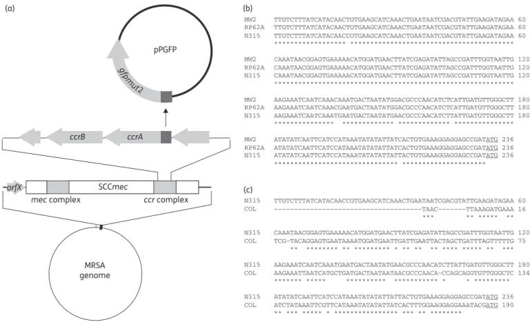
The expression of ccrAB genes was analysed using gfp transla-tional fusions as depicted in Figure 1(a). This required first the characterization of the ccr gene promoter regions. Thus, the complete intergenic regions upstream of ccrA, which contain the putative promoter of the ccrAB transcriptional unit, were analysed for all the strains examined in this study (Table 3). Figure1(b and c) depicts the homologies between these inter-genic regions in different strains. The 233 bp promoter regions of strains N315, MW2 and RP62A, which all carry the ccrAB allo-type 2, were well conserved, with a sequence similarity of 97% (Figure1b). In contrast, the promoter region of strain COL was shorter (i.e. 187 bp), missing the 5′part, and showing a sequence similarity of 77% when compared with N315 (Figure1c).
To test whether the activity of the promoters could be modu-lated in trans by genes from the core chromosome, rather than in
cis or in trans by elements from the SCCmec cassette, we also studied the expression of the ccrAB promoters in S. aureus N315EX, from which SCCmec had been deleted.
Microscopy analysis
The activity of the ccrAB promoters was first evaluated by epifluorescence microscopy during the exponential phase of growth (i.e. samples taken after 3 h of inoculation). Figure2
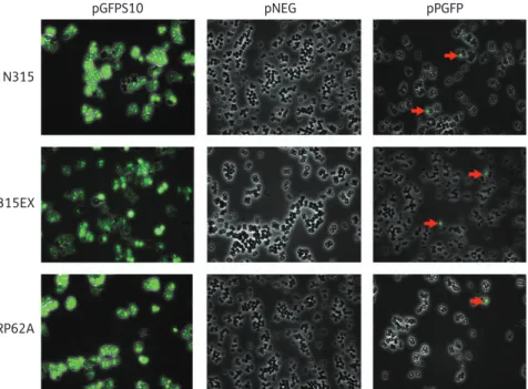
exemplifies such results with strains N315 and RP62A. It can be seen that both strains expressed GFP uniformly from the posi-tive control vector pGFPS10, whereas cells carrying the negaposi-tive control pNEG were devoid of fluorescence. In sharp contrast, only
pPGFP
orfX
mec complex ccr complex ccrB SCCmec MRSA genome ccrA gfpm ut2 (a) (b) (c)
Figure 1. (a) Construction scheme of reporter plasmids (pPGFP). The dark grey box represents the intergenic region containing the cloned ccrAB promoter regions. (b and c) Alignments of the intergenic regions containing the ccrAB promoter regions of the strains analysed in the study. The start codon of the ccrA gene is underlined.
Table 3. ccrAB allotype and SCCmec type of the strains used in the study
Strain ccrAB allotype SCCmec type
S. aureus COL 1 I S. aureus N315 2 II S. aureus N315EX — — S. aureus MW2 2 IV S. epidermidis RP62A 2 II
Stojanov et al.
a small proportion of the two cell populations showed GFP expression when they carried their specific pPGFP-N315 or pPGFP-RP62A. Most interestingly, roughly similar proportions of cells expressing GFP were observed by microscopy when pPGFP was transformed into the SCCmec-deleted mutant N315EX, indi-cating that the SCCmec of strain N315 did not contain determi-nants affecting the activity of the promoter. On the other hand, fluorescence signals were not observed by microscopy when pPGFP-MW2 and pPGFP-COL were introduced into strains MW2 and COL, respectively (data not shown). However, because mi-croscopy results are qualitative and may be dependent on the observer, the experiments described above were repeated using flow cytometry analysis as described below. These, for instance, confirmed the absence of fluorescence in strains MW2 and COL (see Figure4).
Quantification of fluorescent cells by flow cytometry
and influence of stress conditions on ccrAB expression
Flow cytometry was used to quantitatively assess the proportion of cells expressing the ccrAB promoter under different growth conditions. Figure 3depicts prototype fluorescence profiles for N315 cells carrying the negative control pNEG (left panel) as well as the same strain expressing pPGFP (right panel). As observed using epifluorescence microscopy, only a minor propor-tion of the cell populapropor-tion expressed GFP. In order to determine the proportion of GFP-positive cells, we delineated a threshold using the GFP-negative control, as shown in Figure3.
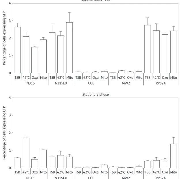
The experiment was repeated with each of the strains described in Table3, tested in either the exponential or station-ary phases of growth, and exposed to various stress conditions including growth at 428C and exposure to oxacillin or mitomycin C. Figure 4 presents the details of these results. As a general
feature, strains N315, N315EX and RP62A expressed GFP at a sizable level under all test conditions, whereas COL (which had a shorter promoter region of 187 bp versus 233 bp in the other strains) and MW2 (which had an N315-like promoter region) remained essentially below the limit of detection (Figure4).
In the exponential growth phase strains N315, N315EX and RP62A exhibited subpopulations expressing GFP at rates between 2% and 3%, which is compatible with epifluorescence microscopy (Figure2). Similar results were obtained under stress conditions, except for oxacillin treatment, which significantly reduced the GFP expression to 1.5% in strain N315. In contrast, in the stationary growth phase GFP expression was significantly reduced under most conditions, with some exceptions, i.e. for N315 at 428C and RP62A with mitomycin C.
N315
pGFPS10 pNEG pPGFP
N315EX
RP62A
Figure 2. GFP expression. Digital overlay of GFP fluorescence image with the respective phase contrast image in cells carrying pPGFP, pNEG (negative control) and pGFPS10 (positive control). Red arrows indicate cells carrying pPGFP in which the ccrAB promoter is active.
pNEG Fluor escence Fluor escence pPGFP
Forward scatter Forward scatter
Figure 3. Quantification of N315 cells expressing GFP from the ccrAB promoter. Fluorescence expression profiles of cells carrying pNEG and pPGFP. Gated cells show the subpopulation expressing GFP. For each measurement, gates were arbitrarily determined with respect to the negative control.
It is noteworthy that the results were very similar when plasmid pPGFP-N315 was expressed in parent N315 or in its SCCmec-negative mutant N315EX, thus confirming the results obtained by epifluorescence microscopy and the fact that ccrAB genes were at least partially affected by non-SCCmec genetic determinants.
Finally, to test whether the shorter promoter region of strain COL was responsible for the absence of promoter activity, we transferred its specific expression plasmid (pPGFP-COL-Tc; Table1) into the N315 background and repeated the expression experiment. Figure5shows that transfer of COL-specific pPGFP-COL-Tc into N315 or N315EX completely restored promoter
activity. Thus, promoter activity was highly dependent on the bac-terial background and the shorter COL promoter region was not responsible for its inactivity in the COL background.
Discussion
The present experiments employed a GFP expression system cloned into a low copy number plasmid in order to test the pro-moter activity of type 1 and 2 ccrAB allotypes in different staphylococcal backgrounds. The results yielded several findings that may help better understand the excision and transmission
Exponential phase Stationary phase 4 3 2 P e
rcentage of cells expr
essing GFP 1 0 4 3 2 P e rc
entage of cells expr
essing GFP
1
0
TSB 42°C
N315 N315EX COL MW2 RP62A
Oxa Mito TSB 42°C Mito TSB 42°C Oxa Mito TSB 42°C Oxa Mito TSB 42°C Oxa Mito
TSB 42°C
N315 N315EX COL MW2 RP62A
Oxa Mito TSB 42°C Mito TSB 42°C Oxa Mito TSB 42°C Oxa Mito TSB 42°C Oxa Mito
Figure 4. Flow cytometry analysis of the effects of various stresses on ccrAB promoter activity. Promoter constructions were transformed and expressed in their original strain. Cultures were grown either in plain TSB at 378C (TSB) or in TSB submitted to various stress conditions from the very beginning of growth [i.e. 428C, 4 mg/L oxacillin (Oxa) or 0.5 mg/L mitomycin C (Mito)]. Samples were removed from the cultures after 3 h (exponential phase) or 24 h (stationary phase) of growth, diluted in PBS in order not to exceed 800 events per second during flow cytometry, and the fluorescence of 20 000 events was recorded for each sample. The bars represent the averages of three independent measurements in three different cultures and error bars represent the standard deviations.
processes of SCCmec. First, as predicted by the two-step trans-mission hypothesis discussed in the Introduction section, ccrAB promoter activity was not present simultaneously in all the cells of a culture, but only in a minority of them that represented a small fraction of the whole population. This is logical econom-ics in a relatively primitive transfer system where the donor cell must lose its (presumably) beneficial genomic island in order to transfer it further. The results show that only a few cells in the population were activated for this purpose at a given time, while the remaining bacteria kept their advantageous genotype. This stochastic gene expression system has been referred to as ‘bistability’ and was described in the transfer of other genomic islands,26,27 including the ICEclc in Pseudomonas knackmussi.28ICEclc confers the capability to use aromatic
com-pounds (e.g. chlorobenzoates and aminophenols) as carbon sources. Conjugative transfer of this island begins with its exci-sion from the chromosome, which is driven by the activity of the IntB13 integrase promoter. Upstream of that, IntB13 pro-moter activity is controlled by the InrR protein, which is encoded on the island. Stochastic activation of IntB13 was observed by GFP expression in proportions of cells that increased from 0.1% to 3% between 24 h and 96 h of incubation. This sug-gested a dependency on cell concentration, and perhaps also on nutrient availability.
The present observations are very reminiscent of the bistabil-ity of ICEclc-related transfer in terms of frequency and depend-ency on growth phase. However, in contrast to ICEclc, the activity of the ccrAB promoters tested herein appeared to be influenced by determinants that were not encoded on the SCCmec cassette, but rather elsewhere on the genome. This was clear from the fact that the type 2 ccrAB promoter region of MRSA N315 was expressed similarly when transformed either into its N315 parent strain or into the isogenic N315EX mutant missing the whole SCCmec cassette. Thus, deletion of SCCmec did not affect promoter activity, which was therefore
regulated by other genetic determinants. Moreover, the type 1 ccrAB promoter region of MRSA COL, which was totally inactive in its parental strain, regained full N315-like activity when trans-formed into MRSA N315 and MSSA N315EX. Thus, factors driving ccrAB gene expression were not only located in the remaining genetic background, but could vary between different strains. Eventually, the fact that the promoter region of the COL ccrAB was functional in N315 restricted critical parts of this region to the 3′end of≤187 bp.
Bose et al.29described the regulatory excision system of the conjugative island ICEBs1 of Bacillus subtilis. The excisionase gene appeared regulated by an original repressor/antirepressor (immR/immA) system, where the antirepressor acted by proteo-lytic degradation of the repressor. The system responded to changes in population density, and to DNA-damage-induced SOS response as well. Like in the Pseudomonas ICEclc example, the two genes were located on the ICEBs1. However, they were rather redundant in other B. subtilis mobile genetic elements (MGEs), suggesting that they could act in trans. Moreover, a homologue of the system was found in Staphylococcus haemolyticus.29
In the present case, we did not find any homologues of the immR/immA system in our strains, either on the SCCmec cassette or on the chromosome. Moreover, we also asked whether there might be differences in the MGE contents of differ-ent strains, which might iddiffer-entify a specific MGE that would be present in strain N315 (which could activate ccrAB), but not in strains COL and MW2 (which lacked ccrAB activity), and so drive ccrAB expression in trans in N315. However, this was not the case. Finally, we also asked whether the ccrAB promoter region could carry specific operators for alternative sigma factors, as recently shown in certain S. aureus prophages,30but we did not find homologous regions either. Thus, the ccrAB regu-latory elements of SCCmec have yet to be identified.
In this regard, and in the context of bistability, one might speculate that the activity of ccrAB in only a minority of cells could serendipitously accompany phenotypic variants in hetero-geneous bacterial populations. Population heterogeneity appeared critical to ensure survival of a minority of cells (called persisters) under certain stress conditions, such as antibiotic treatment.31,32 Gene expression is different in antibiotic persisters, which are dormant and tolerant to antibiotics, than in vegetative cells, which are active and killed by the drugs. Analogously, it could be that subpopulations expressing ccrAB occur only in specific phenotypic variants along with other housekeeping genes. The question would then arise as to how SCCmec cassettes managed to take advantage of this bacterial regulation system, and why was it different in different strains. More insights into the phylogeny of SCCmec cassette precursors might help to solve this issue.
While the present experiments were not aimed at identifying the precise molecular mechanisms responsible for ccrAB regula-tion, some clues for them were identified. Indeed, aside from the fact that trans-acting determinants were located outside the SCCmec cassette, the rate of bistability could vary as a function of growth phase. For instance, it decreased by more than five times (from approx. ≥2.5% to ,0.5%; P,0.0001, unpaired t-test) between the logarithmic and stationary growth phases in S. aureus N315 and coagulase-negative Staphylococcus RP62A. Moreover, stress conditions such as high temperature or
5 4 3 P e rc
entage of cells expr
essing GFP 2 1 0 N315 pPGFP-N315 N315 pPGFP-COL-Tc N315EX pPGFP-N315 N315EX pPGFP-COL-Tc
Figure 5. Flow cytometry analysis of N315 and N315EX transformed with either pPGFP-N315 or pPGFP-COL-Tc. The experimental protocol was as described in Figure 4. The bars represent the averages of three independent measurements in three different cultures and error bars represent the standard deviations.
treatment with mutagenic mitomycin C also showed some strain-specific trends toward increased rates of bistability, as ap-parent in Figure4. Therefore, considering the fact that bistability was affected by various environmental stress conditions it is pos-sible that excision of SCCmec is regulated in a way similar to the excision of other MGEs or bacteriophages. Since major differ-ences between the genomes of S. aureus strains lay in their MGEs, this could account for differences in ccrAB expression in different genetic backgrounds.
The fact that frequency of bistability decreased in late growth was consistent with a recent study showing that spontaneous excision of SCCmec in MRSA N315 occurred transiently and early in the logarithmic growth phase, and much less there-after.33These results were obtained by determining the propor-tion of chromosomes from which the SCCmec cassette had been excised compared with total bacterial chromosomes at dif-ferent timepoints during growth. On the other hand, the present experiments did not reveal an increase in ccrAB promoter activity during treatment with b-lactams, as was previously described for transcription of ccrA (using b-galactosidase and quantitative RT–PCR) after exposure to b-lactams and vancomycin.16 However, the two studies differed in design in several ways that may render them complementary rather than contradictory. One of them is that we cloned the promoter region between the start of ccrAB and its preceding open reading frame, whereas Higgins et al.16 added the first 21 bp of the ccrA gene plus 20 bp of the preceding open reading frame to this region. There-fore, it is conceivable that this larger ‘promoter region’ contained additional regulatory elements that could alter ccrAB promoter activity. Another difference was related to the induction proto-cols. Indeed, while Higgins et al.16looked at promoter activity and mRNA expression 15 min after antibiotic addition or tem-perature shift to 428C, the present study determined promoter expression in cultures that had experienced much more pro-longed exposure to experimental stresses, i.e. throughout growth. Thus, one cannot exclude that ccrAB activity was punc-tually increased at the time of stress exposure, as in the Higgins et al.16study, but returned to baseline afterwards. Moreover, the
present study determined the heterologous ‘on’ or ‘off’ activity of the ccrAB promoter, whereas Higgins et al.16measured the sum of all of these individual activities. Although both studies used low copy number plasmids (with a range of approx. 15 to 22 copies per cell), these measurements are not strictly comparable and are amenable to methodology biases. A more thorough comparison would have required integrating each individual data point of light intensity produced by the flow cytometry ana-lysis in the present study. This anaana-lysis was attempted, but did not modify the overall results.
Taken together, the most important common message of the two experimental works is that activation of ccrAB expression does occur at a substantial rate in vivo and is affected by envir-onmental stresses. Moreover, the present results add the notion of heterogeneous gene expression and bistability to the system, and suggest that ccrAB activity depends on the microbial back-ground. Therefore, both environmental factors and bacterial background are pertinent with regard to SCCmec mobilization, and there might be more or fewer good donors and good recipi-ents present in the environment. In order to achieve a more comprehensive view of SCCmec transfer, further work should focus on the molecular mechanisms driven by these two
factors as well as on the mechanism (general phage transduc-tion?) underlying the transfer of SCCmec from one cell to another.
Acknowledgements
We thank T. Ito for providing plasmid pSR3-1.
Funding
This work was supported by grant 3200B0-113854 to P. M. and by Marie Heim Vo¨gtlin grant PMPDA-106195 to O. S., both from the Swiss National Science Foundation.
Transparency declarations
None to declare.
References
1 Llarrull LI, Fisher JF, Mobashery S. Molecular basis and phenotype of methicillin resistance in Staphylococcus aureus and insights into new b-lactams that meet the challenge. Antimicrob Agents Chemother 2009; 53: 4051– 63.
2 Kreiswirth B, Kornblum J, Arbeit RD et al. Evidence for a clonal origin of methicillin resistance in Staphylococcus aureus. Science 1993; 259: 227–30.
3 Fitzgerald JR, Sturdevant DE, Mackie SM et al. Evolutionary genomics of Staphylococcus aureus: insights into the origin of methicillin-resistant strains and the toxic shock syndrome epidemic. Proc Natl Acad Sci USA 2001; 98: 8821– 6.
4 Robinson DA, Enright MC. Evolutionary models of the emergence of methicillin-resistant Staphylococcus aureus. Antimicrob Agents Chemother 2003; 47: 3926– 34.
5 Enright MC, Robinson DA, Randle G et al. The evolutionary history of methicillin-resistant Staphylococcus aureus (MRSA). Proc Natl Acad Sci USA 2002; 99: 7687– 92.
6 Nubel U, Roumagnac P, Feldkamp M et al. Frequent emergence and limited geographic dispersal of methicillin-resistant Staphylococcus aureus. Proc Natl Acad Sci USA 2008; 105: 14130 –5.
7 Smyth DS, Wong A, Robinson DA. Cross-species spread of SCCmec IV subtypes in staphylococci. Infect Genet Evol 2011; 11: 446– 53. 8 Katayama Y, Ito T, Hiramatsu K. A new class of genetic element, staphylococcus cassette chromosome mec, encodes methicillin resistance in Staphylococcus aureus. Antimicrob Agents Chemother 2000; 44: 1549– 5.
9 International Working Group on the Classification of Staphylococcal Cassette Chromosome Elements (IWG-SCC). Classification of staphylococcal cassette chromosome mec (SCCmec): guidelines for reporting novel SCCmec elements. Antimicrob Agents Chemother 2009; 53: 4961–7. 10 Diep BA, Stone GG, Basuino L et al. The arginine catabolic mobile element and staphylococcal chromosomal cassette mec linkage: convergence of virulence and resistance in the USA300 clone of methicillin-resistant Staphylococcus aureus. J Infect Dis 2008; 197: 1523–30.
11 Ito T, Ma XX, Takeuchi F et al. Novel type V staphylococcal cassette chromosome mec driven by a novel cassette chromosome recombinase, ccrC. Antimicrob Agents Chemother 2004; 48: 2637–51.
12 Noto MJ, Archer GL. A subset of Staphylococcus aureus strains harboring staphylococcal cassette chromosome mec (SCCmec) type IV
Stojanov et al.
is deficient in CcrAB-mediated SCCmec excision. Antimicrob Agents Chemother 2006; 50: 2782–8.
13 Bloemendaal AL, Brouwer EC, Fluit AC. Methicillin resistance transfer from Staphylocccus epidermidis to methicillin-susceptible Staphylococcus aureus in a patient during antibiotic therapy. PLoS ONE 2010; 5: e11841. 14 Boundy S, Zhao Q, Fairbanks C et al. Spontaneous staphylococcal cassette chromosome mec element excision in Staphylococcus aureus nasal carriers. J Clin Microbiol 2011; 50: 469–71.
15 Francois P, Uckay I, Iten A et al. In vivo detection of clonally derived methicillin-resistant/methicillin-susceptible Staphylococcus aureus strains is not a rare event. J Clin Microbiol 2008; 46: 1890– 1.
16 Higgins PG, Rosato AE, Seifert H et al. Differential expression of ccrA in methicillin-resistant Staphylococcus aureus strains carrying staphylococcal cassette chromosome mec type II and IVa elements. Antimicrob Agents Chemother 2009; 53: 4556–8.
17 Berglund C, Soderquist B. The origin of a methicillin-resistant Staphylococcus aureus isolate at a neonatal ward in Sweden–possible horizontal transfer of a staphylococcal cassette chromosome mec between methicillin-resistant Staphylococcus haemolyticus and Staphylococcus aureus. Clin Microbiol Infect 2008; 14: 1048–56.
18 Wisplinghoff H, Rosato AE, Enright MC et al. Related clones containing SCCmec type IV predominate among clinically significant Staphylococcus epidermidis isolates. Antimicrob Agents Chemother 2003; 47: 3574–9. 19 Kreiswirth BN, Lofdahl S, Betley MJ et al. The toxic shock syndrome exotoxin structural gene is not detectably transmitted by a prophage. Nature 1983; 305: 709– 12.
20 Kuroda M, Ohta T, Uchiyama I et al. Whole genome sequencing of methicillin-resistant Staphylococcus aureus. Lancet 2001; 357: 1225–40. 21 Gill SR, Fouts DE, Archer GL et al. Insights on evolution of virulence and resistance from the complete genome analysis of an early resistant Staphylococcus aureus strain and a biofilm-producing methicillin-resistant Staphylococcus epidermidis strain. J Bacteriol 2005; 187: 2426–38. 22 Baba T, Takeuchi F, Kuroda M et al. Genome and virulence determinants of high virulence community-acquired MRSA. Lancet 2002; 359: 1819–27.
23 Charpentier E, Anton AI, Barry P et al. Novel cassette-based shuttle vector system for gram-positive bacteria. Appl Env Microbiol 2004; 70: 6076– 85.
24 Bae T, Glass EM, Schneewind O et al. Generating a collection of insertion mutations in the Staphylococcus aureus genome using bursa aurealis. Methods Mol Biol 2008; 416: 103–16.
25 Stojanov M, Moreillon P, Sakwinska O. Cassette chromosome recombinase expression in MRSA. In: Abstracts of the Forty-ninth Interscience Conference on Antimicrobial Agents and Chemotherapy, San Francisco, CA, 2009. Abstract B-032, p. 3. American Society for Microbiology, Washington, DC, USA.
26 Dubnau D, Losick R. Bistability in bacteria. Mol Microbiol 2006; 61: 564–72.
27 Smits WK, Kuipers OP, Veening JW. Phenotypic variation in bacteria: the role of feedback regulation. Nat Rev Microbiol 2006; 4: 259–71. 28 Minoia M, Gaillard M, Reinhard F et al. Stochasticity and bistability in horizontal transfer control of a genomic island in Pseudomonas. Proc Natl Acad Sci USA 2008; 105: 20792–7.
29 Bose B, Auchtung JM, Lee CA et al. A conserved anti-repressor controls horizontal gene transfer by proteolysis. Mol Microbiol 2008; 70: 570–82. 30 Tao L, Wu X, Sun B. Alternative sigma factor sHmodulates prophage integration and excision in Staphylococcus aureus. PLoS pathog 2010; 6: e1000888.
31 Balaban NQ, Merrin J, Chait R et al. Bacterial persistence as a phenotypic switch. Science 2004; 305: 1622–5.
32 Gefen O, Balaban NQ. The importance of being persistent: heterogeneity of bacterial populations under antibiotic stress. FEMS Microbiol Rev 2009; 33: 704– 17.
33 Stojanov M, Moreillon P, Sakwinska O. Dynamics of excision of the staphylococcal cassette chromosome mec (SCCmec) in methicillin-resistant Staphylococcus aureus (MRSA). In: Abstracts of the Fifty-second Interscience Conference on Antimicrobial Agents and Chemotherapy, San Francisco, CA, 2012. Abstract C1–1746, p. 169. American Society for Microbiology, Washington, DC, USA.