Cardnogenesis vol.8 no.9 pp. 1301-1307, 1987
Immunochemical determination of an initial step in thymine dimer
excision repair in xeroderma pigmentosum variant fibroblasts and
biopsy material from the normal population and patients with
basal cell carcinoma and melanoma
Michael Roth, Hansjakob Miiller and John M.Boyle
1 Laboratory of Human Genetics, Department of Research of the University Clinics, Basel, CH^4O31 Basel, Switzerland, and 'Paterson Institute for Cancer Research, Christie Hospital and Holt Radium Institute, Manchester M20 9BX, UKA monoclonal antibody specific for u.v.-induced
thymine-thymine dimers in single-stranded DNA has been used in an
enzyme immunoassay to investigate the loss of antigenicity
associated with repair of this lesion in the first 2 h following
10 J/m
2254 nm radiation. Variances of ± 10% for the
method and ±6.5% for individuals were established using
primary cultures of biopsies from healthy individuals. No
dif-ferences in the rate of loss of antigenicity was observed
bet-ween 20 normal lymphocyte samples and 10 normal skin
biopsies. Of three xeroderma pigmentosum (XF) variant cell
lines tested, GM3617 could not be distinguished from
nor-mal cells but GM1227 and GM3053 showed lower rates of
loss than any of the healthy samples. When the group mean
values were compared there was no significant difference
bet-ween normals and biopsies from sun-shielded skin areas from
16 basal cell carcinomas but similar material from 10
melanoma patients showed a significantly reduced (P = 0.001)
rate of loss of antigenicity. Since the rate of loss of antigenicity
in normal and XP variant cells reflected their relative abilities
to perform unscheduled DNA synthesis, our results suggest
that some melanoma patients may also have a minor
defi-ciency in an early stage of excision repair.
Introduction
The recent development of sensitive enzyme immunoassays
(EIA*) capable of quantifying low levels of specific types of DNA
damage (1) has prompted their use for monitoring human
en-vironmental exposure to potential carcinogens (2-4) or for
monitoring absorbed doses of chemotherapeutic agents (5).
Another potential use of these assays is the screening of the human
population for individuals having reduced DNA-repair capacity
that may predispose them towards an increased risk of
develop-ing cancer. Such a relationship has been reported for u.v.-induced
photoproducts in xeroderma pigmentosum (XP) cells defective
in excision repair of diymine dimers (6,7).
In this paper we present the results of a study to test the
feasibility of using an immunoassay to screen primary cell
cultures for differential rates of die initial steps in thymine
exci-sion following 10 J/m
2of u.v.-C radiation. An EIA was
developed using a monoclonal antibody highly specific for the
conformational change in single-stranded DNA (ss-DNA), caused
by thymine-thymine dimers (8,9). The loss of antigenicity
associated with the repair of dimers was investigated in groups
of primary cultures derived from patients with XP variant
syn-drome, basal cell carcinoma (bcc) and melanoma, and compared
•Abbreviations: EIA, enzyme immunoassays; XP, xeroderma pigmentosum; SS-DNA, single-stranded DNA; bcc, basal cell carcinoma; uds, unscheduled DNA synthesis.
with similar data obtained from lymphocytes and skin biopsies
of normal healthy volunteers. The results demonstrate the
abili-ty of die assay to detect the minor impairments in photoproduct
repair reported for some XP variants and melanomas. Basal cell
carcinomas and melanomas are the most frequent skin rumors
developed in XP variant patients and may be taken as an
indica-tion for heterozygote relatives (10).
Materials and methods
ChemicalsAdenine, calf thymus ss-DNA, polythymidilic acid, protamine sulphate grades II and X, protease and thymidine were obtained from Sigma. Phenol, pronase E, proteinase K, protease, ribonuclease, 1,2-phenylenediamine and thiomersa] were obtained from Merck; skim milk powder from Fluka; fetal calf serum from Animed; MEM, vitamins, L-glutamine, non-essential amino acids and HBSS from Gibco; peroxidase-conjugated goat IgG raised against mouse IgG + IgM from Tago and Dulbecco's PBS 'A' tablets from Oxoid.
Cell culture and irradiation of cells
Three XP variant cell lines were obtained from NIGMS Human Genetic Mutant Cell Repository. Their DNA-repair capacity as determined by unscheduled DNA synthesis (uds) was 3 0 - 6 0 , 56 and 88% that of normal controls for GM1227, GM3O53 and GM3617 respectively (11).
Control samples were obtained from normal, healthy volunteers as 20 blood samples and 10 skin biopsies from 19 males and 11 females aged 25—57 years. Biopsies from 16 basal cell carcinomas and 10 melanomas were supplied by the departments of Dermatology of the Kantonsspital, Basel and the University Hospital, Zurich respectively. All biopsies were taken from sun-shielded and non-malignant parts of the skin.
Monolayers. Biopsy samples were chopped and teased out under sterile
condi-tions and explant cells were cultivated in MEM supplemented with 10% fetal calf serum, 1 % non-essential ammo acids, 2 mM L-glutamine and 2% vitamins. No antibiotics or antimycotics were added and the medium was changed every other day. After ~ 4 weeks the cultures were divided and grown in two 75-cm flasks (Falcon Plastics) for an additional 2 - 3 weeks until confluent. The cultures were again trypsinized and the cells were divided into five Petri dishes (60 X 15 mm, Falcon) and cultured in fresh medium at 37°C and 5% COj in a humidified incubator.
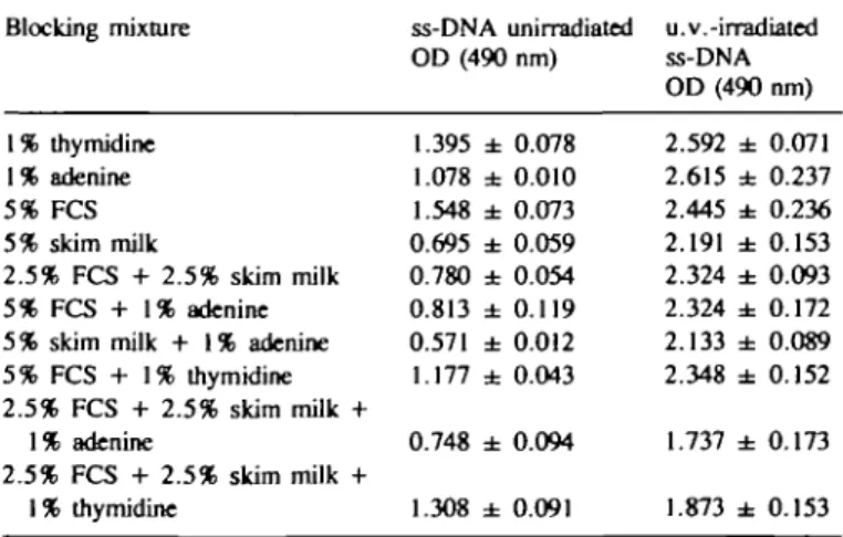
Table I. Effect of blocking mixtures Blocking mixture 1% thymidine 1% adenine 5% FCS 5% skim milk 2.5% FCS + 2.5% skim milk 5% FCS + 1% adenine 5% skim milk + 1% adenine 5% FCS + 1% thymidine 2.5% FCS + 2.5% skim milk + 1% adenine 2.5% FCS + 2.5% skim milk + 1% thymidine on binding of antibody ss-DNA OD (490 1.395 ± 1.078 ± 1.548 ± 0.695 ± 0.780 ± 0.813 ± 0.571 ± 1.177 ± 0.748 ± 1.308 ± unirradiated nm) 0.078 0.010 0.073 0.059 0.054 0.119 0.012 0.043 0.094 0.091 in EIA u.v.-irradiated ss-DNA OD(490 2.592 ± 2.615 ± 2.445 ± 2.191 ± 2.324 ± 2.324 ± 2.133 ± 2.348 ± 1.737 ± 1.873 ± nm) 0.071 0.237 0.236 0.153 0.093 0.172 0.089 0.152 0.173 0.153 PBSAT supplemented with the compounds shown was used as blocking agent in EIA (Materials and methods) with wells coated with 1 pg calf thymus ss-DNA that was unirradiated or irradiated with 10 J/m2 u.v.-light. Values shown
10 90 100
Fig. 1. Fluence dependency of EIA. Poly(dT) (O) and calf thymus ss-DNA (D) were irradiated with u.v.-C radiation and the increase in antigenicity caused by thymine dimer formation was assayed by EIA using triplicate samples. Points are mean values ± SD and curves are fitted by least squares.
.5
1.01.5 2 0
DNA per well (ug)
3 0 3 5 4 0 4 5
TaWe II. An example for the calculation of the loss of antigenicity measured by the bonding of the antibody, as observed in a healthy control (male, aged 38 years)
Time' DNA (/ig/ml)b OD (490 nm)c OD (/*g/ml)d AnUgenicity*
Fig. 2. Linearity of the antibody response (OD = 490 nm) with amount of DNA Oig). Each point represents the mean of five samples. Blood cultures. Samples of 10 ml heparinized peripheral blood were centrifuged
for 20 min at 300 g. Lymphocytes were isolated with a syringe, diluted 1:1 with prewarmed HBSS then centrifuged again on a Ficoll-Ronpacon gradient (d = 1.077, 300 g, 25 min) and washed twice with HBSS. The cells were resuspend-ed in 10 ml culture mresuspend-edium and incubatresuspend-ed for 4 h under the same conditions used for monolayers.
Irradiation. Irradiation was from a Phillips 6-V germicidal lamp emitting
predominantly 253.7 nm radiation at 0.2 J/m2/s at a distance of 38 cm. Each
cell strain was divided into one non-irradiated and four irradiated aliquots. After pouring off the medium from the Petri dishes the samples were irradiated with 10 J/m . Fresh medium was added immediately and the cells were incubated at 37°C for 0, 10, 30 or 60 min. At each time point the repair processes were stop-ped by removing the medium and freezing the cells with liquid nitrogen.
Extraction and preparation of DNA
DNA was extracted by a phenol—ether method (12). The frozen cells were thawed and 1 ml of a solution which has the final concentration of 2% SDS, 0.1 mg/ml protease, 0.05 mg/ml proteinase K, 0.1 mg/ml pronase E and 0.1 mg/ml ribonuclease was added and the mixture was incubated at 37°C for between 6 h and overnight. One millilitre phenol saturated with buffer (0.1 M T r i s - H Q , pH = 7.5, 0.75% hydroxyguanine) was added and vigorously shaken for 1 min, then slowly stirred for 10 min after which the mixture was centrifuged at 4000 g for 5 min and the aqueous phase was pipetted into a clean glass tube. After repeating
0 10 30 60 90 120 150 12.0 15.5 9.1 18.6 12.0 40.4 32.9 0.378 0.336 0.137 0.204 0.114 0.343 0.260 ± ± ± ± ± ± ± 0.040 0.003 0.007 0.026 0.019 0.055 0.009 0.0315 0.02 0.0151 0.0110 0.0095 0.0085 0.0079 100 69 48 35 30 27 25 Time points after u.v.-irradiation. 'O' was immediately following irradia-tion and the other times in minutes after u.v.-irradiairradia-tion.
''Extracted amount of DNA as /ig/ml of each sample.
cMean of triplicate readings of the bound peroxidase-labelled antibody.
''The quotient of column (3) divided by column (2).
eThe relative percentage of bound antibody/jig DNA using the 'O' time
point as the 100% of antigenicity.
Screening of genetic mutations by EIA
25 30 35 40 45
Repair time(min)
50
55 60 65 70
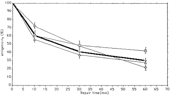
Fig. 3. The percentage loss of antigenicity in 30 normal cell lines after an u.v.-irradiation of 10 J/m2. The dark line represents the mean ± SD of all 30
healthy individuals. To show the range of variance among healthy controls the response of three individuals ( O , D and A) are presented. the phenol extraction the final aqueous phase was mixed with an equal volume
of ether, shaken briefly, centrifuged and the ether layer discarded. This procedure was repeated until the aqueous phase was clear when a double volume of ab-solute ethanol was added. After mixing, the preparation was stored at —20°C overnight before centrifugation at 10 000 g in an Eppendorf centrifuge (Hettich). The pellet was dried and stored in a desiccator until used.
For quantitation, DNA was dissolved in 50 mM Tris—HC1, pH 7.5 contain-ing 10 mM EDTA and 10 mM EGTA (13) and the optical density was measured at - 2 6 0 and - 2 8 0 nm. The ratio of the optical density at these wavelengths was reproducibly to 2.0 indicating that most protein had been removed. The amount of DNA was estimated by the formula: 1.0 OD unit at 260 nm = 50 ^g/ml.
Enzyme immunoassay
EIAs were performed in 96 multiwell plates (Nunc) precoated with protamine sulphate to optimize the binding of DNA (13). To obtain ss-DNA, 0.5 ml of each sample was boiled for 10 mill and quenched in ice water. Aliquots of 100 /J were added to the precoated wells and incubated overnight at 37°C. Non-adsorbed DNA was removed by five washes with blocking buffer (PBSAT containing 5% skim milk powder, 1 % adenine). Non-specific binding sites were blocked by the addition of blocking buffer and incubation for 3 h at room temperature in the dark. After washing five times, the wells were incubated for 90 min at room temperature in the dark with anti-u.v. ss DNA-1 monoclonal antibody (8) diluted in blocking buffer, washed five times and incubated similarly with peroxidase-conjugated anti-mouse second antibody. Wells were washed five times with dcioniz-ed water and incubatdcioniz-ed for 30 min at room temperature in the dark with 50 /J peroxidase reagent (40 mg 1,2-phenylenediamine in 100 ml 0.2 M citric acid, 0.1 M NaH2PO4, pH 5.0 to which was added 40 /U fyOj. The reaction was
stopped after 30 min with 25 /d of 2.5 N H2SO4 and the optical density of each
well was measured at 490 nm. Individual samples were assayed in triplicate unless otherwise specified.
Results
Establishing the EIA
Preliminary experiments showed high non-specific binding of
an-tibody due to the suboptimal blocking conditions. Improved
con-ditions were found by testing the binding of antibody to irradiated
and non-irradiated ss-DNA after blocking with a variety of agents.
The best was Dulbecco's PBS 'A' formulation plus 0.01%
thiomersal (PBSAT) supplemented with 5 % skim milk powder
and 1 % adenine (Table I), and it was used in subsequent
ex-periments.
The linearity of the EIA as a function of u.v dose over the
Table HI. Cell lines
GM1227 GM3O53 GM3617
Loss of antigenicity in three Incubation 10 46" 62 84
XP variant cell lines time (min) 30 55 57 79 60 54 65 84 "Values are percentage antigenicity lost compared with controls.
range 0 - 9 0 J/m
2was examined using calf thymus ss-DNA and
poly(dT) containing —18-20 residues of thymidine per chain
(Figure 1). A nearly linear response was obtained with poly(dT)
over the whole range and the slope of the response is not
significantly different to that of ss-DNA over the range
0—40 J/m . However, the displacement of the two response
curves due to the greater binding of antibody to ss-DNA
com-pared with poly(dT) in the absence of photoproducts is
obvious-ly evident. The standard deviation for poobvious-ly(dT) was ±5.4% and
for ss-DNA ±4.8%. A variance analysis with the Friedman test
did not show any significant difference between the two antigens.
The response curves above clearly indicate that it is possible
to use EIA to determine antigenicity of DNA in cells irradiated
with 10 J/m
2, a dose known to kill - 1 5 - 2 0 % of normal
human fibroblasts (15). To simplify the handling of many
dif-ferent DNA samples it was necessary to demonstrate the linearity
of the antibody response with the amount of DNA applied per
well (Figure 2).
The loss of antigenicity in u. v. -irradiated normal human cells
Table n illustrates how the loss of antigenicity measured by the
binding of the antibody in the DNA of a single individual is
deter-mined. For each time point three cell cultures were irradiated
and each culture was carried out in triplicate. The standard
devia-tion (SD) for the three independently repeated cultures of the same
individual was ±6.5%. For the triplicate readings of bound
an-tibody the standard deviation was ± 10%; this gives the
stan-dard error of the method.
• o
§
* - o40
30
20
10
100.-
90-80.
70.
60-
50-
40-30.
20-10• 8
50
40
30
20
10
-o. « . °°
0°
c. 60 min.
5 10 15 20 25 30
individuals
X . x X 5- o ' • • .
o°
»o . • • o ° o
°Ofl O ° -
o | 0OD
Q X OB X 0 °o « O0 5 10 15 20 25 30
individuals
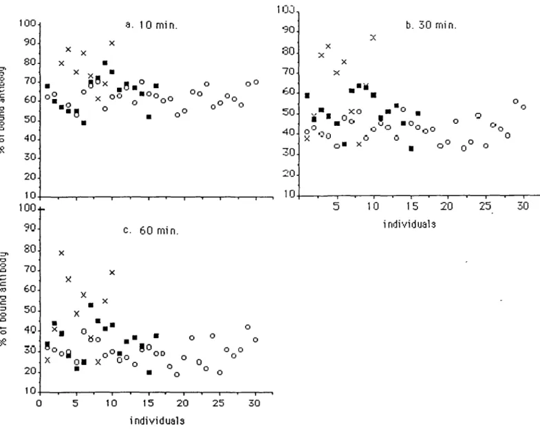
Fig. 4. Individual values of antigenicity at each time point, measured as % of bound antibody. Each individual is represented at the same point at all three time points (10, 30 and 60 min after u.v.-irradiation). O , Healthy controls; M, bec-patients; x , melanoma patients. Melanoma cases 3, 4, 5, 6 and 10 show-ed an obvious higher amount of bound antibody. Among the bec-groups 7, 8, 9 and 10 may belong to a subgroup with higher antigenicity, which would in-dicate a reduced loss of antigenicity, the calculation is shown in Table n.
The analysis of antigenicity included skin biopsies and
lym-phocytes from 30 healthy controls for whom the mean of the
individual SD was ± 8 % . With these samples half of the specific
antigenicity was lost during the first 30 min following
irradia-tion (Figure 3). Figure 3 illustrates the variance in the normal
response of three different individuals at 10, 30 and 60 min
following irradiation. The mean for percentage of loss of
an-tigenicity observed in normal controls at 10, 30 and 60 min after
u.v.-irradiation are 62 ± 9%, 41 ± 12% and 30 =b 11.5%
respectively (Figure 3).
The influence of disease states on loss of antigenicity
Cells from three XP variant patients showing reduced
u.v.-light-induced uds with normal strand incision were tested for the loss
of antigenicity (Table HI). In comparison with the healthy
con-trols GM1227 and GM3O53 showed reduced rates of loss,
whereas GM3617 is near the lower limits of the controls and
would not have been identified as distinct from them.
These data are compared in Figures 4 and 5 with preliminary
results obtained from cultivated fibroblasts of 16 patients with
bec and 10 with melanoma. Figure 4 represents the individual
values of antigenicity at each time point for all the probands.
Each individual is represented at the same point at all three time
points. Figure 5 shows the kinetics of the loss of antigenicity
as a function of time for all three groups and the three XP lines.
Means, standard deviation, standard error and variance of each
of the three groups are presented in Table IV for each time point.
To distinguish whether the three groups show the same kinetics
of antibody binding, an analysis of variance with the
Mann—Whitney test (C/-test) was performed. The results are
presented in Table V.
Discussion
A number of syndromes that involve increased cancer risk have
been shown to display abnormal cellular responses to u.v. light.
Thus XP cells show hypersensitivity and altered repair of dimers
(6,7,16-18) while altered repair synthesis is associated with
mammary carcinoma (19,20) and Cockayne's syndrome shows
hypersensitivity and altered RNA synthesis (21,22).
Hypersen-sitivity to cell killing by u.v. is observed with some strains derived
from Gardner's syndrome (23) and familial melanoma (24,25),
Screening of genetic mutations by E1A
o c
10
20 25
30 35 40
Repair time
45 50 55 60 65 70
Fig. 5. Kinetics of loss of antigenicity in typical and atypical individuals. The extreme cases are found in the XP cell lines GM1227 ( • ) and GM3O53 (A) followed by the melanoma patients, represented by the mean of 10 individuals (X). The kinetics of the third XP cell line GM3617 ( o ) is within the normal kinetic range. The mean of the kinetics of the 16 bec fibroblasts is represented by ( • ) . The mean kinetic of the normal individuals is represented by the dark line (O) and the range is represented by the stippled area.
Several immunoassays have recently been described which can
measure dimers produced by fluences in the range 1-10 J/m
2(8,28—35). In most cases the assays are of the competitive type.
The assay we have developed is a solid-phase non-competitive
assay similar to that described by Leipold et al. (32).
Using this assay we have observed, in agreement with others
(28,29,34), a rapid loss of antigenicity that is faster than that
reported for disappearance of dimers or endonuclease sites. The
reason for this peculiar characteristic remains obscure, but
because the antigen-antibody interaction of monoclonal
an-tibodies is very precise we assume it implies a topological change
at, or close to, the dimer site. The monoclonal antibody used
does not seem to be specific for the dimer itself but for the
con-formational changes induced by the dimer as discussed by
Strickland and Boyle (8) earlier. This possibility is supported by
the findings of Paterson et al. (36) who suggested a new model
for excision repair in humans. Whereas the pyrimidine
dimer—DNA phosphodiesterase activity may be the earliest step
in excision repair and causes a conformational change in the DNA
before the classical repair steps (strand incision; lesion excision;
patch insertion; strand ligation) take place.
Alternatively, the monoclonal antibody may be excluded by
proteins of the repair complex that are possibly covalently
link-ed to DNA. The loss of antigenicity is inhibitlink-ed in excision
defec-tive XP cells in parallel with the inhibition of repair synthesis
(30). Similarly we have observed a correlation between the
percentage of uds reported for the three XP variant strains used
here and the inhibition of the rate of loss of antigenicity. The
comparative indices of antigenicity and the uds relative to
nor-mal control values are 51 ± 9.8% versus 30-60%, 61 ± 7 . 1 %
versus 56% and 82 ± 4.1 % versus 88% for GM1227, GM3O53
and GM3617 respectively. Evidently the loss of antigenicity is
associated with an early step in excision rather than with
post-replication repair, a process defective in all XP variant cells
(37-39). We have investigated the kinetics of this early event
during the first hour after irradiation of normal cells and
unin-Table IV. Mean, standard healthy, bec and melanomi Repair time (min) 10 30 60 •Values Table V all three Group Healthy Bec Melanoma Healthy Bec Melanoma Healthy Bec Melanoma represent the mean
deviation, standard error and variance i groups at each time point
n 30 16 10 30 16 10 30 16 10 1 Of Mean4 62.17 64.19 74.80 ± SD SE ± 5.06 0.92 ± 8.73 2.16 ± 10.56 3.34 42.43 ± 5.98 1.09 50.93 ± 9.06 2.26 63.1 ± 18.78 5.94 29.80 35.37 50.50 ± 5.69 1.04 ± 9.00 2.25 ± 18.24 5.77 percentage of bound antibody.
'. Summary of the analysis of variance (Mann-Whitney investigated patient groups tested one against the other Combination of
Healthy : bec Healthy : melanoma Bec : melanoma
Repair time (min) 10 0.323 0.001 0.106 30 0.040 0.001 0.036 for Variance 25.39 76.16 111.51 35.70 82.19 352.7 32.44 81.05 332.94 tf-test) for 60 0.020 0.001 0.092
P values are shown for each time point and each combination of the three
groups.
while cells of dysplastic naevus syndrome are hypermutable by
u.v (26).
At present it is unclear what role reduced DNA repair plays
in the development of malignancy in these diseases; whether it
is the primary cause or merely an associated phenomenon (18,27).
fying the histological bcc categories (10) to which the patients
were assigned. Individuals 7 , 8 , 9 and 10 have unique variances
that may belong to a subgroup of bcc patients who will develop
a bcc-related tumor in the forseeable future. Or they may have
a genetic predisposition to one of those DNA-repair-deficient
diseases.
Some of the 10 melanoma samples overlapped with the
nor-mal group (Figure 4), but the means of the two groups were
significantly different, which suggest an abnormal response in
some of the patients. Hypersensitivity has been reported in
familial melanoma (25) and the present results suggest that this
could be associated with defective DNA repair. Our findings did
not enable us to use this test for diagnostic analysis. But in Figure
4, it is obvious that five of the melanoma patients (cases 3, 4,
5, 6 and 10) are different from all other investigated probands.
This may be also due to the fact that we could not verify the
differential diagnosis of each of the melanoma cases.
Consequent-ly we were unable to differentiate between familial, single and/or
multiple melanomas.
By comparison of all three groups, in Table IV it is apparent
that the normal controls showed the lowest variance, followed
by the bcc and finally the melanoma patients. The summary of
the statistical analysis is presented in Table V.
Thus the assay provides a useful non-isotopic method for
screening of genetic mutations that effect cellular responses to
u.v., particularly those involved early in excision repair. More
precise analysis of additional data will involve the collection of
a larger number of samples to enable age, sex, differential
diagnoses, familial genetic predisposition for matching and
con-firmation of possible correlation with a reduction in the kinetics
of the initial steps in thymine dimer excision repair. There was
no significant difference between the results obtained with
nor-mal blood and skin samples suggesting that this assay is not
gross-ly affected by heterogeneity of tissue types. A further
consideration for future study is the influence of therapy regimes
on the results obtained.
Acknowledgements
The authors wish to express their gratitude to Professor Lyman Randlett Emmons for his critical review and assistance in the preparation of the manuscript. This work was supported by grants from SNF 3.818.0.84 and the Regional Cancer League, Basel, and the Cancer Research Campaign, UK Cancer Research Campaign.
References
1. Strickland,P.T. and BoyleJ.M. (1984) Immunoassay of carcinogen-modified DNA. Prog. Res. Mol. Bioi, 31, 1-58.
2. Perera.F.P., Poirier,M.C, Yuspa,S.H., NakayamaJ., Jarctslri,A., Cumen, M.M., Knowles.D.M. and Weinstein.I.B. (1982) A pilot project in molecular cancer epidemiology: determination of benzo(a]pyrene—DNA adducts in animal and human tissues by immunoassays. Cardnogenesis, 3, 1405-1410. 3. Umbenhauer.D., Wild.C.P., Montesano.R., Saffhill.R., BoyleJ.M., Kirs-tein.U., ThomaleJ., Rajewski.M.F. and Liu.S.H. (1985) O-methyldeoxy-guanosine in oesophageal DNA among populations at high risk of oesophageal cancer. Int. J. Cancer, 36, 661 - 6 6 5 .
4. Wild.C.P., Umbenhauer.D., Chapot.R. and Montesano.R. (1986)
Monitor-8. Strickland,P.T. and BoyleJ.M. (1981) Characterisation of two monoclonal antibodies specific for dimerised and non-dimerised adjacent thymidines in single stranded DNA. Photochem. Photobiol., 34, 595-601.
9. Strickland.P.T. (1985) Immunoassay of DNA modified by ultraviolet radia-tion: a review. Environ. Mutagen., 7, 185-199.
10. Butterworth.T. and Ladda.R.L. (1981) Clinical Genodermatology, Vol. 2.
Light-Sensitive Genodermatoses. Praeger, New York, pp. 1-17.
11. Cleaver.J.E. (1981) Xeroderma pigmentosum variants. Cytogenet. Cell.
Genet., 31, 188-189.
12. Maniatis.T., Fritsch.E.F. and SambrookJ. (1982) Molecular Cloning. A
Laboratory Manual. Cold Spring Harbor Laboratory Press, New York,
pp. 280-281, 438-439.
13. KlotzJ.L., Minami.R.M. and Teplitz.R.L. (1979) An enzyme linked im-munosorbent assay for antibodies to native and denatured DNA. J. Immunol.
Methods, 29, 155-165.
14. Keyse.S.M. and Tyrell.R.M. (1985) Excision repair in permeable arrested human skin fibroblasts damaged by u.v. (254 nm) radiation: evidence that alpha- and beta-polymerases act sequentially at the repolymerisation step.
Mutat. Res., 146, 109-119.
15. Wells.R.L. and Han.A. (1985) Differences in sensitivity between human, mouse and Chinese hamster cells to killing by monochromatic ultraviolet light.
Int. J. Radial. Bioi, 47, 1 7 - 2 1 .
16. Kraemer.K.H., de Weerd-Kastelein.E.A., RobbinsJ.H., Kreijzer.W., Bar-rett.S.F., Petinga.R.A. and Bootsma.D. (1975) Five complementation groups in xeroderma pigmentosum. Mutat. Res., 33, 327—340.
17. PattonJ.D., Rowan.L.A., MendraJa.A.L., HowelU.N., Maher.V.M. and McCormickJ. (1984) Xeroderma pigmentosum fibroblasts including cells from XP variants are abnormally sensitive to the mutagenic and cytotoxic action of broad spectrum simulated sunlight. Photochem. Photobiol., 39, 3 7 - 4 2 . 18. Takebe,H., Tatsumi.K. and Batch.Y. (1985) DNA repair and its possible involvement in the origin of multiple cancer. Jap. J. Clin. Oncol., 15, 299-305.
19. RussoJ., Tay.L.K., Ciocca.D.R. and Russo,l.H. (1983) Molecular and cellular basis of the mammary gland susceptibility to cardnogenesis. Environ.
Health Perspect., 49, 185-199.
20. Kovacs.E., Stucki.E., Weber.W. and MueUer.Hj. (1986) Impaired DNA-repair synthesis in lymphocytes of breast cancer patients. Eur. J. Cancer Clin.
Oncol., 22, 863-869.
21. Deschavaime,PJ.,Chavaudra,N., Fertil.B. and Malaise.E.P. (1984) Abnormal sensitivity of some Cockayne's syndrome cell strains to UV- and gamma-rays. Mutat. Res., 131, 6 1 - 7 0 .
22. Lehmann,A.R. (1982) Three complementation groups in Cockayne's syn-drome. Mutat. Res., 106, 347-356.
23. Kinsella.TJ., LMe,J.B., Nove,J., Weichselbaum.R.R., U,F.P., Meyer.RJ., Marcetto,D.J. and Patterson.W.B. (1982) Heterogenous response to X-ray and ultraviolet light irradiation of cultured skin fibroblasts in two families with Gardner's syndrome. / . NatL Cancer lnst., 68, 697-701. 24. Teppo.L., Pakkanen.M. and Hakulinen.T. (1978) Sunlight as a risk factor
of malignant melanoma of the skin. Cancer, 41, 2018—2027.
25. Ramsay.R.G., Chen.Ph., Imray.F.P., Kidson.C, Lavin.M.F. and Hockey,A. (1982) Familial melanoma associated with dominant ultraviolet radiation sen-sitivity. Cancer Res., 42, 2909-2912.
26. Jung,E.G., Bohnert.E. and Boonen.H. (1986) Dysplastk nevus syndrome: ultraviolet hypermutability confirmed in vitro by elevated sister chromatid exchanges. Dermatologica, 173, 297-300.
27. CollinsA and Squires.Sh. (1986) The time course of ultraviolet-induced DNA damage: implications of the structural organisation of repair. Mutat. Res., 166, 113-119.
28. Mitchell.D.L. and ClarksonJ.M. (1981) The development of a radioim-munoassay for the detection of photoproducts in mammalian cell DNA.
Biochim. Biophys. Ada, 655, 5 4 - 6 0 .
29. Mitchell.D.L., Naim.R.S., AlvillarJ.A. and Clarkson,J.M. (1982) Loss of thymine dimers from mammalian cell DNA. The kinetics for antibody-binding sites are not the same as that for T4 endonuclease V sites. Biochim. Biophys.
Aaa, 697, 270-277.
30. ClarksonJ.M., Mitchell,D.L. and Adair.G.M. (1983) The use of an
Screening of genetic mutations by EIA munological probe to measure the kinetics of DNA repair in normal and
UV-sensitive mammalian cell lines. Mutat. Res., 112, 287-299.
31. Ley,R.D. (1983) Immunological detection of two types of pyrimidine dimers in DNA. Cancer Res., 43, 4 1 - 4 5 .
32. Leipold.B., Remy.W. and Adelmann-Grill,B. (1983) Measurement of ultraviolet light-induced photolesions in mammalian DNA by micro-ELISA.
J. Immunol. Methods, 60, 6 9 - 7 6 .
33. Wani.A.A., Gibson-d'Ambrosk>,R.E. and d'Ambrosio.S. (1984) Antibodies to UV irradiated DNA: the monitoring of DNA damage by ELISA and in-direct immunofluorescence. Photochem. Photobiol., 40, 465—471. 34. KJccker.H., Auer.B., BurtscherJl.J., Hofmann^., Hirsch-Kaufmann.M. and
Schweiger,M. (1982) A sensitive radioimmunoassay for thymine dimers. Mol.
Gen. Genetics, 186, 475-477.
35. Klocker.H., Burscher.H.J., Auer.B., Hirsch-Kaufmaroi.M. and Schweiger.M. (1985) FibroWasts from patients with Fanconi's anaemia are not defective in excision of thymine dimers. Eur. J. Cell Bioi, 37, 240-242. 36. Paterson.M.C, Middlestadt.M.V., MacFarlane.S.J., Gentner.N.E. and
Weinfeld.M. (1987) Molecular evidence for cleavage of intradimer phos-photriester linkage as a novel step in excision repair of cyclobutylpyrimidine photodimers in cultured human cells. J. Cell. Sci. Suppl., 6, 161 — 176. 37. Lehmann,A.R., Kirk-BeU,S., Arlett.C.F., Paterson,M.C, Lohman.P.H.M., de Weerd-Kastelein.E.A. and Bootsma.D. (1975) Xeroderma pigmentosum cells with normal levels of excision repair have a defect in DNA synthesis after UV-irradiation. Proc. Nail. Acad. Sci. USA, TZ, 219-223. 38. Jaspers.N.GJ., Jansen-van de Kuilen.G. and Bootsma,D. (1981)
Complemen-tation analysis of xeroderma pigmentosum variants. Exp. Cell Res., 136, 81-90.