Cite this article as: De Paulis R, Czerny M, Weltert L, Bavaria J, Borger MA, Carrel TPet al. Current trends in cannulation and neuroprotection during surgery of the aortic arch in Europe. Eur J Cardiothorac Surg 2015;47:917–23.
Current trends in cannulation and neuroprotection
during surgery of the aortic arch in Europe
†‡
Ruggero De Paulis
a,*, Martin Czerny
b, Luca Weltert
a, Joseph Bavaria
c, Michael A. Borger
d, Thierry P. Carrel
e,
Christain D. Etz
d, Michael Grimm
f, Mahmoud Loubani
g, Davide Pacini
h, Timothy Resch
i,
Paul P. Urbanski
jand Ernst Weigang
k(EACTS Vascular Domain Group)
a Department of Cardiac Surgery, European Hospital, Rome, Italy b Department of Cardiac Surgery, University Hospital, Zurich, Switzerland c Department of Cardiac Surgery, University Hospital, Philadelphia, PA, USA d Department of Cardiac Surgery, Heart Center, Leipzig, Germany e Department of Cardiac Surgery, University Hospital, Berne, Switzerland f Department of Cardiac Surgery, Medical University, Innsbuck, Austria g Department of Cardiac Surgery, Castle Hill Hospital, Hull, UK h Department of Cardiac Surgery, Policlinico Sant’ Orsola, Bologna, Italy i Department of Cardiac Surgery, Skane University Hospital, Malmö, Sweden j Department of Cardiac Surgery, Herz und Gefaess Klinik, Bad Neustadt, Germany k Department of Vascular Surgery, Academic Hospital Hubertus, Berlin, Germany
* Corresponding author. Department of Cardiac Surgery, European Hospital, Via Portuense 700, 00149 Rome, Italy. Tel: +39-06-65975224; fax: +39-06-65975112; e-mail: [email protected] (R. De Paulis).
Received 11 March 2014; received in revised form 3 June 2014; accepted 10 June 2014
Abstract
OBJECTIVES: To conduct a survey across European cardiac centres to evaluate the methods used for cerebral protection during aortic surgery involving the aortic arch.
METHODS: All European centres were contacted and surgeons were requested tofill out a short, comprehensive questionnaire on an internet-based platform. One-third of more than 400 contacted centres completed the survey correctly.
RESULTS: The most preferred site for arterial cannulation is the subclavian–axillary, both in acute and chronic presentation. The femoral artery is still frequently used in the acute condition, while the ascending aorta is a frequent second choice in the case of chronic presentation. Bilateral antegrade brain perfusion is chosen by the majority of centres (2/3 of cases), while retrograde perfusion or circulatory arrest is very seldom used and almost exclusively in acute clinical presentation. The same pumping system of the cardio pulmonary bypass is most of the time used for selective cerebral perfusion, and the perfusate temperature is usually maintained between 22 and 26°C. One-third of the centres use lower temperatures. Perfusateflow and pressure are fairly consistent among centres in the range of 10–15 ml/kg and 60 mmHg, respectively. In 60% of cases, barbiturates are added for cerebral protection, while visceral perfusion still receives little attention. Regarding cerebral monitoring, there is a general tendency to use near-infrared spectroscopy associated with bilateral radial pressure measurement. CONCLUSIONS: These data represent a snapshot of the strategies used for cerebral protection during major aortic surgery in current practice, and may serve as a reference for standardization and refinement of different approaches.
Keywords:Aortic arch• Neuroprotection
INTRODUCTION
Patients with dissecting or aneurysmal disease involving the aortic arch represent a unique challenge for the cardiac surgeon. The use of valid surgical and endovascular techniques and appropriate
methods of cerebral protection are crucial to obtaining satisfac-tory postoperative results.
Since the late 1960s hypothermic circulatory arrest has been used during aortic arch repair with acceptable neurological out-comes [1]. Hypothermic circulatory arrest has also been used as a method of organ protection in the repair of thoraco-abdominal aortic aneurysms. Through the years, the effects of deep hypo-thermia on brain metabolism and perfusion have been studied both in animal models and in surgical patients [2,3].
†Presented at the 27th Annual Meeting of the European Association for Cardio-Thoracic Surgery, Vienna, Austria, 5–9 October 2013.
‡Contributing centres and Departments of Cardiovascular Surgery are available asSupplementary Material.
© The Author 2014. Published by Oxford University Press on behalf of the European Association for Cardio-Thoracic Surgery. All rights reserved.
A O RTIC SURGE R Y
European Journal of Cardio-Thoracic Surgery 47 (2015) 917–923
ORIGINAL ARTICLE
selective antegrade cerebral perfusion are presently under examination in several aortic centres, the ischaemic tolerance of the spinal cord during lower-body circulatory arrest at higher temperature levels has developed as a new focus of concern [11]. A definitive consensus has not yet been reached, and now-adays many different approaches in terms of the site of arterial cannulation, temperature of the body and the cerebral perfus-ate, flow, monitoring methods and added visceral protection are pursued.
To obtain further information on current practice, we con-ducted a survey across European cardiac centres to evaluate the methods used for cerebral protection during aortic surgery involv-ing the aortic arch.
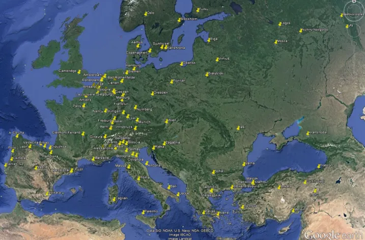
less represented than others, as shown in Fig.1. The average case load reported from each centre was 28.6 ± 20.1 and 26.8 ± 16.8 for chronic and acute patients, respectively. The standard variation reflects the inclusion of small- and large-volume centres.
Data were collected centrally at the EACTS Secretary in Windsor, and then analysed at a core statistical laboratory. Descriptive statis-tics and graphics are detailed in the Results section.
For an easy understanding of plotted data, answers for acute and chronic pathologies are plotted together for each category and a brief comment is given for each set of graphics. Data are presented either via a Pie Chart, Bar Chart or Scatter Plot, and cat-egories are identical in acute and chronic cohorts to facilitate an immediate appraisal of differences.
RESULTS
In both acute and chronic settings, the right subclavian–axillary approach is the favourite site for cannulation (54 and 48%, re-spectively). The second favoured choice differs depending on the clinical presentation: for acute conditions, the femoral approach is preferred, while, for chronic conditions, the ascending aorta is preferred, both accounting for 28% of the cases (Fig.2A). As a second choice for cannulation, percentages are very similar for acute and chronic patients with the subclavian artery (a combined direct and interposed graft accounting for 40% in both cohorts) preferred slightly over a femoral approach (33 and 28%, respect-ively) (Fig.2B).
Bilateral antegrade cerebral perfusion is the most frequent method for brain protection in both types of clinical presentation. Unilateral perfusion is utilized in 1/3 of the cases, while deep hypothermic arrest and retrograde perfusion are very rarely used in Europe and almost exclusively in acute presentation (Fig.3).
Selective cerebral perfusion is implemented in 3/4 of the cases via the same pumping system utilized for the cardiopulmonary bypass system. In the remaining cases, a dedicated pump is utilized (Fig.4).
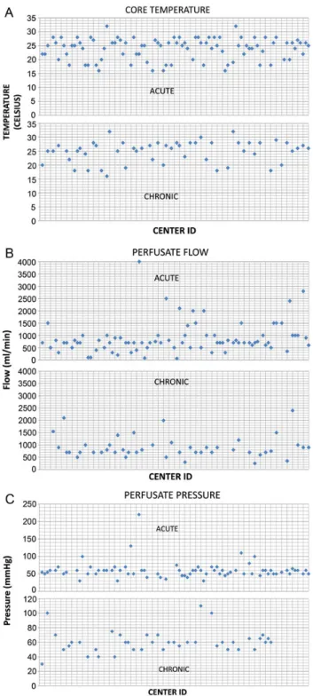
Roughly two-thirds of centres prefer a core temperature between 22 and 26°C, while one-third of them still use colder temperature (down to 15°C in some cases). Only two centres use mild temperature above 30°C (Fig.5A). Perfusateflow is fairly con-sistent among all centres with an average of 10–15 ml/kg with a few exceptions (Fig.5B). Perfusate pressure is almost always main-tained around 60 mmHg in both acute and chronic settings (Fig.5C). There have been markedly fewer answers to this question; possibly due to the fact that many surgeons did not know the pres-sure or did not know where it was meapres-sured (e.g. radial artery versus resistance in the perfusion line). These fewer answers might as well mean that surgeons valueflow rates more than maximal pressure. Adjunctive use of barbiturates to protect the brain is adopted in around 60% of centres, both in acute and chronic settings.
Additional visceral perfusion during circulatory arrest has received little attention and is routinely performed in a minority of centres only (Fig.6). The site of cannulation for visceral perfu-sion is predominantly the femoral artery or the descending aorta, limited to 17% in the case of ascending acute aortic dis-section type A and 30% of chronic aneurysms (Fig.7).
Figure 2:Preferred arterial cannulation sites as afirst choice (A) or as a second choice (B) (see text for details).
Figure 3:Preferred mode of brain perfusion.
Figure 4:Pump system for cerebral perfusion.
A O RTIC SURGE R Y
Near-infrared spectrography is used by two-thirds of centres, and bilateral radial pressure by half of the centres. Often the two techniques are used together. Other methods of monitoring are only used occasionally (Fig.8).
DISCUSSION
Cannulation
The majority of centres prefer an axillary cannulation either in acute or in chronic cases. The axillary route remains the most
frequent also as a second choice. This is clearly different from more than a decade ago when the femoral artery was considered the main access route in the case of dissection and very often also in the case of chronic aneurysm involving the arch.
In fact, in the past years several studies have demonstrated the ad-vantage of axillary cannulation in the standardization and simpli fica-tion of cerebral perfusion [12] while at the same time they have speculated on the potential advantages in terms of stroke reduction [13]. Axillary cannulation can be performed either directly or via the interposition of a small Dacron graft to optimize haemostasis and decrease the chance of damage to the vessel. In both cases, by Figure 5:Management of temperature (A), perfusate (B) and pressure (C) (see
text for details).
Figure 6:Visceral perfusion during circulatory arrest.
simply clamping the three cerebral vessels at their bases, and utiliz-ing a properflow rate, it is possible to smoothly switch from standard cardio pulmonary bypass (CPB) to unilateral antegrade cerebral per-fusion, reducing any potential risk related to vessel manipulation, cannulation or air embolism. The increasing experience with axillary cannulation has therefore prompted the use of the innominate artery as a site for cannulation in order to decrease the number of in-cision sites and further simplify the procedure [14]. More importantly, the use of axillary cannulation, by providing an antegrade body per-fusion, decreases the risk of retrogradeflow both in the acute setting (malperfusion) and in the chronic condition (atheroembolism).
Type of cerebral perfusion
The majority of centres prefer bilateral perfusion. However, a large proportion, as high as 38% in the acute condition, and 33% in the chronic condition, adopts a simple unilateral perfusion.
Selective antegrade cerebral perfusion (SACP) can be delivered bilaterally or unilaterally, wherein contralateral perfusion depends on collateral pathways, most prominently the circle of Willis (CoW). However, the clinical impact of an incomplete CoW is not clear. Merkkola et al. [15] performed autopsies on 98 human brains and determined that as much as 17% of specimens had anatomical evidence of an incomplete CoW. They recommended computed tomography (CT) angiography to identify patients who might require bilateral SACP. Another study [16] suggested that 42.4% of eastern Europeans have an incomplete CoW; in addition to preoperative CT angiography, these authors recommended intraoperative monitoring with near-infrared spectroscopy, tran-scranial Doppler and electroencephalography.
These anatomical studies may underestimate the importance of collateral vessels, such as the ophthalmic artery, leptomeningeal vessels and external carotid arteries. Urbanskiet al. [17] published a series of 99 arch replacements using unilateral SACP (30°C; × 18 min) via the left common carotid artery. Although preoperative
CT angiography documented a complete CoW in only 60% of patients, intraoperative monitoring indicated good contralateral perfusion in all patients, and there was only 1 minor embolic stroke: based on these results, the authors concluded that preoperative CT angiography is unnecessary. Leshnoweret al. [18] corroborated thesefindings in a series of 412 arch procedures using hypother-mic unilateral SACP (26°C; 45 min), with a stroke rate of 3.6%. Nevertheless, concern remains when the duration of SACP is more than 50 min. Krahenbuhl et al. [19] followed 292 patients after aortic arch surgery under deep hypothermic circulatory arrest and unilateral and bilateral cerebral perfusion. The 36-Item Short-Form health survey questionnaires after a mid-term follow-up showed that patients whose SACP times exceeded 40 min had a better quality of life if bilateral SACP had been used. Consequently, the authors recommended using bilateral perfusion for an anticipated SACP interval of more than 50 min.
Many questions remain to be clarified; for example, surgeons performing arch repair under unilateral protection seem to need significantly shorter ischaemic time than those using bilateral pro-tection, though it is not understood why this is the case. Results of both modalities are similar, even if reports describing bilateral cerebral perfusion time exceeds considerably those times periods considered to be normal for an arch repair. All-in-all, there is in the literature a distinct lack of evidence, at least at a higher level, justifying this variety of practices in European centres.
Temperature
Data from the present survey indicated that the majority of centres still rely on some level of hypothermic perfusion, with the average of the perfusate temperature being 22°C.
As the efficacy of hypothermic SACP became accepted, many experienced centres began to explore the use of warmer SACP. In a porcine model, Khaladjet al. [20] compared the temperature of the perfusate for SACP (90 min) at 10, 20 and 30°C with Figure 8:Methods of cerebral monitoring. AP: arterial pressure.
A O RTIC SURGE R Y
Flow
In general, the results of this survey show that most centres main-tained their cerebral flow within margins generally considered safe.
Experiments also focused on cerebrovascular bloodflow (CBF) during SACP, searching for the ideal maximal and minimal limits. Haldenwanget al. [22], administering hypothermic SACP at 8 and 18 ml/kg/min, found a pattern similar to the Halstead pressure experiments: equivalent cerebral metabolic suppression but ‘luxury’ regional blood flow at the higher flow rates, with elevated ICP and sagittal sinus pressures. Other studies defined the lower limit of hypothermic SACP. Tanaka et al. [23] for example, decreased SACP (25°C) from the baseline (100%) towards zero in a canine model, evaluating cerebral function with continuous somatosensory evoked potential monitoring and histological outcomes. The groups with a 100 or 50% decrease offlow showed no signs of cerebral compromise. However, decreasingflow to 25% (mean arterial pressure, 25 mmHg) produced a loss of somatosensory evoked potential and mild cellular injury. Thus, perfusion pressure andflow studies indicate that excessive SACP perfusion should be avoided. The clinical application of this principle in the elderly warrants a note of caution, becausefixed atherosclerotic lesions and chronic hypertension may alter normal autoregulation of CBF, requiring some distal compensatory in-crease in SACP pressure.
Pressure
In general, the results of this survey show that overpressure is generally avoided. Halsteadet al. [24] addressed optimum perfu-sion pressure for hypothermic SACP in the porcine model. In this experimental setting, SACP (20°C) delivered at 50, 70 and 90 mmHg, revealed that although increasing perfusion pressure increased cerebral bloodflow, cerebral metabolic suppression was similar in each group. In the 90-mmHg group, ICP increased throughout the duration of SACP, and in the period after CPB, the metabolic rate was elevated; moreover, these animals demonstrated inferior neurobehavioural recovery. It was hypothesized that thesefindings resulted from cerebral oedema, resulting from the high perfusion pressure. Also, excess or ‘luxury’ CBF during SACP may increase the risk of thrombo-embolic material being directed to the brain.
than 60 min. Etz et al. [11] performed moderately hypothermic SACP (28°C) in a pig model for 90 and 120 min and used fluores-cent microspheres to quantify spinal cord bloodflow. After initiat-ing SACP, bloodflow was nearly absent below the T4–T13 region. All animals showed evidence of paraparesis or paraplegia, but the effect was more severe in the 120-min group. Histological speci-mens demonstrated moderate to severe ischaemic injury in the lumbar spine, even in animals that regained normal function, with the severity of injury increasing in progressively distal spinal seg-ments; the effect was most pronounced in the 120-min group. This study demonstrated that spinal injury is not an all-or-none phenomenon: Prolonged ischaemia causes injury to a certain amount of motor neurons, so that some degree of spinal cord injury may be undetectable on routine clinical examination. Thus, the margin of safety for spinal ischaemia during moderately hypo-thermic SACP may be less than widely assumed.
CONCLUSIONS
Based on experimental and clinical studies, the use of non-pulsatile SACP can be recommended. The results of the present survey show that the majority of responding centres are in line with the data present in the literature. Pressure is kept between 40 and 60 mmHg, with aflow of 6–10 ml/kg/min, avoiding excessive perfusion pressure that may be detrimental. Temperature is kept for core cooling between 20 and 30°C, with a perfusate temperature between 20 and 26°C. SACP is frequently unilateral; however, if SACP duration is sup-posed to exceed 30–40 min, a bilateral perfusion might be preferred. Cerebral monitoring is routinely performed and the adoption of near infra red spectroscopy associated with bilateral monitoring of perfusion pressure seems to be preferred by a majority of surgeons.
In general, this survey reveals that it takes time for new evidence in the literature to transfer into common practice. It is evident that the majority of centres still rely on a certain degree of hypothermia, and that warmer circulatory arrest is being adopted more frequently.
We hope that these data may be of help to standardize and further develop the optimal strategy of cerebral protection.
SUPPLEMENTARY MATERIAL
Supplementary material is available atEJCTS online. Conflict of interest: none declared.
REFERENCES
[1] Dumanian AV, Hoeksema TD, Santschi DR, Greenwald JH, Frahm CJ. Profound hypothermia and circulatory arrest in the surgical treatment of traumatic aneurysm of the thoracic aorta. J Thorac Cardiovasc Surg 1970; 59:541–5.
[2] Fisk GC, Wright JS, Turner BB, de C Baker W, Hicks RG, Lethlean AKet al. Cerebral effects of circulatory arrest at 20 degrees c in the infant pig. Anaesth Intensive Care 1974;2:33–42.
[3] Greeley WJ, Kern FH, Meliones JN, Ungerleider RM. Effect of deep hypo-thermia and circulatory arrest on cerebral bloodflow and metabolism. Ann Thorac Surg 1993;56:1464–6.
[4] Frist WH, Baldwin JC, Starnes VA, Stinson EB, Oyer PE, Miller DCet al. A re-consideration of cerebral perfusion in aortic arch replacement. Ann Thorac Surg 1986;42:273–81.
[5] Bachet J, Guilmet D, Goudot B, Termignon JL, Dreyfus G, Teodori G. Cold cerebroplegia: a new technique of cerebral protection during surgery of the transverse aortic arch. J Thorac Cardiovasc Surg 1991;102:85–94. [6] Kazui T, Inoue N, Yamada O, Komatsu S. Selective cerebral perfusion
during operation for aneurysms of the aortic arch: a reassessment. Ann Thorac Surg 1992;53:109–14.
[7] Ueda Y, Miki S. Retrograde cerebral perfusion. Ann Thorac Surg 1992;53: 364–5.
[8] Di Eusanio M, Schepens MA, Morshuis WJ, Di Bartolomeo R, Pierangeli A, Dossche KM. Antegrade selective cerebral perfusion during operations on the thoracic aorta: factors influencing survival and neurologic outcome in 413 patients. J Thorac Cardiovasc Surg 2002;124:1080–6.
[9] Sakahashi H, Hashimoto A, Aomi S, Tokunaga H, Koyanagi T, Imamaki M et al. Transcranial Doppler measurement of middle cerebral artery blood flow during continuous retrograde cerebral perfusion. Nihon Kyobu Geka Gakkai Zasshi 1994;42:1851–7.
[10] Rigg CD, Clutton-Brock TH. Near-infrared spectroscopy changes during hypothermic circulatory arrest with retrograde cerebral perfusion. Anaesthesia 1997;52:356–9.
[11] Etz CD, Luehr M, Kari FA, Lin HM, Kleinman G, Zoli Set al. Selective cere-bral perfusion at 28 degrees C—is the spinal cord safe? Eur J Cardiothorac Surg 2009;36:946–55.
[12] Sabik JF, Lytle BW, McCarthy PM, Cosgrove DM. Axillary artery: an alterna-tive site of arterial cannulation for patients with extensive aortic and peripheral vascular disease. J Thorac Cardiovasc Surg 1995;109:885–90. discussion 890–1.
[13] Svensson LG, Blackstone EH, Rajeswaran J, Sabik JF III, Lytle BW, Gonzalez-Stawinski G et al. Does the arterial cannulation site for
circulatory arrest influence stroke risk? Ann Thorac Surg 2004;78:1274–84. discussion 1274–84.
[14] Di Eusanio M, Ciano M, Labriola G, Lionetti G, Di Eusanio G. Cannulation of the innominate artery during surgery of the thoracic aorta: our experi-ence in 55 patients. Eur J Cardiothorac Surg 2007;32:270–3.
[15] Merkkola P, Tulla H, Ronkainen A, Soppi V, Oksala A, Koivisto Tet al. Incomplete circle of Willis and right axillary artery perfusion. Ann Thorac Surg 2006;82:74–9.
[16] Papantchev V, Stoinova V, Aleksandrov A, Todorova-Papantcheva D, Hristov S, Petkov Det al. The role of Willis circle variations during unilat-eral selective cerebral perfusion: a study of 500 circles. Eur J Cardiothorac Surg 2013;44:743–53.
[17] Urbanski PP, Lenos A, Blume JC, Ziegler V, Griewing B, Schmitt Ret al. Does anatomical completeness of the circle of Willis correlate with suf fi-cient crossperfusion during unilateral cerebral perfusion? Eur J Cardiothorac Surg 2008;33:402–8.
[18] Leshnower BG, Myung RJ, Kilgo PD, Vassiliades TA, Vega JD, Thourani VH et al. Moderate hypothermia and unilateral selective antegrade cerebral perfusion: a contemporary cerebral protection strategy for aortic arch surgery. Ann Thorac Surg 2010;90:547–54.
[19] Krahenbuhl ES, Clement M, Reineke D, Czerny M, Stalder M, Aymard T et al. Antegrade cerebral protection in thoracic aortic surgery: lessons from the past decade. Eur J Cardiothorac Surg 2010;38:46–51.
[20] Khaladj N, Peterss S, Oetjen P, von Wasielewski R, Hauschild G, Karck M et al. Hypothermic circulatory arrest with moderate, deep or profound hypothermic selective antegrade cerebral perfusion: which temperature provides best brain protection? Eur J Cardiothorac Surg 2006;30:492–8. [21] Pacini D, Leone A, Di Marco L, Marsilli D, Sobaih F, Turci Set al. Antegrade
selective cerebral perfusion in thoracic aorta surgery: safety of moderate hypothermia. Eur J Cardiothorac Surg 2007;31:618–22.
[22] Haldenwang PL, Strauch JT, Amann I, Klein T, Sterner-Kock A, Christ Het al. Impact of pumpflow rate during selective cerebral perfusion on cerebral hemodynamics and metabolism. Ann Thorac Surg 2010;90:1975–84. [23] Tanaka H, Kazui T, Sato H, Inoue N, Yamada O, Komatsu S et al.
Experimental study on the optimumflow rate and pressure for selective cerebral perfusion. Ann Thorac Surg 1995;59:651–7.
[24] Halstead JC, Meier M, Wurm M, Zhang N, Spielvogel D, Weisz D. Optimizing selective cerebral perfusion: deleterious effects of high perfu-sion pressures. J Thorac Cardiovasc Surg 2008;135:784–91.
[25] Kamiya H, Hagl C, Kropivnitskaya I, Bothig D, Kallenbach K, Khaladj N et al. The safety of moderate hypothermic lower body circulatory arrest with selective cerebral perfusion: a propensity score analysis. J Thorac Cardiovasc Surg 2007;133:501–9. A O RTIC SURGE R Y