Does retrograde cerebral perfusion via superior vena cava
cannulation protect the brain?
§Andreas Ku
¨nzli
*
, Patrick O. Zingg, Gregor Zu
¨nd,
Boris Leskosek, Ludwig K. von Segesser
Department of Cardio-Vascular Surgery, Centre Hospitalier Universitaire Vaudois (CHUV), Lausanne, Switzerland Received 13 February 2006; received in revised form 11 July 2006; accepted 9 August 2006; Available online 30 October 2006
Abstract
Objective: The retrograde cerebral perfusion via cannulation of the superior vena cava is a widespread method for optimising protection of the brain during hypothermic circulatory arrest. Methods: In 14 cadavers (8 females, 6 males) of the local department of pathology, an examination was performed to check the competence of the valves of the internal jugular veins. After a complete preparation of the superior vena cava, the innominate vein and both internal jugular veins, ligating all side branches, a retrograde perfusion on 7 cadavers was installed, documenting flow and pressure of each internal jugular vein (IJV) in vitro. Afterwards, the veins were opened and their valves inspected. Results: In all 14 cadavers, anatomically and functionally competent valves on the right proximal IJV were found. Only 1/14 cadaver had no valve in the left proximal IJV. Additional rudimentary and incompetent valves could be identified in 1/14 cadaver on the distal right IJV, and in 2/14 cadavers on the left IJV. Retrograde flow measurement of 7/14 cadavers revealed 0 ml/min in 4/7 cadavers, 6 ml/min in 1/7, 340 ml/min in 1/7 and 2500 ml/min in 1/7 cadaver. Conclusions: As a rule, anatomically and functionally competent valves in the proximal IJV are present. In human beings, they obstruct the direct retrograde inlet to the intracranial venous system, which suggests an unbalanced and unreliable perfusion of the brain. Therefore, retrograde cerebral perfusion by cannulating the superior vena cava may help flushing out embolism and supporting ‘the cold jacket’ of the brain. However, its effect of retrograde backflow cannot be a sign of adequate cerebral perfusion.
#2006 Elsevier B.V. All rights reserved.
Keywords: Cardiopulmonary bypass; Deep hypothermic circulatory arrest; Experimental; Retrograde cerebral perfusion
1. Introduction
The technique of retrograde cerebral perfusion (RCP) was first described by Mills and Ochsner [1] in 1980, but not especially for aortic operations. They used RCP for a surgical treatment of massive air embolism during cardiopulmonary bypass. Air returning from the aortotomy has confirmed the effectiveness of this technique.
In 1982, Lemole et al.[2]published intermittent RCP as a method to improve cerebral oxygen delivery during opera-tions using hypothermic cardiac arrest (HCA). They reported using profound hypothermia for cardiac arrest (CA) and prolonging it by an intermittent retrograde perfusion via the superior vena cava for 2 min out of every 10, providing in the way cerebral oxygenation during this period with decreased risk for neurological sequel and bleeding. They concluded
that this would be the method of choice for repair of dissecting aortic aneurysm.
First, Lin et al.[3]have suggested in 1996 that with RCP, deep hypothermia may not be necessary and had applied the technique successfully in a clinical series of 23 patients with a rectal temperature of only 23.3 8C. They concluded that there is no evidence of ischemia of the brain during prolonged moderate hypothermic CA with the aid of RCP, and retrograde cerebral perfusion would effectively extend the safe time of CA.
With animal models, investigators tried to answer the questions, whether RCP has a benefit for cerebral protection or not.
In a chronic porcine model, Juvonen et al.[4]compared anterograde cerebral perfusion (ACP) versus retrograde cerebral perfusion and versus RCP with occlusion of the inferior vena cava (RCP-O) and versus hypothermic cardiac arrest with the head packed in ice. The histological findings suggest that RCP without inferior vena cava (IVC) occlusion affords a better cerebral protection than HCA alone, even when the head is packed in ice. Furthermore, conventional RCP without (IVC) occlusion results in a significantly better outcome than RCP-O after prolonged HCA, despite more
www.elsevier.com/locate/ejcts European Journal of Cardio-thoracic Surgery 30 (2006) 906—909
§
The first data were presented at the 5th postgraduate course of the EACTS in Glasgow, 1999.
* Corresponding author. Address: Rue du Bugnon 46, CH-1011 Lausanne, Switzerland. Tel.: +41 21 314 22 80; fax: +41 21 314 22 78.
E-mail address:[email protected](A. Ku¨nzli).
1010-7940/$ — see front matter # 2006 Elsevier B.V. All rights reserved. doi:10.1016/j.ejcts.2006.08.024
efficient cerebral perfusion. These experiments showed the maintenance of metabolic function by doing a RCP, providing of oxygen, removal of lactic acid and flushing out of emboli, but there is less evidence of a sufficient cerebral perfusion. Retrograde cerebral perfusion via cannulation of the superior vena cava is a widespread method [5] and was described as a promising method by many authors[1,2,4,6— 9]for optimising protection of the brain during hypothermic circulatory arrest, which is necessary for the surgery of the ascending and/or transverse aortic arch, and for acute type A aortic dissection. This technique is easy to handle and compared with anterograde cerebral perfusion through the open aorta, there might be an important exposure advantage and no need of a risky direct cannulation of a dissected vessel for repair of dissecting aortic aneurysm.
The idea of retrograde cerebral perfusion is (1) to extend the safe period of circulatory arrest, (2) to support the cold jacket of the brain, (3) to prevent cerebral embolism and (4) to improve the surgical exposure. It makes deep hypothermia probably not mandatory and avoids the disadvantage of oedema and increased bleeding.
The purpose of the present study was (1) to measure the in vitro flow of the internal jugular vein, (2) to assess the competence of the valves of the internal jugular vein and (3) to give the answer to the question whether blood does reach the brain reliably in human beings through the internal jugular vein or not.
2. Material and methods
In our laboratory, we studied the function of explanted jugular vein valves of 14 human cadavers. Beside a control of the presence of valves, we perfused the jugular vein through the superior vena cava retrogradly ligating all side branches. In that way, we could measure in vitro the maximal flow reaching the brain in any case.
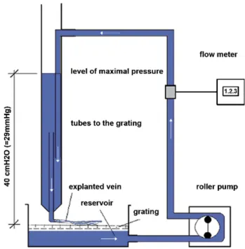
For this experiment, we laid the prepared veins on a grating. With a roller pump, we adjusted the maximal flow possible, with a maximal pressure of 40 cm of water, corresponding to 29 mmHg. The perfusion pressure itself could be read directly on the scale. (Figs. 1 and 2)
3. Results
Of 14 cadavers, we found valves in 100% on the right, and in 92% on the left side.
In all 14 cadavers, anatomically and functionally compe-tent valves on the right proximal IJV were found. Only 1/14 cadaver had no valve in the left proximal IJV. Additional rudimentary and incompetent valves could be identified in 1/ 14 cadavers on the distal right IJV, and in 2/14 cadavers on the left IJV.
The selective flow measurement through the internal jugular vein of seven cadavers revealed a maximal flow of 2500 ml/min only in one cadaver. In this case, a remarkable dilatation of the isolated venous preparation was seen under pressure. Due to this unphysiological increase of diameter and any probable competence, the jugular valves were destroyed. In one cadaver, we measured a flow of
A. Ku¨nzli et al. / European Journal of Cardio-thoracic Surgery 30 (2006) 906—909 907
Fig. 1. In vitro arrangement with roller pump, flow meter, tubes to the grating with veins lying on it. The superior vena cava is connected to the tubing system, the open jugular veins are delivering any volume through the grating to the system, which refer to the maximal volume retrogradly to the brain.
Fig. 2. Seeing on great view the grating with the explanted jugular vein of a human cadaver, connected at the superior vena cava to the tube. The clamp demonstrates the ligated side branches to the neck and the head.
340 ml/min. One cadaver had 6 ml/min, and four cadavers had absolutely no flow through the system because of competent valves although, we elevate the inlet pressure to 29 mmHg. (Table 1). Also, remarkably more pressure than ever used clinically did not increase the flow in these in vitro cases.
4. Discussion
Bavaria et al.[7]evaluated 60 consecutive patients who underwent repair of acute type A aortic dissection over a 6 years period. Patients were grouped according to intrao-perative circulatory management strategies. In Group I, 41 patients were managed by CPB (n = 21) with conversion to HCA (n = 20). Group II consisting of 19 patients managed with routine open distal anastomosis and HCA with RCP. Patients undergoing RCP experienced statistically significant reduc-tions in rates of confirmed cerebrovascular accidents and mortality.
In a period between 1987 and 1995, Ennker et al. [8]
treated 437 patients surgically for aortic disease (ascending and/or transverse aortic arch), 361 operated with deep hypothermic circulatory arrest, 75 patients (20.8%) with acute aortic dissection. His statistic of this inhomogeneous group clearly revealed, that early mortality and, in particular, stroke are reduced using RCP.
In a cadaver study, De Brux et al. [9] examined the retrograde flow distribution of the brain by injection of opacified latex through a cannula in the superior vena cava. They demonstrated that even if the internal jugular veins are competently valvulated, the brain was perfused through the azygos and collateral veins. In Group III, with ligation of the azygos vein, no opafication was observed except that of the superior vena cava, right and left brachiocephalic veins, and internal jugular veins up to the valves. On the contrary, the distribution via azygos vein in Groups I and II feeded the intercostal veins of the thorax, the inferior vena cava and their inlets and also the cervical spinal cord.
In different clinical articles, Ueda et al.[10]from Japan described the RCP as his favoured adjunct to HCA for aortic arch surgery: in a three-centre retrospective review of 249 patients undergoing HCA and RCP for aortic arch surgery, his studies suggested a limit of safe duration of RCP and HCA with up to 80 min of HCA. Although, an other study from Ogino et al.[11]revealed an oxygen delivery to the intracranium using near-infrared spectroscopy during HCA with RCP, it is
not proven that the RCP took always the shortest way and passed the jugular valve.
Together with the clinical observation that only a small fraction comes back through the carotid artery, we must conclude that the greatest portion of RCP perfuses the body but not the brain. In our study, jugular vein valves were found in 100% and in the great majority there were competent. Consecutively, we could demonstrate that no relevant flow through the IJV can be installed with a maximal pressure of more than 25 mmHg. In addition, due to the explantation of the venous system in our test arrangement, no other factor could influence the retrograde flow than the human extracranial venous anatomy itself. The influence of intracranial venous resistance in particular, which could lead to a dominant flow to other (extracranial or brachial) regions in the in vivo use, was eliminated.
The method of retrograde cerebral perfusion is simple and safe, helps keeping a good exposure and statistically improves results in patients undergoing aortic surgery with HCA. Obviously, RCP does not reach the brain by the shortest and directest way and in its full quantity: by cannulation of the superior vena cava, the blood does not reach the brain reliably through the internal jugular vein, but this technique may reach a perfusion of the brain trough the collateral venous net.
Its effects of flushing out embolism, deairing, supporting the cold jacket and perfusing the brain may be present, but are unpredictable. Particularly, a homogenous distribution in the brain for supply is questionable. For effective cerebral security, in our opinion, RCP reminds not an alternative to HCA, but a valuable adjunct to it. On the contrary, antegrade cerebral perfusion by its ‘natural’ arterial way will have the most reliable access to the brain and therefore, from an anatomical point of view, will have the best and safest protecting capacities[3,12].
Acknowledgements
This study was supported by the Institute of Pathology and the Laboratories of Clinical Research, University Hospital Zurich, Switzerland.
References
[1] Mills NL, Ochsner JL. Massive air embolism during cardiopulmonary bypass. J Thoracic Cardiovasc Surg 1980;80:708—17.
[2] Lemole GM, Strong MD, Spagna PM, Karmilowicz NP, Mills B. Improved results for dissecting aneurysms. J Thorac Cardiovasc Surg 1982;83:249— 55.
[3] Lin PJ, Chang Ch, Tan PPC, Chang CN, Lee ST, Wang CC, Chang JP, Liu DW, Chu JI, Tsai KT, Kao CL, Hsieh MJ, Hua MS. Prolonged circulatory arrest in moderate hypothermia with retrograde cerebral perfusion. Circulation 1996;94(Suppl. II):169—72.
[4] Juvonen T, Zhang N, Wolfe D, Weisz DJ, Bodian CA, Shiang HH, McCullough JN, Griepp RB. Retrograde cerebral perfusion enhances cerebral protec-tion during prolonged hypothermic circulatory arrest: a study in a chronic porcine model. Ann Thorac Surg 1998;66:38—50.
[5] Safi HJ, Brien HW, Winter JN, Thomas AC, Maulsby RL, Doerr HK, Svensson LG. Brain protection via cerebral retrograde perfusion during aortic arch aneurysm repair. Ann Thorac Surg 1993;56(2):270—6.
[6] Deeb GM, Jenkins E, Bolling SF, Brunsting LA, Williams DM, Quint LE, Deeb ND. Retrograde cerebral perfusion during hypothermic circulatory arrest A. Ku¨nzli et al. / European Journal of Cardio-thoracic Surgery 30 (2006) 906—909
908
Table 1
In vitro assessment showed no evidence of reliable and sufficient blood flow to the brain via the internal jugular veins
14 preparations of 8 females and 6 males
Right internal jugular vein 14 valves (100%) Left internal jugular vein 13 valves (92%) Selective flow measurement through the internal jugular veins of seven patients (max. inlet pressure 29 mmHg)
Four cadavers 0 ml/min
One cadaver 2500 ml/min
One cadaver 340 ml/min
reduces neurologic morbidity. J Thorac Cardiovasc Surg 1995;109: 259—68.
[7] Bavaria JE, Woo YS, Hall RA, Wahl PM, Acker MA, Gardner TJ. Circulatory management with retrograde cerebral perfusion for acute type A aortic dissection. Circulation 1996;94(Suppl. II):173—6.
[8] Ennker J, Coselli JS, Treasure T, editors. Retrograde cerebral perfusion in surgery for aortic arch aneurysm. Cerebral protection in cerebrovascular and aortic surgery. Springer; 1997. p. 239—49.
[9] De Brux JL, Subayi JB, Pegis JD, Pillet J. Retrograde cerebral perfusion: anatomic study of the distribution of blood to the brain. Ann Thorac Surg 1995;60:1294—8.
[10] Ueda Y, Okita Y, Aomi S, Koyanagi H, Takamoto S. Retrograde cerebral perfusion for aortic arch surgery: analysis of risk factors. Ann Thorac Surg 1999;67(6):1879—82 [discussion 1891—4].
[11] Ogino H, Ueda Y, Sugita T, Morioka K, Sakakibara Y, Matsubayashi K, Nomoto T. Monitoring of regional cerebral oxygenation by near-infra-red spectroscopy during continuous retrograde cerebral perfusion for aortic arch surgery. Eur J Cardiothorac Surg 1998;14(4):415— 418.
[12] Bachet J, Guimet D, Goudot B, Dreyfus GD, Delentdecker P, Brodaty D, Dubois C. Antegrade cerebrale perfusion with cold blood: a 13-year experience. Ann Thorac Surg 1999;67(6):1874—8 [discussion 1891—4]. A. Ku¨nzli et al. / European Journal of Cardio-thoracic Surgery 30 (2006) 906—909 909