THE JOURNAL OF INFECTIOUS DISEASES· VOL. 158. NO.1· JULY 1988 © 1988 by The University of Chicago. All rights reserved. 0022-1899/88/5801-0010$01.00
Fibronectin-Cleaving Activity in Bronchial Secretions of Patients with Cystic Fibrosis
Susanne Suter,U. B. Schaad,J. J. Morgenthaler, I.Chevallier, and H. P. Schnebli
From the Department of Pediatrics, Children's Hospital, University of Geneva, Geneva; the Department of Pediatrics, Children's Hospital, University of Bern, and the Central Laboratory, Swiss Red Cross, Bern; and the CIBA-GEIGY Research Institute, Basel, Switzerland
In cystic fibrosis, colonization of the airways withPseudomonas aeruginosafollows coloni-zation withStaphylococcus aureusand is related to accelerated deterioration of pulmonary function. BecauseP.aeruginosaadheres better to cell surfaces devoid of fibronectin, we searched for fibronectin-cleaving activity in bronchial secretions and saliva from24 pa-tients with cystic fibrosis who were followed up for 4.5 y and from two control groups. Proteolytic activity against 12sI-labeled fibronectin was continuously present in cysticfibrosis bronchial secretions; significantly higher fibronectin-cleaving activity was found in older vs. younger patients, in patients with advanced disease stages determined by a five-stage scoring system, and in those colonized withP.aeruginosa.The fibronectin-cleaving ac-tivity was due to neutrophil elastase and cathepsin G. Cystic fibrosis bronchial secretions had proteolytic activity against surface fibronectin of airway mucosal cells. Thus fibro-nectin-cleaving activity of bronchial secretions rather than of saliva may favorP.aeruginosa colonization of the upper respiratory tract in individuals with cystic fibrosis.
Most individuals with cystic fibrosis who survive be-yond the neonatal period die from respiratory fail-ure, which is the consequence of chronic, pro-gressively destructive bronchitis [1-3].Clinical and pathological observations show extensive tissue de-struction in the airways, which first causes obstruc-tion and obliteraobstruc-tion of small airways and thereafter destruction of the walls of large airways [4, 5]. Early in the disease course the bacterial pathogens fre-quently found in the bronchial secretions are
Staph-ylococcus aureus and Haemophilus influenzae. Later
the airways of these subjects are invariably colonized with Pseudomonas aeruginosa, an event that initi-ates an acclerated deterioration of lung functions [3] as well as an excessive systemic immune response [6]. These pathogens cannot be eliminated from the air-ways by antimicrobial therapy, and because the del-eterious effect ofP.aeruginosa infection in
individ-uals with cystic fibrosis is well established, the identification of factors favoring colonization of the airways withP.aeruginosa is important.
Coloniza-Received for publication 29 October 1987 and in revised form 27 January 1988.
This work was supported by grants 3.816-0.84 and 3.850-0.86 from the Swiss National Research Foundation.
We thank Dr. A. Baici for suggestions and P. Schilling for manu-script preparation.
Please address requests for reprints to Dr. Susanne Suter, Cli-nique Universitaire de Pediatrie, 30, bd de la Cluse, 1211 Geneva 4, Switzerland.
89
tion of the upper respiratory tract with bacterial pathogens is achieved by binding of bacteria to epi-thelial cell surfaces. Severalexperimental studies have shown that the type of bacteria binding to epithelial surfaces is influenced by the presence of fibronec-tin, a glycoprotein on the cell surface [7-11]. The ab-sence of fibronectin on epithelial cell surfaces from patients with cystic fibrosis [9] as well as from seri-ouslyillpatients with acute respiratory failure [10] correlated with increased adherence ofP.aeruginosa
to buccal epithelial cells. In these studies a parallel increase in activity of salivary proteases was demon-strated, and it was suggested that salivary proteases were the source of fibronectin-cleaving activity in these patients. However, in individuals with cystic fibrosis, not only saliva but also bronchial secretions may be the source of proteolytic activity [11-13].
We therefore attempted to quantitate and identify the fibronectin-cleaving enzymes in cystic fibrosis bronchial secretions and saliva in comparison with those from a control group of patients with chronic bronchitis. We also determined the proteolytic ef-fect of cystic fibrosis bronchial secretions on cell sur-face fibronectin of airway mucosal cells of human lung tissue.
Patients and Methods
Study group. Twenty-four patients with cystic fibrosis attending the cystic fibrosis clinic or hospital-ized at the Children's Hospitals of the Universities
90
of Bern (Switzerland) and Geneva were prospectively included in the study since 1982. The diagnosis of cystic fibrosis had been established by clinical fea-tures of the disease and was confirmed by sweat con-centrations of Na" andCl of >70 mEq/L. The mean age of the patients was 12.7y (range, 15mo to 21y),
Cultures of sputum from nine patients were positive mainly for S.
aureus;
other pathogens isolated in-cludedH. influenzae,
~-hemolyticStreptococcus
group A, andEscherichia coli.
The mean age of these patients was 8.1 y (range, 15mo to 15y). Fifteen pa-tients had cultures of sputum positive forP.aerugi-nosa;
other pathogens isolated from these patients were S.aureus
andAspergillus fumigatus.
The mean age of this group was 15.5 y (range, 5.5-20 y). All patients who were colonized withP.aeruginosa
were hospitalized three times per year for iv antimicrobial therapy.Clinical and radiographic staging of cystic fibro-sis was done by one of us, unaware of the labora-tory results, with use of a five-stage scoring system [14], based on physical activity, general physical con-dition and bacteriology of sputum, signs of pulmo-nary disease, radiological lung involvement, and gas-trointestinal manifestations. Each parameter was assigned a maximum of 5 points, with the highest score indicating the best status. Severity of the dis-ease was graded in five stages (I-V). Radiographic scoring was done with the scoring system of Chrispin and Norman [15]. The highest stage (V) corresponds to poor pulmonary condition and advanced radio-graphic alterations of cystic fibrosis.
Control groups.
Two control groups were also included in the study: one group included 16patients with acute exacerbations of chronic bronchitis and bronchiectases, as documented by chronic cough, purulent sputum, positive chest findings, and roent-genograms. In nine patients, bronchiectases had been documented by bronchography or bronchoscopy. The mean age of the patients was 45 y (range, 9-56 y). Pathogens isolated from sputum includedH. influenzae, Streptococcus
pneumoniae,E. coli,
S.aureus,
andAspergillus.
The second control group included 10 patients who had chronic bronchitis without acute exacerbation (all of whom were smokers), as documented by chronic cough and in-creased amounts of nonpurulent sputum. These pa-tients produced sputum that was negative when cul-tured for bacterial pathogens. The mean age of these patients was 68 y (range, 41-90 y).Specimens.
From all these patients, bronchialSuter et at.
secretions were collected according to the following protocol: Fresh morning sputum was obtained dur-ing morndur-ing chest physiotherapy. For the patients with cystic fibrosis the number of samples collected per patient ranged from four to 22 over a period of 4.5 y. From two patients with cystic fibrosis and six patients with chronic bronchitis a sample of morn-ing sputum and a sample obtained by bronchosco-py were collected on the same day. None of the pa-tients received mucolytic treatment during the study period.
Cultures and identifications of bacteria from bronchial secretions were done according to well-established methods [16]. The secretions were mixed with a half volume of sterile 0.9070 NaCI and shaken by hand for 1 min; the mixture was then centrifuged at 1000g until the supernatant was clear. The super-natants were stored at - 70 C until tested, and the total protein concentration was determined by a stan-dard method (Biorad, Richmond, Calif). Ten bron-chial secretions had been subjected to ultracentrifu-gation at 30000 g for 30 min without prior dilution, and the enzymatic activities were measured in these supernatants.
We also collected saliva from 18 healthy individ-uals and from 14 patients with cystic fibrosis (11 colonized withP.
aeruginosa
and three not colonized withP.aeruginosa);
these samples were centrifuged without prior dilution. The saliva of patients with cystic fibrosis was obtained during a cough-free period after chest physiotherapy, and only saliva that was macroscopically free from bronchial secretions was examined. No stimulus was used for stimulat-ing production of saliva.Lung tissue was obtained at autopsy from an eight-month-old child without cystic fibrosis and without lung disease.
Three strains ofP.
aeruginosa,
isolated from our patients with cystic fibrosis, that secreted bacterial elastase [11] were cultured in Mueller-Hinton broth for 24 h; the culture supernatant was obtained by centrifugation at 8000 g for 20 min.Assessment oj fibronectin-cleaving activity oj
bronchial secretions and saliva.
Fibronectin was purified from human plasma [17] and radiolabeled with 1251 [18] (Amersham Corp., Amersham, En-gland). About 250000 cpm of 1251was incorporated into 1 ug of purified fibronectin. 125I-Labeled fibro-nectinC
251-fibronectin) was then coupled with CNBr-activated Sepharose" 4B (Pharmacia Fine Chemicals, Uppsala, Sweden).1251-Fibronectin-Fibronectin Cleavage in Cystic Fibrosis
Sepharose beads corresponding to 250 000 cpm were then suspended in 500 ul, of Dulbecco's PBS (pH 7.4) and incubated at 37 C with 10 ul, of bronchial secretion, saliva, or purified neutrophil enzymes for 4 h. 1251-Fibronectin fragments were separated from 1251-fibronectin still bound to Sepharose by centrifu-gation at 8000gfor 5 min. Proteolysis of fibronec-tin was expressed as a percentage of the cpm in the supernatant divided by the cpm of the total sample. Spontaneous release of 1251 from fibronectin was measured simultaneously «2010) and subtracted from the results. The time course of degradation of 1251-fibronectin by bronchial secretions and by puri-fied neutrophil enzymes was similar and showed that the enzymatic reaction was not linear; 1251_fibronectin fragments were rapidly released over the first min-ute of incubation, followed by slower release over a 4-h period.
For characterizing the fibronectin fragments generated by bronchial secretions, the fragments pro-duced by proteolysis of purified (unlabeled) fibro-nectin by a pool of cystic fibrosis bronchial secre-tions over time was compared with the fragments generated by a mixture of purified elastase (three parts) and cathepsin G (one part). The fragments were separated with PAGE [19]. After electrophore-sis the gels were incubated in 25mMTris and 20% methanol (pH 10.4)for 15min, and the proteins were transferred to Novilon" membranes (Millipore Corp., Bedford, Mass) by using a Multiphor II Novablot" system (LKB, Bromma, Sweden) at 150 rnA for 90 min [20]. The membrane was then slowly shaken in a blocking buffer (10mMTris, 1mMEDTA, 133 mMNaCI, 10010 skim milk, 0.05% Triton X-l00, and 2% sodium azide) at room temperature (1'\.123C)for 2 h. Subsequently the membrane was incubated in skim milk (Difco, Detroit) containing an antibody to fibronectin raised in rabbits (Behring, Marburg, Federal Republic of Germany) at a dilution of 1:4000 followed by three washing steps in PBS (pH 7.4),Ca" and Mg" free. During the next step the Novilon membrane was incubated in skim milk containing an antibody to rabbit IgG bound to alkaline phos-phatase (Sigma, St. Louis) at a dilution of 1:2000 for 2 h. After three washing steps inCa"-and Mg"-free PBS, the membrane was incubated in 50mM Tris (pH 8) for 10 min and exposed to the chromo-genic substrate (naphthol AS-MX [40 mg] and fast red salt [80 mg; both from Sigma] in 50mMTris). The reaction was stopped by washing the Novilon membrane in tap water for 10min. Molecular weights
91
were monitored with standard proteins (Biorad), which were revealed with amido-black staining.
Zymographs. To identify fibronectin-cleaving enzymes in cystic fibrosis bronchial secretions, we modified previously published methods [21, 22] for zymographs. Purified fibronectin (1.5 mg/mL) was added to a 7.5% (wt/vol) polyacrylamide gel before polymerization. The stacking gel contained no fi-bronectin. One microliter of a pool of cystic fibro-sis bronchial secretions, purified elastase, cathepsin G, or a mixture (3:1) of purified elastase and cathep-sin G was added to 9 ul, of sample buffer [21] and electrophoresed through the fibronectin-containing gel at 4 C for 5 h at 170 V without prior heating of the samples. SDS was then removed from the gel by incubation of the gel in 2.5% Triton X-100 for 1 h. Thereafter the gel was incubated at 37 C for 16 h in 0.1M glycine, pH 8.3, 7.4, or 6.8. Proteins were transferred to Novilon membranes, and fibronectin was revealed as described above. With this procedure the location to which a fibronectin-degrading pro-tease had migrated was seen as a clear band, in which copolymerized fibronectin had been degraded. In control experiments, samples heated to 90 C for 15 min were tested, and in others the Novilon membrane was incubated in 2mMphenylmethylsulfonyl fluo-ride (PMSF) dissolved in methanol for 1 h before incubation with the antibodies.
Determination ofelastolytic activityofbronchial
secretions.
The elastolytic activity was determined as described by Hornebeck and Schnebli [23] with purified bovine elastin (Sigma) radioactively labeled with 3H (New England Nuclear Corp., Boston). Ten microliters of the bronchial secretion was incubated with 500 ug of 3H-Iabeled elastin suspended in 1 mL of 0.1M Tris(pH 8.2)containing 0.01% 23 lauryl ether (Brij" 35; Sigma) and 0.02% sodium azide for 16 h at 37C.The samples were centrifuged at 3300gfor 10 min. The radioactivity of 100 ul, of the superna-tant was then measured and used for calculating the amount of solubilized elastin. One milligram of com-pletely solubilized 3H-Iabeled elastin corresponded to 145 000 cpm. Elastolytic activity (DE) was ex-pressed as the number of milligrams of labeled elastin solubilized by 1 mL of enzyme solution. This enzymatic reaction was linear over the interval tested. Purified human neutrophil elastase [24] and ca-thepsin G [25] were used as controls. For determin-ing the concentration of inhibitors that achieved complete inhibition of purified leukocyte elastase and cathepsin G, the activity of these enzymes was92
measured with the synthetic substrates Succ-Ala-Ala-Pro-Val-pNA and Succ-Ala-Pro-Phe-pNA (both from Calbiochem Behring, Luzern, Switzerland) [26].
- Inhibition experiments.
The 1251-fibronectin-cleaving activity of cystic fibrosis bronchial secre-tions was also determined after prior incubation of bronchial secretions with the following inhibitors: partially purified aI-proteinase inhibitor (arPI; Sigma); PMSF (Sigma); Eglin C, an inhibitor of neu-trophil elastase and cathespin G [27]; Ac-Ala-Ala-Pro-Val-ChCI2 [26], a specific inhibitor of neutro-phil elastase; Ac-Ala-Ala-Pro-Phe-ChCl., a specific inhibitor of cathepsin G [26] (both peptides from Enzyme Systems Products, Livermore, Calif); and o-phenanthroline (Sigma), an inhibitor of metal-loproteases such as pseudomonas elastase [28]. We also inactivated the proteolytic activity of cystic fibrosis bronchial secretions by heating at different temperatures.Assessment of proteolysis of cellular
fibronec-tin.
Because plasma fibronectin and cellular fibro-nectin have different subunit structures[29],we stud-ied the proteolytic effect of cystic fibrosis bronchial secretions on cellular fibronectin of airway mucosa. Cellular fibronectin was revealed by direct immuno-fluorescence with an FITC-conjugated [30] antibody to fibronectin that had been raised in rabbits. Sec-tions of frozen lung tissue 4urn
thick were fixed on glass slides coated withpoly-t-lysine
(Sigma)by ex-posure to acetone for 2 min. The preparation was rinsed three times with PBS, and a drop of FITC-conjugated antiserum to fibronectin was deposited on the tissue section. Fluorescence was observed in a fluorescent light microscope after incubation for 30 min with the antibody, subsequent rinsing with PBS, and exposure to 90010glycerol [31]. Immuno-fluorescent fibronectin was observed after incuba-tion of lung tissue secincuba-tions with cystic fibrosis bron-chial secretions. No fluorescence was observed when the antibody to fibronectin was adsorbed with puri-fied fibronectin before incubation with the lung tis-sue. Quenching of fluorescence by bronchial secre-tions was excluded by an experiment in which the cystic fibrosis bronchial secretion had been incubated with an excess amount of Eglin C (100!lM)before incubation with lung tissue sections.Statistical analysis.
Results are given as mean ± SD values. Comparisons were made with the Mann-Whitney test, and correlations were analyzed with the Spearman correlation coefficient.Suter et al.
Results
Elastolytic and 125I-fibronectin-cleaving activities
and protein concentration of bronchial secretions
and saliva.
Bronchial secretions from patients with cystic fibrosis and patients with acute exacerbations of chronic bronchitis had a significantly higher mean fibronectin-cleaving activity than did bronchial se-cretions from patients with chronic bronchitis with-out acute exacerbation(P<.01) and than did saliva from patients with cystic fibrosis(P<
.01) and nor-mal individuals(P<
.01; table 1). Saliva from pa-tients with cystic fibrosis had significantly higher mean fibronectin-cleaving activity than that from normal individuals (P<
.01).The elastolytic activity of cystic fibrosis bronchial secretions (1.61 ± 0.85 UE) was significantly higher than the elastolytic activity of bronchial secretions from patients with chronic bronchitis and acute exacerbation (0.42 ± 0.33 UE)' No measureable elastolytic activity was found in bronchial secretions from those with chronic bronchitis without acute ex-acerbation and in saliva from patients with cystic fibrosis and normal individuals. The elastolytic ac-tivity of the bronchial secretions obtained by bron-choscopy of two patients with cystic fibrosis and six patients with chronic bronchitis correlated well with that of the morning sputum sample of the same day, and no significant differences were observed in sam-ples of bronchial secretions diluted with 0.9% NaCI and shaken by hand before centrifugation and those prepared by ultracentrifugation starting from the same sample. Because the precise quantitation of leu-kocytes in bronchial secretions is not feasible owing to the viscosity of the samples and because our previ-ous studies had shown a good correlation between semiquantitative determination of leukocyte counts and elastolytic activity of bronchial secretions [11, 12],we made no attempt to quantitate neutrophils in the secretions.
None of the culture supernatants of the three elastase-producing strains ofP.
aeruginosa
had sig-nificant 1251-fibronectin-cleaving activity.The total protein concentration was significantly higher in bronchial secretions from patients with cys-tic fibrosis and those with chronic bronchitis with acute exacerbations than in the bronchial secretions from those with chronic bronchitis without acute ex-acerbation and in the samples of saliva from patients with cystic fibrosis and normal individuals(P< .01). The mean protein concentration of cystic fibrosis
Fibronectin Cleavage in Cystic Fibrosis 93
Table 1. Elastolytic and fibronectin-cleaving activities and protein concentration of bronchial secretions and saliva.
No. tested
Elastolytic Fibronectin-cleaving Total protein Specimen, group Patients Samples activity (VE) activity(070) concentration (g/L) Bronchial secretions
Cystic fibrosis 24 194 1.61 ± 0.85* 39.1 ± 15.2 5.8 ± 3.8
Chronic bronchitis
With acute exacerbations 16 24 0.42 ± 0.33*t 37.5 ± 16.2t 4.0 ± 3.6
Without acute exacerbations 10 12 <0.05t 3.3 ± 3.4t 2.4 ± 2.7
Saliva
Cystic fibrosis 14 24 <0.05 8.2 ± 19.0 2.4 ± 2.1
Normal 18 18 <0.05 4.0 ± 9.0 1.4 ± 0.7
NOTE. Elastolytic activity is mg of 3H-Iabeled elastin solubilized (16 h at 37C)by I mL of undiluted sample. Fibronectin-cleaving activity is the percentage of the cpm of 1Z51-fibronectin released in the supernatant divided by the radioactivity of the total sample. All tests were done in triplicate, and the background (determined by incubation of the substrates without the source of enzymes) was subtracted. Data are mean ± SD values. For statistical evaluation the Mann-Whitney test was used.
*p < .01.
t p < .01.
t p < .01.
bronchial secretions was significantly higher (P
<
.05)than that of bronchial secretions from patients with chronic bronchitis and acute exacerbations.Inhibition experiments.
The elastolytic activity of all samples of bronchial secretions was inhibited 86%-95070by 2 mMPMSF, a finding demonstrating that most of it was due to serine proteases (table 2). To determine which enzyme(s) might be respon-sible for the fibronectin-cleaving activity, we used specific inhibitors of neutrophil elastase, cathepsin G, or both. The inhibition as a percentage of fibro-nectin-cleaving activity of cystic fibrosis bronchialsecretions, purified neutrophil elastase, and cathep-sin G is shown in table 2. Purified lIt-PI, PMSF, and Eglin C inhibited the fibronectin-cleaving activity of cystic fibrosis bronchial secretions substantially (80%-90%) as well as that of purified neutrophil elastase and cathepsin G, with one exception: As ex-pected [32], the fibronectin-cleaving activity of puri-fied cathepsin G was inhibited only 37% by partially purified lIt-PI at the concentration used. We then tried to distinguish fibronectin-cleaving activity due to elastase from that due to cathepsin G by using the elastase-specific [26] inhibitor
Ac-Ala-Ala-Pro-Table 2. Inhibition of I25I-fibronectin-cleaving activity by various inhibitors and inactivation by heating.
Inhibition(070)of "'I-fibronectin - cleaving activity of
Inhibitor (concentration) a.-PI (4~M) PMSF (2mM) Eglin C (12.5 l!M) Ac-Ala-Ala-Pro-Val-ChCI, (I mM) Ac-Ala-Ala-Pro-Phe-ChCl, (I mM) Heating at 60 C for 15 min 80
c
for 15 minCystic fibrosis Purified Purified
bronchial secretions neutrophil elastase cathepsinG
80 ± 7 (9) 89 ± 3 (4) 37 ± 9 (4) 90 ± 5 (11) 95 ± 2 (7) 91 ± 2 (7) 87 ± 8 (11) 98 ± 3 (7) 99±1(7) 67 ± 9 (10) 82 ± 3 (4) 28 ± 9 (4) 84 ± 9 (9) 89 ± 3 (4) 93 ± 3 (4) II ± 6 (2) 22 ± 8 (3) 87 ± 3 (3) 96 ±2 (2) 98 ± I (3) 94 ±2 (3)
NOTE. Fibronectin-cleaving activity of bronchial secretions and purified neutrophil enzymes was tested simultaneously in the presence and in the absence of the indicated inhibitors or after heat inactivation of the samples. Inhibition is expressed as 100x (activity of the sample with inhibitor/activity of the sample without the inhibitor). Bronchial secretions were preincubated with the inhibitor for 15 min. Data are mean ± SD values (no. of bronchial secretions tested in triplicate).
94
Val-ChCI2and the cathepsin G-specific inhibitor [26]
Ac-Ala-Ala-Pro-Phe-ChCl.. We first determined the concentration of inhibitor inhibiting the esterolytic activity of purified elastase and cathepsin G; the es-terolytic activity on the specific substrates of both enzymes was inhibited efficiently (>95070) when the inhibitor was used at 20~M.However, the fibronec-tin-cleaving activity of purified elastase and cathep-sin G was not substantially reduced when we used the inhibitor at this concentration, and we therefore increased the concentration of inhibitor to 1mM. Under these conditions the elastase-specific inhibi-tor partially inhibited the fibronectin-cleaving activ-ity of cathepsin G (28%), whereas the cathepsin G-specific inhibitor also inhibited the fibronec-tin-cleaving activity of purified elastase by 84%. The fibronectin-cleaving activity of cystic fibrosis bron-chial secretions was inhibited 67% by the elastase-specific and 84% by the cathepsin G-elastase-specific inhib-itor. Because cathepsin G is less resistant to heat than is elastase [33], we also tested the heat resistance of the fibronectin-cleaving activity of cystic fibrosis bronchial secretions and of purified elastase and cathepsin G. Whereas the fibronectin-cleaving ac-tivity of purified elastase and the cystic fibrosis bron-chial secretions was inactivated only after heating of the samples at 80 C for 15 min, that of purified cathepsin G was inactivated 87% after heating at 60 C for 15 min.
From these inhibition experiments we concluded that (1) the fibronectin-cleaving activity of cystic fibrosis bronchial secretions was inhibited best by PMSF and Eglin C (90% and 87%, respectively), which inhibit both cathepsin G and elastase; (2) the specific chloromethylketone inhibitors of cathepsin G and elastase did not allow us to distinguish be-tween these two enzymes as a source of fibronectin-cleaving activity in cystic fibrosis bronchial secretions because they lost their specificity at a concentration of 1mM;and(3)the heat resistance of the fibronec-tin-cleaving activity of cystic fibrosis bronchial secre-tions was similar to that of elastase rather than of cathepsin G. Phenanthroline (l mM), which in-hibited the elastolytic activity of
P.
aeruginosa
cul-ture supernatant by >90%, did not inhibit the fibro-nectin-cleaving or the elastolytic activity of cystic fibrosis bronchial secretions.Kinetics ofproteolysisofpurified
fibronectinby
cysticfibrosis bronchial secretions, purifiedelastase,
and cathepsin
G. To test further whether cathep-sin G, other enzymes, or both contributed to theSuter et at.
fibronectin-cleaving activity of cystic fibrosis bron-chial secretions in addition to elastase, we examined the time course and the kinetics of the breakdown of purified fibronectin by these bronchial secretions and compared the apparent molecular weights of fibronectin breakdown products generated by these three sources of enzymatic activity. The breakdown pattern of fibronectin by a pool of cystic fibrosis bronchial secretions is shown in figure 1, top, and that of a mixture of purified elastase and cathepsin G (3:1) is shown in figure 1, bottom, at 0, 1, 5, 10, 15, 30, and 60 min of incubation at 37 C. After 1 min of incubation with the cystic fibrosis bronchial secretion the two monomers of fibronectin with ap-parent molecular weights of 220 000 were split into multiple fragments. The molecular weights of some fragments were identical to those generated by elastase and cathepsin G: 181000, 164000, 152000, 141 000, 138000, 108000, and 84000. Although ma-jor breakdown products had similar molecular weights, the time course of appearance was clearly different in figure 1, top and bottom. These experi-ments showed that(1)purified fibronectin was com-pletely hydrolyzed by the cystic fibrosis bronchial secretion as wellas by elastase and cathepsin G within 1 min and (2) although some breakdown products of fibronectin generated by the cystic fibrosis bron-chial secretion had molecular weights identical to those generated by elastase and cathepsin G, the time course of degradation and the molecular weights of several fragments were different.
Zymographs. On the zymographs (figure 2) copolymerized fibronectin appeared as dark stain-ing. In figure 2 the first four lanes were loaded with 1.2 and 2.4 ug of fibronectin, lanes 5 and 6 with 3 ~Lof purified neutrophil elastase, lanes 7 and 8 with a combination of 3~Lof purified elastase and 1~L of purified cathepsin G, lanes 9 and 10 with 1~L of purified cathepsin G, and lanes 11 and 12 with 1~Lof a pool of cystic fibrosis bronchial secretions. Figure 2 shows an identically migrating, clear dou-ble band for lanes 7 and 8 and lanes 11 and 12. The results were identical for a pH of the incubation buffer of 6.8, 7.4, and 8.3. Thus this experiment demonstrated that the fibronectin-cleaving activity of cystic fibrosis bronchial secretions comigrated with purified elastase and cathepsin G. No other zones of fibronectin cleavage were observed under our experimental conditions. Control experiments confirmed that the clear bands corresponded to degradation of fibronectin by proteases. When the
95
Figure 1. Top,proteolytic activity of I ul, of a pool of cystic fibrosis bronchial secretions against purified fibro-nectin after 0, 1,5, 10, IS, 30, and 60 min of incubation at 37C,The proteolysis of fibronectin was examined by PAGE (running gel,7,5l1Jo[wt/volJ) and subsequent im-munoblotting. The molecular weights were monitored using standard proteins (Biorad).Bottom,an identical ex-periment, in which 3 ul, of purified elastase and I ul, of purified cathepsin G were incubated with fibronectin. Ma-jor fragments with molecular weights of 181000, 164000, 152000, 141000, 138000, 108000, and 84 000 appeared after incubation of fibronectin with the bronchial
secre-tion (top) and with purified enzymes (bottom), but the
time course of appearance of fragments and the molecu-lar weights of other fragment s were different. Lanes I,
2, 10,andIIwere loaded with 1.211g of purified
fibronec-tin (FN) .
bronchial secretion and the purified neutrophil en-zymes were heated at 90 C for 15 min before elec-trophoresis, no clear bands appeared. Degradation of the rabbit antibody to fibronectin or the antibody
Figure 2. Zymograph in which the proteolytic effect of I ul, of purified elastase(E; lanes5 and 6), 3 ul, of puri-fied elastase plus I ul, of puripuri-fied cathepsin G(E
+
C;fanes7 and 8), I ul, of purified cathepsin G(C; fanes 9
and10),and I ul, of a pool of cystic fibrosis (CF) bron-chial secretions(lanes IIand 12)on purified fibronectin copolymerized in a polyacrylamide gel (7.5% [wt/voll) was evaluated after electrophoresis of the unheated sam-ples (40 C) through the gel. Purified fibronectin (1.2 or 2.4 I1g) was loaded onfanes1-4. The twoarrowspoint to areas of proteolysis of fibronectin, which appear as clear bands. The fibronectin-cleaving activity of cystic fibrosis bronchial secretions comigrated with the combination of elastase plus cathepsin G in a double band.
to rabbit IgG by enzymes transferred to the Novilon membrane was excluded because the clear zones ap-peared also when the Novilon membrane was treated with 2mMPMSF before incubation with the anti-bodies.
Because free granulocyte elastase can be quanti-tated in cystic fibrosis bronchial secretions by mea-suring elastolytic activity, we measured the mean elastolytic activities in these bronchial secretions in relation to bacterial colonization (with and without
P.
aeruginosa),
age, and disease stage (table 3) to de-termine precisely the amount of enzymes responsi-ble for fibronectin-cleaving activity. Bronchial secre-tions from patients with cysticfibrosis colonized withP.
aeruginosa
had significantly higher mean elasto-lytic activity than those from patients not colonized withP.
aeruginosa
(P<
.01), patients> 17 y old had significantly higher mean elastolytic activities than96
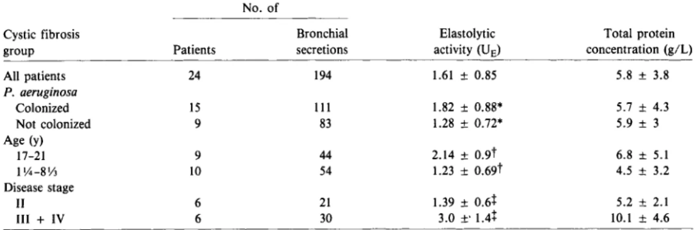
Table 3. Elastolytic activity and protein concentration of cystic fibrosis bronchial secretions.
Suter et al.
No. of
Cystic fibrosis Bronchial Elastolytic Total protein
group Patients secretions activity(UE) concentration (giL)
All patients 24 194 1.61± 0.85 5.8 ± 3.8 P. aeruginosa Colonized 15 111 1.82± 0.88* 5.7 ± 4.3 Not colonized 9 83 1.28± 0.72* 5.9 ± 3 Age (y) 17-21 9 44 2.14 ± 0.9t 6.8 ± 5.1 1\4-8Y3 10 54 1.23± 0.69t 4.5 ± 3.2 Disease stage II 6 21 1.39± 0.6+ 5.2 ± 2.1 ll1 + IV 6 30 3.0 ±'1.4+ 10.1 ± 4.6
NOTE. Elastolyticactivity ismgof 3H-labeled elastin solubilized(16h at37 C)by1mL of undiluted sample. Data aremean ± SO
values. The Mann-Whitney test was used for comparisons between groups. *P
<
.01.t
P<
.01.+
P <.01.did those less than nine years old(P
<
.01),and bron-chial secretions from patients who remained at dis-ease stageIIthroughout the observation period had significantly lower mean elastolytic activity than those from patients with disease stages IIIand IV, regardless of their bacterial colonization status(P<
.01). Two patients not colonized withP.
aerugi-nosa
were in the group including disease stages IIIand IV.
The mean ± SD elastolytic activity of the culture supernatants of three elastase-producing strains of
P.
aeruginosa
was 1.2± 0.35VE. Purified leuko-cyte elastase had an elastolytic activity of 7.78± 0.59 VE, and the fibronectin-cleaving activity of 1IJLof purified elastase and cathepsin G was 33070 ± 4% and 30% ± 3%, respectively.Proteolysis ofcellularfibronectin by
cysticfibro-sis bronchial secretions.
We used frozen sections of lung tissue from an individual without cystic fibro-sis to reveal cellular fibronectin on the surface of mucosal cells before and after incubation of the tis-sue with a cystic fibrosis bronchial secretion for I min. The results of three separate experiments were similar. Figure 3 shows the effect of the cystic fibro-sis bronchial secretion of fibronectin of airway mucosal cells. Figure 3A shows the lung tissue sec-tion stained with hematoxylin and eosin. Immuno-fluorescent fibronectin appears in figure 3B. After incubation of the lung tissue section with a cystic fibrosis bronchial secretion for 1 min, the immuno-fluorescent fibronectin had disappeared (figure 3C).When the cystic fibrosis bronchial secretion was preincubated with an excess amount of Eglin C, in-cubation of this secretion with the lung tissue had no effect on tissue fibronectin fluorescence. A con-trol experiment had shown that the bright immuno-fluorescence observed before incubation with the cys-tic fibrosis bronchial secretion could not be observed when the FITC-conjugated antibody to fibronectin was adsorbed with purified fibronectin before use.
Discussion
The results of our study show that bronchial secre-tions from patients with cystic fibrosis and patients with chronic bronchitis with acute exacerbations con-tained large amounts of 1251-fibronectin-cleavingac-tivity, whereas only small amounts were found in saliva from patients with cystic fibrosis and normal individuals (table 1). In contrast to the patients with chronic bronchitis, in whom the 1251-fibronectin-cleaving activity was present only during acute ex-acerbations, fibronectin-cleaving activity was found in all 194 samples of bronchial secretions from pa-tients with cystic fibrosis who were followed up for a period of4.5y.
The test that we developed for measuring fibro-nectin-cleaving activity with 1251-fibronectin bound to Sepharose beads as a substrate did not allow us to quantitate the enzyme(s) involved precisely be-cause the enzymatic reaction was not linear, regard-less of the enzyme source tested (bronchial secretions
Fibronectin Cleavage in CysticFibrosis 97
Figure 3. A, hematoxylin and eosin staining of a 4-~m-thick section of deep-frozen lung tissue. Immunoflu-orescent staining of fibronectin was done on a similar section (B) before and (C)after I min of incubation in a moist chamber with 200 ILL of a cys-tic fibrosis bronchial secretion. After incubation, most of the immunofluo-rescent fibronectin had disappeared.
or purified neutrophil elastase and cathepsin G). This observation may be explained by the presence of sev-eral cleavage sites on the fibronectin molecule, as shown by the degradation pattern of unlabeled fibronectin (figure1).The time course of degrada-tion of unlabeled fibronectin confirms the rapidity of proteolysis of fibronectin and argues against the hypothesis that the radioactively labeled fibronec-tin was more susceptible to proteolysis because of the labeling procedure.We therefore attempted first to identify the enzymes responsible for fibronectin cleavage by bronchial secretions. For this purpose we used inhibitors and determined the degradation .
pattern of purified fibronectin by bronchial secre -tions in comparison to purified elastase and cathep-sin G, which were the enzymes most likely involved in fibronectin cleavage [29, 34]. Finally, we used zymographs [21, 22] to estimate the migration of fibronectin-degrading enzymes in bronchial secre-tions, in comparison to the migration of purified elastase and cathepsin G.
The inhibition of 90070 and 87070 of 125I-fibronec -tin-cleaving activity by PMSF and Eglin C, respec-tively, showed that the enzymes involved were most likely serine proteases, such as neutrophil elastase and cathepsin G [24-26, 32, 33] (table 2). The two inhibitors Ala-Pro-val-ClrCl, and Ac-Ala-Pro-Phe-ChCI2, which are specific for neutrophil elastase and cathepsin G, respectively, when used with purified enzymes [26], did not allow us to dis-tinguish further the enzymes involved in the 1251_ fibronectin-cleaving activity of cystic fibrosis bronchial secretions because the inhibitors lost their specificity at a concentration of ImM, as demon-strated by inhibition of the activity of purified elastase and cathepsin G (table 2). We observed this problem with Eglin C [12], for which the
concentra-tions required for inhibiting the elastolytic activity of cystic fibrosis bronchial secretions was much higher than for an identical amount of elastolytic activity of purified neutrophil elastase. These obser-vations suggest that such specific inhibitors are not always suitable tools for identifying enzymatic ac-tivities within biologic fluids.In biologic fluids these inhibitors may bind unspecifically to components other than enzymes or to enzymes without activity on the substrate tested. When we compared the time course of degradation of purified fibronectin by cys-tic fibrosis bronchial secretions and purified neutro-phil elastase and cathepsin G (either alone or in com-bination), we found that seven breakdown products with identical molecular weights were generated by cystic fibrosis bronchial secretions (figure
1,
top) as well as by the combination of elastase and cathep -sin G (figure1,bottom). Because human neutrophils contain "-'3 ug of elastase for 1ug of cathepsin G, we used purified elastase and cathepsin G in a mix-ture of 3:1[33].
However, the time course of degra-dation and the molecular weights of other fragments were clearly different. This finding raises the ques-tion of whether other enzymes, either from bacteria or other endogenous protease(s), were involved in the degradation of fibronectin or of its fragments generated by neutrophil proteases in cystic fibrosis bronchial secretions or whether this activity was related to a different proportion of elastase/cathepsin G in these bronchial secretions than the proportion of3:1
that was used for the purified enzymes. The best evidence that neutrophil elastase and cathepsin G were the main enzymes involved in fibronectin-cleaving activity of cystic fibrosis bronchial secre-tions is shown on the zymographs (figure2).These experiments showed that over a pH range of the in-cubation buffer ranging from 6.8 to 8.3,fibronectin-98
cleaving activity of cystic fibrosis bronchial secre-tions comigrated with the fibronectin-cleaving ac-tivity of a mixture (3:1) of purified neutrophil elastase and cathepsin G.
Taken together, these results suggest that neutro-phil elastase and cathepsin G are both involved in the fibronectin-cleaving activity of cystic fibrosis bronchial secretions.
In the experiments described above we used fibronectin purified from plasma [17]. Although plasma and cellular fibronectins are very similar in molecular properties (they have nearly identical amino-acid compositions and secondary and tertiary structures), cellular fibronectin, in contrast to plasma fibronectin, is present not only in dimers but also in multimers [8]. We therefore examined the effect of cystic fibrosis bronchial secretions on microscopic sections of lung tissue and especially of airway mucosa by revealing cellular fibronectin before and after incubation of the tissue with a cystic fibrosis bronchial secretion. Figure 3 clearly shows a strik-ing decrease of fibronectin fluorescence on the sur-face of mucosal cells as well as of intercellular fibronectin after exposure to the cystic fibrosis bron-chial secretion. This experiment demonstrated that cystic fibrosis bronchial secretions also had proteo-lytic activity on cell-bound fibronectin.
We knew from previous studies [11-13] that bron-chial secretions from patients with cystic fibrosis in-fected withP.
aeruginosa
contained high amounts of free neutrophil elastase (a mean of55 ug/ml, of sputum). In contrast to the kinetics of proteolysis of 125I-fibronectin by the neutrophil enzymes elastase and cathepsin G, the proteolytic degradation of3H_ labeled elastin by these enzymes was linear over the incubation period tested. To determine precisely the amount of neutrophil elastase, we measured elasto-lytic activity in bronchial secretions from patients colonized withP. aeruginosa as well as patients notcolonized with this organism and compared free elastolytic activity of these two groups, as well as that of bronchial secretions of younger vs. older pa-tients and papa-tients with high vs. low disease stages (table 3). Significantly higher elastolytic activity in bronchial secretions was found in older vs. younger patients, in patients with high vs. low disease stages, and in those colonized with P.
aeruginosa
vs. the patients colonized with other bacterial pathogens. These values, however, represent elastolytic activity per milliliter of sputum and do not reflect the total elastase burden to which the airways of the patientsSuter et al.
wereexposed. As weshowed previously [12], the spu-tum quantity and therefore the total elastase bur-den decrease significantly after antimicrobial treat-ment in patients with cystic fibrosis colonized with
P.
aeruginosa.
Because the kinetics of 125I-fibronectin degradation were not comparable to those of3H_ labeled elastin degradation, no correlation between the enzymatic activities on these two substrates could be calculated.Because in all these patients the 125I-fibronec-tin-cleaving activity of saliva was much lower than that of bronchial secretions, it is conceivable that the fibronectin-cleaving activity from bronchial secretions rather than from saliva led to degradation of surface fibronectin of buccal epithelial cells. In-deed, bronchial secretions reach the oropharynx dur-ing coughdur-ing and durdur-ing chest physiotherapy, and because they contain high amounts of potent pro-teolytic enzymes, they might contribute to the alter-ation of epithelial surfaces of the upper respiratory tract. An increase in elastase content in saliva of postoperative patients was found to precede coloni-zation with gram-negative bacilli[35]. The origin of the elastase was most likely from neutrophils, accord-ing to the inhibition experiments shown in this study, but whether elastase was released by neutrophils within the upper respiratory tract - for example, in crevicular fluid - or whether it may have originated from the lower respiratory tract was not clear.
Although the correlation between colonization with gram-negative bacilli of the upper respiratory tract and the absence of fibronectin on buccal epi-thelial surfaces is well documented [7-10,35], the significance of the absence of surface fibronectin from the lower-respiratory-tract epithelium as a fac-tor favoring colonization with gram-negative bacte-ria is unknown. Acid injury favors adherence of mucoid and nonmucoid P. aeruginosa to tracheal
epithelium [36], but whether acid injury destroys fibronectin on cell surfaces is not known. Although we have shown that fibronectin was present on the surface of epithelial cells of small airways of normal human lung tissue and that surface fibronectin dis-appeared after incubation of the tissue for I min with cystic fibrosis bronchial secretions, the correlation of adherence ofP.
aeruginosa
to epithelial cell sur-faces of the lower respiratory tract of patients with cystic fibrosis and absence of surface fibronectin re-mains to be established. That the proteolytic activity of neutrophil elastase and cathepsin G is not limited to fibronectin is certain: Elastase destroys the threeFibronectin Cleavage in Cystic Fibrosis
major structural proteins of the lung and the airways, which are elastin, collagen, and proteoglycans [37, 38]. Therefore, it is likely that these proteases pro-duce extensive damage to airway epithelia. Other fac-tors favoring colonization with
P.
aeruginosaof the lower respiratory tract, such as adherence to mucins, have been found [39], but the peculiar shift from colonization with gram-positive to gram-negative bacilli in patients with cystic fibrosis cannot be ex-plained by this finding alone, unless the binding properties of cystic fibrosis mucin also changed with time. What does change with time is the elastase bur-den to which the airways are continuously exposed (table 3).In summary, we have shown that bronchial secre-tions, much more than saliva, from patients with cys-tic fibrosis contain fibronectin-cleaving activity over prolonged periods, in contrast to those from patients with chronic bronchitis, in which fibronectin-cleav-ing activity was present only durfibronectin-cleav-ing acute exacerba-tions. This fibronectin-cleaving activity was mostly due to neutrophil elastase and cathepsin G and was shown to degrade surface fibronectin on epithelial cells of small airways of normal lung tissue. Our results suggest that fibronectin-cleaving activity of bronchial secretions rather than saliva may favor the colonization with
P.
aeruginosa
of the upper respi-ratory tract of patients with cystic fibrosis and that the role of proteolytic activity on surface fibronec-tin of the lower respiratory tract in the persistence of colonization withP.
aeruginosashould be further studied. Prevention of colonization withP.
aerugi-nosa
of the upper respiratory tract of patients with cystic fibrosis could be attempted by using topical elastase and cathepsin G inhibitors such as Eglin C to protect surface fibronectin of buccal epithelial cells, as was suggested for the prevention of coloni-zation with gram-negative bacilli in postoperative pa-tients [35].Ifthe adherence ofP.
aeruginosa
to epi-thelial cell surfaces of the lower respiratory tract is mediated by a similar mechanism, systemic rather than local treatment with protease inhibitors would be required. No synthetic inhibitor of neutrophil elastase and cathepsin G is currently available for clinical studies. To our knowledge, only one ther-apeutic approach was shown to decrease the fre-quency of colonization withP.
aeruginosa
of the re-spiratory tract of patients with cystic fibrosis: In a placebo-controlled study, patients with cystic fibro-sis receivingalternate-day prednisone treatment were less often colonized with P. aeruginosa [40]. The99
anti-inflammatory effect of prednisone may have led to a decrease of migration of neutrophils into the respiratory tract, with a concomitant decrease of the protease burden originating from neutrophils within the airways. We believe that compounds that poten-tially lead to a decrease of the protease burden in the airways of patients with cystic fibrosis should be evaluated in clinical studies.
References
I. Pennington JE, Wolff SM, Puziss M. Summary of a work-shop on infections in patients with cystic fibrosis. J Infect Dis 1979;140:252-6
2. Marks MI. The pathogeneis and treatment of pulmonary infections in patients with cystic fibrosis. J Pediatr 1981; 98:173-9
3. Pier GB. Pulmonary disease associated withPseudomonas aeruginosain cystic fibrosis: current status of host-bacte-rium interaction. J Infect Dis 1985;151:575-80 4. Bruce MC, Poncz L, Klinger JD, Stern RC, Tomashefski JF
Jr, Dearborn DG. Biochemical and pathologic evidence for proteolytic destruction of lung connective tissue in cystic fibrosis. Am Rev Respir Dis 1985;132:529-35
5. Sobonya RE, TaussigLM. Quantitative aspects of lung pathol-ogy in cystic fibrosis. Am Rev Respir Dis 1986;134:290-5 6. Wheeler WB, Williams M, Matthews W, Colten HR. Progres-sion of cystic fibrosis lung disease as a function of serum immunoglobulin G levels: a 5-year longitudinal study. J Pediatr 1984;104:695-9
7. Woods DE, Straus DC, Johanson WG, Bass JA. Role of fibronectin in the prevention of adherence of Pseudomo-nasaeruginosato buccal cells. J Infect Dis 1981;143:784-90 8. Abraham SN, Beachey EH, Simpson WA. Adherence of Streptococcus pyogenes, Escherichia coliand Pseudomo-nas aeruginosato fibronectin-coated and uncoated epi-thelial cells. Infect Immun 1983;41:1261-8
9. Woods DE, Bass JA, Johanson WG, Straus DC. Role of ad-herence in the pathogenesis ofPseudomonas aeruginosa lung infection in cystic fibrosis patients. Infect Immun 1980;30:694-9
10. Woods DE, Straus DC, Johanson WG, Bass JA. Role of sal-ivary protease activity in adherence of gram-negative bacilli to mammalian buccal epithelial cells in vivo. J Clin Invest 1981;68:1435-40
11. Suter S, SchaadVB,Roux L, NydeggerVE,Waldvogel FA. Granulocyte neutral proteases and pseudomonas elastase as possible causes of airway damage in patients with cys-tic fibrosis. J Infect Dis 1984;149:523-31
12. Suter S, SchaadVB,Tegner H, Ohlsson K, Desgrandchamps 0, Waldvogel FA. Levels of free granulocyte elastase in bronchial secretions from patients with cystic fibrosis: ef-fect of antimicrobial treatment againstPseudomonas
aeru-ginosa. J Infect Dis 1986;153:902-9
13. Goldstein W, Doring G. Lysosomal enzymes from polymor-phonuclear leukocytes and proteinase inhibitors in patients with cystic fibrosis. Am Rev Respir Dis 1986;134:49-56 14. Kraemer R, Riideberg A, Klay M, Rossi E. Relationship be-tween clinical conditions, radiographic findings and
pul-100
monary functions in patients with cystic fibrosis. Helv Paediatr Acta 1979;34:417-28
15. Chrispin AR, Norman AP. The systematic evaluation of the chest radiograph in cystic fibrosis. Pediatr Radiol 1974; 2:101-6
16. Washington JA. Laboratory procedures in clinical micro-biology. New York: Springer-Verlag, 1981
17. Morgenthaler J J. Hydrophobic chromatography of fibronec-tin. FEBS Lett 1982;150:81-4
18. McConahey PJ, Dixon FJ. A method of trace iodination of proteins for immunological studies. Int Arch Allergy Appl Immunol 1966;29:185-9
19. Laemmli UK. Cleavage of structural proteins during the as-sembly of the head of bacteriophage T4. Nature 1970; 227:680-5
20. Gershoni JM, Palade GE. Protein blotting: principles and applications. Anal Biochem 1983;131:1-15
21. Heussen C, Dowdle EB. Electrophoretic analysis of plasmino-gen activators in polyacrylamide gels containing sodium dodecyl sulfate and copolymerized substrates. Anal Bio-chern 1980;10.2:196-202
22. Chen J-M, Chen W-T. Fibronectin-degrading proteases from the membranes of transformed cells. Cell 1987;48:193-203 23. Hornebeck W, Schnebli HP. Effect of different elastase inhibitors on leukocyte elastase preadsorbed to elastin. Hoppe Seylers Z Physiol Chern 1982;363:455-8 24. Baugh RJ, Travis J. Human leukocyte granule elastase: rapid
isolation and characterization. Biochemistry 1976;15: 836-41
25. Feinstein G, Janoff A. A rapid method for purification of human granulocyte cationic neutral proteases: purification and characterization of human granulocyte chymotrypsin-like enzyme. Biochim Biophys Acta 1975;403:477-92 26. Nakajima K, Powers JC, Ashe BM, Zimmerman M.
Map-ping the extended substrate binding site of cathepsin G and human leukocyte elastase. Studies with peptide sub-strates related to the ell-protease inhibitor reactive site. J Bioi Chern 1979;254:4027-32
27. Rink H, Liersch M, Sieber P, MeyerF.A large fragment ap-proach to DNA synthesis: total synthesis of a gene for the protease inhibitor Eglin C from the leechHirudo medici-nalisand its expression in E. coli. Nucleic Acid Res 1984;12:6369-87
28. Hudgin RL, Charleson SE, Zimmerman M, Mumford R,
Suter et al.
Wood PL. Enkephalinases: selective peptide inhibitors. Life Sci 1981;29:2593-601
29. Yamada KM. Biochemistry of fibronectin. In: Horowitz MI, ed. The glycoconjugates. Vol. 3. New York: Academic Press, 1983:331-62
30. Hudson L, Hay FC. Practical immunology. Oxford: Black-well Scientific Publications, 1976
31. Zardi L, Siri A, Carnemolla B, Cosulich E, Viale G, Santi L. A simplified procedure for the preparation of antibod-ies to serum fibronectin. J Immunol Methods 1980;34: 155-65
32. Schmidt W, Havemann K. Chymotrypsin-like neutral pro-teases from Iysosomes of human polymorphonuclear leu-kocytes. In: Havemann K, Janoff A, eds. Neutral proteases of human polymorphonuclear leukocytes. Baltimore: Ur-ban and Schwarzenberg, 1978:150-60
33. Schmidt W. Neutrale Proteasen aus menschlichen Leukozyten: Isolierung, Charakterisierung und biologische Wirkungen. Inaugural-dissertation, Marburg, Federal Republic of Ger-many: University of Marburg, 1975
34. McDonald JA, Baum BJ, Rosenberg DM, Kelman JA, Brin SC, Crystal RG. Destruction of a major extracellular adhe-sive glycoprotein (fibronectin) of human fibroblasts by neu-tral proteases from polymorphonuclear leukocyte granules. Lab Invest 1979;40:350-7
35. Dal Nogare AR, Toews GB, Pierce AK. Increased salivary elastase precedes gram-negative bacillary colonization in post-operative patients. Am Rev Respir Dis 1987;135:671-5 36. Ramphal R, Pyle M. Adherence of mucoid and non-mucoid
Pseudomonas aeruginosato acid-injured tracheal epithe-lium. Infect Immun 1983;41:345-51
37. Gadek JE, Fells GA, Zimmerman RL, Rennard SI, Crystal RG. Antielastases of the human alveolar structures. Im-plications for the protease-antiprotease theory of emphy-sema. J Clin Invest 1981;68:889-98
38. Sandberg LB, Soskel NT, Leslie JG. Elastin structure, bio-synthesis, and relation to disease states. N Engl J Med 1981;304:566-79
39. Ramphal R, Pyle M. Evidence for mucins and sialic acid as receptors forPseudomonas aeruginosain the lower respi-ratory tract. Infect Immun 1983;41:339-44
40. Auerbach HS, Kirkpatrick JA, Williams M, Colten HR. Alternate-day prednisone reduces morbidity and improves pulmonary function in cystic fibrosis. Lancet 1985;2:686-8