Detecting and Understanding the Roles of Nitric Oxide in biology
The MIT Faculty has made this article openly available. Please sharehow this access benefits you. Your story matters.
Citation Tonzetich, Zachary J., Lindsey E. McQuade, and Stephen J. Lippard.
“Detecting and Understanding the Roles of Nitric Oxide in Biology.” Inorganic Chemistry 49.14 (2010): 6338–6348.
As Published http://dx.doi.org/10.1021/ic9022757
Publisher American Chemical Society (ACS)
Version Author's final manuscript
Citable link http://hdl.handle.net/1721.1/74072
Terms of Use Creative Commons Attribution-Noncommercial-Share Alike 3.0
1
Detecting and Understanding the Roles of Nitric Oxide
in Biology
Zachary J. Tonzetich, Lindsey E. McQuade and Stephen J. Lippard*
Department of Chemistry, Massachusetts Institute of Technology, Cambridge, MA 02139
lippard@mit.edu
RECEIVED DATE
TITLE RUNNING HEAD: Detecting and Understanding the Roles of Nitric Oxide
ABSTRACT: We are pursuing a dual strategy for investigating the chemistry of nitric oxide as a biological signaling agent. In one approach, metal-based fluorescent sensors for the detection of NO in living cells are evaluated, and a sensor based on a copper fluorescein complex has proved to be a valuable lead compound. Sensors of this class permit identification of NO from both inducible and constitutive forms of nitric oxide synthase and facilitate investigation of different NO functions in response to external stimuli. In the other approach, we employ synthetic model complexes of iron-sulfur clusters to probe their reactivity toward nitric oxide as biomimics of the active sites of iron-sulfur proteins. Our studies reveal that NO disassembles the Fe-S clusters to form dinitrosyl iron complexes (DNICs).
2
Introduction
Nitric oxide (NO) has long been of interest to inorganic chemists because of the rich variety of coordination modes and spectroscopy displayed by transition metal nitrosyls.1
In addition to its appealing inorganic chemistry, a biological role for NO as the endothelium derived relaxation factor (EDRF) was identified nearly 30 years ago.2-4 NO serves as a secondary messenger by activating soluble
guanylyl cyclase (sGC), inducing a downstream pathway that leads to vascular smooth muscle relaxation. The mode of action of NO involves coordination to a heme-iron in the active site of sGC.5
Since these initial discoveries, the role of nitric oxide in biology has been studied extensively. A small free radical, NO elicits many physiological events.6
As for all signaling agents, NO homeostasis is crucial to its proper function, and misregulation of NO production is implicated in a large number of pathologies.6
In order to study the many roles of NO in biology, we are pursuing a dual strategy whereby we employ both cellular fluorescence imaging to monitor endogenously produced NO in a variety of biological contexts and we apply synthetic coordination chemistry to study the fundamental reactivity of NO with non-heme iron-sulfur complexes as biomimics of its cellular targets. Through this approach, we are able to probe chemical reactivity at crucial NO interaction sites under conditions of biological stimulation and, at the same time, introduce novel, bio-compatible visualization techniques to identify and establish possible new roles of NO in ex vivo systems.
Probes for detecting nitric oxide in live cells
Because nitric oxide is implicated in numerous biological processes significant to health and disease, investigating the roles of this signaling agent is critical to revealing biological function and continues to be an active area of research. Although many techniques are available to detect NO, including electrochemistry, magnetism, chemiluminescence, and absorbance, these methods often require electrodes or further chemical manipulations to accomplish detection. It is difficult to design such sensors to be suitable for direct, rapid, and resolved detection in living specimens.7
3
microscopy, however, is amenable to cellular and in vivo analyte sensing. The best biologically relevant fluorescent probes are cell membrane permeable, non-toxic, water-soluble, and excitable at low-energy wavelengths that do not harm cells or cause interfering autofluorescence. Additional desirable photophysical properties are a large dynamic range, selectivity, diffusibility, cell-trappability, reversibility, and rapid response times. In the best scenario, these probes can detect nitric oxide directly at its biological concentrations.
The field of NO probe design has improved dramatically over the past decades. Most new probes are designed with biocompatibility that renders them useful for cell, tissue, and possibly animal research. Despite this progress, some important goals remain to be achieved. Cell-trappability is an important probe feature that has been accomplished for organic NO-sensors but not metal-based NO-sensors. In order to understand the signaling properties of nitric oxide it would be valuable to identify the NO-production origin and subsequent cellular paths of the molecule. Sub-cellular localization of probes would assist in this regard. Quantitative reactive nitrogen species (RNS) detection using ratiometric probes would be valuable for measuring NO production upon cellular exposure to various stimuli. A major issue that needs to be addressed in future probe design is reversibility, which is required to get true spatiotemporal resolution. To be most useful, a reversible probe should report the time-dependent change in local NO concentration and not include other chemical reactions that restore the probe to its original off state.
Small-Molecule Fluorescent NO-sensing Strategies
Small-molecule fluorescent nitric oxide probes fall into two main categories, organic-based and metal-based. The organic probes are quenched fluorophores functionalized to create a species that is only emissive following reaction with NO or a derivative thereof. The most widely used organic probes contain an o-phenylenediamine moiety (Scheme 1a).8-13
Quenching is accomplished by a photoinduced electron transfer (PeT) mechanism in the excited state. The electron-rich vicinal amines alter the energy of the frontier orbitals in such a way as to facilitate PeT. In the presence of NO oxidized to N2O3, an
4
electron-poor aryl triazole is formed, which sufficiently alters the energy of the frontier orbitals to abolish the PeT pathway and restore emission.14
These probes have been used extensively in biological experiments. A major limitation of these sensing agents, however, is the need to oxidize NO before it can react with the sensor, rendering them indirect detection systems unable to provide spatial or temporal information. Moreover, these probes are irreversible and offer no prospect for improvement in that regard.
Metal-based probes take advantage of either direct NO reactivity at the metal center or reactivity at chelated ligand atoms.15 These probes provide an opportunity to explore direct and reversible sensing,
because, unlike o-phenylenediamines, metals can interact reversibly with nitric oxide. Typically, metal-ligand constructs are assembled where the fluorophore is part of the metal-ligand. Fluorophore emission is quenched by one of several PeT mechanisms. There are then three main strategies for eliciting a fluorescence response from these quenched systems in response to NO.
Fluorophore displacement. The simplest strategy to regain fluorophore emission is to remove it from
the quenching site (Scheme 1b). This task can be accomplished either by releasing the ligand entirely or by removing a chelating arm to a sufficient distance from the metal. In both cases, ligand displacement accompanies nitric oxide binding to form a metal nitrosyl. Many metal-based NO-probes utilize this approach, including those containing Co(II),16
Fe(II),16,17
Ru(II)18
or Rh(II).19,20
Metal reduction. Reduction of the metal to form a diamagnetic species can alleviate quenching caused
by paramagnetic metal ions (Scheme 1c).21
Typically, this mechanism operates in protic solvents (ROH) and results in transfer of NO to the solvent to form an alkyl nitrite (RONO). This strategy has been employed primarily with Cu(II) probes, which are readily reduced by NO but fail to form stable Cu(I)-NO species.22,23
Copper(II) has also been incorporated into conjugated polymers to fashion NO-sensitive films using the intrinsic fluorescence of the polymer as the emitter.24-26
This tactic accommodates a wide range of modifications, such as incorporating water-soluble functional groups to facilitate biological compatibility.27
5
Fluorophore displacement and metal reduction. A third mechanism for restoring fluorescence
emission is both to displace the fluorophore and to reduce the paramagnetic metal center by reductive nitrosylation (Scheme 1d). In this process, NO can either coordinate to the metal28-30
or nitrosate either the ligand or solvent, when the latter is protic.31-34
The first example of this approach involved a paramagnetic cobalt(II) aminotroponiminate complex incorporating a dansyl fluorophore as part of the ligand.28
Upon reaction with NO in dichloromethane, one of the chelating arms was displaced and a diamagnetic cobalt-dinitrosyl complex formed. This reaction was accompanied by an ~8-fold emission enhancement over the course of 6 h. This strategy remains the most promising for biological NO detection and is further discussed below.
Biologically Relevant Direct NO Detection
A fluorescent nitric oxide probe employing the third metal-based sensing strategy is CuFL1 (Scheme 2), a molecule that can detect NO in live cells.31,32
The advantages of this probe are its brightness, cell membrane permeability, minimal cytotoxicity, selectivity, and rapid fluorescent enhancement in the presence of NO. Upon exposure to excess nitric oxide, there is an immediate 11±2-fold increase in fluorescence that is complete within 5 min (16±1-fold total fluorescence enhancement).31,32
CuFL1 is selective for NO over a wide range of reactive oxygen and nitrogen species (RONS), including nitrate, nitrite, hydrogen peroxide, hypochlorite, peroxynitrite, superoxide, and nitroxyl.31
Importantly, endogenously produced NO can be visualized using CuFL1 in both iNOS- and cNOS-expressing cell lines in a time-dependent fashion when incubated with the appropriate NOS-inducing agent (Figure 1).32
Recently, a Cu(II) complex of 4-methoxy-2-(1H-naphtho[2,3-d]imidazol-2-yl)phenol (MNIP) operating by the same mechanism as CuFL1 was employed as a specific probe in liver slices to study NO-induced inflammation during hepatic injury,35
demonstrating the applicability of this strategy in ex vivo tissue experiments.
The mechanism by which fluorescence enhancement occurs when CuFL1 is exposed to nitric oxide involves nitrosation of the secondary amine of the ligand and loss of copper. The ligand by itself is only
6
weakly fluorescent. DFT calculations suggest that quenching of FL1 is due to PeT from one of the lone pair electrons on the aminoquinaldine into a half-filled fluorescein molecular orbital in the excited state.31
This electron-transfer event is possible because the lone pair of the aniline nitrogen atom on the quinoline (HOMO-1 orbital) occupies an sp3
orbital that is similar in energy to that of the HOMO orbital (Figure 2a).31
In the excited state, when electronic relaxation of the HOMO orbital occurs, the HOMO-1 orbital remains largely undisturbed because the nitrogen lone pair is delocalized onto the quinoline ring and increases in energy due to π-antibonding interactions (Figure 2b).31
As a result, the HOMO-1 orbital is higher in energy than the half-filled HOMO orbital in the excited state and therefore a competent donor orbital for PeT quenching. The emission of the ligand is further reduced when it forms a Cu(II) complex, due to the quenching by the paramagnetic center. Fluorescence studies confirmed that it is the CuFL1 complex, not the FL1 ligand, which senses NO. EPR experiments revealed that Cu(II) is reduced to Cu(I) during the reaction, but formation of a diamagnetic metal species alone is not responsible for the fluorescence enhancement because of PeT-induced self-quenching of the ligand.31,32
LC-MS and UV-vis studies of the product of the reaction revealed that Cu(I) is released and an N-nitrosated version of the ligand, FL1-NO, is formed.31,32 MS and UV-vis spectroscopic characterization of independently
synthesized FL1-NO corroborated these results. 15
N NMR spectral characterization of the product of the reaction of FL1 with 15
NO2 under acidic conditions confirmed that the ligand could be nitrosated at the
secondary amine to produce an emissive derivative, FL1-NO.31,32
The quantum yield of FL1-NO is 7.5-fold greater than that of FL1 and 9.2-7.5-fold greater than that of the copper complex, supporting the conclusion that FL1-NO is the species responsible for the fluorescence emission in these reactions.31,32
DFT calculations confirmed that FL1-NO has lost the ability to self-quench. When the aniline nitrogen atom is nitrosated, it adopts a trigonal planar geometry (Figure 2a), causing the molecular orbital containing the lone pair of the quinoline (HOMO-14 orbital) to be much lower in energy than in the un-nitrosated version (Figure 2b).31 The interaction of the lone pair on the aniline nitrogen atom with the
7 into the π-system of the formally NO+
fragment, producing a stabilized NO moiety of lower energy.31
It is assumed that this situation will also apply to the excited state, which would cause FL1-NO to emit.31
There is more than one plausible intimate mechanism for this reaction. It is possible that, upon exposure of CuFL1 to NO, a transient copper-nitrosyl forms with attendant reduction of Cu(II) to Cu(I) and oxidation of NO to NO+
. Upon deprotonation of the ligand aniline nitrogen atom, the NO+
would nitrosate the amine, resulting in ligand dissociation from the metal (Scheme 3a). It is possible that, instead of direct Cu(II)-NO bond formation, outer sphere electron transfer from NO occurs to reduce Cu(II) to Cu(I), forming NO+
, which reacts with the ligand upon deprotonation. An alternative mechanism involves initial deprotonation of the aniline nitrogen atom, followed by attack of NO at that site to form the N-nitrosamine. Subsequent inner-sphere electron transfer to copper by the coordinated amido ligand would result in reduction to Cu(I) and ligand dissociation (Scheme 3b). Another possible result of ligand deprotonation is conversion of the resulting Cu(II)-NR2 to an aminyl radical Cu(I)–·NR2
species, which would readily react with NO.36
The mechanism by which a copper(II) dianthracenyl-cyclam complex (CuDAC) reacts with NO in a manner similar to CuFL1 has been investigated.33,34
CuDAC, like CuFL1, is non-emissive in the absence of NO, but in its presence fluoresces due to reduction of the paramagnetic Cu(II) center. Like CuFL1, the DAC ligand is N-nitrosated and dissociates from the metal center after reaction with NO.34
The reaction of CuDAC and NO is pH-dependent, and no copper-nitrosyl was observed during infrared studies of the complex.34
These results support a reaction mechanism like the second postulated scenario for CuFL1 (Scheme 3b).
Application of CuFL1: Determination of NO as a Virulence Factor in Bacillus anthracis
Most nitric oxide produced in vivo is generated by the enzyme nitric oxide synthase (NOS), which converts L-arginine to L-citrulline and NO via N-hydroxy-L-arginine.37
In eukaryotes, there are two major classes of the enzyme (euNOSes), constitutive and inducible, which produce different concentrations of NO after induction by different stimuli.38 Neuronal (nNOS) and endothelial (eNOS)
8
produce NO, a process that occurs after an increase in the intracellular Ca2+
ion concentration activates calmodulin (CaM). Calcium binding to CaM induces a conformational change in this protein, which primes it for subsequent action on a variety of targets, including nNOS and eNOS, activating their catalytic functions.39,40
The NO-forming reaction occurs in the oxidase domain of cNOSes, which contains a heme responsible for L-arginine hydroxylation. A reductase domain supplies electrons required for turnover. The electrons are shuttled from flavin moieties in the reductase to the heme center after calmodulin binding alters the protein conformation to bring the oxidase and reductase domains into close proximity.39 Recently, a NOS from the Gram-negative bacterium Sorangium cellulosum has been
isolated that is similar to eukaryotic NOSes.41
This enzyme uses a 2Fe2S ferrodoxin domain in the reductase instead of flavins to transfer electrons, and it utilizes either tetrahydrobiopterin (H4B) or
tetrahydrofolate (H4F) as cofactors, rather than only H4B in the euNOSes. 41
Many Gram-positive bacteria also express a nitric oxide synthase, bNOS, which contains an oxidase that is homologous to that of the euNOSes. These enzymes differ from the eukaryotic ones because they lack a reductase domain in the protein. As a result, it was long thought that bNOS-expressing bacteria were incapable of producing NO in vivo. However, NO production has been demonstrated by three Gram-positive bacteria, Bacillus subtilis, Bacillus anthracis, and Staphylococcus aureus.42-44
It is hypothesized that these bNOS-expressing bacteria recruit non-committed reductases to provide electrons for catalysis.42
Anthrax infection is caused by Bacillus anthracis, one of the NO-generating bacteria. Cutaneous, gastrointestinal, and inhalation anthrax infections are all potentially fatal, but inhalation anthrax is the most deadly because of its ease of spread, rapid uptake into the body, and potent action. When these bacteria enter the body they are taken up by the host’s macrophage cells, which attempt to kill the bacteria by attacking them with an onslaught of reactive oxygen species including hydrogen peroxide and hydroxyl radical. B. anthracis is potent in part because it can defend itself against such an attack by using nitric oxide as an important virulence factor.43
B. anthracis (Sterne) pre-treated with NO have increased viability against hydrogen peroxide exposure and a bNOS deficient strain of the bacterium has
9
increased susceptibility to attack by cultured macrophages.43
Moreover, the bNOS knockout strain of B.
anthracis exhibits reduced virulence in mice.43
It is hypothesized that the bacterium uses NO in a dual mechanism to assuage attack by the host. Firstly, NO activates bacterial catalase, which decomposes hydrogen peroxide to water and dioxygen.43,44
Secondly, NO helps to suppress Fenton chemistry, thereby reducing hydroxyl radical production and subsequent nucleic acid, protein and lipid damage.43,44
In order for Fenton chemistry to operate catalytically, the ferric iron generated in the reaction must be converted back to ferrous iron, a process that is accomplished by using cysteine as the reductant in bacteria. Nitric oxide inhibits the thioredoxin/thioredoxin reductase (Trx/TrxRed) system, the only system for thiol reduction in many bacteria.44
NO-mediated protection is crucial to bacterial survival because it does not rely on the initiation of protein synthesis, which is time- and energy-consuming. Instead, NO production affects the target proteins and protects the bacterial cells immediately, providing a valuable defense weapon for bacteria germinating inside macrophages.43,44
The observation that NO is required for bacterial survival in a host suggests that a bNOS inhibitor would make a good antibacterial agent against Bacillus anthracis and related pathogens, provided that the compound was specific for bNOS over the eukaryotic enzymes. Bacterial NO synthase is a novel antibacterial target, because NO production occurs in a number of Gram-positive bacteria and, as such, might prove to be a general virulence factor. Because the bNOS enzyme lacks a reductase domain it is possible that inhibition of the reduction step would prove to be useful as a strategy for selective inhibition of bNOS. Using CuFL1, a protocol for measuring NO production in E. coli transfected with the B. anthracis bNOS was established.42 This protocol revealed that the NO production times in
bacteria vs. macrophages are very different. The latter use inducible nitric oxide synthase (iNOS), which, unlike cNOSes, must be activated by immunogenic agents, such as cytokines.40
Inducible nitric oxide synthase is regulated at the transcription level and its basal expression levels are negligible. Activation involves upregulation of the iNOS mRNA, which after a few hours is translated into the functional enzyme.40
10
opportunity for using CuFL1 to discover selective inhibitors of the former (Figure 3).43
Chemistry of nitric oxide with non-heme iron
In this and the following sections we turn our attention from the detection of NO to a discussion of its reactivity at non-heme iron sites. We focus on non-heme iron because the NO chemistry of this class of compounds is less developed than that of its heme-containing counterparts. Unlike the interaction of nitric oxide with heme iron, the chemistry of NO with non-heme iron targets in biology can result in dramatic modifications of the metallocofactor.45
As a consequence, detailed knowledge of physiologically relevant reactions at these sites is challenging to obtain because both the coordination number and nature of the ligands can change during reaction. Despite such challenges, studies of several proteins that contain non-heme iron centers have identified possible physiological roles for NO in transcriptional regulation46-52
and iron mobility.53-55
Non-heme iron has also been recognized as a target of NO toxicity.56,57
Among non-heme sites targeted by NO, iron-sulfur clusters have received special attention because of their propensity to react with nitric oxide and their involvement in both physiological and pathological processes mediated by NO.58-62
The inorganic chemistry of NO with iron-sulfur cluster compounds has a rich history, dating back to Roussin’s preparation of the eponymous black salts discovered while studying the action of sulfur on solutions of sodium nitroprusside.63
Chart 1 displays several common iron-sulfur nitrosyl compounds, many of which still bear Roussin’s name.
In most instances, reaction of an iron-sulfur cluster protein with nitric oxide leads to disassembly of the Fe-S core and formation of dinitrosyl iron complexes (DNICs). DNICs can exist in a variety of forms (Chart 1) and oxidation states. Prototypical DNICs, which contain a single iron atom, are S = ½ paramagnets designated as {Fe(NO)2}
9
species in the Enemark-Feltham notation.64
The doublet ground state gives rise to a very characteristic axial EPR signal at gav = 2.03.
65
This common spectroscopic signature has aided the identification of DNICs in both synthetic and biological contexts, dating back several decades to early studies on human liver samples displaying extra-hepatic obstructive jaundice.66
DNICs typically take the form [Fe(NO)2(X)2] −
11
a protein-based cysteinate residue or a mobile species such as glutathione. The nature of the non-nitrosyl ligands in DNICs is uncertain, however, and ligand substitution reactions with N or O atom donors can be facile.67
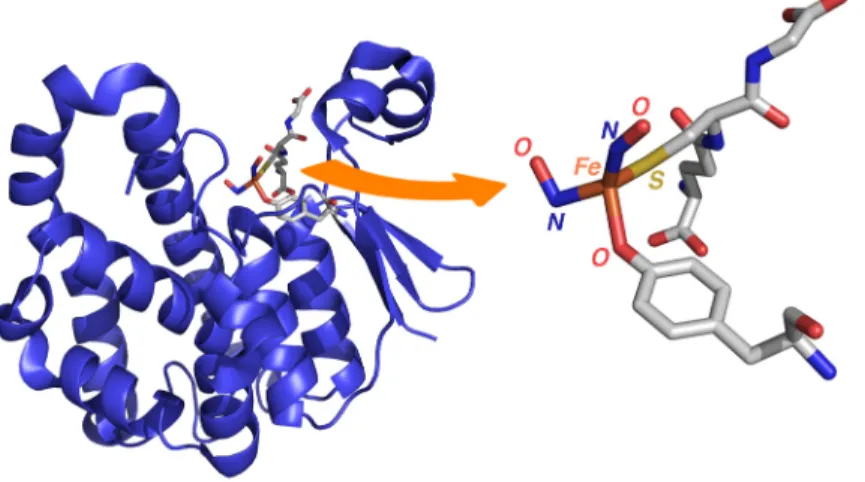
The single example of a crystallographically characterized, protein-bound DNIC contains a tyrosinate ligand (Figure 4).68
EPR spectroscopy is not always capable of distinguishing between potential ligands for the {Fe(NO)2}
9
fragment, and numerous examples of synthetic DNICs containing non-thiol ligands have been reported.69,70
DNICs can exist in equilibrium with dimeric analogs known as Roussin’s red esters (RRE, Chart 1).71
Several factors including solvent polarity, concentration, and the nature of the thiolate ligand can influence which species predominates in solution. Unlike DNICs, RRE derivatives are EPR silent due to antiferromagnetic coupling between the two {Fe(NO)2}
9
units, which gives rise to a diamagnetic ground state. Consequently, identification of these species biologically is more difficult. Both UV-vis and Mössbauer spectroscopy have been employed to aid in the identification of protein-bound RREs,60,72
although assignments are tenuous because many of the spectroscopic properties of the DNIC and RRE are similar.
DNICs display an electrochemically reversible one-electron reduction corresponding to the {Fe(NO)2}
9/10
couple.73
Chemical reduction of thiolate-bound DNICs, however, results in formation of the reduced Roussin’s red ester derivative (rRRE Chart 1).74
Certain rRREs can also exist in equilibrium with the corresponding DNICs by autoxidaion of the coordinating thiolate ligands.75
As with the DNIC-RRE equilibrium, the nature of the solvent and the thiolate ligand is important in determining which species predominates. Not surprisingly, one-electron reduction of RRE derivatives also gives rise to the corresponding rRREs.76-78
Reduced Roussin’s red esters have been detected in nitrosylated protein samples that have been subjected to reduction.60,79
In older reports, this species was sometimes erroneously referred to as a “d9
DNIC”, the prototypical DNIC having been designated as a “d7
DNIC”. Recent work has unambiguously identified the reduced DNIC as a bimetallic species containing antiferromagnetically coupled {Fe(NO)2}
9
−{Fe(NO)2} 10
centers.75
12
rise to an S= ½ ground state that displays an axial EPR signal centered at gav = 1.99. This signal has
been detected when iron-sulfur clusters are exposed to NO, but only after subsequent treatment with a reducing agent. It therefore remains to be determined whether the presence of rRRE in protein samples arises from reduction of pre-existing RREs or from transformation of protein bound DNICs.
Apart from their appearance as products of iron-sulfur cluster nitrosylation, DNICs have garnered a great deal of interest due to their potential role as NO storage and transfer units.80-82
The transfer of nitrosyl ligand(s) between metal centers is not only an intriguing reaction from the standpoint of inorganic chemistry but may also be an important way that biology stores the reactivity of NO for targeted delivery to a remote site of action. Several NO-mediated responses can be turned on with DNICs,83
and formation of dinitrosyl iron complexes has been suggested as a possible means of attenuating NO toxicity in cells.84
DNICs also play an important role in protein nitrosation,85
a reaction that cannot be performed by NO alone.86,87
Dinitrosyl iron complexes therefore represent an interesting bioinorganic cofactor, being both the end products of nitrosylation and mobile units that deliver nitric oxide in a controlled manner.
Mononuclear Fe thiolates
As an entry point into the chemistry of NO with non-heme iron, we chose to examine reactions of iron(II) thiolates.88
We reasoned that the reactivity of these simple complexes with NO would provide a framework with which to understand the chemistry of more complex iron-sulfur clusters. Treatment of [Fe(SR)4]
2−
with NO gas or the conveniently prepared thionitrite, trityl-S-nitrosothiol, leads to formation of the corresponding DNICs, [Fe(NO)2(SR)2]
−. During the course of the reaction, Fe(II) is reduced to
{Fe(NO)2} 9
, which formally contains Fe(I). The necessary reducing equivalent is provided by the thiolate ligand, which is converted to the disulfide. Formation of the DNIC occurs through the intermediacy of a mononitrosyl iron complex (MNIC), [Fe(NO)(SR)3]
−
(Scheme 4). This species, designated as {Fe(NO)}7
in the Enemark-Feltham notation, has an S = 3
/2 ground-state in contrast to
heme-type {Fe(NO)}7
species for which S = 1
13
obtained upon treatment of mammalian ferritin with gaseous NO, suggesting that under appropriate biological conditions these species may form in addition to DNICs.89
Four-coordinate mononitrosyl iron complexes such as [Fe(NO)(SR)3]
−
are rare but have been known for several decades.71,90
Typically, thiolate-bound MNICs are challenging to isolate because of their propensity to disproportionate into DNICs and Fe(III) thiolates.91
If two equivalents of NO+
are used in place of NO, [Fe(SR)4] 2−
transforms to the RRE derivative (Scheme 4). This reaction also occurs via the intermediacy of the MNIC, but results in oxidation of additional equivalents of thiolate.92
Chemistry of the Fe(III) thiolate, [Fe(SR)4] −
, with NO has also been examined and proceeds to the DNIC through the MNIC as with the Fe(II) complex.92
The reactivity of these iron thiolates demonstrates that RS−
ligands are capable of providing the necessary reducing equivalents to transform Fe(II) or Fe(III) to {Fe(NO)2}
9
species.
Fe2S2 clusters
The reaction of synthetic iron-sulfur clusters (both Fe2S2 and Fe4S4) with nitric oxide was first
communicated in 1985.93
These studies demonstrated the propensity for cluster disassembly by NO gas and NO2
−
, but left unanswered the precise nature of the nitrosylation products. Subsequent work from our laboratory94
and elsewhere95
has definitively identified dinitrosyl iron complexes (DNICs) as the products of synthetic iron-sulfur cluster nitrosylation (Scheme 5). This finding appears to be general for a variety of synthetic Fe2S2 and Fe4S4 clusters, and is in good agreement with the large amount of data
obtained from studies of iron-sulfur proteins.
Oxidized versions of synthetic Fe2S2 clusters react in a straightforward manner with NO (g) or
nitrosothiol to generate two equivalents of the respective DNIC and elemental sulfur (eq 1).
(1)
Unlike the reaction of NO with homoleptic iron thiolates, [Fe(SR)4] 2−/1−
, which proceeds with oxidation of the coordinated thiolate, the NO chemistry of Fe2S2 clusters appears to involve modification of only
14
the sulfide ligands. We have not been able to observe any intermediates in this reaction nor have we identified the allotrope of sulfur that is formed. Addition of fewer than four equivalents of NO to solutions of [Fe2S2(SPh)4]
2−
results in a mixture of [Fe(NO)2(SPh)2] −
and unreacted Fe2S2 cluster,
suggesting that cluster disassembly by NO is rapid and that intermediate nitrosylation products are too unstable to observe. The elemental sulfur that is formed by destruction of the Fe2S2 cluster can further
react with the DNIC, resulting in oxidation of the thiolate ligands and formation of the nitrosylated Fe4S3 cluster known as Roussin’s black salt (Chart 1).
91,96
The exact stoichiometry of this reaction is unknown, although a formal balanced equation can be written that accounts for formation of RBS and disulfide (eq 2).
(2)
For Fe2S2 clusters containing aryl thiolates (PhS −
, p-tolylS−
), subsequent oxidation of the thiolate ligands by sulfur is slow and the DNIC can be isolated from the reaction mixture. With alkyl thiolates such as [(SCH2)2-o-C6H4]
2−
(S2-o-xyl 2−
) oxidation is facile and the thiolate ligands are lost as disulfides resulting in formation of [Fe4S3(NO)7]
−
. Trapping the elemental sulfur as the phosphine sulfide can obviate this additional reactivity and allow for isolation of alkyl thiolate-bound DNICs.97
The fate of the sulfide ligands in reactions of biological iron-sulfur clusters with NO remains unknown, although the results with synthetic systems suggest that sulfur byproducts must be sequestered from the vicinity of the iron atoms in order for the DNICs to remain stable. Interestingly, the reverse of cluster nitrosylation has been demonstrated in aerobically growing E. coli cells in the presence of L-cysteine and cysteine
desulfurase (IscS).98
The fact that only sulfur is required for this chemistry is consistent with results using synthetic clusters. Dioxygen may play an important role in these reactions, trapping NO as NO2 or
other nitrogen oxides and effectively removing NO. The synthetic cluster, [Fe2S2(S5)2]
2−, can be
regenerated from the DNIC, [Fe(NO)2(S5)] −
15 serves as a trap for NO.95
Transformation of the synthetic DNIC, [Fe(NO)2(SEt)2] −
, into the corresponding Fe2S2 cluster can also occur via the intermediacy of Roussin’s red salt (Chart 1).
97
Because this chemistry requires additional thiolate equivalents in the form of [Fe(SR)4] −
, it is unlikely to be a true model of Fe2S2 repair in vivo, albeit a most interesting reaction.
Fe4S4 clusters
Reaction pathways of synthetic Fe4S4 clusters with nitric oxide are more difficult to describe in detail
because of the added complexity of the larger {Fe4S4} 2+
core. Reaction of Fe4S4 clusters from
aconitase,79 HiPIP,60 and endonuclease99 with nitric oxide all lead to formation of protein bound DNICs.
As with Fe2S2 clusters, the repair of Fe4S4 clusters by DNICs has been demonstrated in both protein and
synthetic systems.99,100
Our investigations of the reactivity of several synthetic Fe4S4 clusters with
varying equivalents of NO gas or trityl-S-nitrosothiol revealed the thermodynamically stable Roussin’s black salt to be the major product of cluster nitrosylation (Scheme 5).94
Both the corresponding DNIC and the nitrosylated Fe4S4 clusters, [Fe4S4(NO)4]
n−
(n = 1, 2),100,101
were also detected in minor quantities by IR spectroscopy. Whether or not these mononitrosyl iron-sulfur clusters represent intermediates in the conversion of [Fe4S4(SR)4]
2−
to RBS100
or result from alternative reaction pathways is not known. In all cases, the corresponding disulfides were isolated as reaction products, indicating that the thiolate ligands were oxidized in the process. These results stand in contrast to work with Fe4S4 cluster proteins,
where treatment with NO leads to the formation of protein-bound DNICs. However, when the reaction of NO with the prototypical synthetic cluster [Fe4S4(SPh)4]
2−
was carried out in the presence of added PhS−, [Fe(NO)
2(SPh)2]
− was detected as the major iron-containing reaction product. These results again
suggest that at least two thiolate ligands are required to stabilize DNICs and prevent further reactivity in the presence of elemental sulfur. By comparison, when the sulfide-free Fe4 cluster, [Fe4(SPh)10]
2−
, was treated with NO, the DNIC and RRE were formed in a 2:1 ratio (eq 3), consistent with the thiolate ligands providing all of the necessary reducing equivalents.
16
(3)
It therefore appears that in both Fe2S2 and Fe4S4 clusters the sulfide ligands are oxidized during
disassembly of the Fe-S core. Without a means of sequestering the sulfur byproducts, however, the resulting DNICs react further with elemental sulfur to form the thermodynamically stable Roussin’s black salt. RBS is toxic to living cells and without ligand substitution lacks an available site for attachment to proteins.45
It is therefore unlikely that RBS plays an important role in biological iron-sulfur cluster nitrosylation. As observed with HiPIP from C. vinsoum, iron can be lost during nitrosylation of protein-bound Fe4S4 clusters, resulting in a thiolate-to-iron ratio sufficient to stabilize
DNICs against transformation to RBS.
Non-thiolate Fe2S2 clusters
While studying the reactions of synthetic Fe2S2 clusters, we discovered that the abiological cluster,
[Fe2S2Cl4] 2−
, reacts with NO in a fashion identical to that of its [Fe2S2(SR)4] 2−
thiolate analogues (eq 4).102
(4)
This result suggested a commonality in the chemistry of compounds containing the {Fe2S2} 2+
corewith nitric oxide. In a biological context, very little work has been devoted to the reactivity of nitric oxide with iron-sulfur clusters containing non-thiolate ligands.103
Such reactions are of interest because a variety of important Fe2S2 clusters contain at least one non-cysteinate ligand.
104
Furthermore, many potentially ligating amino acid residues other than cysteine lack the capacity for redox chemistry and therefore may display different chemistry in reactions with NO. Based on our results with the thiolate- and chloride-bound Fe2S2 clusters, we hypothesized that the chemistry of other Fe2S2 might be expected
17 cluster, [Fe2(μ-S)2(BIPM)(S2-o-xyl)]
2−
,105
could be nitrosylated to the corresponding N-bound and S-bound DNICs (Scheme 6).102
Although many examples of DNICs coordinated by nitrogen-donor ligands exist, [Fe(NO)2(BIPM)] −
is the first such complex to be prepared by nitrosylation of an iron-sulfur cluster.67,106-110
As for prototypical DNICs, [Fe(NO)2(BIPM)]
−
demonstrates a reversible one-electron reduction in acetonitrile at −1.98 V (vs Fc/Fc+
), assigned to the {Fe(NO)2} 9/10
couple (Figure 5). Unlike DNICs containing thiolate ligands, DNICs containing nitrogen ligands might be expected to undergo redox reactions without decomposition because of a lesser propensity for ligand oxidation. With a related DNIC containing a 2,6-diisopropylphenyl-substituted β-diketiminate ligand we isolated a homologous set of {Fe(NO)2}
9/10
redox partners (Figure 6). These species will facilitate investigations into how changes in the redox state of the DNIC influences both structure and reactivity, particularly in NO transfer processes. Such a redox switch would be a useful means by which nature could control the storage and release of NO equivalents from DNICs.
Conclusions and Outlook
Detailed knowledge of the chemical reactivity of nitric oxide in the biological milieu is required for a complete understanding of its physiology and pathology. We have explored this reactivity by identifying where NO is produced and how it is utilized in cells using fluorescent probes and, in biomimetic chemistry, by investigating the fundamental inorganic chemistry of NO with iron-sulfur protein active site models. This dual approach is well suited to the study of small molecules of biological interest like nitric oxide, and we envision that it may also be successful in studies of other such species including peroxynitrite, nitroxyl, carbon monoxide, and hydrogen sulfide.
ACKNOWLEDGMENT
18
Invitrogen. Z.J.T. thanks the National Institute of General Medical Science for postdoctoral fellowship support (F32 GM082031-03).
19 REFERENCES
(1) Eisenberg, R.; Meyer, C. D. Acc. Chem. Res. 1975, 8, 26-34. (2) Furchgott, R. F.; Vanhoutte, P. M. FASEB J. 1989, 3, 2007-2018.
(3) Ignarro, L. J.; Buga, G. M.; Wood, K. S.; Byrns, R. E.; Chaudhuri, G. Proc. Natl. Acad. Sci.
U.S.A. 1987, 84, 9265-9269.
(4) Rapoport, R. M.; Draznin, M. B.; Murad, F. Nature 1983, 306, 174-176.
(5) Cary, S. P. L.; Winger, J. A.; Derbyshire, E. R.; Marletta, M. A. Trends Biochem. Sci. 2006, 31, 231-239.
(6) Nitric Oxide: Biology and Pathobiology; Ignarro, L. J., Ed.; Academic Press: San Diego, 2000.
(7) Hetrick, E. M.; Schoenfisch, M. H. Ann. Rev. Anal. Chem. 2009, 2, 409-433.
(8) Gabe, Y.; Ueno, T.; Urano, Y.; Kojima, H.; Nagano, T. Anal. Bioanal. Chem. 2006, 386, 621-626.
(9) Gabe, Y.; Urano, Y.; Kikuchi, K.; Kojima, H.; Nagano, T. J. Am. Chem. Soc. 2004, 126, 3357-3367.
(10) Izumi, S.; Urano, Y.; Hanaoka, K.; Terai, T.; Nagano, T. J. Am. Chem. Soc. 2009, 131, 10189-10200.
(11) Kojima, H.; Hirotani, M.; Nakatsubo, N.; Kikuchi, K.; Urano, Y.; Higuchi, T.; Hirata, Y.; Nagano, T. Anal. Chem. 2001, 73, 1967-1973.
(12) Kojima, H.; Nakatsubo, N.; Kikuchi, K.; Kawahara, S.; Kirino, Y.; Nagoshi, H.; Hirata, Y.; Nagano, T. Anal. Chem. 1998, 70, 2446-2453.
(13) Sasaki, E.; Kojima, H.; Nishimatsu, H.; Urano, Y.; Kikuchi, K.; Hirata, Y.; Nagano, T. J. Am.
Chem. Soc. 2005, 127, 3684-3685.
(14) Nagano, T.; Yoshimura, T. Chem. Rev. 2002, 102, 1235-1269. (15) Lim, M. H.; Lippard, S. J. Acc. Chem. Res. 2007, 40, 41-51.
(16) Hilderbrand, S. A.; Lippard, S. J. Inorg. Chem. 2004, 43, 5294-5301.
(17) Soh, N.; Imato, T.; Kawamura, K.; Maeda, M.; Katayama, Y. Chem. Commun. 2002, 2650-2651. (18) Lim, M. H.; Lippard, S. J. Inorg. Chem. 2004, 43, 6366-6370.
(19) Hilderbrand, S. A.; Lim, M. H.; Lippard, S. J. J. Am. Chem. Soc. 2004, 126, 4972-4978. (20) Smith, R. C.; Tennyson, A. G.; Lippard, S. J. Inorg. Chem. 2006, 45, 6222-6226. (21) Ford, P. C.; Fernandez, B. O.; Lim, M. D. Chem. Rev. 2005, 105, 2439-2455. (22) Lim, M. H.; Lippard, S. J. J. Am. Chem. Soc. 2005, 127, 12170-12171. (23) Lim, M. H.; Lippard, S. J. Inorg. Chem. 2006, 45, 8980-8989.
(24) Smith, R. C.; Tennyson, A. G.; Lim, M. H.; Lippard, S. J. Org. Lett. 2005, 7, 3573-3575. (25) Smith, R. C.; Tennyson, A. G.; Won, A. C.; Lippard, S. J. Inorg. Chem. 2006, 45, 9367-9373. (26) Xing, C.; Yu, M.; Wang, S.; Shi, Z.; Li, Y.; Zhu, D. Macromol. Rapid Commun. 2007, 28,
241-245.
(27) Do, L.; Smith, R. C.; Tennyson, A. G.; Lippard, S. J. Inorg. Chem. 2006, 45, 8998-9005. (28) Franz, K. J.; Singh, N.; Spingler, B.; Lippard, S. J. Inorg. Chem. 2000, 39, 4081-4092. (29) Hilderbrand, S. A.; Lippard, S. J. Inorg. Chem. 2004, 43, 4674-4682.
(30) Lim, M. H.; Kuang, C.; Lippard, S. J. Chembiochem 2006, 7, 1571-1576.
(31) Lim, M. H.; Wong, B. A.; Pitcock, W. H.; Mokshagundam, D.; Baik, M. H.; Lippard, S. J. J.
Am. Chem. Soc. 2006, 128, 14364-14373.
(32) Lim, M. H.; Xu, D.; Lippard, S. J. Nat. Chem. Biol. 2006, 2, 375-380.
(33) Tsuge, K.; DeRosa, F.; Lim, M. D.; Ford, P. C. J. Am. Chem. Soc. 2004, 126, 6564-6565. (34) Khin, C.; Lim, M. D.; Tsuge, K.; Iretskii, A.; Wu, G.; Ford, P. C. Inorg. Chem. 2007, 46,
9323-9331.
(35) Ouyang, J.; Hong, H.; Shen, C.; Zhao, Y.; Ouyang, C.; Dong, L.; Zhu, J.; Guo, Z.; Zeng, K.; Chen, J.; Zhang, C.; Zhang, J. Free Radical Biol. Med. 2008, 45, 1426-1436.
(36) Mankad, N. P.; Antholine, W. E.; Szilagyi, R. K.; Peters, J. C. J. Am. Chem. Soc. 2009, 131, 3878-3880.
20
(38) Wink, D. A.; Vodovotz, Y.; Laval, J.; Laval, F.; Dewhirst, M. W.; Mitchell, J. B.
Carcinogenesis 1998, 19, 711-721.
(39) Stuehr, D. J. Biochim. Biophys. Acta-Bioenerg. 1999, 1411, 217-230.
(40) MacMicking, J.; Xie, Q. W.; Nathan, C. Ann. Rev. Immunol. 1997, 15, 323-350.
(41) Agapie, T.; Suseno, S.; Woodward, J. J.; Stoll, S.; Britt, R. D.; Marletta, M. A. Proc. Natl. Acad.
Sci. U.S.A. 2009, 106, 16221-16226.
(42) Gusarov, I.; Starodubtseva, M.; Wang, Z. Q.; McQuade, L.; Lippard, S. J.; Stuehr, D. J.; Nudler, E. J. Biol. Chem. 2008, 283, 13140-13147.
(43) Shatalin, K.; Gusarov, I.; Avetissova, E.; Shatalina, Y.; McQuade, L. E.; Lippard, S. J.; Nudler, E. Proc. Natl. Acad. Sci. U.S.A. 2008, 105, 1009-1013.
(44) Gusarov, I.; Nudler, E. Proc. Natl. Acad. Sci. U.S.A. 2005, 102, 13855-13860. (45) Butler, A. R.; Megson, I. L. Chem. Rev. 2002, 102, 1155-1165.
(46) Strube, K.; de Vries, S.; Cramm, R. J. Biol. Chem. 2007, 282, 20292-20300. (47) D'Autréaux, B.; Tucker, N. P.; Dixon, R.; Spiro, S. Nature 2005, 437, 769-772. (48) Gonzalez, D.; Drapier, J.-C.; Bouton, C. J. Biol. Chem. 2004, 279, 43345-43351.
(49) D'Autréaux, B.; Horner, O.; Oddou, J.-L.; Jeandey, C.; Gambarelli, S.; Berthomieu, C.; Latour, J.-M.; Michaud-Soret, I. J. Am. Chem. Soc. 2004, 126, 6005-6016.
(50) D'Autréaux, B.; Touati, D.; Bersch, B.; Latour, J.-M.; Michaud-Soret, I. Proc. Natl. Acad. Sci.
U.S.A. 2002, 99, 16619-16624.
(51) Ding, H.; Demple, B. Proc. Natl. Acad. Sci. U.S.A. 2000, 97, 5146-5150. (52) Bouton, C.; Raveau, M.; Drapier, J.-C. J. Biol. Chem. 1996, 271, 2300-2306.
(53) Toledo, J. C., Jr.; Bosworth, C. A.; Hennon, S. W.; Mahtani, H. A.; Bergonia, H. A.; Lancaster, J. R., Jr. J. Biol. Chem. 2008, 283, 28926-28933.
(54) Watts, R. N.; Hawkins, C.; Ponka, P.; Richardson, D. R. Proc. Natl. Acad. Sci. U.S.A. 2006, 103, 7670-7675.
(55) Watts, R. N.; Richardson, D. R. Eur. J. Biochem. 2002, 269, 3383-3392.
(56) Asanuma, K.; Iijima, K.; Ara, N.; Koike, T.; Yoshitake, J.; Ohara, S.; Shimosegawa, T.; Yoshimura, T. Nitric Oxide 2007, 16, 395-402.
(57) Bogdan, C. Nat. Immunol. 2001, 2, 907-916. (58) Drapier, J.-C. Methods 1997, 11, 319-329.
(59) Duan, X.; Yang, J.; Ren, B.; Tan, G.; Ding, H. Biochem. J. 2009, 417, 783-789. (60) Foster, M. W.; Cowan, J. A. J. Am. Chem. Soc. 1999, 121, 4093-4100.
(61) Kiley, P. J.; Beinert, H. Curr. Opin. Microbiol. 2003, 6, 181-185.
(62) Szacilowski, K.; Chmura, A.; Stasicka, Z. Coord. Chem. Rev. 2005, 249, 2408-2436. (63) Roussin, M. L. Ann. Chim. Phys. 1858, 52, 285-303.
(64) Enemark, J. H.; Feltham, R. D. Coord. Chem. Rev. 1974, 13, 339-406.
(65) Vanin, A. F.; Serezhenkov, V. A.; Mikoyan, V. D.; Genkin, M. V. Nitric Oxide 1998, 2, 224-234.
(66) Commoner, B.; Ternberg, J. L. Proc. Natl. Acad. Sci. U.S.A. 1961, 47, 1374-1384. (67) Tsai, M.-L.; Hsieh, C.-H.; Liaw, W.-F. Inorg. Chem. 2007, 46, 5110-5117.
(68) Cesareo, E.; Parker, L. J.; Pedersen, J. Z.; Nuccetelli, M.; Mazzetti, A. P.; Pastore, A.; Federici, G.; Caccuri, A. M.; Ricci, G.; Adams, J. J.; Parker, M. W.; Bello, M. L. J. Biol. Chem. 2005,
280, 42172-42180.
(69) Bryar, T. R.; Eaton, D. R. Can. J. Chem. 1992, 70, 1917-1926.
(70) Woolum, J. C.; Tiezzi, E.; Commoner, B. Biochim. Biophys. Acta-Protein Struct. 1968, 160, 311-320.
(71) Butler, A. R.; Glidewell, C.; Li, M.-H. Adv. Inorg. Chem. 1988, 32, 335-393.
(72) Lobysheva, I. I.; Serezhenkov, V. A.; Stucan, R. A.; Bowman, M. K.; Vanin, A. F. Biochemistry
(Moscow) 1997, 62, 801-808.
(73) Crayston, J. A.; Glidewell, C.; Lambert, R. J. Polyhedron 1990, 9, 1741-1746. (74) Tsou, C.-C.; Lu, T.-T.; Liaw, W.-F. J. Am. Chem. Soc. 2007, 129, 12626-12627.
21
(75) Lu, T.-T.; Tsou, C.-C.; Huang, H.-W.; Hsu, I.-J.; Chen, J.-M.; Kuo, T.-S.; Wang, Y.; Liaw, W.-F. Inorg. Chem. 2008, 47, 6040-6050.
(76) Chau, C.-N.; Wojcicki, A. Polyhedron 1992, 11, 851-852. (77) Chau, C.-N.; Wojcicki, A. Polyhedron 1993, 12, 1261-1263. (78) Glidewell, C.; Lambert, R. J. W. Polyhedron 1992, 11, 2803-2804.
(79) Kennedy, M. C.; Antholine, W. E.; Beinert, H. J. Biol. Chem. 1997, 272, 20340-20347. (80) Vanin, A. F. Biochemistry (Moscow) 1998, 63, 782-793.
(81) Ueno, T.; Suzuki, Y.; Fujii, S.; Vanin, A. F.; Yoshimura, T. Biochem. Pharmacol. 2002, 63, 485-493.
(82) Chen, Y.-J.; Ku, W.-C.; Feng, L.-T.; Tsai, M.-L.; Hsieh, C.-H.; Hsu, W.-H.; Liaw, W.-F.; Hung, C.-H.; Chen, Y.-J. J. Am. Chem. Soc. 2008, 130, 10929-10938.
(83) Vanin, A. F. Nitric Oxide 2009, 21, 1-13.
(84) Kim, Y.-M.; Chung, H.-T.; Simmons, R. L.; Billiar, T. R. J. Biol. Chem. 2000, 275, 10954-10961.
(85) Foster, M. W.; Liu, L.; Zeng, M.; Hess, D. T.; Stamler, J. S. Biochemistry 2009, 48, 792-799. (86) Boese, M.; Mordvintcev, P. I.; Vanin, A. F.; Busse, R.; Mülsch, A. J. Biol. Chem. 1995, 270,
29244-29249.
(87) Bosworth, C. A.; Toledo, J. C., Jr.; Zmijewski, J. W.; Li, Q.; Lancaster, J. R., Jr. Proc. Natl.
Acad. Sci. U.S.A. 2009, 106, 4671-4676.
(88) Harrop, T. C.; Song, D.; Lippard, S. J. J. Am. Chem. Soc. 2006, 128, 3528-3529. (89) Lee, M.; Arosio, P.; Cozzi, A.; Chasteen, N. D. Biochemistry 1994, 33, 3679-3687. (90) Connelly, N. G.; Gardner, C. J. Chem. Soc., Dalton Trans. 1976, 1525-1527.
(91) Butler, A. R.; Glidewell, C.; Hyde, A. R.; Walton, J. C. Polyhedron 1985, 4, 797-809. (92) Lu, T.-T.; Chiou, S.-J.; Chen, C.-Y.; Liaw, W.-F. Inorg. Chem. 2006, 45, 8799-8806.
(93) Butler, A. R.; Glidewell, C.; Hyde, A. R.; Walton, J. C. Inorg. Chim. Acta 1985, 106, L7-L8. (94) Harrop, T. C.; Tonzetich, Z. J.; Reisner, E.; Lippard, S. J. J. Am. Chem. Soc. 2008, 130,
15602-15610.
(95) Tsai, F.-T.; Chiou, S.-J.; Tsai, M.-C.; Tsai, M.-L.; Huang, H.-W.; Chiang, M.-H.; Liaw, W.-F.
Inorg. Chem. 2005, 44, 5872-5881.
(96) Chen, T.-N.; Lo, F.-C.; Tsai, M.-L.; Shih, K.-N.; Chiang, M.-H.; Lee, G.-H.; Liaw, W.-F. Inorg.
Chim. Acta 2006, 359, 2525-2533.
(97) Lu, T.-T.; Huang, H.-W.; Liaw, W.-F. Inorg. Chem. 2009, 48, 9027-9035. (98) Yang, W.; Rogers, P. A.; Ding, H. J. Biol. Chem. 2002, 277, 12868-12873. (99) Rogers, P. A.; Eide, L.; Klungland, A.; Ding, H. DNA Repair 2003, 2, 809-817.
(100) Tsou, C.-C.; Lin, Z.-S.; Lu, T.-T.; Liaw, W.-F. J. Am. Chem. Soc. 2008, 130, 17154-17160. (101) Ting-Wah Chu, C.; Yip-Kwai Lo, F.; Dahl, L. F. J. Am. Chem. Soc. 1982, 104, 3409-3422. (102) Tonzetich, Z. J.; Do, L. H.; Lippard, S. J. J. Am. Chem. Soc. 2009, 131, 7964-7965.
(103) Welter, R.; Yu, L.; Yu, C.-A. Arch. Biochem. Biophys. 1996, 331, 9-14. (104) Beinert, H.; Holm, R. H.; Münck, E. Science 1997, 277, 653-659.
(105) Ballmann, J.; Albers, A.; Demeshko, S.; Dechert, S.; Bill, E.; Bothe, E.; Ryde, U.; Meyer, F.
Angew. Chem. Int. Ed. Engl. 2008, 47, 9537-9541.
(106) Reginato, N.; McCrory, C. T. C.; Pervitsky, D.; Li, L. J. Am. Chem. Soc. 1999, 121, 10217-10218.
(107) Wang, X.; Sundberg, E. B.; Li, L.; Kantardjieff, K. A.; Herron, S. R.; Lim, M.; Ford, P. C.
Chem. Commun. 2005, 477-479.
(108) Hung, M.-C.; Tsai, M.-C.; Lee, G.-H.; Liaw, W.-F. Inorg. Chem. 2006, 45, 6041-6047. (109) Tsai, M.-L.; Liaw, W.-F. Inorg. Chem. 2006, 45, 6583-6585.
23
Figure 1. (a) CuFL1 detection of NO in SK-N-SH neuroblastoma cells. [CuFL1] = 1 μM,
[17β-estradiol] = 100 nM. From left to right: 25 min exposure to CuFL1, no 17β-estradiol; 5 min; 10 min; 15 min; 25 min exposure to CuFL1 and 17β-estradiol. Top: Fluorescence images; Bottom: DIC images. Scale bars = 50 μm. (b) CuFL1 detection of NO in Raw 264.7 murine macrophages. [CuFL1] = 1 μM, [lipopolysaccharide (LPS)] = 500 ng/mL, [interferon- γ (IFN-γ)] = 250 U/mL. From left to right: 12 h exposure to CuFL1, no LPS/IFN-γ; 6 h; 8 h; 10 h; 12 h exposure to CuFL1 and LPS/IFN-γ. Top: Fluorescence images; Bottom: DIC images. Scale bars = 50 μm. For additional details see ref. 32
24 E (eV)
a.
-2 -3 -4 -5 -6 -7 LUMO (-2.725) HOMO (-4.593) HOMO-1 (-4.665) N R1 H R2 E LUMOb.
HOMO (-4.582) HOMO-14 (-6.400) R1 N N R2 O LUMO (-2.648) HOMO N-lone pair hν quenching N R1 H R2 N R1 H R2 N R1 H R2Figure 2. (a) Relative energies of the molecular orbitals for the ground states of FL1 (left) and FL1-NO
(right). (b) Qualitative molecular orbital diagram for the ground (left), excited (middle), and charge-transfer (right) states of FL1. R1 = 2-methylquinoline; R2 = fluorophore. For additional details see ref. 31.
25
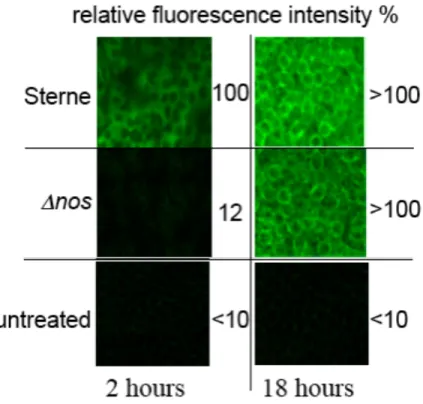
Figure 3. Visualization of fluorescence enhancement by CuFL1 in J774A.1 infected macrophages at 2
and 18 h post-infection. Top row: bNOS-expressing B. anthracis (Sterne) cells taken up by macrophages. Sterne cells produce NO using bNOS within 2 h of uptake. The host macrophages produce NO using iNOS between 2 and 18 h. Middle row: bNOS-deficient B. anthracis cells taken up by macrophages. No fluorescence is observed after 2 h because these bacteria cannot generate NO. The host macrophages produce NO using iNOS as usual. Bottom row: Control, no bacteria and no induction of iNOS
26
Figure 4. Structure of human glutathione S-transferase P1-1 monomer containing a bound dinitrosyl
27
Figure 5. Cyclic voltammogram of (Et4N)[Fe(NO)2(BIPM)] displaying the reversible {Fe(NO)2} 9/10
couple. Conditions: 3 mM in CH3CN; glassy carbon electrode; 0.1 M Bu4NPF6 electrolyte; 100 mV/s
28
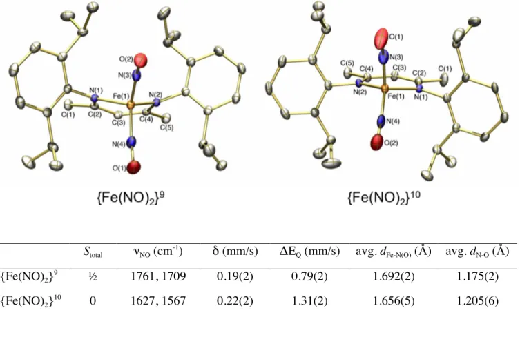
Stotal νNO (cm -1
) δ (mm/s) ΔEQ (mm/s) avg. dFe-N(O) (Å) avg. dN-O (Å)
{Fe(NO)2} 9 ½ 1761, 1709 0.19(2) 0.79(2) 1.692(2) 1.175(2) {Fe(NO)2} 10 0 1627, 1567 0.22(2) 1.31(2) 1.656(5) 1.205(6)
Figure 6. Comparison of selected spectroscopic and structural properties for two homologous DNIC
redox partners. Thermal ellipsoids are drawn at 50% probability. Hydrogen atoms and the PPN+
cation in the {Fe(NO)2}
10
29
Scheme 1.
30
31
32
33
34
Chart 1. Common {Fe(NO)2}
35 TOC GRAPHIC
SYNOPSIS: A dual strategy for investigating the chemistry of nitric oxide as a biological signaling agent is described. In one approach, metal-based fluorescent sensors for the detection of NO in living cells are devised and evaluated. In the other approach, synthetic model complexes of the active sites of iron-sulfur proteins are employed to probe their reactivity toward nitric oxide.
![Figure 1. (a) CuFL1 detection of NO in SK-N-SH neuroblastoma cells. [CuFL1] = 1 μM, [17β- [17β-estradiol] = 100 nM](https://thumb-eu.123doks.com/thumbv2/123doknet/14723866.571233/24.918.271.652.146.495/figure-cufl-detection-sk-neuroblastoma-cells-cufl-estradiol.webp)

![Figure 5. Cyclic voltammogram of (Et 4 N)[Fe(NO) 2 (BIPM)] displaying the reversible {Fe(NO) 2 } 9/10 couple](https://thumb-eu.123doks.com/thumbv2/123doknet/14723866.571233/28.918.189.728.123.739/figure-cyclic-voltammogram-et-bipm-displaying-reversible-couple.webp)