HAL Id: inserm-01480026
https://www.hal.inserm.fr/inserm-01480026
Submitted on 26 Sep 2017
HAL is a multi-disciplinary open access
archive for the deposit and dissemination of
sci-entific research documents, whether they are
pub-lished or not. The documents may come from
teaching and research institutions in France or
abroad, or from public or private research centers.
L’archive ouverte pluridisciplinaire HAL, est
destinée au dépôt et à la diffusion de documents
scientifiques de niveau recherche, publiés ou non,
émanant des établissements d’enseignement et de
recherche français ou étrangers, des laboratoires
publics ou privés.
Distributed under a Creative Commons Attribution - ShareAlike| 4.0 International
Host-related factors explaining interindividual
variability of carotenoid bioavailability and tissue
concentrations in humans
Torsten Bohn, Charles Desmarchelier, Lars Dragsted, Charlotte Nielsen,
Wilhelm Stahl, Ralph Rühl, Jaap Keijer, Patrick Borel
To cite this version:
Torsten Bohn, Charles Desmarchelier, Lars Dragsted, Charlotte Nielsen, Wilhelm Stahl, et al..
Host-related factors explaining interindividual variability of carotenoid bioavailability and tissue
concen-trations in humans. Molecular Nutrition and Food Research, Wiley-VCH Verlag, 2017, 61 (6), Epub
ahead of print. �10.1002/mnfr.201600685�. �inserm-01480026�
R
EVIEWHost-related factors explaining interindividual variability
of carotenoid bioavailability and tissue concentrations
in humans
Torsten Bohn
1, Charles Desmarchelier
2, Lars O. Dragsted
3, Charlotte S. Nielsen
3,
Wilhelm Stahl
4, Ralph R ¨uhl
5,6, Jaap Keijer
7and Patrick Borel
21Luxembourg Institute of Health, Strassen, Luxembourg
2NORT, Aix-Marseille Universit ´e, INRA, INSERM, Marseille, France
3Department of Nutrition, Exercise and Sports, University of Copenhagen, Frederiksberg C, Denmark
4Institute of Biochemistry and Molecular Biology I, Heinrich-Heine-University D ¨usseldorf, D ¨usseldorf, Germany 5Paprika Bioanalytics BT, Debrecen, Hungary
6MTA-DE Public Health Research Group of the Hungarian Academy of Sciences, Faculty of Public Health,
University of Debrecen, Debrecen, Hungary
7Human and Animal Physiology, Wageningen University, Wageningen, The Netherlands
Received: August 5, 2016 Revised: December 19, 2016 Accepted: January 4, 2017
Carotenoid dietary intake and their endogenous levels have been associated with a decreased risk of several chronic diseases. There are indications that carotenoid bioavailability depends, in addition to the food matrix, on host factors. These include diseases (e.g. colitis), life-style habits (e.g. smoking), gender and age, as well as genetic variations including single nucleotide polymorphisms that govern carotenoid metabolism. These are expected to explain interindivid-ual differences that contribute to carotenoid uptake, distribution, metabolism and excretion, and therefore possibly also their association with disease risk. For instance, digestion enzymes fostering micellization (PNLIP, CES), expression of uptake/efflux transporters (SR-BI, CD36, NPC1L1), cleavage enzymes (BCO1/2), intracellular transporters (FABP2), secretion into chy-lomicrons (APOB, MTTP), carotenoid metabolism in the blood and liver (LPL, APO C/E, LDLR), and distribution to target tissues such as adipose tissue or macula (GSTP1, StARD3) depend on the activity of these proteins. In addition, human microbiota, e.g. via altering bile-acid concentrations, may play a role in carotenoid bioavailability. In order to comprehend individ-ual, variable responses to these compounds, an improved knowledge on intra-/interindividual factors determining carotenoid bioavailability, including tissue distribution, is required. Here, we highlight the current knowledge on factors that may explain such intra-/interindividual differences.
Keywords:
Absorption / Biodistribution / Genetic polymorphisms / Intestine / Macula lutea
Correspondence: Torsten Bohn E-mail: torsten.bohn@gmx.ch
Abbreviations: ADME, absorption, distribution, metabolism and
excretion; AMD, age related macular degeneration; AMY1, sali-vary amylase gene 1; ABCA1, ATP binding cassette ily A, member 1; ABCG5/G8, ATP binding cassette subfam-ily G, member 5/8; ADH7, Alcohol dehydrogenase 7; ALDH1, aldehyde dehydrogenase 1; APOA, 1–4, apolipoprotein A, 1– 4; APOB/C2/E/48, apolipoprotein B/C2/E/48; AUC, area under (plasma/serum concentration-time) curve; BCO1/2, -carotene oxygenase 1/2; CD36, cluster of differentiation 36 molecule;
CES1/2, human carboxyl-esterase 1/2; CETP, cholesteryl
es-ter transfer protein; CLPS, colipase; COBLL1, cordon-bleu WH2 repeat protein like 1; 9CRA, cis-retinoic acid; 9CDHRA,
9-cis-13, 14-dihydro-retinoic acid; CRISPR/CAS9, clustered
reg-ularly interspaced short palindromic repeats/protein-9 nucle-ase; CXCL8, C-X-C motif chemokine ligand 8; CYP26B1, cy-tochrome P450 family 26 subfamily B member 1; CYP7A1, bile acid synthetic enzyme; ELOVL2, elongation of very long chain fatty acids like 2; GI, gastro-intestinal; GPS, protein path-way suppressor; GSTP1, glutathione S-transferase pi 1; HNF4A, hepatocyte nuclear factor 4, alpha; FABP2/I-FABP, fatty acid binding protein, intestinal; FGF4/19, fibroblast growth factor 4/19; FOXO1, forkhead box O1; FXR, farnesoid X receptor; IL8, interleukin 8; INSIG2, insulin induced gene 2; IRS1, insulin
1
Introduction
Carotenoids are natural pigments with a C-30 or C-40 backbone. They can be produced by most plants, bacteria, and fungi, but not by animals or humans, making diet their sole source. Carotenoids have recently been investigated with much interest, as their dietary intake and endogenous con-centrations have been associated with a reduced risk of several chronic diseases. For example, carotenoid intake has been positively associated with a reduced risk of cancer [1], type 2 diabetes mellitus (T2D) [2], cardiovascular diseases [3], and asthma [4], while plasma carotene concentration was shown to be significantly associated with reduced total mortality [5]. In addition, some carotenoids, including␣-, -carotene and -cryptoxanthin (Fig. 1), are vitamin A precursors, constitut-ing the predominant source of vitamin A in most developconstitut-ing countries (up to 90% [6]) as well as in Western countries es-pecially with respect to vegetarians. Recently, it has also been suggested that cis-carotenoids are even more beneficial for the prevention of atherosclerosis and T2D than their all-trans isomers [7, 8]. Finally, it is now acknowledged that lutein and zeaxanthin play a role in vision by improving contrast sensi-tivity and visual acuity [9] and participate in the prevention of age-related macular degeneration [10].
However, several intervention trials with carotenoid sup-plements have not supported these beneficial associations, or even found negative health effects [11, 12]. To explain this discrepancy, it is hypothesized that the food matrix
(miss-receptor substrate 1; ISX, intestine specific homeobox; KD, equilibrium dissociation constant; LCAT, lecithin-cholesterol acyl-transferase; LDLR, low density lipoprotein receptor; LIPC, pase C, hepatic type; LIPF, gastric lipase; LPL, lipoprotein li-pase; LRAT, lecithin-retinol acyltransferase; LRP1, low den-sity lipoprotein receptor-related protein 1; LXR, liver X re-ceptor; MC4R, melanocortin 4 rere-ceptor; MTTP/MTP, microso-mal triglyceride transfer protein/gene; NF-B, nuclear factor kappa-B; NRF2/NFE2L2, nuclear factor (erythroid-derived 2) like 2; NPC1L1, NPC1 like intracellular cholesterol transporter 1; PGA3/4/5, pepsinogen3/4/5; PGC, progastricsin; PGC1␣, per-oxisome proliferator-activated receptor gamma coactivator 1-alpha; PKD1L2, polycystin 1 like 2; PLRP2, pancreatic lipase-related protein-2; PNLIP, pancreatic lipase; PPAR, peroxisome proliferator-activated receptor; PXR, pregnane X receptor; RAR, retinoic acid receptor; RBP1/3/4, retinol binding protein 1/3/4;
RPE65, retinal pigment epithelium specific protein 65kDa; RSD,
relative standard deviation (RSD= SD/mean), equal to CV (co-efficient of variation); RXR, retinoid X receptor; RXRA, retinoid X receptor alpha; SAR1B, secretion associated Ras related GT-Pase 1B; SR-BI/SCARB1, scavenger receptor class B member 1, protein/gene; SHP, short heterodimer partner; SNP, single nu-cleotide polymorphism; SETD7, SET domain containing lysine methyltransferase 7; SLC27A6, solute carrier family 27 (fatty acid transporter), member 6; SOD2, superoxide dismutase 2, mito-chondrial; StARD3, StAR related lipid transfer domain containing 3; STRA6, stimulated by retinoic acid gene 6 protein homolog;
T2D, type II diabetes mellitus; TCF7L2, transcription factor 7 like
2; TRL, triacylglycerol-rich lipoprotein fraction; WT, wild-type
Colour online: See the article online to view Fig. 2 in colour.
Figure 1. Predominant carotenoids in our diet, common
metabo-lites and nomenclature.
ing synergistic effects, e.g. with other antioxidants such as polyphenols), a larger array of natural occurring carotenoids compared to single carotenoids in high amounts, presenta-tion in the form of carotenoid supplements (powder, solid matrix) and continuous intake in case of supplements, in-fluence absorption, distribution, metabolism and excretion (ADME), and therefore also their bioactivity. However, it has also been emphasized that ADME-related factors, including digestion and matrix release, solubilisation in mixed micelles, epithelial uptake in the (small) intestine, and further bio-distribution, all prerequisites for exerting potential biologi-cal effects, can be different between individuals. This likely results in variable blood/tissue concentrations [13–15]. How-ever, blood plasma/serum alone may not constitute the best indicator to assess carotenoid status, and additional methods, such as isotopic labelling, similar as for retinoids [16] or easy accessible compartments such as white blood cells [17] or buccal cells [18], may allow for more insights regarding en-dogenous carotenoid levels, carotenoid compartments, and turnover [19].
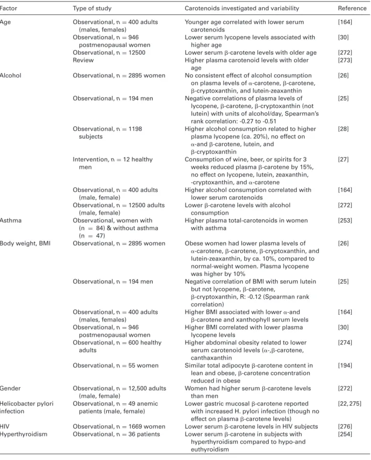
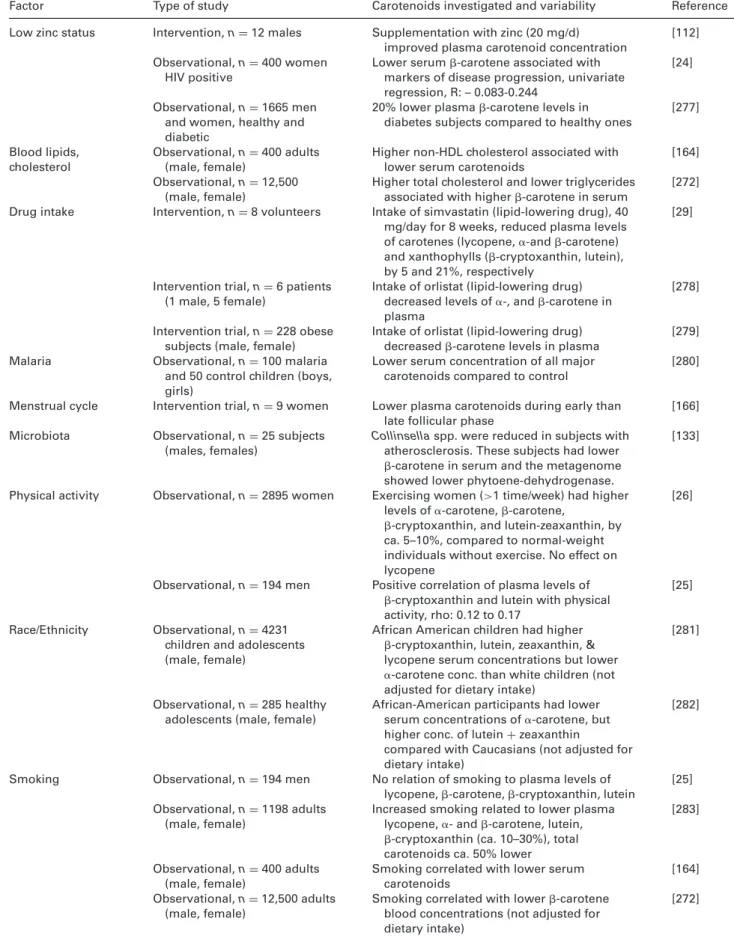
This intra- and interindividual variability can be attributed, in addition to dietary habits [20, 21], to host-related factors (Table 1) including disease state [22–24], possibly physical activity [25, 26], being overweight/obese [26], alcohol use [25, 27, 28], smoking habits [26], drug intake [29], age [30], and genetic aspects [31, 32]. However, the underlying mecha-nisms for this variability – e.g. lower bioaccessibility, reduced
Table 1. Overview of host (non-dietary) factors proposed to influence (in addition to genetic make-up and malabsorption diseases of the
GI) intra-and interindividual differences regarding carotenoid ADME
Factor Type of study Carotenoids investigated and variability Reference Age Observational, n= 400 adults
(males, females)
Younger age correlated with lower serum carotenoids
[164] Observational, n= 946
postmenopausal women
Lower serum lycopene levels associated with higher age
[30] Observational, n= 12500 Lower serum-carotene levels with older age [272] Review Higher plasma carotenoid levels with older
age
[273] Alcohol Observational, n= 2895 women No consistent effect of alcohol consumption
on plasma levels of␣-carotene, -carotene, -cryptoxanthin, and lutein-zeaxanthin
[26]
Observational, n= 194 men Negative correlations of plasma levels of lycopene,-carotene, -cryptoxanthin (not lutein) with units of alcohol/day, Spearman’s rank correlation: -0.27 to -0.51
[25]
Observational, n= 1198 subjects
Higher alcohol consumption related to higher plasma lycopene (ca. 20%), no effect on ␣-and -carotene, lutein, and
-cryptoxanthin
[28]
Intervention, n= 12 healthy men
Consumption of wine, beer, or spirits for 3 weeks reduced plasma-carotene by 15%, no effect on lycopene, lutein, zeaxanthin, -cryptoxanthin, and␣-carotene
[27]
Observational, n= 400 adults (male, female)
Higher alcohol consumption correlated with lower serum carotenoids
[164] Observational, n= 12500 adults
(male, female)
Lower-carotene levels with alcohol consumption
[272] Asthma Observational, women with
(n = 84) & without asthma (n = 47)
Higher plasma total-carotenoids in women with asthma
[253]
Body weight, BMI Observational, n= 2895 women Obese women had lower plasma levels of ␣-carotene, -carotene, -cryptoxanthin, and lutein-zeaxanthin, by ca. 10%, compared to normal-weight women. Plasma lycopene was higher by 10%
[26]
Observational, n= 194 men Negative correlation of BMI with serum lutein but not lycopene,-carotene,
-cryptoxanthin, R: -0.12 (Spearman rank correlation)
[25]
Observational, n= 400 adults (males, females)
Higher BMI associated with lower␣-and -carotene and xanthophyll serum levels
[164] Observational, n= 946
postmenopausal women
Higher BMI correlated with lower plasma lycopene levels
[30] Observational, n= 600 healthy
adults
Higher abdominal obesity related to lower serum carotenoid levels (␣-,-carotene, canthaxanthin
[274]
Observational, n= 55 women Similar total adipocyte-carotene content in lean and obese,-carotene concentration reduced in obese
[194]
Gender Observational, n= 12,500 adults (male, female)
Women had higher serum-carotene levels than men
[272] Helicobacter pylori
infection
Observational, n= 49 anemic patients (male, female)
Lower gastric mucosal-carotene reported with increased H. pylori infection (though no effect on plasma-carotene levels)
[22, 275]
HIV Observational, n= 1669 women Lower serum-carotene levels in HIV subjects [276] Hyperthyroidism Observational, n= 36 patients Lower serum-carotene in subjects with
hyperthyroidism compared to hypo-and euthyroidism
Table 1. Continued
Factor Type of study Carotenoids investigated and variability Reference Low zinc status Intervention, n= 12 males Supplementation with zinc (20 mg/d)
improved plasma carotenoid concentration
[112] Observational, n= 400 women
HIV positive
Lower serum-carotene associated with markers of disease progression, univariate regression, R: – 0.083-0.244
[24]
Observational, n= 1665 men and women, healthy and diabetic
20% lower plasma-carotene levels in diabetes subjects compared to healthy ones
[277]
Blood lipids, cholesterol
Observational, n= 400 adults (male, female)
Higher non-HDL cholesterol associated with lower serum carotenoids
[164] Observational, n= 12,500
(male, female)
Higher total cholesterol and lower triglycerides associated with higher-carotene in serum
[272] Drug intake Intervention, n= 8 volunteers Intake of simvastatin (lipid-lowering drug), 40
mg/day for 8 weeks, reduced plasma levels of carotenes (lycopene,␣-and -carotene) and xanthophylls (-cryptoxanthin, lutein), by 5 and 21%, respectively
[29]
Intervention trial, n= 6 patients (1 male, 5 female)
Intake of orlistat (lipid-lowering drug) decreased levels of␣-, and -carotene in plasma
[278]
Intervention trial, n= 228 obese subjects (male, female)
Intake of orlistat (lipid-lowering drug) decreased-carotene levels in plasma
[279] Malaria Observational, n= 100 malaria
and 50 control children (boys, girls)
Lower serum concentration of all major carotenoids compared to control
[280]
Menstrual cycle Intervention trial, n= 9 women Lower plasma carotenoids during early than late follicular phase
[166] Microbiota Observational, n= 25 subjects
(males, females)
Collinsella spp. were reduced in subjects with
atherosclerosis. These subjects had lower -carotene in serum and the metagenome showed lower phytoene-dehydrogenase.
[133]
Physical activity Observational, n= 2895 women Exercising women (>1 time/week) had higher levels of␣-carotene, -carotene,
-cryptoxanthin, and lutein-zeaxanthin, by ca. 5–10%, compared to normal-weight individuals without exercise. No effect on lycopene
[26]
Observational, n= 194 men Positive correlation of plasma levels of -cryptoxanthin and lutein with physical activity, rho: 0.12 to 0.17
[25]
Race/Ethnicity Observational, n= 4231 children and adolescents (male, female)
African American children had higher -cryptoxanthin, lutein, zeaxanthin, & lycopene serum concentrations but lower ␣-carotene conc. than white children (not adjusted for dietary intake)
[281]
Observational, n= 285 healthy adolescents (male, female)
African-American participants had lower serum concentrations of␣-carotene, but higher conc. of lutein+ zeaxanthin
compared with Caucasians (not adjusted for dietary intake)
[282]
Smoking Observational, n= 194 men No relation of smoking to plasma levels of lycopene,-carotene, -cryptoxanthin, lutein
[25] Observational, n= 1198 adults
(male, female)
Increased smoking related to lower plasma lycopene,␣- and -carotene, lutein, -cryptoxanthin (ca. 10–30%), total carotenoids ca. 50% lower
[283]
Observational, n= 400 adults (male, female)
Smoking correlated with lower serum carotenoids
[164] Observational, n= 12,500 adults
(male, female)
Smoking correlated with lower-carotene blood concentrations (not adjusted for dietary intake)
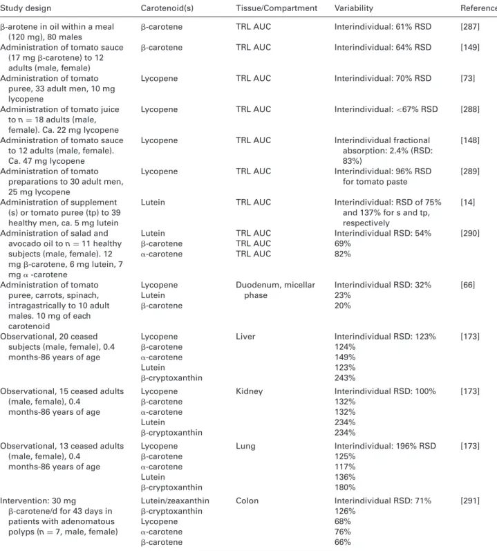
absorption, altered tissue distribution, turnover, and excre-tion as well as possible interacexcre-tions of individual carotenoids on absorption and bio-activation of other carotenoids [33, 34], are only poorly understood. These factors can result in huge variability of carotenoid absorption and circulating plasma levels (Table 2). In a double tracer study [13] with D6
-carotene (37 mol), lowest AUC (area under the plasma-concentration-time curve,mol h/L) versus highest AUC were found to be 0.01 and 30.00, respectively. Major host factors influencing carotenoid ADME patterns are likely to include:
(i) Factors influencing carotenoid release from the food ma-trix, and their transition from lipid droplets to mixed mi-celles, i.e. factors impacting bioaccessibility. This includes genes responsible for the expression of digestive enzymes (e.g. gastric lipase, cholesterol esterase, pancreatic lipase, etc.), and bile acid formation aiding in carotenoid micel-lization [20, 35];
(ii) Factors altering carotenoid uptake into (or efflux out of) the intestinal epithelium. This encompasses up-take/efflux transporters such as scavenger receptor class B member 1 (SR-BI), cluster of differentiation 36 (CD36), and Niemann-Pick C1 like intracellular cholesterol trans-porter 1 (NPC1L1), and perhaps other ATP-binding cas-sette (ABC) proteins such as ABCG5/G8, or ABCA1 [31, 36], but also small intestinal surface available for ab-sorption;
(iii) Factors contributing to intracellular cleavage, especially BCO1/2 (-carotene oxygenase 1/2), responsible for cen-tric/asymmetric cleavage of carotenoids, respectively, pro-ducing a variety of retinoids and potential endogenous occurring apocarotenoids [37–39];
(iv) Factors that impinge on carotenoid intracellular transport in the gut epithelium, i.e. lecithin-retinol acyltransferase (LRAT) for retinol (and perhaps other-carotene cleavage products), and maybe intestinal fatty acid binding protein (FABP2/I-FABP);
(v) Factors altering the secretion of carotenoid-containing chylomicrons into the lymphatic system, such as apolipoprotein B 48 (APOB48), APOAIV, SAR1B (secre-tion associated Ras related GTPase1B), and microsomal triglyceride transfer protein (MTTP) [38];
(vi) Factors influencing carotenoid transport in the blood plasma/serum, such as their further distribution into lipoproteins, e.g. lipoprotein lipase (LPL), APOA-I, APOB, APOE and perhaps APOC3, and low density lipoprotein receptor (LDLR) [40]; and the liver such as by hepatic li-pase (LIPC);
(vii) Deposition of carotenoids in “target tissues”, e.g. the macula (lutein, zeaxanthin), influenced by SR-BI, glu-tathione S-transferase P1 isoform (GSTP1), StAR-related lipid transfer domain protein 3 (StARD3), BCO1, choles-terol transporters (SR-BI, ABCA1, ABCG5/8), retinal pig-ment epithelial-specific protein (RPE65), elongation of very long chain fatty acids like 2 (ELOVL2), and those
involved in visual pigment metabolism [41]. However, also deposition in adipocytes, involving e.g. LDLR, could play a role [42];
(viii) Any factor associated with carotenoid catabolism and ex-cretion (in addition to BCO1/2), possibly those involv-ing cytochrome P450 enzymes or the aryl-hydrocarbon-receptor [43];
(ix) The microbiota. This may alter e.g. patterns and concen-trations of secondary bile acids [44], or possibly carotenoid absorption or degradation patterns [45], though it is not sure if a significant fraction of carotenoids can be ab-sorbed from the colon [46].
(x) Effects of individual carotenoids on the absorption, bind-ing, transport and bioactivation of other carotenoids as well as selective absorption, binding, transport and bioac-tivation of individual carotenoids [33].
(xi) Any factor associated with vitamin A/retinoid storage and metabolism.
Thus, as individual responses can depend on many vary-ing factors, it is paramount to understand these and their influence on the biological variability of carotenoid ADME. In this review, it is aimed to highlight known host-related fac-tors that predispose for variations in carotenoid metabolism, such as genetic factors (e.g. single nucleotide polymorphisms (SNPs)), though additional ones (disease state, body weight, smoking, physical activity etc.) are also briefly reviewed. The manuscript structure is oriented around the metabolic path of carotenoids, from digestion (chapter 2) to intestinal absorp-tion (chapter 3) and further transport to the liver (chapter 4) and distribution to target tissues (chapter 5) to storage and excretion related pathways (chapter 6). Searched databased in-cluded Pubmed and Scopus, for all years, in English language, employing the following search terms (abstract and title) to start with: “Human* AND (lutein OR lycopene OR xantho-phyll OR carotene*) AND (bioavailab* OR pharmacokinetic* OR kinetic* OR absorption OR postprandial OR metabol* OR microb* OR microflora OR biliary OR enterhohepatic* OR chylomicron OR plasma OR tissue OR metabolism OR ente-rocyte OR lipoproteins OR transporters OR Single nucleotide polymorphism* OR genetic varia* OR SNP OR cleavage OR enzym* OR intestine) AND (intra* OR inter*) NOT (drug-interaction OR in-vitro)”, though additional literature follow-ing the primary search results were surveyed.
2
Host factors influencing digestion
aspects – from matrix release to
bioaccessibility
2.1 General aspects and oral phase of digestion
Bioavailability of carotenoids depends on their bioacces-sibility, i.e. the release from the food matrix and subse-quent availability for absorption. As carotenoids are apolar, with octanol/water partition coefficients of 8–12 [47], their
Table 2. Studies investigating the variability of carotenoids in blood and target tissues, following intervention trials and observational
studies
Study design Carotenoid(s) Tissue/Compartment Variability Reference
Observational: 901adult subjects during 4 years (male, female)
Lycopene, lutein, ␣-carotene, -carotene, -cryptoxanthin
Plasma conc. Intraindividual variance: Lutein/zeaxanthin: 20.7% -carotene: 21.0% ␣ -carotene: 21.9% Lycopene: 35.0% -cryptoxanthin: 27.1% Interindividual variance: Lutein/zeaxanthin: 70.5% -carotene: 70.7% ␣ -carotene: 67.5% Lycopene: 61.0% -cryptoxanthin: 66.6% [284] Observational: 381 adult women, 4-month intervals, 4 visits
Lutein Plasma conc. Interindividual: 47% RSD Intraindividual: 44% of
interind. variation
[243]
-carotene Plasma conc. Interindividual: 80%RSD Intraindividual: 34% of
interind. variation Lycopene Plasma conc. Interindividual: 41% RSD
Intraindividual: as interind. variation
Observational: 21 adult subjects over 1 year (male, female), 6 measurements
-carotene Plasma conc. Interindividual: 100% RSD Intraindividual: 21% of
interind. variation
[244]
Lycopene Plasma conc. Interindividual: 42% Intraindividual: 72% of
interind. variation Double stable isotope to 11
healthy men (37mol -carotene)
-carotene Plasma AUC Interindividual: 137%%RSD 300 fold differences in AUC dose response observed
[13]
Intervention: 8 adult subjects (4 males, 4 females), 0.5 mol/kg bw.
-carotene Plasma AUC Intraindividual: 68% RSD [159] Intervention: 8 adult subjects,
0.5mol/kg bw.
Lutein Plasma AUC Intraindividual: 43% RSD Administration of isotopically
labelled lycopene (10.2 mg) to 8 subjects (4 males, 4 females)
Lycopene Absorption % based on plasma AUC
Interindividual: 504% RSD for all trans-lycopene
[15]
Administration of lycopene (10-120 mg) in a tomato beverage (5 male adults)
Lycopene Absorption (%) Interindividual: 77% RSD for highest dose, 53% RSD for lowest dose
[160]
Administration of soup, juice or tablets to 6 adult males (ca. 20 mg lycopene)
Lycopene Plasma AUC Interindividual:<28% RSD [285]
Feeding trial (5 weeks, 9 mg lutein/d) to young males
Lutein Plasma conc. Interindividual: ca. 70% RSD
[286] Administration of tomato
puree, spinach (12 mg -carotene, 8 mg lutein), and pills containing-carotene and lutein (20 young females)
Lycopene -carotene Lutein Plasma Plasma TRL AUC Interindividual: 40 % RSD Interindividual: 40% RSD Interindividual: ca. 45% RSD from spinach [147]
Administration of tomato puree to 33 adult men (0.4 mg -carotene)
Table 2. Continued
Study design Carotenoid(s) Tissue/Compartment Variability Reference
-arotene in oil within a meal (120 mg), 80 males
-carotene TRL AUC Interindividual: 61% RSD [287] Administration of tomato sauce
(17 mg-carotene) to 12 adults (male, female)
-carotene TRL AUC Interindividual: 64% RSD [149] Administration of tomato
puree, 33 adult men, 10 mg lycopene
Lycopene TRL AUC Interindividual: 70% RSD [73]
Administration of tomato juice to n= 18 adults (male, female). Ca. 22 mg lycopene
Lycopene TRL AUC Interindividual:<67% RSD [288]
Administration of tomato sauce to 12 adults (male, female). Ca. 47 mg lycopene
Lycopene TRL AUC Interindividual fractional absorption: 2.4% (RSD: 83%)
[148]
Administration of tomato preparations to 30 adult men, 25 mg lycopene
Lycopene TRL AUC Interindividual: 96% RSD
for tomato paste
[289]
Administration of supplement (s) or tomato puree (tp) to 39 healthy men, ca. 5 mg lutein
Lutein TRL AUC Interindividual: RSD of 75%
and 137% for s and tp, respectively
[14]
Administration of salad and avocado oil to n= 11 healthy subjects (male, female). 12 mg-carotene, 6 mg lutein, 7 mg␣ -carotene Lutein -carotene ␣-carotene TRL AUC TRL AUC TRL AUC Interindividual RSD: 54% 69% 82% [290] Administration of tomato puree, carrots, spinach, intragastrically to 10 adult males. 10 mg of each carotenoid Lycopene Lutein -carotene Duodenum, micellar phase Interindividual RSD: 32% 23% 20% [66] Observational, 20 ceased subjects (male, female), 0.4 months-86 years of age
Lycopene -carotene ␣-carotene Lutein -cryptoxanthin Liver Interindividual RSD: 123% 124% 149% 123% 243% [173]
Observational, 15 ceased adults (male, female), 0.4
months-86 years of age
Lycopene -carotene ␣-carotene Lutein -cryptoxanthin Kidney Interindividual RSD: 100% 132% 132% 234% 234% [173]
Observational, 13 ceased adults (male, female), 0.4
months-86 years of age
Lycopene -carotene ␣-carotene Lutein -cryptoxanthin Lung Interindividual: 196% RSD 125% 117% 136% 180% [173] Intervention: 30 mg
-carotene/d for 43 days in patients with adenomatous polyps (n= 7, male, female)
Lutein/zeaxanthin -cryptoxanthin Lycopene ␣-carotene -carotene Colon Interindividual RSD: 71% 126% 68% 76% 66% [291]
incorporation into mixed micelles is necessary prior to their cellular uptake, which is assumed to take place predominantly in the small intestine.
Mastication during oral digestion results in enhanced sur-face area and the breakdown into smaller particles. In ad-dition, saliva appears to contain some lipase activity [48],
though not necessarily lingual lipase (a triacylglycerol-lipase, EC 3.1.1.3) [49, 50] (Table 3). As exposure in the oral cavity is rather short (usually less than 1 min), the enzymatic ef-fect on carotenoid bioavailability is presumably small, though smaller particle size has been related to improved carotenoid bioavailability [51]. To our knowledge, no mutagenesis on or
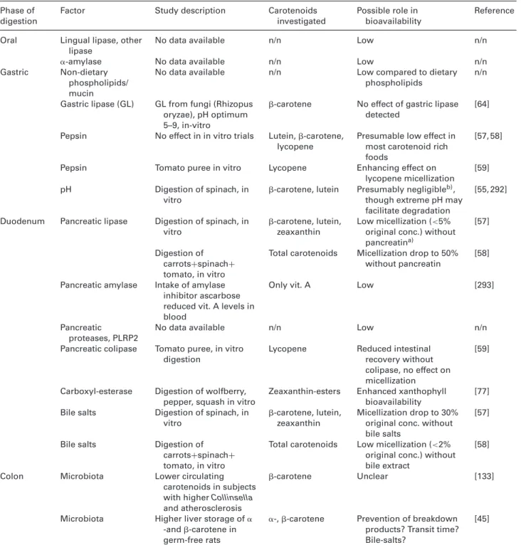
Table 3. Host factors influencing carotenoid release from food matrix and bioaccessibility
Phase of digestion
Factor Study description Carotenoids investigated
Possible role in bioavailability
Reference
Oral Lingual lipase, other lipase
No data available n/n Low n/n
␣-amylase No data available n/n Low n/n
Gastric Non-dietary phospholipids/ mucin
No data available n/n Low compared to dietary phospholipids
n/n
Gastric lipase (GL) GL from fungi (Rhizopus oryzae), pH optimum 5–9, in-vitro
-carotene No effect of gastric lipase detected
[64]
Pepsin No effect in in vitro trials Lutein,-carotene, lycopene
Presumable low effect in most carotenoid rich foods
[57, 58]
Pepsin Tomato puree in vitro Lycopene Enhancing effect on lycopene micellization
[59] pH Digestion of spinach, in
vitro
-carotene, lutein Presumably negligibleb), though extreme pH may facilitate degradation
[55, 292]
Duodenum Pancreatic lipase Digestion of spinach, in vitro
-carotene, lutein, zeaxanthin
Low micellization (<5% original conc.) without pancreatina)
[57]
Digestion of carrots+spinach+ tomato, in vitro
Total carotenoids Micellization drop to 50% without pancreatin
[58]
Pancreatic amylase Intake of amylase inhibitor ascarbose reduced vit. A levels in blood
Only vit. A Low [293]
Pancreatic
proteases, PLRP2
No data available n/n Low n/n
Pancreatic colipase Tomato puree, in vitro digestion
Lycopene Reduced intestinal recovery without colipase, no effect on micellization
[59]
Carboxyl-esterase Digestion of wolfberry, pepper, squash in vitro
Zeaxanthin-esters Enhanced xanthophyll bioavailability
[77] Bile salts Digestion of spinach, in
vitro
-carotene, lutein, zeaxanthin
Micellization drop to 30% original conc. without bile salts
[57]
Bile salts Digestion of carrots+spinach+ tomato, in vitro
Total carotenoids Low micellization (<2% original conc.) without bile extract
[58]
Colon Microbiota Lower circulating carotenoids in subjects with higher Collinsella and atherosclerosis
-carotene Unclear [133]
Microbiota Higher liver storage of␣ -and-carotene in germ-free rats
␣-, -carotene Prevention of breakdown products? Transit time? Bile-salts?
[45]
n/n: no data available.
a) though containing also other enzymes, pancreatic lipase is presumably the enzyme most important for carotenoid digestion. b) except for epoxy-carotenoids (violaxanthin, neoxanthin).
polymorphisms with effects on oral lipases and lipid diges-tion has been reported to date.
As salivary alpha-amylase (EC 3.2.1.1) participates in the break-down of starch, food matrices rich in both starch and carotenoids, such as sweet potato, may be influenced by
alterations in alpha-amylase levels. It has been reported that in populations traditionally exposed to high levels of starch, more copies of the salivary amylase gene (AMY1) and higher enzyme levels were found [52], though its influence on the digestion of carotenoids has never been investigated.
2.2 Gastric phase of digestion
In the stomach, the primary digestion enzymes include pepsin (3.4.23.1) and gastric lipase (3.1.1.3), though orally se-creted lipases may still be active. In addition, a small amount of phospholipids [53] is released from the mucus layer [54], aiding in the emulsification of lipophilic constituents. The pH may have an influence, as low pH can result in the degra-dation of epoxy-carotenoids (e.g. violaxanthin, neoxanthin), resulting in epoxide-furanoid transitions [55]. Human gas-tric pH is influenced mostly by meal, with a complex meal increasing the pH from initially 2 to 3–5, though interindi-vidual differences in fasting pH exist [35].
A few native foods are rich in both proteins and carotenoids, including egg yolk, salmon, and some types of cheese, and protein digestion could contribute to the release of carotenoids. In addition, (partly) digested proteins may aid in emulsifying carotenoids [51]. Expression of pepsin has been reported to depend on the pepsinogen genes PGA3,
PGA4, PGA5, and progastricsin (PGC) [56]. However,
vary-ing the amount of pepsin in in vitro trials did not appear to have measurable effects on carotenoid bioaccessibility from leafy vegetables [57], and at least for such and similar sources, variations in pepsin are not expected to contribute to plasma level variability. Similarly, using a test meal composed of meat, carrots, spinach and tomato paste, gastric digestion (in vitro) had no significant influence on carotenoid bioac-cessibility [58], suggesting rather small effects on carotenoid bioavailability at this step, though in these trials, gastric li-pase was not involved. By contrast, Periago et al. [59] reported a positive effect of pepsin on lycopene micellization from a puree in vitro. It is possible that for this very apolar carotenoid, protein degradation products added to the emulsifying effect, or aided in matrix breakdown.
The genes related to the production and secretion of mu-cus containing phospholipids, which could aid in the emul-sification process, are not clearly identified. Concentration variations of phospholipids between 0.03 and 0.6 mM have been reported, [60] and may be expected to have some influ-ence on carotenoid micellization. However, their influinflu-ence and strengths of effect are unknown and would also be su-perseded by dietary phospholipids, which are expected to play a more important role. This would be true especially follow-ing follow-ingestion of lipid-rich meals (a mean intake of 2–8 g/d of phosphatidylcholine has been reported [61], which would translate into ca. 8 mM (if taken within 1 out of 3 major meals per day, and dissolved in 1 L gastric fluid).
Gastric lipase, encoded by the LIPF (lipase F, gastric type) gene [62] and secreted by gastric chief cells, can digest up to 25% of the ingested lipids [35]. It thus could be expected to influence the accumulation of carotenoids in lipid droplets, and their degradation, important for the following transition of carotenoids from lipid droplets to mixed micelles. This occurs mostly in the small intestine. Unfortunately, gastric lipase cannot, at present, be studied in vitro, due to the un-availability of human gastric lipase. Other sources, such as
those from fungi, have different cleavage kinetics, differing in their pH optimum and also the type sequence of cleavage [63]. Rabbit lipase would be an interesting option, but is not commercially available. Some, such as lipase from the fun-gus Rhizopus oryzae have been tested (cleavage optimum pH 5–9), though no significant improvement in bioaccessibility was found [64].
2.3 Small intestinal phase of digestion
The most crucial step influencing carotenoid bioacessibility is the small intestinal phase. Here, micellization occurs or is completed, following the secretion of bile salts, in addi-tion to pancreatic lipase, and addiaddi-tional enzymes (pancreatic amylase, nucleosidases, trypsinogen, chymotrypsinogen, car-boxypeptidase, elastases, phospholipases, and carboxyl ester lipase). Bile salts aid in the emulsification process and for-mation and stability of the mixed micelles, while pancreatic lipase produces free fatty acids and monoglycerides, fostering emulsification. Thus, it can be expected that modifications of both bile-acid and pancreatic lipase secretions have strong effects on the micellization of carotenoids, a pre-requisite for their diffusion to the unstirred water layer prior to absorp-tion [65]. This has been confirmed by several in vitro stud-ies, where micellization and resulting bioaccessibility was very much compromised when either bile salts or pancre-atic lipase were missing. Without bile, bioaccessibility of to-tal carotenoids fell to 30%, and without pancreatic lipase or both, to below 5% of their original value [57]. Similar strong effect were found by Garret et al. [58], where total carotenoid micellization dropped below 5% of the original values without bile salts. The effect of pancreatin was less drastic (reduction by approximately 50%), possibly due to differences between test meals. In order to study factors influencing lycopene bioaccessibility, tomato puree was digested under various conditions, testing among other factors gastric pH, gastric digestion time, pepsin concentration, intestinal pH, pancreatin concentration, bile salt concentration, colipase ad-dition and intestinal digestion time [59]. It was found that only pepsin positively influenced micellization, while olive oil had a slightly negative effect, likely due to entrapment of lycopene by non-hydrolysed olive oil.
Following intragastric in vivo administration of carotenoid rich meals, duodenal fluid was aspirated, and micellization determined [66]. Variability between subjects’ micellization efficacy (fractional bioaccessibility) was considerably lower compared to plasma or triacylglycerol-rich lipoprotein (TRL) carotenoid variability following interventions, being 20, 23, and 32%, respectively for -carotene, lutein and lycopene, vs. typically 50–80% for plasma, though variations between studies can be considerable (Table 3). This may point out that, although enzyme or bile salt concentrations surely play a role in interindividual variation, an additional and about equal portion of variability is added during and after absorption.
Bile acid production by the liver is governed by a variety of genes, involving for instance bile acid synthetic enzyme (CYP7A1), activators of CYP7A1 expression such as HNF4␣ (hepatocyte nuclear factor 4 alpha, encoded by HNF4A), and PGC1␣ (encoded by PPARGC1A), repressors of CYP7A1 (farnesoid X receptor (FXR, encoded by NR1H4)), short het-erodimer partner (SHP, encoded by NR0B2), G protein path-way suppressor 2 (GPS 2, encoded by GPS2), pregnane X receptor (PXR, encoded by NR1I2), fibroblast growth factor 19 (FGF19; encoded by FGF19), fibroblast growth factor re-ceptor 4 (FGFR4; encoded by FGFR4), klotho B (encoded by
KLB), and forkhead box O1 (FOXO1; encoded by FOXO1)
[67], however, their role in carotenoid absorption and tissue variability has not been examined.
At least three lipases are secreted from the pancreas, including pancreatic triglyceride lipase (encoded by
PN-LIP), which is the most abundant lipase (producing
sn2-monoacylglycerol and free fatty acids), but also two homo-logues, pancreatic lipase-related proteins 1 (not apparently ac-tive regarding lipolysis) and 2 (PLRP1 and PLRP2) [68]. PLRP2 possess a broader substrate specificity, also cleaving, unlike PNLIP, phospholipids and galactolipids. The frequency of a SNP in the PLRP2 gene (rs4751995) has been associated with populations historically consuming a diet rich in cereals, and may have repercussions on lipid digestion [69].
Though pancreatic triglyceride lipase activity is usually reduced by bile-salts, this effect is offset by colipase, also se-creted by the pancreas [70]. Formation of colipase preprotein is regulated by the CLPS gene, and mice deficient for CLPS showed lower survival and weight gain on a high-fat diet, suggesting the inability to cope with lipids on a high fat diet [71]. A polymorphism for the gene encoding procolipase has been related to lipid metabolism and diabetes risk [72], and would be an interesting candidate also regarding carotenoid metabolism. In a recent study, a SNP in PNLIP (rs11197742) was found in a combination of SNPs associated with chy-lomicron secretion of lycopene [73], although its contribution was rather low and did not reach statistical significance when investigated individually (p= 0.086). Several SNPs in PNLIP have been reported in children, and the latter was related to al-tered plasma lipoprotein and total cholesterol concentrations [74], as well as with lycopene bioavailability (Table 4).
Carboxyl-ester lipase (CEL), also termed cholesterol-esterase, typically cleaves cholesterol esters in the gut, and its ability to cleave carotenoid esters, such as of lutein, present in many leafy vegetables, has been controversially discussed [75]. At least five types of CEL are known, though human carboxylesterases CES1 and CES2 may play the most impor-tant role during digestion [76]. These are situated on the gut mucosa (brush border enzymes), and have shown to cleave carotenoid esters [77]. Its origin (pancreatic vs. enterocyte) remains somewhat unclear. However, this cleavage is ex-pected to influence bioavailability, as the more apolar esters are characterized by lower micellization efficiency and ab-sorption than the cleaved carotenoids [78]. In fact, in plasma and circulating chylomicrons, free xanthophylls are almost
exclusively found, suggesting that cleavage is in fact quite complete [79], though reduced absorption of the esters could play a role. A number of SNPs have been described in humans for CES1 and CES2 [80], though not in relation to carotenoid or lipophilic phytochemical/micronutrient metabolism.
Certain diseases such as pancreatitis may also result in lower secretion of digestion enzymes [81]. Also during older age reduction of lipid absorption has been reported, perhaps also due to reduced epithelial surface [82], which may thus be expected to correlate with lower carotenoid absorption, as suggested by some, though not all studies (Table 1).
3
Host factors determining aspects
of intestinal absorption
3.1 Factors influencing cellular uptake and cleavage
Following their extraction from the food matrix and incorpo-ration, at least in part, into mixed micelles, carotenoids are taken up by enterocytes. This process is not only passive, as previously thought [83], and several apical membrane pro-teins have been shown to facilitate carotenoid uptake [36]. SR-BI, encoded by SCARB1, is involved in the uptake of -carotene [84,85], lutein [86] and lycopene [87]. CD36 facilitates -carotene [84] uptake and could facilitate lycopene uptake [88], while NPC1L1 participates in the uptake of lutein [89]. All of these proteins have SNPs in their encoding genes asso-ciated with carotenoid plasma concentrations (Table 4), and their contribution to carotenoid uptake has been confirmed in cellular models (e.g. human Caco-2 cell line), but also in models employing transfected kidney (HEK) cells. After en-terocyte uptake, carotenoids can be metabolized by BCO1 [90] and BCO2 [39]. BCO1 catalyses the oxidative cleavage of provitamin A carotenoids (chieflycarotene, ␣-carotene, -cryptoxanthin), apo-carotenals, and lycopene, but not that of lutein [91]. BCO1 is presumably the main cleaving-enzyme for-carotene [92]. Lycopene was suggested to be predomi-nantly cleaved by BCO2 [93], while recently lycopene cleavage by BCO1 was also reported [94]. However, until now no ly-copene derived BCO1-products were determined [95, 96] and were only postulated [97, 98]. BCO2 has also been shown to be involved in lutein metabolism [99]. Most-carotene con-version (>70%) is thought to occur in the intestine; by using stable isotope techniques it was estimated that about 20–30% occurs after absorption [100], contributing to overall vitamin A homeostasis. In addition, a controlled temporal and spa-tial conversion of carotenoids to bioactive retinoids is also of physiological importance, indicated by a specific pattern of BCO1 expression in various tissues [101]. This expression is linked to RAR-mediated signaling [39, 102].
The involvement of several proteins in the intestinal ab-sorption of carotenoids (apical uptake) suggests that varia-tions in the genes encoding these proteins could modulate carotenoid absorption efficiency. This has been confirmed in an association study by Borel et al. [103] where the influence
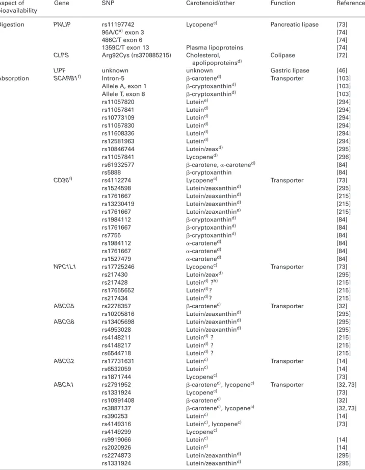
Table 4. List of SNPs known, or speculated, to influence carotenoid metabolism
Aspect of bioavailability
Gene SNP Carotenoid/other Function Reference
Digestion PNLIP rs11197742 Lycopenec) Pancreatic lipase [73]
96A/Ca)exon 3 [74]
486C/T exon 6 [74]
1359C/T exon 13 Plasma lipoproteins [74]
CLPS Arg92Cys (rs370885215) Cholesterol, apolipoproteinsd)
Colipase [72]
LIPF unknown unknown Gastric lipase [46]
Absorption SCARB1f) Intron-5 -carotened) Transporter [103]
Allele A, exon 1 -cryptoxanthind) [103]
Allele T, exon 8 -cryptoxanthind) [103]
rs11057820 Luteine) [294] rs11057841 Luteind) [294] rs10773109 Luteind) [294] rs11057830 Luteind) [294] rs11608336 Luteind) [294] rs12581963 Luteind) [294] rs10846744 Lutein/zeaxd) [295] rs11057841 Lycopened) [296] rs61932577 -carotene, ␣-carotened) [84] rs5888 -cryptoxanthin [84] CD36f) rs4112274 Lycopenec) Transporter [73] rs1524598 Lutein/zeaxanthind) [295] rs1761667 Lutein/zeaxanthind) [215] rs13230419 Lutein/zeaxanthind) [215] rs1761667 Lutein/zeaxanthine) [215] rs1984112 -cryptoxanthind) [84] rs1761667 -cryptoxanthind) [84] rs7755 -cryptoxanthind) [84] rs1984112 ␣-carotened) [84] rs1761667 ␣-carotened) [84] rs1527479 ␣-carotened) [84] NPC1L1 rs17725246 Lycopenec) Transporter [73] rs217430 Lutein/zeaxd) [295] rs217428 Luteind)?h) [215] rs17655652 Luteind)? [215] rs217434 Luteind)? [215]
ABCG5 rs2278357 -carotenec) Transporter [32]
rs10205816 Lutein/zeaxanthind) [295] ABCG8 rs13405698 Lutein/zeaxanthind) [295] rs4953028 Lutein/zeaxanthind) [295] rs4148211 Luteind)? [215] rs4148217 Luteind)? [215] rs6544718 Luteind)? [215]
ABCG2 rs17731631 Luteinc) Transporter [14]
rs6532059 Luteinc) [14]
rs1871744 Lycopenec) [73]
ABCA1 rs2791952 -carotenec), lycopenec) Transporter [32, 73]
rs1331924 Lycopenec) [73] rs10991408 -carotenec) [32] rs3887137 -carotenec), lycopenec) [32, 73] rs390253 Luteinc) [14] rs4149316 Luteinc), lycopenec) [73] rs4149299 Lycopenec) rs9919066 Luteinc) [14] rs2020926 Luteinc) [14] rs2274873 Lutein/zeaxanthind) [295] rs1331924 Lutein/zeaxanthind) [295]
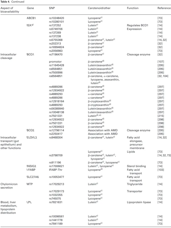
Table 4. Continued
Aspect of bioavailability
Gene SNP Carotenoid/other Function Reference
ABCB1 rs10248420 Lycopenec) [73]
rs10280101 Lycopenec) [73]
ISXg) rs137252 Luteinc) Regulates BCO1 [14]
rs5749706 Luteinc) Expression [14] rs137269 Luteinc) [14] rs137238 Luteinc) [14] rs5755368 -carotenec), luteinc) [14, 32] rs202313 -carotenec) [32] rs16994824 -carotenec) [32] rs2056983 Lycopenec) [73] Intracellular cleavage
BCO1 rs7196470 -carotenec) Cleavage enzyme [32]
promotor -carotened) [107] rs11645428 Lutein/zeaxanthind) [295] rs6564851 Lutein/zeaxanthind) [295] rs7500996 Lutein/zeaxanthind) [295] rs6564851 -carotene, ␣-carotene, lycopene, zeaxanthin, luteind) [32, 104] rs4889286 -carotened) [297] rs12934922 -carotened) [297] rs4889293 ␣-carotened) [297] rs4889286 ␣-carotened) [297] rs12918164 -cryptoxanthind) [297] rs4889293 -cryptoxanthind) [297] rs56389940 Lutein/zeaxanthind) [297] rs10048138 Lutein/zeaxanthind) [297] rs7501331 Luteind),e) [215] rs12934922 -carotened) [298] rs7501331 -carotened) [298] rs12934922 -carotened) [298]
BCO2 rs12796114 Association with AMD Cleavage enzyme [295]
rs2250417 Association with AMD [295]
Intracellular transport (gut epithelium) and other functions
ELOVL2 rs9468304 -carotenec), luteinc) Fatty acid
elongase, precursor membrane [14, 32] Lycopenec) Lipids [73] rs3798709 -carotenec), luteinc), lycopenec) [14, 32, 73] rs911196 -carotenec), lycopenec) [73]
INSIG2 rs17006621 Luteinc), lycopenec) Sterol binding [14]
I-FABP IFABP-Thr Lycopened) Fatty acid
transport
[103]
SLC27A6 rs10053477 Lycopenec) Fatty acid
transport [73] Chylomicron secretion MTP rs17029213 Luteinc) Triglyceride [14] rs17029173 Lycopenec) Transporter [73] rs1032355 Lycopenec) [73] rs745075 Lycopenec) [73] Blood, liver metabolism, lipoprotein distribution
LPL rs7821631 Luteinc) Lipoprotein lipase [14]
rs10096561 Luteinc) [14]
rs1441778 Luteinc) [14]
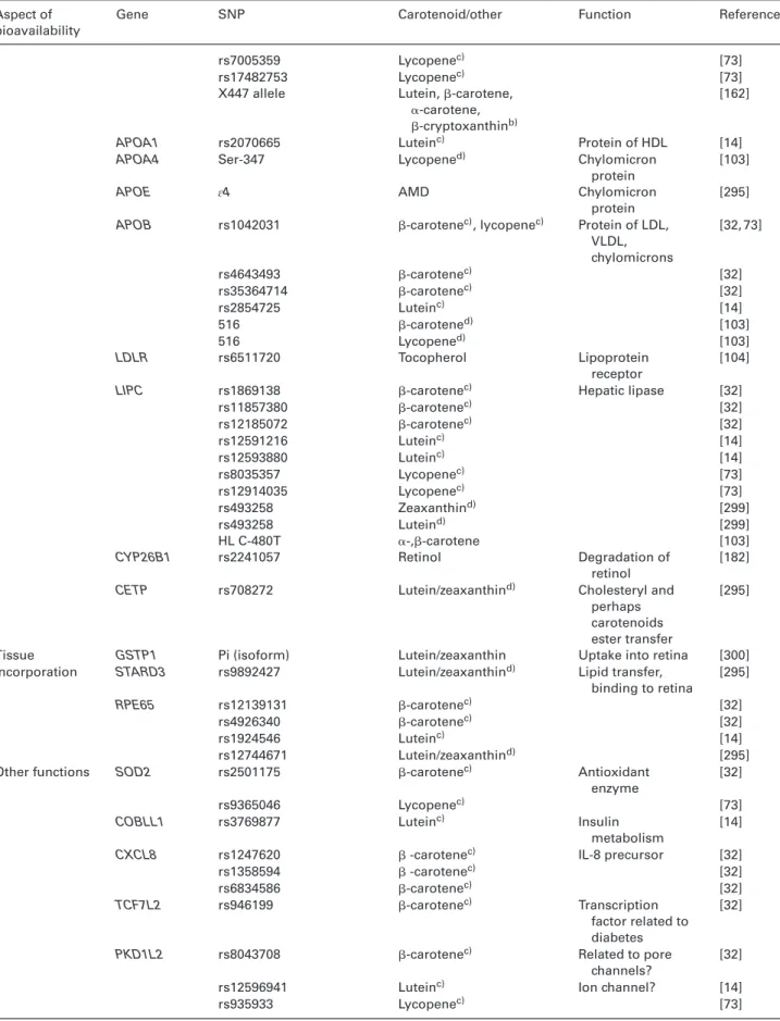
Table 4. Continued
Aspect of bioavailability
Gene SNP Carotenoid/other Function Reference
rs7005359 Lycopenec) [73]
rs17482753 Lycopenec) [73]
X447 allele Lutein,-carotene, ␣-carotene, -cryptoxanthinb)
[162]
APOA1 rs2070665 Luteinc) Protein of HDL [14]
APOA4 Ser-347 Lycopened) Chylomicron
protein
[103]
APOE ɛ4 AMD Chylomicron
protein
[295]
APOB rs1042031 -carotenec), lycopenec) Protein of LDL,
VLDL, chylomicrons [32, 73] rs4643493 -carotenec) [32] rs35364714 -carotenec) [32] rs2854725 Luteinc) [14] 516 -carotened) [103] 516 Lycopened) [103] LDLR rs6511720 Tocopherol Lipoprotein receptor [104]
LIPC rs1869138 -carotenec) Hepatic lipase [32]
rs11857380 -carotenec) [32] rs12185072 -carotenec) [32] rs12591216 Luteinc) [14] rs12593880 Luteinc) [14] rs8035357 Lycopenec) [73] rs12914035 Lycopenec) [73] rs493258 Zeaxanthind) [299] rs493258 Luteind) [299] HL C-480T ␣-,-carotene [103]
CYP26B1 rs2241057 Retinol Degradation of
retinol
[182]
CETP rs708272 Lutein/zeaxanthind) Cholesteryl and
perhaps carotenoids ester transfer
[295]
Tissue GSTP1 Pi (isoform) Lutein/zeaxanthin Uptake into retina [300]
incorporation STARD3 rs9892427 Lutein/zeaxanthind) Lipid transfer,
binding to retina [295] RPE65 rs12139131 -carotenec) [32] rs4926340 -carotenec) [32] rs1924546 Luteinc) [14] rs12744671 Lutein/zeaxanthind) [295]
Other functions SOD2 rs2501175 -carotenec) Antioxidant
enzyme
[32]
rs9365046 Lycopenec) [73]
COBLL1 rs3769877 Luteinc) Insulin
metabolism
[14]
CXCL8 rs1247620  -carotenec) IL-8 precursor [32]
rs1358594  -carotenec) [32] rs6834586 -carotenec) [32] TCF7L2 rs946199 -carotenec) Transcription factor related to diabetes [32]
PKD1L2 rs8043708 -carotenec) Related to pore channels?
[32]
rs12596941 Luteinc) Ion channel? [14]
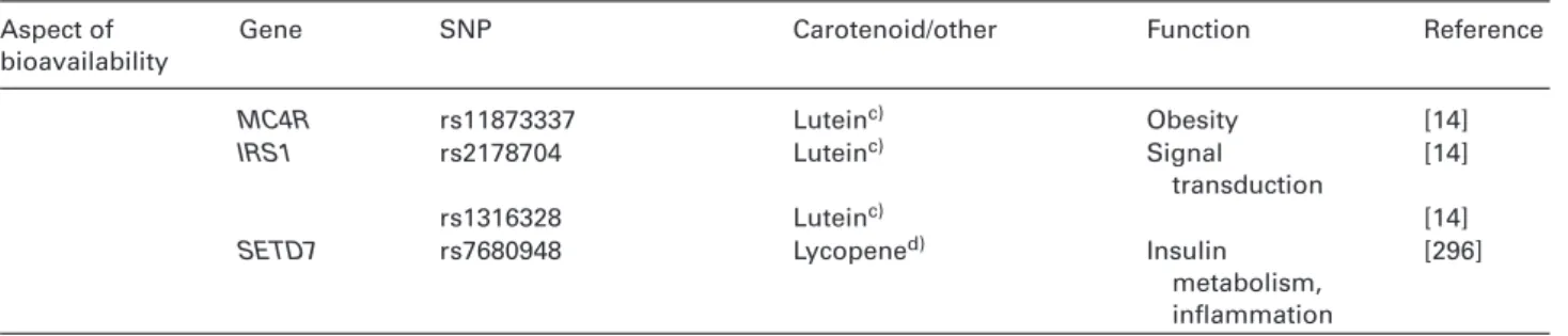
Table 4. Continued
Aspect of bioavailability
Gene SNP Carotenoid/other Function Reference
MC4R rs11873337 Luteinc) Obesity [14]
IRS1 rs2178704 Luteinc) Signal
transduction
[14]
rs1316328 Luteinc) [14]
SETD7 rs7680948 Lycopened) Insulin
metabolism, inflammation
[296]
a) base-pairs: A: adenine, C: cytosine, T: thymine, G: guanine. b) in animals, not humans.
c) Measured by chylomicron response. d) Measured by plasma levels.
e) Related to AMD, Measured as macula pigment optical density (MPOD). f) also involved in uptake in other tissues
g) Intestine Specific Homeobox.
h) Question mark indicating assumed influence.
of candidate SNPs of genes involved in lipid metabolism on the fasting blood concentration of several carotenoids was investigated. More specifically, SNPs in SCARB1 were asso-ciated with-carotene but not with lycopene concentrations. These SNPs explained differences in-carotene plasma con-centrations by up to 50%. Several additional SNPs have mean-while been identified, including several in BCO1 in genome-wide association studies [31, 104, 105]. Three recent studies have reported associations of combinations of SNPs involved in interindividual variability of the bioavailability of lutein [14], lycopene [73] and-carotene [32], employing a candidate gene approach in postprandial studies. In these, plasma-TRL carotenoids, representing newly absorbed carotenoids, were measured in healthy male adults. These combinations were associated with 73, 72, and 69% of the interindividual variabil-ity of the bioavailabilvariabil-ity of lutein, lycopene and-carotene, respectively. While some SNPs were located in genes ex-pressed in other tissues or were closely involved in plasma-TRL metabolism, others were involved with carotenoid trans-port or metabolism at the enterocyte level. These included
ABCA1, ABCG5, BCMO1, CD36, ELOVL2, and ISX
(intes-tine specific homeobox). Interestingly, one SNP in ELOVL2 (rs9468304) was very strongly associated with all three phenotypes, possibly due to the inhibitory effect of eicosapen-taenoic acid, which is further elongated to docosapeneicosapen-taenoic acid and docosahexaenoic acid by ELOVL2, on carotenoid ab-sorption, as has been shown with-carotene [106].
3.2 Influence of nutritional status
Host vitamin A status has been linked with-carotene ab-sorption variability. Lobo et al. [107] demonstrated that the in-testinal transcription factor ISX acts as a repressor of SCARBI and BCO1 expression following retinoic acid induction. This mechanism is thought to serve as a negative feedback loop regulating retinal and further retinoic acid, retinyl esters and
retinol status through modulation of provitamin A carotenoid absorption and cleavage efficiencies. Interestingly, the same team has reported the existence of an SNP in the ISX binding site in the BCO1 promoter (rs6564851) which was associated with decreased conversion rates by 50% and increased fasting blood levels of-carotene [108].
Though the mechanisms are not fully elucidated, low iron status was suggested to interact with retinol homeostasis, re-sulting in decreased mobilization of liver vitamin A and thus low serum concentrations [109], possibly involving altered BCO1 activity [110]. Also a low zinc status appears to reduce -carotene absorption from the gut [111], perhaps as phos-pholipase A2 can bind zinc and may be more active. These effects were confirmed in human studies, where supplemen-tation with iron and zinc following a vitamin A deficient diet improved retinol and carotenoid plasma appearance, respec-tively [112]. Also low protein status appears to hinder conver-sion of-carotene to vitamin A, contributing to carotenoid variability [113].
BCO1 and BCO2 were also described to be controlled by peroxisome proliferator-activated receptor (PPAR) – retinoid X receptor (RXR) mediated signaling [114, 115]. The endoge-nous ligands of the PPARs␣, /␦ and ␥ are ranging from free fatty acids to various eicosanoids such as prostaglandins, leukotrienes and mono-hydroxylated fatty acids [116]. The RXR was described to be activated by 9-cis-retinoic acid (9CRA) [117], as well as the newly found endogenous rele-vant ligand 9-cis-13, 14-dihydro-retinoic acid/9CDHRA [118]. It is debated whether 9CRA occurs endogenously [119]. Cur-rently, 9CRA is considered mainly as a ligand that is present after high non-physiological and non-nutritional relevant vita-min A intake, leaving 9CDHRA as the principal endogenous and the nutritional relevant RXR ligand. PPAR ligands are mainly food derived [116], while the nutritional precursors of the endogenous RXR ligand 9CDHRA were not yet iden-tified. The PPAR-regulatory pathway of BCO1/2 expression and further carotenoid bioactivation is thus controlled by the
amount and fractional distribution of lipids present in the food matrix. In addition to genomic regulation of BCO1/2 expression, carotenoid cleavage can also be modulated by in-hibitory effects of lutein on-carotene cleavage [120]. This indicated that not just the individual carotenoid concentra-tion is of relevance to bioactivaconcentra-tion towards retinoic acid and further transcriptomic regulation, but also the concentration of carotenoids inhibiting this metabolic step, as well as their concentration relative to-carotene. The consequences of BCO1/2 mediated regulation of retinoic acid synthesis and further transcriptional signaling by additional factors and its consequences for our health will be discussed later (chapter 7), highlighting the special importance of BCO1/2 on explain-ing interindividual variability, likely related to the beneficial health effects of carotenoids.
3.3 Colonic fermentation as an interindividual source
To date, it is unclear to what extent the microbiota con-tributes to carotenoid metabolism, and whether carotenoids/ their metabolites can be taken up in the colon. It is known that a large proportion of carotenoids reaches the colon, as only 5–50% are absorbed in the small intestine. It is also known that carotenoids are partly bioaccessible in the colon [121]. However, only 10–50% of the carotenoids re-main intact after fermentation, while the rere-mainder reacts to unknown compounds [121–123]. This was supported by carotenoid standards as the only fermentation source in vitro, as>98% losses for -carotene and zeaxanthin were reported
[123].
Very little is known on carotenoid interaction with the mi-crobiota [124]. Unlike polyphenols, which are heavily metab-olized, no carotenoid degradation products/bacterial metabo-lites have been identified. In general, bacteria in the colon are able to deglycosylate, hydrolyse, deglucuronidate, demethy-late, and cause ring-fission in some molecules, among other [46, 125]. However, in germ-free rats, higher carotenoid uti-lization (of␣- and -carotene) as measured by their liver lev-els, has been reported compared to rats with intact microbiota [45]. It was suggested that indirect effects, such as decreased intestinal transit time and an altered bile pool in the absence of bacteria could have played a role, though a reduced level of bacterial breakdown products and more remaining native compounds could have been involved. In support of a poten-tial absorption of carotenoids in the colon, a study in mice found BCO1 to be expressed in many cells including mu-cosal, glandular cells in the stomach, small intestine, and the colon [126]. BCO2 is known to be expressed in almost all cell types known to express BCO1. However, BCO2 was not found in the colon, suggesting that only symmetric cleav-age of carotenoids may happen in the mucosal cells in the colon.
In a previous study,-carotene uptake into human ex-foliated epithelial cells of the colon, separated from feces,
has been demonstrated [127]. Following the consumption of-carotene rich spirulina, the concentration of -carotene in the cells increased approximately 3-fold, demonstrating colonic cellular presence. However, this may have occurred not necessarily through direct cellular uptake via colonocytes, as carotenoids could have been absorbed via the small intes-tine and then distributed via the circulatory system to the colonocytes. Furthermore, the same constituents known to enhance carotenoid bioavailability, namely bile salts, emul-sifiers such as lecithin [128, 129], enhanced colonic cellular uptake. Though carotenoids can be taken up by colonic de-rived Caco-2 cells, direct colonic uptake is not easy to prove, and studies so far have not suggested a strong correlation be-tween dietary intake of carotenoids and colon concentrations [130]. Oshima et al. [131] investigated colonic absorption and distribution of lycopene in rats with or without a colostomy at mid colon that diverted the fecal stream but without resec-tion of the distal colon. In rats given intragastric treatment, lycopene was found in the mucosa in the proximal colon and in the distal colon, also of the colostomized rats, whose dis-tal colon was isolated from the faecal stream, indicating that lycopene may be transported via the blood into the colon. Moreover, lycopene reached the liver to an appreciable ex-tent even when administered into the isolated distal colon, indicating that absorption is possible from the distal colon in rats.
Taken together, these results indicate that carotenoid ab-sorption from the colon could be relevant and contribute to interindividual variation in carotenoid bioavailability, de-pending on the food matrix and microbiota. Furthermore, as faecal transplants have shown to be able to trigger obesity, at least in animal models [132], and obese subjects having gener-ally lower concentrations of circulating carotenoids (Table 1), a potential direct or indirect link between the microbiota and carotenoid tissue levels may exist. In a study with atheroscle-rotic subjects, patients showed a metagenome with reduced phytoene-dehydrogenase and lower-carotene serum levels compared to healthy controls, which was associated with a higher level of Collinsella spp. in diseased subjects [133], highlighting the potential role of the microbiota.
3.4 Diseases and medical intervention effecting the intestine and colon
Any condition reducing the intestinal mucosal surface area can be expected to reduce carotenoid absorption. As most studies do not directly measure carotenoid absorption effi-ciency but rather look at blood carotenoid levels (or a plasma fraction), it is important to distinguish between direct effects on carotenoid absorption (i.e. through reduced mucosal sur-face area or limited transport capacity) and indirect effects (through dietary adaptations, e.g. high fiber or low fat diet). This is usually achieved by controlling for carotenoid dietary intake.
A study with 20 Crohn’s disease patients reported lower fasting blood carotenoid concentrations, independent of di-etary intake [134], suggesting that malabsorption affected carotenoid uptake, though increased turnover rate and colonic losses via e.g. bleeding could not be excluded. Similar results were obtained by Geerling et al. [135] in a study with 32 Crohn’s disease patients and Genser et al. [136] with 24 patients. Crohn’s disease usually affects the ileum but only three of the 20 patients in the study had ileal inflammation, indicating the importance of the colonic mucosal integrity for carotenoid absorption. Patients undergoing bariatric surgery (Roux-en-Y gastric bypass and biliopancreatic diversion) also displayed lower blood carotenoid levels [137]. Since fruit and vegetable consumption was apparently normal, the effect was attributed to malabsorption due to reduced mucosal surface area and also due to limited capacity of transport related to decreased lipoprotein concentration. Also reduced gastric di-gestion (via gastric lipase, or mechanic dispersion), could have played a role, as could have biliopancreatic diversion, af-fecting bile and pancreatic enzyme concentrations in the gut. In another study, subjects with Celiac disease and Crohn’s disease (n = 22) showed significantly 37% decreased levels of macular carotenoids compared to controls (n= 25 [138].
Short bowel syndrome, usually due to large resections of the small intestine to treat pathologies such as Crohn’s disease or gastrointestinal tumors, have also been associ-ated with carotenoid malabsorption. Edes et al. [139] reported undetectable-carotene blood levels following supplemen-tation, despite adequate fat absorption, in a patient with ex-tensive small intestinal resection (serum vitamin A levels ap-peared normal). Perhaps carotenoid absorption occurred in a more limited section of the intestine, or absorbed-carotene was fully converted to vitamin A. Luo et al. [140] reported no increase in blood carotenoid levels in subjects with short bowel syndrome undergoing intestinal rehabilitation, despite a 12-week-long supplementation with-carotene, lutein and lycopene. This was attributed to low fat absorption (about 30 versus>95% in healthy subjects) in these patients. However, no estimates of the contribution of the colon to the observed differences in absorption efficiencies were reported. There-fore, it is uncertain if it is the disease affecting the lower gut, the limited length of residual ileum, the presence or absence of the colon, the patient’s lifestyle, or a combination that results in low plasma carotenoids. Similar low levels were observed in 63 patients with total gastrectomy [141], possibly due to duodenal bypass and short interposition of a small intestine loop.
Intestinal parasites and bacterial overgrowth can also dam-age mucosal cells and result in increased permeability and decreased absorption of nutrients. In Indonesian children receiving red sweet potato, serum retinol concentrations creased to a greater extent when children infected with in-testinal helminths were dewormed, than when the intensity of infection was high [142], though the effect may have been also due to improved fat absorption. In tropical countries, also enteropathies, resulting in inflamed epithelium and reduced
surface available for absorption, are likely to contribute to low carotenoid and vitamin A status [143].
4
Host factors influencing intracellular
transport and transport to the liver
4.1 Intracellular transport within the enterocyte
After their uptake at the apical side of the enterocyte by mem-brane proteins, which are involved in the uptake of other li-posoluble micronutrients, e.g. vitamin E/D [144], carotenoids have to cross the aqueous environment of the cell to reach its basolateral side. As carotenoids are very hydrophobic [21] it is assumed that they need to be associated with intracel-lular proteins to move through this medium [36]. Though candidate proteins have been suggested, limited evidence of their involvement is available yet. A first one is human retinal lutein-binding protein [145], as it shows a good cross-reactivity with antibodies raised against carotenoid-binding protein, which has been shown to transport carotenoids in the midgut cytosol of the silkworm Bombyx mori [146]. However, its ex-pression in the enterocyte should be verified. Other candi-dates could be the enterocyte FABPs (FABP2/I-FABP and FABP1/L-FABP) that allow the transport of various lipids. Fi-nally, it can be hypothesized that the main enzyme responsi-ble for carotenoid cleavage in the enterocyte, i.e. BCO1 [39,96], could also be involved, as it attracts and binds carotenoids for further cleavage, and it may also function as a non-identified but predicted selective carotenoid-transporter. The involve-ment of some of these candidate proteins in carotenoid trans-port within the enterocyte is suptrans-ported by studies that have observed associations between SNPs in genes encoding these proteins and carotenoid status or bioavailability. This is the case for FABP and lycopene [103] and BCO1 and-carotene [32], though this second association can also be due to the catalytic activity of this protein. Functional studies employing cell cultures or transgenic mice should be performed to iden-tify the respective proteins. Nevertheless, it can be hypothe-sized that variations in genes encoding proteins involved in the transport of carotenoids within the enterocyte contribute to the observed interindividual variability in carotenoid bioavailability.
The previously described interaction of lutein and -carotene was not investigated further in detail, but it was pre-dicted also to be of relevance regarding mutual interferences during absorption [33,120,147]. A different fractional absorp-tion efficacy was also suggested for cis-isomers of lycopene [20,148–150]. Unfortunately, for lutein and-carotene as well as for lycopene and-carotene cis-isomers, the mechanism of this altered transport efficiency was not examined further, but it appears to have an important physiological importance due to the different and possibly augmented health beneficial ef-fects of especially 9-cis--carotene versus all-trans- -carotene, at least in respect to atherosclerosis [151].