HAL Id: inserm-00869452
https://www.hal.inserm.fr/inserm-00869452
Submitted on 3 Oct 2013
HAL is a multi-disciplinary open access
archive for the deposit and dissemination of
sci-entific research documents, whether they are
pub-lished or not. The documents may come from
teaching and research institutions in France or
abroad, or from public or private research centers.
L’archive ouverte pluridisciplinaire HAL, est
destinée au dépôt et à la diffusion de documents
scientifiques de niveau recherche, publiés ou non,
émanant des établissements d’enseignement et de
recherche français ou étrangers, des laboratoires
publics ou privés.
Ornello, Antonio Carolei, Tobias Kurth
To cite this version:
Simona Sacco, Patrizia Ripa, Davide Grassi, Francesca Pistoia, Raffaele Ornello, et al.. Peripheral
vascular dysfunction in migraine: a review.. Journal of Headache and Pain, SpringerOpen, 2013, 14
(1), pp.80. �10.1186/1129-2377-14-80�. �inserm-00869452�
R E V I E W A R T I C L E
Open Access
Peripheral vascular dysfunction in migraine:
a review
Simona Sacco
1*, Patrizia Ripa
1, Davide Grassi
2, Francesca Pistoia
1, Raffaele Ornello
1, Antonio Carolei
1and Tobias Kurth
3,4Abstract
Numerous studies have indicated an increased risk of vascular disease among migraineurs. Alterations in
endothelial and arterial function, which predispose to atherosclerosis and cardiovascular diseases, have been
suggested as an important link between migraine and vascular disease. However, the available evidence is
inconsistent. We aimed to review and summarize the published evidence about the peripheral vascular
dysfunction of migraineurs.
We systematically searched in BIOSIS, the Cochrane database, Embase, Google scholar, ISI Web of Science, and
Medline to identify articles, published up to April 2013, evaluating the endothelial and arterial function of
migraineurs.
Several lines of evidence for vascular dysfunction were reported in migraineurs. Findings regarding
endothelial function are particularly controversial since studies variously indicated the presence of
endothelial dysfunction in migraineurs, the absence of any difference in endothelial function between
migraineurs and non-migraineurs, and even an enhanced endothelial function in migraineurs. Reports
on arterial function are more consistent and suggest that functional properties of large arteries are
altered in migraineurs.
Peripheral vascular function, particularly arterial function, is a promising non-invasive indicator of the
vascular health of subjects with migraine. However, further targeted research is needed to understand
whether altered arterial function explains the increased risk of vascular disease among patients
with migraine.
Keywords: Migraine; Migraine with aura; Arterial stiffness; Endothelial function; Flow mediated dilation;
Pulse wave velocity; Augmentation index; Stroke; Coronary artery disease; Cardiovascular disease;
Risk factors
Introduction
Numerous studies have indicated that migraineurs have
an increased risk of vascular diseases. The association
between migraine and ischemic stroke was the earliest
to be recognized [1-3]. Thereafter, migraine has been
associated also with myocardial infarction, hemorrhagic
stroke, retinal vasculopathy, cardiovascular mortality,
incidental brain lesions, including infarct-like lesions,
and in some instances also with peripheral artery disease
(Table 1) [4-7]. Mechanisms which may explain these
associations have been discussed previously [5,8-10]. One
of the plausible mechanisms predisposing individuals to
atherosclerosis and vascular diseases is endothelial and
arterial dysfunction [11]. Consequently, several studies
have evaluated the vascular function of migraineurs
out-side the brain, providing equivocal results. The aim of
the present study was to review the published evidence
linking peripheral arterial function with migraine.
Background information about arterial and endothelial
function and its measurements
The endothelium, i.e. the inner layer of cells of blood
vessels, has physiologically favorable and atheroprotective
* Correspondence:[email protected]
1Department of Neurology and Regional Headache Center, University of
L’Aquila, Piazzale Salvatore Tommasi 1, L’Aquila 67100, Italy Full list of author information is available at the end of the article
© 2013 Sacco et al.; licensee Springer. This is an Open Access article distributed under the terms of the Creative Commons Attribution License (http://creativecommons.org/licenses/by/2.0), which permits unrestricted use, distribution, and reproduction in any medium, provided the original work is properly cited.
Sacco et al. The Journal of Headache and Pain 2013, 14:80 http://www.thejournalofheadacheandpain.com/content/14/1/80
effects [12]. It works as both a receptor and effector organ
and responds to each physical or chemical stimulus with
the release of substances, including nitric oxide (NO), with
which the endothelium maintains vasomotor balance and
vascular-tissue homeostasis [13]. The established
cardio-vascular risk factors cause oxidative stress initiating a
chronic inflammatory process which alters the endothelial
cells capacity. This leads to “endothelial dysfunction” with
a reduction in endothelium-dependent vasodilation and
the induction of a specific state of “endothelial activation,”
characterized by a proinflammatory, proliferative, and
procoagulatory milieu which favors all stages of
athero-genesis, pathological inflammatory processes, and vascular
disease [14]. Endothelial dysfunction represents an early
step in the development of atherosclerosis [15]. Endothelial
function can be investigated by invasive and non-invasive
tests [16]. Invasive tests are based upon intra-arterial
infu-sion of vasoactive agents. Among the several non-invasive
methods of measuring endothelial function, flow-mediated
dilation (FMD), laser Doppler flowmetry, and digital pulse
amplitude tonometry (PAT) are the most widely adopted
[16-18]. Figure 1 shows details about measurement of
FMD [16-19].
Measures of arterial elasticity are also being
investi-gated as non-invasive means of gauging vascular health
[14]; the most commonly considered parameters are
pulse wave velocity (PWV), augmentation index (AIx)
and local arterial distensibility in the carotid artery or
in the aorta. While FMD addresses functional status of
the endothelium, PWV, AI, and local arterial
distensi-bility are better related to structural changes in the
vessel wall. PWV is an indicator of segmental arterial
stiffness. Carotid-femoral PWV is the gold standard of
assessment (Figure 2), but it is difficult to measure, so
in clinical practice it can be replaced by the measurement
of brachial-ankle PWV (although this measurement
in-cludes some muscular arteries and not only the elastic
ones). The AIx (Figure 3) is an index related to the
stiffness of the systemic arterial tree. An increase in
arterial stiffness leads to a more proximal point of
reflection, thus altering the wave profile as measured
by the instruments. Local arterial distensibility is assessed
by measuring the minimum and maximum diameter of
central arteries – such as the carotid artery or aorta –
by ultrasound or MRI with simultaneous recording of
the blood pressure in the arterial district from which the
diameters are obtained.
Search strategy and selection criteria
Data for this review were obtained through searches in
BIOSIS, the Cochrane database, Embase, Google scholar,
ISI Web of Science, and Medline from their first
availabil-ity up to April 2013. The search terms included “migraine”
OR “headache” AND (“endothelial function”, “endothelial
injury”, “arterial stiffness”, “arterial distensibility”, “vascular
resistance”, “vascular reactivity”, “flow-mediated dilation”,
“forearm blood flow”, “arterial pulse wave velocity”,
“aug-mentation index”). We also searched reference lists of
identified articles and papers quoting identified articles.
We reviewed only studies published in English.
Table 1 Evidence referring to the association between
migraine and the risk of vascular disease
Ischemic stroke
Numerous studies demonstrating an association with any migraine [e1-e22];
Definite association with migraine with aura [e1,e3-e4,e7-e8,e11-e12,e15-e16,e19-e21]; No definite association with migraine without aura [e1,e3,e7,e11-e12,e16,e19-e20];
Association with migraine with aura confirmed by three meta-analyses [e23-e25].
Transient ischemic attack
The risk seems to be increased in migraineurs, although this issue has not been extensively investigated due to a challenging overlap of symptoms with migraine aura [e6,e19]. Hemorrhagic stroke
Several studies addressing the topic and providing inconsistent results [e5,e8-e10,e26-e29];
A meta-analysis of those studies indicating a small but significant association [e30];
No definite conclusion regarding migraine type. Cardiac events
Two large studies indicating an association with any migraine in men and women and with migraine with aura in women (data not available for men) [e1,e2]; Conflicting results provided by other available studies [e4,e31-e33]; No association with any migraine in meta-analysis of data but few studies available [e23]; No analysis according to migraine type due to lack of data.
A recent study reporting an association between migraine (any migraine, migraine with aura and migraine without aura) and myocardial infarction [e4].
Vascular death
A meta-analysis and a large study supporting an association with migraine with aura [e23];
No association with any migraine according to meta-analysis of data [e23].
Other vascular diseases
Studies indicating a possible association with any migraine and retinal disease [e34,e35] and peripheral artery disease [e36-e37]. Brain lesions
Migraine has been associated with white-matter hyperintensities and infarct-like lesions [e38-e42];
Association of migraine with white matter hyperintensities confirmed in two meta-analyses [e41,e43]; no definite association with infarct-like lesions [e43]
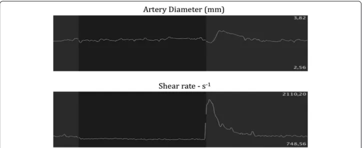
Figure 1 Flow-mediated dilation. Flow-mediated dilation (FMD) measures endothelial function by inducing a temporary ischemia in a brachial artery and observing the amount of vasodilation following the stressor event. After a baseline measurement of the artery diameter via an ultrasound probe, a sphygmomanometer blood pressure cuff is positioned on the right forearm 2 cm below the elbow and inflated to 250 mmHg to produce ischemia in the forearm. The cuff is deflated after some minutes, usually 5, thus causing a reactive hyperemia which in turn produces a shear stress stimulus that induces the endothelium to release nitric oxide as a vasodilator. FMD is considered as diameter after reactive hyperemia - basal diameter/basal diameter × 100. The figure shows the diameter (upper part) and shear rate (lower part) of brachial artery during FMD before (grey-shaded part on the left), during (black-shaded part), and after (grey-shaded part on the right) ischemia.
Figure 2 Carotid-Femoral Pulse Wave Velocity. Increased arterial stiffness leads to increased velocity of the pulse wave generated in the arteries by the contraction of the left ventricle. Pulse wave velocity (PWV) consists in measuring pulse wave profiles by tonometry at two distant locations (carotid and femoral) and measuring the delay in the onset of the wave between those two locations. PWV is calculated as the distance traveled by the wave divided by the time taken to travel that distance. Surface distance between the two recording sites and simultaneously recorded electrocardiograms are used to calculate wave transit time. The figure shows tonometric (white lines) recordings of the carotid (above) and femoral (below) artery waves according with simultaneous electrocardiographic (yellow lines) ‘R’ wave of the electrocardiogram as a timing reference.
Sacco et al. The Journal of Headache and Pain 2013, 14:80 Page 3 of 10 http://www.thejournalofheadacheandpain.com/content/14/1/80
Review
Arterial and endothelial function in migraineurs
Several lines of evidence for vascular dysfunction have been
identified in migraineurs and there is increasing evidence to
suggest that in migraineurs the vascular system is impaired
not only within the brain.
Endothelial function in migraineurs
The many studies assessing endothelial function in
migraineurs have used heterogeneous techniques and
indicators as shown in Tables 2, 3 and 4. Most of them
assessed endothelial function by measuring FMD
(Table 2) [20-30], some used PAT (Table 3) [31,32],
some measured peripheral vasodilation in response
to pharmacological stimuli (Table 3) [20,30,33-35], and
others assessed endothelial progenitor cells (EPCs)
(Table 4) [23,36,37]. Findings were not consistent
across the studies. Several among the available studies
did not reveal any alteration in the endothelial function
of migraineurs. Six of them did not find any difference
in FMD between migraineurs and control subjects
(Table 2) [20-23,25,26] and a further study confirmed
the lack of any alteration in endothelial function
assessed by PAT ratio (Table 3) [32]. Three studies
assessing peripheral vasodilation after pharmacological
stimuli found comparable responses in subjects with
and without migraine (Table 3) [20,33,35]. However,
other studies supported the presence of endothelial
dysfunction in migraineurs. Four studies reported
de-creased FMD in migraineurs [24,27,29,30]; in one of
those studies, FMD correlated with dysfunction of the
autonomic nervous system (Table 2) [24]. Another
study, using PAT, reported that subjects with chronic
migraine had worse endothelial function (Table 3) [31].
A further study found an abnormal vascular response
to dilating pharmacological stimuli (Table 3) [34]. The
possible underlying mechanisms of endothelial
dys-function in those studies were unclear. Since the
im-pairment of FMD was demonstrated also in individuals
with recent onset of migraine it is unlikely that the
dis-turbance was a consequence of longstanding and
re-peated exposure to the vasoconstrictor agents used for
migraine treatment [27]. In addition, the results of one
study suggested that the impaired vascular reactivity
in migraineurs is entirely attributable to a reduced
response of vascular smooth muscle cells to NO while
the endothelial response to NO appeared intact [34].
Some additional studies supported the presence of
an endothelial dysfunction in migraineurs by
docu-menting a decreased number of EPCs [23,36] or a
higher number of more mature EPCs (which could
be associated with potential endothelial damage) in
migraineurs (Table 4) [37]. EPCs maintain the
integ-rity of damaged endothelium and are considered a
marker of endothelial function [38]. At variance with
other studies, Vernieri et al., found that subjects
with migraine with aura had higher FMD than both
control subjects and migraine without aura subjects
indicating an excessive arterial response to hyperemia.
It is suggested that this is probably an effect of an
increased sensitivity to endothelium-derived NO or of
increased release of NO induced by shear stress (Table 2)
[28]. In agreement, Yetkin et al., found that migraineurs
have an increased nitrate-mediated response showing
a major role of NO supersensitivity in migraine
patho-physiology (Table 3) [30].
Several studies support the presence of biochemical
changes in the endothelial activation of migraineurs.
These biochemical changes may precede structural
damage. In particular, alterations in the NO pathway
have been described [30,39,40] as well as increased
levels of calcitonin gene-related peptide (CGRP),
vascu-lar endothelial growth factors (VEGF), von Willebrand
factor (vWF) activity, tissue plasminogen activator
anti-gen, C-reactive protein and endothelin-1 [21,23,40].
Re-cently, a study involving 40 non-hypertensive subjects
with migraine without aura randomized to treatment
with enalapril 5 mg twice a day for 3 months or to
pla-cebo, showed that active treatment was associated with
an increase in FMD values [41].
Figure 3 Augmentation Index. The interaction between the incident pulse wave and the reflected pulse wave, which is generated by the arterial resistance, is expressed by the augmentation index, which is the amount of pulse wave induced only by arterial resistance. In the figure, the upper arrow indicates the first systolic peak (incident wave), while the lower arrow indicates the second systolic peak (reflected wave). The ratio between the reflected wave/incident wave × 100 is the augmentation index.
Arterial function in migraineurs
Compared with studies on endothelial function, reports
on arterial function measured with PWV, AIx, and local
arterial distensibility are more consistent and suggest
that functional properties of large arteries are altered in
migraineurs [27,42-46] (Table 5).
Four studies addressed PWV in migraineurs (Table 5)
[27,42-44]. Ikeda et al. evaluated peripheral PWV [42]
while the other authors assessed central PWV [27,43,44].
Three studies on PWV [27,42,43] found higher values
of this parameter in migraineurs with respect to
non-migraineurs. However, in one of the studies PWV did
not correlate with the presence of migraine after
ad-justment for age and mean arterial pressure [27]. The
inclusion in the study of patients with a short history
of migraine and with less use of vasoconstrictive agents
may explain the difference [27]. Schillaci et al., also
addressed PWV according to migraine type and reported
that subjects with migraine either with or without aura
had increased PWV with respect to controls while
mi-graine with aura subjects had higher aortic PWV than
those without aura [43]. A more recent
population-based study, involving a larger number of migraineurs,
has challenged the suggestion of possible impairment
Table 2 Studies on endothelial function using flow-mediated dilation in migraineurs
Study (First author, year) Study design Patients included (n) % women Exclusion criteria Migraine diagnosis Results
De Hoon, 2006 [20] CC Migraine: 10 60 CVD, HYP, DM, dyslipidemia, CS ICHD-I No differences in FMD between migraineurs and controls Controls: 10
Hamed, 2010 [21] CC Migraine: 38 89 CVD and vascular RF, DM, CS, alcoholism, active gastrointestinal disease, gout, epilepsy, recent infection, renal failure, pregnancy or lactation, regular use FANS or antimigrainous drugs and OC
ICHD-II No differences in FMD between migraineurs and controls (MwA: 14; MwoA: 24) Controls: 35
Perko, 2011 [22] CC MwA: 20 80 CVD, obesity, HYP, dyslipidemia, pregnancy or lactation and regular use of vasoactive drugs
ICHD-II No differences in FMD between migraineurs and controls MwoA: 20
Controls: 20
Rodriguez-Osorio, 2012 [23] CC Migraine : 47 98 CAD, inflammatory disease, obesity, HYP, DM, dyslipidemia, CS, infectious disease, severe systemic disease, ovarium pathology, pregnancy or lactation, use of vasoactive drugs
ICHD-II No differences in FMD between migraineurs and controls (MwA:14; MwoA: 33) Controls: 23
Rossato, 2011 [24] CC Migraine: 20 75 Age ≥50, vascular RF, CS, use of vasoactive drugs
Not reported Decreased FMD in migraineurs Controls: 20
Silva, 2007 [25] CC Migraine: 50 (MwA: 25; MwoA: 25)
88 None ICHD-II No differences in FMD between migraineurs and controls Controls: 25
Thomsen, 1996 [26] CC MwoA: 12 100 Use of any daily drugs ICHD-I No differences in FMD between migraineurs and controls Controls: 12
Vanmolkot, 2007 [27] CC Migraine: 50 78 Age >50, CVD, HYP, obesity, DM, dyslipidemia, pregnancy or lactation, use of vasoactive drugs (except OC)
Validated questionnaire
Decreased FMD in migraineurs Controls: 50
Vernieri, 2010 [28] CC Migraine:21 None ICHD-II FMD increased from controls to MwoA to MwA patients (MwA: 11; MwoA: 10)
Controls: 13
Yetkin, 2006 [29] CC Migraine: 45 80 CAD, HYP, obesity, DM, infectious disease, ovarium pathology
ICHD-I Decreased FMD in migraineurs Controls: 45
Yetkin, 2007 [30] CC Migraine: 24 89 HYP, CAD, DM, infectious disease ICHD-I Decreased FMD in migraineurs Controls: 26
CC case–control; MwA migraine with aura; MwoA migraine without aura; CVD cardiovascular disease; HYP arterial hypertension; DM diabetes mellitus; CS cigarette smoking; RF risk factors; FANS non steroid anti-inflammatory drugs; OC oral contraceptives; CAD coronary artery disease; ICHD International classification headache disorders; FMD flow-mediated dilation.
Sacco et al. The Journal of Headache and Pain 2013, 14:80 Page 5 of 10 http://www.thejournalofheadacheandpain.com/content/14/1/80
of arterial function in migraineurs [44]. In fact, Stam
et al. reported no differences in PWV among subjects
with migraine with aura, migraine without aura, and
controls [44]. In this latter study subjects with known
cardiovascular risk factors were included and this may
explain the differences in the results [44].
Higher values of AIx were found in migraineurs by five
studies [27,31,32,43,45], which variously measured AIx
in the radial artery [45], in the aorta [27,43] or by finger
tonometry [31,32]. One of those studies involved only
women with migraine with aura [32] and one involved
only subjects with chronic migraine [31]. Schillaci et al.
reported increased AI in migraine with and without aura
with respect to controls and no differences between the
two groups of migraineurs [43]. All the studies about
AIx had a case–control design, except for a
population-based one [45] which involved cases and controls older
than those of the other studies.
Two studies assessed static arterial parameters [27,46].
De Hoon et al. found that migraineurs had larger
tem-poral artery diameter compared with control subjects
but a decreased distensibility and buffering capacity of
the brachial artery in the absence of significant
alter-ations in the common carotid and femoral arteries
Table 3 Studies on endothelial function by arterial tonometry or vasodilation in response to pharmacological stimuli
in migraineurs
Study (First author, year) Study design Patients included (n) % women Exclusion criteria Migraine diagnosis Results Arterial tonometryJiménez-Caballero, 2013 [31] CC CM: 21 71 Age ≥50, CVD, inflammatory disease, obesity, HYP, DM, dyslipidemia, CS, active cancer, ovarium pathology, pregnancy or lactation, regular use of vasoactive drugs
ICHD-II Smaller RHI in migraineurs Controls: 21
Liman, 2012 [32] CC MwA: 29 100 CVD, obesity, HYP, DM, pregnancy, use of drugs (statins, anticoagulants or antiplatelet and intake of triptans within the previous 24 h)
ICHD-II No differences in PAT ratio between migraineurs and controls Controls: 30
Vasodilation in response to pharmacological stimuli
De Hoon, 2006 [20] CC Migraine: 10 60 CVD, HYP, DM, dyslipidemia, CS
ICHD-I No differences between migraineurs and controls in vasodilation response to serotonin, sodium nitroprusside, and CGRP Controls: 10
Edvinsson, 2008 [33] CC MwoA: 9 78 None ICHD-I No differences between migraineurs and controls in vasodilation response after local heating and iontophoretic administration of acetylcholine, sodium nitroprusside, and CGRP Controls: 9
Napoli, 2009 [34] CC MwoA: 12 58 HYP, DM,
hypercholesterolemia, CVD, CS
ICHD-I Reduced response to endothelium-dependent vasodilation and production of cGMP in migraineurs; no difference in production of NO Controls: 12
Vanmolkot, 2010 [35] CC Migraine:16 75 Age >50, CVD, HYP, obesity, DM, CS, dyslipidemia, pregnancy or lactation, use of vasoactive drugs (except OC)
ICHD-II No differences between migraineurs and controls to sodium nitroprusside, substance P, and NG-monomethyl-L-arginine
Controls: 16
Yetkin, 2007 [30] CC Migraine: 24 89 HYP, CAD, DM, infectious disease
ICHD-I Higher nitrate-mediated dilation in migraineurs
Controls: 26
CC case–control; CM chronic migraine; MwA migraine with aura; MwoA migraine without aura; CVD cardiovascular disease; HYP arterial hypertension; DM,diabtes mellitus; CS cigarette smoking; OC oral contraceptive; ICHD International classification headache disorders; RHI reactive hyperhemia index; PAT peripheral arterial tonometry; CGRP Calcitonin Gene Related Peptide; cGMP cyclic guanosine monophosphate; NO nitric oxide.
[46]. Vanmolkot et al. found that migraineurs had a
de-creased diameter and compliance of superficial muscular
arteries [27].
Discussion
The results of the systematic review suggest the presence
of a peripheral vascular dysfunction in migraineurs.
However, while several studies support an alteration of
arterial function among subjects with migraine, findings
on the endothelial function are less clear.
Endothelial function in migraineurs has sometimes
been reported as impaired and sometimes as altered,
while some studies found no difference compared with
endothelial function in controls. Differences among
avail-able studies may be explained by various inclusion/
exclusion criteria and the generally small number of
participants. Migraine, and particularly migraine with
aura, has been associated with an elevated
cardiovascu-lar profile. The exclusion of subjects with cardiovascucardiovascu-lar
risk factors from some of the studies may at least partly
explain the described differences. Duration of migraine,
severity of attacks and the active or inactive state of the
disease may also have played a role in the
between-study differences in results. To clarify the status of
endothelial function in migraineurs further, studies are
required that need to involve large number of subjects
of properly selected individuals, applying rigorous
inclusion and exclusion criteria, and including subjects
with a wide range of migraine conditions in terms of
frequency, duration, and severity of the attacks. In
addition, further studies should also adopt a
standard-ized and reliable methodology to address endothelial
function.
Regarding arterial function, most of the available
evi-dence supports greater stiffness or impaired compliance
of the arterial system in migraineurs. The mechanisms
underlying the association between migraine and altered
arterial function are likely to be complex and may
involve both structural and functional changes in the
arterial wall. Since all the studies had a cross-sectional
design, a cause-effect relationship between migraine and
increased arterial stiffness cannot be established.
How-ever, migraine usually starts in early adulthood, at an age
in which arterial wall pathologies are rare. In the general
population alteration in arterial function has been
asso-ciated with the presence of vascular risk factors and
is considered a marker of vascular disease associated
with risk of future vascular events [47,48]. Since
most studies addressing arterial function in migraineurs
excluded subjects with comorbid vascular risk factors,
other mechanisms can be hypothesized. The possible
hypotheses include alterations in vessel wall structure in
migraineurs as supported by the presence of impaired
serum elastase activity [49], higher sympathetic tone
[50,51], use of drugs such as triptans and ergot
deriva-tives, or it may represent a primary alteration linked to
the presence of migraine itself [52]. At the moment it is
unknown whether the duration of migraine and the
frequency and severity of the attacks has an impact on
arterial stiffness. Moreover, gender differences may also
play a role in determining arterial stiffness in migraineurs
with females being more susceptible to changes [43].
In addition, possible clinical implications of arterial
dysfunction in migraineurs have to be clarified. In the
general population, arterial dysfunction has been linked to
an increased risk of vascular disease through an
athero-thrombotic mechanism [47,48]. However, in migraineurs
Table 4 Studies on endothelial function by endothelial progenitor cells in migraineurs
Study (First author, year) Study design Patients included (n) % women Exclusion criteria Migraine diagnosis Results
Lee, 2008 [36] CC Migraine: 92 70 CVD, diabetic retinopathy ICHD-II Decreased number and migratory capacity and higher senescence levels of endothelial progenitor cells in migraineurs versus controls
(MwoA: 67; MwA: 25) Controls: 37
Oterino, 2013 [37] CC CM: 51 73 CVD, inflammatory disease, cancer or treatment with antimitogen agents, pregnancy in the last year
ICHD-I Higher number of activated endothelial progenitor cells in migraineurs
EM: 48 Controls: 35
Rodriguez-Osorio, 2012 [23] CC Migraine : 47 98 CAD, inflammatory disease, obesity, HYP, DM, dyslipidemia, CS, infectious disease, severe systemic disease; ovarium pathology, pregnancy or lactation, use of vasoactive drugs
ICHD-II Lower endothelial progenitor cell counts in migraineurs
(MwA:14; MwoA: 33) Controls: 23
CC case–control; CM chronic migraine; EM episodic migraine; MwA migraine with aura; MwoA migraine without aura; CVD cardiovascular disease; CAD coronary artery disease; HYP arterial hypertension; DM diabetes mellitus; CS cigarette smoking; ICHD International classification
headache disorders.
Sacco et al. The Journal of Headache and Pain 2013, 14:80 Page 7 of 10 http://www.thejournalofheadacheandpain.com/content/14/1/80
the evidence of an atherothrombotic mechanism leading
to vascular events is not supported and much evidence
points against this possibility. [53,54] In fact, intima-media
thickness (IMT), which is a surrogate marker of subclinical
atherosclerosis has not been consistently linked to migraine
status [20,24,27,55,56]. In addition, despite little
informa-tion being available on the frequency of the different
sub-types of ischemic stroke in migraineurs, the available
studies do not support an increased atherosclerotic load
[57,58]. Also the cardiac events observed in migraineurs
did not seem to be attributable to atherothrombosis and
alternative mechanisms have been hypothesized [59-61].
Future studies should also clarify whether the vascular
dysfunction may represent a marker able to identify those
migraineurs who are at higher risk of presenting vascular
events and who might be the target of possible preventive
strategies.
Conclusion
The study of systemic vascular function is a promising
tool for non-invasively investigating the vascular health
of subjects with migraine. At the moment, evidence is too
scarce to support the clinical application of the techniques
and further research studies are needed to better clarify all
the pending issues.
Table 5 Studies on arterial function in migraineurs
Study (First author, year) Study design Patients included (n) % women Exclusion criteria Migraine diagnosis Parameters assessed Results De Hoon, 2003 [46] CC MwA: 11 76 CVD, inflammatory disease, HYP, DM, dyslipidemia
ICHD-I Diameter and compliance parameters of brachial, carotid, femoral, and temporal arteries
Smaller diameter and distension of brachial artery and larger right temporal artery diameter in migraineurs; no differences in carotid and femoral arteries
MwoA: 39 Controls: 50
Ikeda, 2011 [42] CC MwA:22 73 CVD, vascular RF ICHD-II Brachial PWV Higher PWV in migraineurs MwoA:89 Controls:110 Jiménez-Caballero, 2013 [31] CC CM: 21 71 Age ≥50, CVD, inflammatory disease, obesity, HYP, DM, dyslipidemia
ICHD-II Radial AIx Higher AIx in migraineurs Controls: 21
Liman, 2012 [32] CC MwA: 29 100 CVD, obesity, arterial HYP, DM
ICHD-II Peripheral AIx Higher Aix in migraineurs Controls: 30
Nagai, 2007 [45] PB Group A:134 93 Stroke Validated questionnaire
Radial AIx Higher AIx in migraineurs (5% migraineurs)
Group B:138 (17% migraineurs)
Schillaci, 2010 [43] CC MwA:17 85 CVD, inflammatory disease, HYP, DM, dyslipidemia
ICHD-II Aortic PWV and aortic AIx
Higher PWV and AIx in migraineurs, especially in MwA
MwoA: 43 Controls:60
Stam, 2013 [44] PB MwA: 123 75 None ICHD-II Carotid and femoral PWV
No differences between migraineurs and controls MwoA: 167
Controls: 542 Vanmolkot,
2007 [27]
CC Migraine: 50 78 None ICHD-I Diameter and compliance parameters of brachial and femoral arteries, aortic AIx and aortic PWV
Smaller brachial artery diameter and compliance and smaller femoral artery diameter in migraineurs; higher aortic AIx in migraineurs; higher PWV in migraineurs not confirmed after adjustment for age and mean arterial pressure
Controls: 50
CC case–control; PB population-based; CM chronic migraine; MwA, migraine with aura; MwoA, migraine without aura; CVD, cardiovascular disease; HYP, arterial hypertension; DM, diabetes mellitus; RF, risk factors; ICHD, International classification headache disorders; AIx, augmentation index; PWV, pulse wave velocity.
Competing interests
The authors declare that they have no competing interests.
Authors’ contributions
All authors of this manuscript (SS, PR, DG, RO, FP, AC, TK) have made substantial contributions to conception and design of the review, have been involved in drafting the manuscript and revising it critically for important intellectual content and have given final approval of the version to be published.
Author details
1
Department of Neurology and Regional Headache Center, University of L’Aquila, Piazzale Salvatore Tommasi 1, L’Aquila 67100, Italy.2Department of
Life, Health, and Eviromental Sciences, University of L’Aquila, L’Aquila 67100, Italy.3Inserm Research Center for Epidemiology and Biostatistics, Team
Neuroepidemiology, Bordeaux F-33000, France.4University of Bordeaux, Bordeaux F-33000, France.
Received: 5 August 2013 Accepted: 20 September 2013 Published: 1 October 2013
References
1. Connor RC (1962) Complicated migraine. A study of permanent neurological and visual defects caused by migraine. Lancet 2:1072–1075 2. Carolei A, Marini C, Di Napoli M, Di Gianfilippo G, Santalucia P, Baldassarre
M, De Matteis G, di Orio F (1997) High stroke incidence in the prospective community-based L’Aquila registry (1994–1998). First year’s results. Stroke 28:2500–2506
3. Tzourio C, Iglesias S, Hubert JB, Visy JM, Alpérovitch A, Tehindrazanarivelo A, Biousse V, Woimant F, Bousser MG (1993) Migraine and risk of ischaemic stroke: a case–control study. BMJ 307:289–292
4. Kurth T, Diener HC (2012) Migraine and stroke: perspectives for stroke physicians. Stroke 43:3421–3426
5. Kurth T, Chabriat H, Bousser MG (2012) Migraine and stroke: a complex association with clinical implications. Lancet Neurol 11:92–100
6. Sacco S, Ricci S, Carolei A (2012) Migraine and vascular diseases: a review of the evidence and potential implications for management. Cephalalgia 32:785–795
7. Sacco S, Ornello R, Ripa P, Pistoia F, Carolei A (2013) Migraine and hemorrhagic stroke: a meta-analysis. Stroke, in press
8. Bigal ME, Kurth T, Hu H (2009) Migraine and cardiovascular disease: possible mechanisms of interaction. Neurology 72:1864–1871
9. Sacco S, Olivieri L, Bastianello S, Carolei A (2006) Comorbid neuropathologies in migraine. J Headache Pain 7:222–230 10. Sacco S, Cerone D, Carolei A (2008) Comorbid neuropathologies in
migraine: an update on cerebrovascular and cardiovascular aspects. J Headache Pain 9:237–248
11. Brunner H, Cockcroft JR, Deanfield J, Donald A, Ferrannini E, Halcox J, Kiowski W, Lüscher TF, Mancia G, Natali A, Oliver JJ, Pessina AC, Rizzoni D, Rossi GP, Salvetti A, Spieker LE, Taddei S, Webb DJ, Working Group on Endothelins and Endothelial Factors of the European Society of Hypertension (2005) Endothelial function and dysfunction. Part II: Association with cardiovascular risk factors and diseases. A statement by the Working Group on Endothelins and Endothelial Factors of the European Society of Hypertension. J Hypertens 23:233–246
12. Davignon J, Ganz P (2004) Role of endothelial dysfunction in atherosclerosis. Circulation 109:III27–3
13. Tomiyama H, Yamashina A (2010) Non-invasive vascular function tests: their pathophysiological background and clinical application. Circ J 74:24–33 14. Widlansky ME, Gokce N, Keaney JF Jr, Vita JA (2003) The Clinical Implications
of Endothelial Dysfunction. J Am Coll Cardiol 42:1149–1160 15. Ross R (1999) Atherosclerosis-an inflammatory disease. N Engl J Med
340:115–126
16. Flammer AJ, Anderson T, Celermajer DS, Creager MA, Deanfield J, Ganz P, Hamburg NM, Lüscher TF, Shechter M, Taddei S, Vita JA, Lerman A (2012) The assessment of endothelial function: from research into clinical practice. Circulation 126:753–767
17. Al-Qaisi M, Kharbanda RK, Mittal TK, Donald AE (2008) Measurement of endothelial function and its clinical utility for cardiovascular risk. Vasc Health Risk Manag 4:647–652
18. Corretti MC, Anderson TJ, Benjamin EJ, Celermajer D, Charbonneau F, Creager MA, Deanfield J, Drexler H, Gerhard-Herman M, Herrington D, Vallance P, Vita J, Vogel R, International Brachial Artery Reactivity Task Force (2002) Guidelines for the ultrasound assessment of endothelial-dependent flow-mediated vasodilation of the brachial artery: a report of the International Brachial Artery Reactivity Task Force. J Am Coll Cardiol 39:257–265
19. Joannides R, Haefeli WE, Linder L, Richard V, Bakkali EH, Thuillez C, Lüscher TF (1995) Nitric oxide is responsible for flow-dependent dilatation of human peripheral conduit arteries in vivo. Circulation 91:1314–1319
20. De Hoon JN, Smits P, Troost J, Struijker-Boudier HA, Van Bortel LM (2006) Forearm vascular response to nitric oxide and calcitonin gene-related peptide: comparison between migraine patients and control subjects. Cephalalgia 26:56–63
21. Hamed SA, Hamed EA, Ezz Eldin AM, Mahmoud NM (2010) Vascular Risk Factors, Endothelial Function, and Carotid Thickness in Patients with Migraine: Relationship to Atherosclerosis. J Stroke Cerebrovasc Dis 19:92–103 22. Perko D, Pretnar-Oblak J, Šabovič M, Zaletel M, Žvan B (2011) Associations
between cerebral and systemic endothelial function in migraine patients: a post-hoc study. BMC Neurol 11:146
23. Rodríguez-Osorio X, Sobrino T, Brea D, Martínez F, Castillo J, Leira R (2012) Endothelial progenitor cells: a new key for endothelial dysfunction in migraine. Neurology 79:474–479
24. Rossato A, Veronese F, Maggioni F, Vedovetto V, Zancan A, Biasiolo M, Bilora F (2011) Autonomic dysfunction and endothelial changes in migraine sufferers. Panminerva Med 53:13–18
25. Silva FA, Rueda-Clausen CF, Silva SY, Zarruk JG, Guzmán JC, Morillo CA, Vesga B, Pradilla G, Flórez M, López-Jaramillo P (2007) Endothelial function in patients with migraine during the interictal period. Headache 47:45–51 26. Thomsen LL, Daugaurd D, Iversen H (1996) Normal radial artery dilatation
during reactive hyperaemia in migraine without aura. Endothelium 4:199–206
27. Vanmolkot FH, Van Bortel LM, de Hoon JN (2007) Altered arterial function in migraine of recent onset. Neurology 68:1563–1570
28. Vernieri F, Moro L, Altamura C, Palazzo P, Antonelli Incalzi R, Rossini PM, Pedone C (2010) Patients with migraine with aura have increased flow mediated dilation. BMC Neurol 10:18
29. Yetkin E, Ozisik H, Ozcan C, Aksoy Y, Turhan H (2006) Decreased endothelium-dependent vasodilatation in patients with migraine: a new aspect to vascular pathophysiology of migraine. Coron Artery Dis 17:29–33 30. Yetkin E, Ozisik H, Ozcan C, Aksoy Y, Turhan H (2007) Increased dilator
response to nitrate and decreased flow-mediated dilatation in migraineurs. Headache 47:104–110
31. Jiménez Caballero PE, Muñoz Escudero F (2013) Peripheral endothelial function and arterial stiffness in patients with chronic migraine: a case– control study. J Headache Pain 14:8
32. Liman TG, Neeb L, Rosinski J, Wellwood I, Reuter U, Doehner W, Heuschmann PU, Endres M (2012) Peripheral endothelial function and arterial stiffness in women with migraine with aura: a case–control study. Cephalalgia 32:459–466
33. Edvinsson ML, Edvinsson L (2008) Comparison of CGRP and NO responses in the human peripheral microcirculation of migraine and control subjects. Cephalalgia 28:563–566
34. Napoli R, Guardasole V, Zarra E, Matarazzo M, D’Anna C, Saccà F, Affuso F, Cittadini A, Carrieri PB, Saccà L (2009) Vascular smooth muscle cell dysfunction in patients with migraine. Neurology 72:2111–2114
35. Vanmolkot FH, de Hoon JN (2010) Endothelial function in migraine: a cross-sectional study. BMC Neurol 10:119
36. Lee ST, Chu K, Jung KH, Kim DH, Kim EH, Choe VN, Kim JH, Im WS, Kang L, Park JE, Park HJ, Park HK, Song EC, Lee SK, Kim M, Roh JK (2008) Decreased number and function of endothelial progenitor cells in patients with migraine. Neurology 70:1510–1517
37. Oterino A, Toriello M, Palacio E, Quintanilla VG, Ruiz-Lavilla N, Montes S, Vega MS, Martinez-Nieto R, Castillo J, Pascual J (2013) Analysis of endothelial precursor cells in chronic migraine: a case–control study. Cephalalgia 33:236–244
38. George J, Shmilovich H, Deutsch V, Miller H, Keren G, Roth A (2006) Comparative analysis of methods for assessment of circulating endothelial progenitor cells. Tissue Eng 12:331–335
39. Fidan I, Yüksel S, Ymir T, Irkeç C, Aksakal FN (2006) The importance of cytokines, chemokines and nitric oxide in pathophysiology of migraine. J Neuroimmunol 171:184–188
Sacco et al. The Journal of Headache and Pain 2013, 14:80 Page 9 of 10 http://www.thejournalofheadacheandpain.com/content/14/1/80
40. Tietjen GE, Herial NA, White L, Utley C, Kosmyna JM, Khuder SA (2009) Migraine and biomarkers of endothelial activation in young women. Stroke 40:2977–2982
41. Javanmard SH, Sonbolestan SA, Heshmat-Ghahdarijani K, Saadatnia M, Sonbolestan SA (2011) Enalapril improves endothelial function in patients with migraine: A randomized, double-blind, placebo-controlled trial. J Res Med Sci 16:26–32
42. Ikeda K, Hirayama T, Iwamoto K, Takazawa T, Kawase Y, Yoshii Y, Kano O, Kawabe K, Tamura M, Iwasaki Y (2011) Pulse wave velocity study in middle-aged migraineurs at low cardiovascular disease risk. Headache 51:1239–1244 43. Schillaci G, Sarchielli P, Corbelli I, Pucci G, Settimi L, Mannarino MR, Calabresi
P, Mannarino E (2010) Aortic stiffness and pulse wave reflection in young subjects with migraine: A case–control study. Neurology 75:960–966 44. Stam AH, Weller CM, Janssens AC, Aulchenko YS, Oostra BA, Frants RR, van
den Maagdenberg AM, Ferrari MD, van Duijn CM, Gisela MT (2013) Migraine is not associated with enhanced atherosclerosis. Cephalalgia 33:228–235 45. Nagai T, Tabara Y, Igase M, Nakura J, Miki T, Kohara K (2007) Migraine is
associated with enhanced arterial stiffness. Hypertens Res 30:577–583 46. De Hoon JN, Willigers JM, Troost J, Struijker-Boudier HA, Van Bortel LM
(2003) Cranial and peripheral interictal vascular changes in migraine patients. Cephalalgia 23:96–104
47. Grassi D, Desideri G, Ferri C (2011) Cardiovascular risk and endothelial dysfunction: the preferential route for atherosclerosis. Curr Pharm Biotechnol 12:1343–1353
48. Bonetti PO, Lerman LO, Lerman A (2003) Endothelial dysfunction: a marker of atherosclerotic risk. Arterioscler Thromb Vasc Biol 23:168–175 49. Tzourio C, Kittner SJ, Bousser MG, Alpérovitch A (2000) Migraine and stroke
in young women. Cephalalgia 20:190–199
50. Cortelli P, Pierangeli G, Parchi P, Contin M, Baruzzi A, Lugaresi E (1991) Autonomic nervous system function in migraine without aura. Headache 31:457–462
51. Martín R, Ribera C, Moltó JM, Ruiz C, Galiano L, Matías-Guiu J (1992) Cardiovascular reflexes in patients with vascular headache. Cephalalgia 12:360–364
52. Sacco S, Ripa P, Carolei A (2011) Migraine attributed to genetic disorder: proposal of a new category. Cephalalgia 31:760–762
53. Ikeda K, Nakamura Y, Iwamoto K, Kawabe K, Iwasaki Y (2010) Ankle-brachial index in migraineurs. Headache 50:1215
54. Jurno ME, Chevtchouk L, Nunes AA, de Rezende DF, Jevoux Cda C, de Souza JA, Moreira Filho PF (2010) Ankle-brachial index, a screening for peripheral obstructive arterial disease, and migraine – a controlled study. Headache 50:626–630
55. Schwaiger J, Kiechl S, Stockner H, Knoflach M, Werner P, Rungger G, Gasperi A, Willeit J (2008) Burden of atherosclerosis and risk of venous
thromboembolism in patients with migraine. Neurology 71:937–943 56. Tzourio C, Gagniere B, El Amrani M, Alperovitch A, Bousser MG (2003)
Relationship between migraine, blood pressure and carotid thickness. A population-based study in the elderly. Cephalalgia 23:914–920 57. MacClellan LR, Giles W, Cole J, Wozniak M, Stern B, Mitchell BD, Kittner SJ
(2007) Probable migraine with visual aura and risk of ischemic stroke: the stroke prevention in young women study. Stroke 38:2438–2445 58. Rist PM, Buring JE, Kase CS, Schürks M, Kurth T (2010) Migraine and
functional outcome from ischemic cerebral events in women. Circulation 122:2551–2557
59. Ahmed B, Bairey Merz CN, McClure C, Johnson BD, Reis SE, Bittner V, Pepine CJ, Sharaf BL, Sopko G, Kelsey SF, Shaw L, WISE Study Group (2006) Migraines, angiographic coronary artery disease and cardiovascular outcomes in women. Am J Med 119:670–675
60. Kurth T, Gaziano JM, Cook NR, Bubes V, Logroscino G, Diener HC, Buring JE (2007) Migraine and risk of cardiovascular disease in men. Arch Intern Med 167:795–801
61. Rose KM, Carson AP, Sanford CP, Stang PE, Brown CA, Folsom AR, Szklo M (2004) Migraine and other headaches: associations with Rose angina and coronary heart disease. Neurology 63:2233–2239
doi:10.1186/1129-2377-14-80
Cite this article as: Sacco et al.: Peripheral vascular dysfunction in migraine: a review. The Journal of Headache and Pain 2013 14:80.
Submit your manuscript to a
journal and benefi t from:
7 Convenient online submission 7 Rigorous peer review
7 Immediate publication on acceptance 7 Open access: articles freely available online 7 High visibility within the fi eld
7 Retaining the copyright to your article

![Table 5 Studies on arterial function in migraineurs Study (First author, year) Study design Patients included (n) % women Exclusioncriteria Migraine diagnosis Parametersassessed Results De Hoon, 2003 [46] CC MwA: 11 76 CVD, inflammatorydisease, HYP, DM, dy](https://thumb-eu.123doks.com/thumbv2/123doknet/14697142.563539/9.892.86.821.149.798/studies-migraineurs-patients-exclusioncriteria-migraine-diagnosis-parametersassessed-inflammatorydisease.webp)