HAL Id: hal-01999389
https://hal.archives-ouvertes.fr/hal-01999389
Submitted on 15 Feb 2019
HAL is a multi-disciplinary open access
archive for the deposit and dissemination of
sci-entific research documents, whether they are
pub-lished or not. The documents may come from
teaching and research institutions in France or
abroad, or from public or private research centers.
L’archive ouverte pluridisciplinaire HAL, est
destinée au dépôt et à la diffusion de documents
scientifiques de niveau recherche, publiés ou non,
émanant des établissements d’enseignement et de
recherche français ou étrangers, des laboratoires
publics ou privés.
Coxiella burnetii effector CvpB modulates
phosphoinositide metabolism for optimal vacuole
development
Eric Martinez, Julie Allombert, Franck Cantet, Anissa Lakhani, Naresh
Yandrapalli, Aymeric Neyret, Isobel Norville, Cyril Favard, Delphine Muriaux,
Matteo Bonazzi
To cite this version:
Eric Martinez, Julie Allombert, Franck Cantet, Anissa Lakhani, Naresh Yandrapalli, et al.. Coxiella
burnetii effector CvpB modulates phosphoinositide metabolism for optimal vacuole development.
Pro-ceedings of the National Academy of Sciences of the United States of America , National Academy of
Sciences, 2016, 113 (23), pp.E3260-E3269. �10.1073/pnas.1522811113�. �hal-01999389�
Coxiella burnetii effector CvpB modulates
phosphoinositide metabolism for optimal
vacuole development
Eric Martineza, Julie Allomberta, Franck Canteta, Anissa Lakhania, Naresh Yandrapallia, Aymeric Neyreta,
Isobel H. Norvilleb, Cyril Favarda, Delphine Muriauxa, and Matteo Bonazzia,1
aCNRS, Formation de Recherche en Évolution 3689, Centre d’études d’agents Pathogènes et Biotechnologies pour la Santé, Université Montpellier, 34090
Montpellier, France; andbDefence Science and Technology Laboratory, Porton Down SP4 0JQ, United Kingdom
Edited by Yasuko Rikihisa, The Ohio State University, Columbus, OH, and approved April 26, 2016 (received for review November 18, 2015)
The Q fever bacteriumCoxiella burnetii replicates inside host cells within a largeCoxiella-containing vacuole (CCV) whose biogenesis relies on the Dot/Icm-dependent secretion of bacterial effectors. Several membrane trafficking pathways contribute membranes, proteins, and lipids for CCV biogenesis. These include the endo-cytic and autophagy pathways, which are characterized by phos-phatidylinositol 3-phosphate [PI(3)P]-positive membranes. Here we show that theC. burnetii secreted effector Coxiella vacuolar protein B (CvpB) binds PI(3)P and phosphatidylserine (PS) on CCVs and early endosomal compartments and perturbs the activity of the phosphatidylinositol 5-kinase PIKfyve to manipulate PI(3)P me-tabolism. CvpB association to early endosome triggers vacuolation and clustering, leading to the channeling of large PI(3)P-positive membranes to CCVs for vacuole expansion. At CCVs, CvpB binding to early endosome- and autophagy-derived PI(3)P and the concom-itant inhibition of PIKfyve favor the association of the autophago-somal machinery to CCVs for optimal homotypic fusion of the Coxiella-containing compartments. The importance of manipulat-ing PI(3)P metabolism is highlighted by mutations incvpB resulting in a multivacuolar phenotype, rescuable by gene complementa-tion, indicative of a defect in CCV biogenesis. Using the insect modelGalleria mellonella, we demonstrate the in vivo relevance of defective CCV biogenesis by highlighting an attenuated viru-lence phenotype associated withcvpB mutations.
Coxiella burnetii
|
host–pathogen interactions|
phosphoinositidesT
he obligate intracellular bacterium Coxiella burnetii is respon-sible for severe outbreaks of the zoonosis Q fever (1). The pri-mary targets of Coxiella are alveolar macrophages; however, bacteria can invade phagocytic and nonphagocytic cells and disseminate to other tissues and organs, such as the liver and heart, giving rise to hepatitis and endocarditis (1). Because of its remarkable infectivity and its environmental stability, Coxiella is considered a category B biothreat (2). Upon internalization by phagocytic and nonphagocytic cells, bacteria remain confined within Coxiella-containing vacuoles (CCVs), tight-fitting com-partments that mature along the endocytic pathway (1). Key to the successful colonization of host cells is the acidification of CCVs, which activates bacterial metabolism and effectors translocation by a Dot/Icm type IV secretion system (3). Bioinformatics analysis identified more than 300 Coxiella candidate effectors, a third of which have been validated for secretion (1, 4). Many Coxiella ef-fectors remain poorly understood or completely uncharacterized, but it is clear that several of these manipulate host vesicular trafficking, allowing CCVs to intercept and recruit membranes, proteins, and lipids from the endocytic, autophagy, and recycling pathways (4). SNAREs such as VAMP7 and Syntaxin-17 play a key role in this process, mediating the fusion events that lead to the biogenesis of large CCVs that occupy the majority of the host cell cytoplasm (5, 6). Mature CCVs are dynamic acidic compartments (pH 4.8), containing active hydrolases, which Coxiella is able to resist, and are positive for markers of late endosomes, lysosomes,and autophagosomes such as vATPase, LAMP1, CathepsinD, CD63, and microtubule-associated protein 1A/1B-light chain 3 (LC3) (7). Many efforts are being focused on the identification and characterization of the Coxiella effectors involved in CCV bio-genesis, as well as in the identification of the host cell trafficking pathways hijacked by this microbe. It has recently been shown that the Coxiella effector Coxiella vacuolar protein B (CvpB; also referred to as Cig2) is required for CCV homotypic fusion, which seems to be mediated by the autophagy machinery (8, 9). However, the link between CvpB and autophagy remains to be defined.
Phosphoinositides (PIs) are short-lived lipids whose spatio-temporal localization is determined by the activity of specific ki-nases and phosphatases (10). Their presence dictates the identity of cellular organelles and allows the targeted recruitment and activation of many downstream effectors, making them important signaling hubs for the regulation of cellular functions, including membrane traffic and actin rearrangement. In recent years, PIs have emerged as new targets for a number of intracellular bac-terial pathogens (11–13). As for all host/pathogen interactions, the strategies developed by different pathogens to exploit host PIs are extremely diverse. Bacterial effectors may use host PIs as anchors to specific host cell membranes, whereas others have evolved to manipulate their metabolism directly by mimicking eukaryotic kinases or phosphatases or indirectly by enhancing or blocking the activity of host PI-metabolizing enzymes (11–13). The manipulation
Significance
The biogenesis of a replicative vacuole is an essential step of Coxiella burnetii infections and involves the hijack of several host membrane trafficking pathways. Here we describeCoxiella vacuolar protein B (CvpB) as a Coxiella effector that interacts with phosphoinositides on host cell membranes and manipulates phosphatidylinositol 3-phosphate [PI(3)P] metabolism for optimal Coxiella-containing vacuole (CCV) development. This is achieved by perturbing the activity of the phosphatidylinositol 5-kinase PIKfyve, leading to an enrichment of PI(3)P on CCV membranes, which is required for the autophagy machinery to mediate CCV homotypic fusion. The importance of this process is highlighted by a homotypic fusion defect between CCVs in cells infected with CvpBCoxiella mutants, which translates into an attenuated viru-lence in the insect modelGalleria mellonella.
Author contributions: E.M., J.A., C.F., D.M., and M.B. designed research; E.M., J.A., F.C., A.L., N.Y., A.N., I.H.N., and M.B. performed research; E.M., J.A., F.C., N.Y., and M.B. con-tributed new reagents/analytic tools; E.M., J.A., F.C., A.L., I.H.N., C.F., D.M., and M.B. analyzed data; and E.M. and M.B. wrote the paper.
The authors declare no conflict of interest. This article is a PNAS Direct Submission.
1To whom correspondence should be addressed. Email: matteo.bonazzi@cpbs.cnrs.fr. This article contains supporting information online atwww.pnas.org/lookup/suppl/doi:10. 1073/pnas.1522811113/-/DCSupplemental.
of PI metabolism has pleiotropic downstream effects, mediated by the numerous PI effectors, which are collectively beneficial for the establishment/development of infections.
Building on our recent screen of a Coxiella transposon library (14, 15), here we use image-based supervised machine learning to address the mechanisms regulating the fusogenicity of CCVs. We show that the Coxiella effector CvpB localizes at CCVs and early endosomes by interacting with phosphatidylinositol 3-phosphate [PI(3)P] and phosphatidylserine (PS). CvpB binding to PI(3)P interferes with the recruitment of the phosphatidyli-nositol 5-kinase PIKfyve at PI(3)P-positive compartments, thereby preventing PI(3)P phosphorylation to PI(3,5)P2. This allows
CvpB to play a dual role in CCV biogenesis during infections. At early endosomes, CvpB leads to the appearance of large PI(3)P-positive compartments that interact and fuse with CCVs for vacuole expansion. At CCVs, CvpB binding to PI(3)P incoming from early endosomes and autophagosomes, and the concomi-tant inhibition of PIKfyve, is essential to recruit and stabilize components of the autophagy machinery to mediate CCV homo-typic fusion. The significance of this process is highlighted by six independent transposon insertions in cvpB resulting in a multi-vacuolar phenotype, with Coxiella replicating within isolated CCVs that fail to coalesce into a single vacuole. The cvpB::Tn multivacuolar phenotype can be rescued by gene complementa-tion or by inhibiting the phosphatidylinositol 5-kinase PIKfyve, provided that the autophagy machinery is functional. Impor-tantly, despite the lack of an intracellular replication phenotype, cvpB mutation attenuates Coxiella virulence in the insect model Galleria mellonella, demonstrating an important role of CCV biogenesis for Coxiella pathogenesis.
Results
Identification ofCoxiella Factors Involved in CCV Biogenesis Using Image-Based Supervised Machine Learning. In the context of a multiparametric high-content screen of a Coxiella transposon mutant library, we have recently reported the seemingly multi-vacuolar phenotype associated with a transposon insertion in the dot/icm gene icmS (14). The Legionella pneumophila IcmS ho-molog is a chaperone that mediates the translocation of specific effectors (16, 17), suggesting that Coxiella IcmS may regulate the translocation of effectors involved in the homotypic fusion of CCVs. This phenotype was further investigated by challenging Vero cells for 6 d with the Coxiella control transposon mutant Tn1832 [which carries an intergenic transposon insertion and phenocopies WT Coxiella (14)] or the icmS::Tn mutant and an-alyzing the morphology of CCVs by fluorescence microscopy. Cells infected with Tn1832 displayed large CCVs positive for the lysosomal marker LAMP1 and the autophagosomal marker LC3 (SI Appendix, Fig. S1 A and B, Top). Conversely, Coxiella icmS::Tn replicated within clusters of small LAMP1-positive, but LC3-negative, compartments (SI Appendix, Fig. S1 A and B, Bottom). EM confirmed that cells infected with Tn1832 presented large Coxiella-filled vacuoles (SI Appendix, Fig. S1C, Left), as opposed to icmS::Tn-infected cells (SI Appendix, Fig. S1C, Right), which presented several tight-fitting vacuoles enclosing few bacteria. To determine whether the multivacuolar phenotype results from the defective secretion of Coxiella effectors involved in CCV homotypic fusion, we used image-based supervised machine learning to mine the database generated during the screen of our Coxiella transposon library (14) and identify transposon inser-tions resulting in multivacuolar phenotypes. Among others, this approach identified the icmS::Tn mutant as well as all six transposon insertions in cvpB (CBU_0021; SI Appendix, Fig. S2A) previously identified in our screen as affecting the size of intracellular Coxiella colonies (14) and recently reported as im-portant for the homotypic fusion of CCVs (8). The axenic and intracellular replication of six independent cvpB transposon mutants (Tn1146, Tn396, Tn239, Tn170, Tn2579, and Tn2032)
was thus analyzed. When inoculated in the axenic medium acidified citrate cysteine medium-2 (ACCM-2) and allowed to grow for 6 d, cvpB mutants replicated with kinetics comparable to those of Tn1832 (SI Appendix, Fig. S2B). However, when used to challenge epithelial cell lines (Vero, HeLa, U2OS, or A431), cvpB mutants consistently displayed a marked multivacuolar phenotype, with Coxiella replicating within clusters of three or more LAMP1-positive CCVs (SI Appendix, Fig. S2C). The num-ber and size of Coxiella CCVs within a cluster varied depending on the cell line: Vero and A431 cells infected with the cvpB mutants presented a higher number of relatively small CCVs; these were fewer but larger in HeLa cells, and U2OS cells displayed an in-termediate phenotype. More than 50% of cells infected with the cvpB mutants presented a multivacuolar phenotype, regardless of the cell line and of the multiplicity of infection (MOI) used for infections (e.g., 100, 10, 1). As previously reported (8), the pres-ence of multiple, average-sized CCVs in cells infected with the cvpB transposon mutants did not impact the intracellular repli-cation of Coxiella (SI Appendix, Fig. S2D). cvpB encodes an 809-aa protein with a PmrA consensus sequence (18) 102 nt upstream of the start codon and a putative E-block domain (19) at the C terminus (SI Appendix, Fig. S2A). CvpB translocation was thus assessed by using a β-lactamase secretion assay on U2OS cells challenged with Coxiella for 24 and 48 h. β-Lactamase–tagged CvpB was efficiently translocated at both time points (SI Appen-dix, Fig. S3A). Conversely, CvpB translocation was abolished by the deletion of its putative E-block or by a transposon insertion in dotA (SI Appendix, Fig. S3A). The efficient expression of all β-lactamase–tagged CvpB constructs was verified by Western blot (SI Appendix, Fig. S3B). These observations are in agreement with the recently reported translocation of CvpB (8, 9).
The Coxiella cvpB::Tn Mutant Is Attenuated in the in Vivo Model G. mellonella.The in vivo relevance of the CvpB mutation was investigated by using the insect model G. mellonella (20). Larvae were injected in the upper right proleg with 106genome equiv-alents (GE)/mL of WT Coxiella, Tn1832, or the cvpB mutant Tn1146, and the survival rate was determined over 10 d. Larvae injected with PBS solution were used as controls. As previously reported (14, 20), Galleria larvae were efficiently killed by WT Coxiella or Tn1832; however, the injection of the cvpB mutant strain resulted in the killing of 50% of the larvae over the same time course (SI Appendix, Fig. S3C). Importantly, taking into account the lack of obvious intracellular replication phenotypes associated with cvpB mutations, this attenuation in virulence speaks in favor of an important role of CCV biogenesis for Coxiella pathogenesis. The bacterial load in infected larvae was monitored at 96 and 120 h post injection by real-time PCR using the Coxiella com1 gene. At both time points, Coxiella load within infected larvae was significantly affected in the cvpB mutation (SI Appendix, Fig. S3D).
CvpB Mediates Homotypic Fusion of CCVs and Vacuolation.As all six transposon insertions in cvpB result in comparable phenotypes, we focused our study on Tn1146, which carries the most up-stream transposon insertion, 791 nt downup-stream of the starting codon (14). Southern blot analysis validated the presence of a single transposon insertion in Tn1146 (hereafter referred to as cvpB::Tn; SI Appendix, Fig. S3E). The cvpB::Tn mutation was complemented by using a mini-Tn7 transposon system to express an HA-tagged copy of CvpB under the regulation of an anhy-drotetracycline (aTc)-inducible promoter (cvpB::Tn-miniTn7:cvpB; hereafter referred to as cvpB::Tn Comp.). Expression of CvpB by the Coxiella complemented strain was assessed in the presence of aTc by Western blot (SI Appendix, Fig. S3F). U2OS cells were then challenged with the cvpB::Tn complemented strain in the presence of aTc to trigger CvpB expression at the time of in-fection. Expression of CvpB by cvpB::Tn Comp. restored the
Martinez et al. PNAS | Published online May 25, 2016 | E3261
MIC
ROBIOLOGY
PNAS
formation of single, large CCVs (SI Appendix, Fig. S2C). To confirm that the multivacuolar phenotype was indeed caused by an impairment of fusion between CCVs, U2OS cells were chal-lenged with the control transposon mutant Tn1832 or the cvpB::Tn mutant for 24 h and imaged over 36 h. CCV formation and development was assessed by phase contrast, and Coxiella rep-lication was monitored by using the GFP expressed by trans-poson mutants. Cells infected with Coxiella Tn1832 displayed large CCVs undergoing fission and fusion events, whereas cells infected with the cvpB::Tn mutant gradually formed smaller CCVs, which largely failed to coalesce into single CCVs over time (Movie S1). Next, U2OS cells were challenged with the cvpB::Tn complemented strain, and CvpB expression was trig-gered at the time of infection or 72 h after infection. Induction of CvpB at the moment of infection restored the formation of single, large CCVs (Movie S1). Cells infected with the noninduced complemented strain presented the typical multivacuolar phenotype. This rapidly converted into a single CCV phenotype upon induction of CvpB expression (Movie S2), clearly demon-strating a role for CvpB in CCV homotypic fusion. Interestingly, cells challenged with Tn1832 or the cvpB::Tn complemented strains were characterized by the formation of large peripheral vesicles, which migrated toward the forming CCV (Movie S1). This was never observed in noninfected cells or in cells chal-lenged with the cvpB::Tn strain, suggesting that CvpB favors vacuolation (possibly of early endosomes), which may contribute membranes to the forming CCV over the course of the infection.
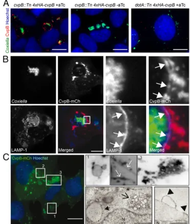
CvpB Localizes at CCVs and Early Endosomes, Triggering Endosome Enlargement and Clustering. To investigate the intracellular lo-calization of CvpB, the cvpB::Tn mutation was complemented by using a mini-Tn7 transposon system to express a 4xHA-tagged copy of CvpB under the regulation of an aTc-inducible promoter (cvpB::Tn 4xHAcvpB). The Coxiella dotA::Tn mutant (14) was transformed with the same construct as negative control (dotA::Tn 4xHA-cvpB). U2OS cells were then challenged with the Coxiella cvpB::Tn or dotA::Tn strains complemented with 4xHA-tagged CvpB in the presence or absence of aTc. Cells were processed for immunofluorescence at different time points postinfection, and an anti-HA antibody was used to reveal the intracellular locali-zation of CvpB. The translocated effector was more efficiently observed at 24 h post infection and mainly labeled CCVs. However, CvpB was also detected as spots scattered throughout the cytoplasm of infected cells (Fig. 1A). Labeling was absent in cells challenged with the noninduced cvpB::Tn 4xHAcvpB strain or the induced dotA::Tn 4xHA-cvpB strain (Fig. 1A). The partic-ular immunofluorescence protocol used to visualize 4xHA-CvpB during infection did not allow the labeling of other intracellular proteins. We thus optimized the codons in the cvpB ORF to facilitate expression in eukaryotic cells and generated CvpB constructs carrying HA, FLAG, or mCherry tags at the car-boxyl-terminal end of the protein to visualize CvpB upon ec-topic expression. Ecec-topically expressed CvpB decorated LAMP1-positive CCVs in cells infected with the Coxiella trans-poson mutant Tn1832 (Fig. 1B). Confirming our initial obser-vations, CvpB also labeled vesicular structures clustered around CCVs or scattered throughout the cytoplasm of infected cells (Fig. 1B), which were LAMP1-negative. Membrane localization was never observed when cells were transfected with plasmids carrying the tags alone.
The nature of CvpB-labeled vesicles was then characterized in noninfected cells. CvpB localized at large vacuolar structures as well as peripheral clusters of smaller vesicles in U2OS cells transfected with HA-FLAG or mCherry-tagged CvpB (Fig. 1C, Left and Top Right). CvpB-HA–transfected U2OS cells were also processed for immuno-EM and analyzed at the ultrastructural level (Fig. 1C, Bottom Right), confirming CvpB localization at large vacuoles and clusters of smaller vacuoles (Fig. 1C, Bottom
Middle). Ultrastructural analysis also revealed a clear plasma membrane localization of CvpB that was elusive at the fluores-cence level (Fig. 1C, Bottom Right). Immunogold labeling of CvpB at the plasma membrane was concentrated at micro-domains and membrane protrusions. Nontransfected or HA-transfected cells did not present large vacuoles or clusters of small vacuoles. The presence of markers of intracellular com-partments at CvpB-induced vacuoles was then assessed. As expected, CvpB-induced vesicles were negative for the lysosomal marker LAMP1 (Fig. 2A). It has been suggested that CvpB may be involved in recruiting autophagosomes to forming CCVs (8). We therefore compared CvpB localization with that of the auto-phagosomal marker LC3. U2OS cells transfected with CvpB-mCherry and LC3C-GFP (Fig. 2B) or LC3B-GFP were mock treated or incubated with chloroquine for 3 h before fixation. CvpB was never found colocalizing with either autophagy marker
Fig. 1. CvpB localizes at host membranes in Coxiella-infected and non-infected cells. (A) U2OS cells were challenged with cvpB::Tn 4xHAcvpB in the presence (Left) or absence (Middle) of aTc or with dotA::Tn 4xHA-cvpB in the presence of aTc (Right). Cells were fixed 24 h post infection, and translocated CvpB was labeled by using an anti-HA antibody (red), bacteria were visual-ized using GFP (green), and host cell nuclei were visualvisual-ized using Hoechst 33258 (blue). (B) U2OS cells were infected for 3 d with Coxiella Tn1832 and transfected with pLVX-CvpB-mCherry. At 12 h post transfection, cells were fixed, and Coxiella colonies, CvpB, and LAMP1 were detected by using green fluorescence (green), mCherry fluorescence (red), and anti-LAMP1 coupled to Alexa Fluor 647 (blue), respectively. White arrows indicate discrete re-gions of the Coxiella vacuole where CvpB and LAMP1 colocalize. (C, Left and Top Right) U2OS cells transiently transfected with pLVX-CvpB-mCherry (green) were fixed and labeled with Hoechst 33258 (blue). CvpB is found on large vacuolar structures (1), tubules (2), and peripheral clusters of smaller vesicles (3). (Insets) Images shown converted to inverted grayscale. (C, Bot-tom Right) U2OS cells transiently transfected with CvpB-HA were fixed and processed for immuno-EM with the HA tag labeled using Nanogold particles. CvpB could be detected in clusters of small vesicles (arrow), enlarged vesicles (filled arrow), or plasma membrane protrusions (arrowheads). (Scale bars: A–C, Left and Top Right, 10 μm; C, Bottom Right, 1 μm).
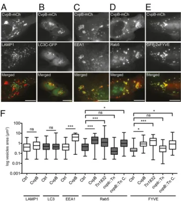
in any of the aforementioned conditions (Fig. 2B). On the con-trary, CvpB vesicles were positive for the early endosomal markers EEA1 (Fig. 2C), Rab5 (Fig. 2D), and the PI(3)P probe GFP-2xFYVE (Fig. 2E). Of note, CvpB expression triggered a significant increase in the size of early endosomes compared with cells expressing mCherry alone (Fig. 2F). Late endosome and autophagosome morphology was unaffected by CvpB expression (Fig. 2F). These findings indicate that CvpB specifically targets CCVs and early endosomal compartments and is capable of trig-gering endosome swelling and clustering.
CvpB Binds PI(3)P and PS. The remarkable colocalization be-tween CvpB and the 2xFYVE probe prompted us to determine whether CvpB localization at CCVs and early endosomes might be dictated by direct binding of PI(3)P on host cell membranes. We thus generated a GST-tagged CvpB construct and used the purified recombinant protein in a PIP Strip assay (Fig. 3A). Purified GST was used as a control and did not interact with any lipid spotted on the PIP Strips (SI Appendix, Fig. S3G). Con-versely, GST-CvpB bound to monophosphorylated PIs [with a preferential binding to PI(3)P] and with PS (Fig. 3A). CvpB binding to PIs was further analyzed by using a PIP array, which confirmed the preferential binding of the Coxiella effector to PI(3)P (Fig. 3B). CvpB binding to PI(3)P was consistent with the colocalization between CvpB and the PI(3)P probe GFP-2xFYVE, whereas the binding to PS was unexpected. U2OS cells were thus cotransfected with CvpB-mCherry and the GFP-tagged C2 domain of lactadherin (GFP-Lact-C2), a specific PS probe. In-deed, CvpB colocalized with the majority of intracellular structures labeled by GFP-Lact-C2 (SI Appendix, Fig. S4A). CvpB binding to
PS and PI(3)P was then validated by using an in vitro cosedi-mentation assay in which histidine-tagged CvpB was incubated with large unilamellar vesicles (LUVs) of lipid composition matching those found on PIP Strips at concentrations compatible with those observed in intracellular vesicles (21). In agreement with our ob-servations using PIP Strips, CvpB did not associate with LUVs containing 100% phosphatidylcholine (PC; Fig. 3C). CvpB in-teraction with LUVs was triggered by the combination of 30% PS and 70% PC or 2% PI(3)P and 98% PC at LUVs membranes, and was seemingly enhanced by a mixture of 68% PC, 30% PS, and 2% PI(3)P (Fig. 3C).
Bioinformatics analysis failed to highlight known lipid-binding domains for CvpB; thus, a mutational analysis of CvpB-mCherry was carried out to identify the domain(s) required for its re-cruitment on host cell membranes. Incremental C-terminal dele-tions up to 500 aa of CvpB did not affect the protein colocalization with FYVE-, Rab5-, and EEA1-positive vesicles (Fig. 3D andSI Appendix, Fig. S5). Further C-terminal deletions completely delo-calized CvpB to the cytoplasm (Fig. 3D and SI Appendix, Fig. S5). Next, incremental deletions from the N terminus of CvpB were generated. The deletion of the first 100 aa was sufficient to delocalize CvpB to the cytoplasm of transfected cells (Fig. 3D andSI Appendix, Fig. S5), indicating that the membrane-binding domain (MBD) of CvpB is encompassed between amino acids 1 and 500 (Fig. 3D). Of note, despite CvpB1–500colocalization with early endosomal markers, cells transfected with CvpB1–500
dis-played an attenuated vacuolation phenotype (Fig. 3D and SI Appendix, Fig. S5), suggesting that the C-terminal domain of the protein plays a role in membrane trafficking. In vitro cosedi-mentation assay with LUVs confirmed these observations. Histidine-tagged CvpB1–500displayed a binding profile comparable to that of
the full-length protein, whereas CvpB500–809did not associate to LUVs, regardless of their lipid composition (Fig. 3C).
Fig. 2. CvpB colocalizes with early endosome markers triggering their clustering and enlargement. U2OS (A–D) or U2OS GFP-2xFYVE (E) transiently transfected with pLVX-CvpB-mCherry (red) and LC3C-GFP (B) were fixed and labeled with anti-LAMP1 (A), anti-EEA1 (C), or anti-Rab5 (D) coupled to Alexa Fluor 488 (green). (Scale bars: 10μm.) (F) The median area of 100 vesicles was calculated in cells transfected with mCherry (Ctrl) or pLVX-CvpB-mCherry (CvpB) and in cells infected with Coxiella Tn1832, the cvpB::Tn mutant, or its complemented strain (cvpB::Tn C.; ***P< 0.0001, *P < 0.05; ns, not significant, one-way ANOVA, Bonferroni’s multiple comparison test).
Fig. 3. CvpB interacts with PI3P and PS via its N-terminal domain. Repre-sentative protein/lipid overlay assays performed, incubating GST-CvpB with PIP Strips (A), whereby several lipids are spotted at 100 pmol per spot, or with PIP Arrays (B), whereby PIs are spotted at decreasing concentrations. (C) Representative Western blot of cosedimentation assays in which histi-dine-tagged CvpB (His-CvpB), its 1–500-aa N-terminal domain (His-CvpB1–500), or
its 500–809-aa C-terminal domain (His-CvpB500–809) were incubated with
LUVs containing 100% PC, 70% PC and 30% PS (PC/PS), 98% PC and 2% PI(3)P [PC/PI(3)P], or 68% PC, 30% PS, and 2% PI(3)P [PC/PS/PI(3)P]. Following LUV centrifugation, anti-Histidine antibodies were used to detect CvpB in the pellet (P; bound to LUVs) or the supernatant (SN; unbound to LUVs) fraction of samples. (D, Left) Schematic representation of the CvpB fragments ec-topically expressed as mCherry fusion proteins. (Right) U2OS GFP-2xFYVE (green) cells were transiently transfected with pLVX-CvpB-mCherry (1–809) or CvpB fragments cloned into pLVX-mCherry (red). (Scale bars: 10μm.)
Martinez et al. PNAS | Published online May 25, 2016 | E3263
MIC
ROBIOLOGY
PNAS
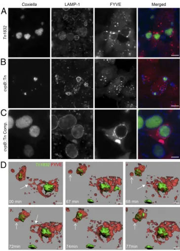
CvpB Mediates PI(3)P Enrichment of CCVs.To investigate the role of CvpB in CCV biogenesis with respect to its PI(3)P binding property and the colocalization with early endosomal markers, the morphology and dynamics of 2xFYVE or mCherry-Rab5 were assessed in cells infected with the Coxiella control mutant Tn1832, the cvpB::Tn mutant, or the cvpB::Tn com-plemented strain. In line with our observations in cells ectopi-cally expressing CvpB, cells infected with Coxiella Tn1832 or the cvpB::Tn complemented strain presented large FYVE- and Rab5-positive vesicles, compared with noninfected cells (Fig. 2F), which clustered around LAMP1-positive CCVs (Fig. 4 A and C andSI Appendix, Fig. S6A). The size of FYVE- and Rab5-positive compartments was unaltered in cells infected with the cvpB::Tn mutant, compared with noninfected cells (Fig. 2F), and vesicles were scattered through the cytoplasm of infected cells. In all cases, Rab5 was largely excluded from lysosome-derived CCVs (SI Appendix, Fig. S6A), indicating that, if early endosome/ CCV fusion occurred, early endosomal markers were likely lost as a consequence of endosomal maturation. Interestingly, however, the FYVE domain probe was clearly found colocalizing with LAMP1 at CCVs in a CvpB-dependent manner (Fig. 4 A–C). This observation suggests that CvpB may alter the metabolism of
incoming PI(3)P (either from the endocytic or the autophagy pathways) at CCV membranes. The distribution of early endo-somal markers with respect to CCVs was also investigated by using cell fractionation. In line with our observations by fluorescence microscopy, the endosomal markers Rab5 and EEA1 were ex-cluded from fractions containing CCVs generated by Coxiella Tn1832 and the cvpB::Tn mutant (SI Appendix, Fig. S6B). Frac-tions containing CCVs from cells infected with the cvpB::Tn complemented strain were mainly concentrated in one fraction, which was also positive for Rab5 and EEA1 (SI Appendix, Fig. S6B), suggesting that early endosomal markers can be found at CCVs, and enrichment of CCVs in a single fraction may facilitate their detection. However, the possibility that early endosomal markers detected by cell fractionation may originate from vesicles clustered around CCVs cannot be excluded. We then used 4D microscopy to investigate the dynamics of CvpB-triggered PI(3)P-positive vesicles in U2OS cells expressing mCherry-2xFYVE. Live imaging confirmed the presence of large PI(3)P-positive vesicles clustered at CCVs generated by the Coxiella control transposon mutant Tn1832 or the cvpB::Tn complemented strain (Fig. 4D andMovies S3andS4). In addition, the FYVE probe clearly defined the contour of CCVs under these conditions. Surface rendering of the acquired time lapses revealed the pres-ence of PI(3)P in large patches, which sometimes completely covered the surface of CCVs (Fig. 4D andMovies S3andS4). A number of PI(3)P-positive tubules also emanated from CCVs and interacted with bystander PI(3)P-positive vesicles, which were captured and merged with CCVs (Fig. 4D andMovies S3andS4). Cells infected with the cvpB::Tn mutant presented regular-sized PI(3)P-positive vesicles that did not interact with CCVs (Movie S5). Importantly, these findings correlate with our initial obser-vation of waves of large vesicles formed at the periphery of the cell and migrating toward CCVs (Movie S1).
CvpB Modulates the Intracellular Levels of PI(3)P.Host PIs and their metabolism are known targets of a number of bacterial patho-gens; we thus investigated whether Coxiella uses CvpB to ma-nipulate pathways controlled by PS and/or PI(3)P. Besides its role as a signaling lipid during apoptosis, it has been reported that PS plays an important role in retrograde traffic from the plasma membrane to the Golgi complex (22). We therefore transfected U2OS cells with mCherry or CvpB-mCherry and monitored retrograde transport by incubating cells with 1μg/mL FITC-labeled Cholera toxin B subunit (CtxB). After 60 min in-cubation on ice, cells were washed and CtxB was chased at 37 °C for 30 and 60 min. Further confirming CvpB localization at early endosomes, CtxB was observed partially localizing at CvpB-positive compartments 30 min post internalization (SI Appendix, Fig. S4B, Top). At later time points, however, CtxB trafficked to the Golgi complex unperturbed (SI Appendix, Fig. S4B, Bottom) in control and CvpB-expressing cells (SI Appendix, Fig. S4C).
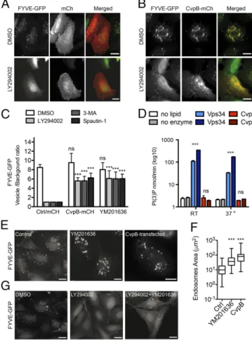
We next focused on the interaction of CvpB with PI(3)P. This key signaling phospholipid is primarily produced at the level of early endosomes and autophagosomes by the phosphorylation of phosphatidylinositol in position 3 of the inositol ring, mediated by class II and III PI3Ks (10). U2OS cells expressing GFP-2xFYVE were thus treated with the PI3Ks inhibitors LY294002, Spautin-1, or 3-methyladenine (3-MA) to deplete the majority of PI(3)P and monitor the effects of PI(3)P depletion on the in-tracellular distribution of CvpB. Upon GFP-2xFYVE relocalization to the cytoplasm, cells were transfected with mCherry alone or with CvpB-mCherry. DMSO-treated cells were used as control. mCherry alone was found in the cytoplasm of DMSO-treated or PI3Ks inhibitor-treated cells (Fig. 5A), whereas CvpB-mCherry was observed labeling large vacuoles and clusters of smaller vacuoles in DMSO-treated cells (Fig. 5B, Top). Interestingly, when CvpB-mCherry was expressed in PI(3)P-depleted cells, the bacterial effector was still observed labeling the same vesicular structures as in control cells
Fig. 4. CvpB induces the presence of PI(3)P at CCV. U2OS mCherry-2xFYVE cells were infected with Coxiella Tn1832 (A), the cvpB::Tn mutant (B), or the complemented strain (C) for 3 d. Coxiella colonies, FYVE-positive vesicles, and LAMP1 were detected by using GFP (green), mCherry (red), and an anti-LAMP1 antibody coupled to Alexa Fluor 647 (blue), respectively. (D) Represen-tative images fromMovie S3illustrating surface rendering of mCherry-2xFYVE (red) surrounding Coxiella Tn1832 colonies (green). Filled arrows point at PI (3)P-positive tubules connected with the CCV. Simple arrows follow a representative fusion event between a large PI(3)P-positive vesicle and the PI(3)P-enriched CCV (Movie S3). (Scale bars: A–C, 10 μm; D, 5 μm.)
(Fig. 5B, Bottom andSI Appendix, Fig. S7). Surprisingly, these vesicles were also labeled by GFP-2xFYVE, indicating the presence of PI(3)P (Fig. 5B, Bottom andSI Appendix, Fig. S7). The levels of intracellular PI(3)P were calculated as the ratio of GFP-2xFYVE intensity between labeled compartments and the cytoplasm, revealing that CvpB expression restored 60% of the PI(3)P probe on endosomal membranes (Fig. 5C). The same results were obtained in cells transfected with CvpB before PI(3)P depletion. Our observations suggest that CvpB may influence the metabolism of PI(3)P in cells infected by Coxiella.
CvpB Increases Intracellular Levels of PI(3)P by Perturbing the Activity of the PI 5-Kinase PIKfyve.Increases in the intracellular levels of PI(3)P may result from an increased PI3-kinase activity or an
inhibition of PI 5-kinase activity (10). However, the restoration of PI(3)P upon CvpB expression in the presence of several PI3Ks inhibitors seems to rule out the possibility that CvpB modulates PI3Ks activity. By using an in vitro PI3-kinase activity assay, we investigated the possibility that CvpB itself may act as a PI3-kinase. In agreement with the lack of known PI3-kinase domains in the sequence of CvpB, the Coxiella effector did not exhibit any kinase activity compared with Vps34, used here as positive control, regardless of the temperature used for the assay or the protein concentration (Fig. 5D). We therefore tested the possi-bility that CvpB perturbs the activity of the PI3 phosphate 5-kinase PIKfyve, which phosphorylates PI(3)P in position 5 of the ino-sitol ring to generate PI(3,5)P2 (10). Indeed, several studies
reported an increase of PI(3)P leading to vacuolation in cells where the activity of PIKfyve has been perturbed (23–25). The effects of PIKfyve inhibition on PI(3)P-positive endosomes were then compared with those of CvpB overexpression. U2OS cells expressing GFP-2xFYVE were incubated with the specific PIK-fyve inhibitor YM201636 (25) or transfected with CvpB-mCherry. In agreement with our hypothesis, PIKfyve inhibition phenocopied the ectopic expression of CvpB (Fig. 5 E and F). We thus tested whether inhibiting PIKfyve in cells pretreated with PI3K inhib-itors restores PI(3)P on early endosomes, as observed with the ectopic expression of CvpB. U2OS cells expressing GFP-2xFYVE were treated with LY294002, Spautin-1, or 3-MA for 4 h. Upon GFP-2xFYVE relocalization to the cytoplasm, the PIKfyve inhibitor YM201636 was added to the culture medium (in the presence of PI3Ks inhibitors). Indeed, the combined treatment of cells with PI3Ks inhibitors and YM201636 restored PI(3)P on endosomal membranes, as indicated by the reappearance of GFP-2xFYVE on these compartments (Fig. 5 C and G). PIKfyve and CvpB are both recruited at membranes by interacting with PI(3)P (26), raising the possibility that CvpB perturbs PIKfyve activity by interfering with its recruitment at early endosomes. U2OS cells were transfected with GFP-PIKfyve in combination with mCherry, CvpB-mCherry, CvpB100–809-mCherry, CvpB1–400-mCherry, and CvpB1–500-mCherry. PIKfyve recruitment at membranes was then measured as the number of GFP puncta in transfected cells for each condition. As previously described, control cells presented a large cytoplasmic pool of PIKfyve as well as a number of small puncta, revealing the protein association with endosomal vesicles (24, 27) (Fig. 6 A and D). The coexpression of PIKfyve with CvpB, significantly reduced the number of GFP puncta (Fig. 6 B and D), indicating that, indeed, CvpB perturbs PIKfyve associ-ation with PI(3)P-positive vesicles. Accordingly, coexpression of PIKfyve with CvpB MBD mutants restored membrane localiza-tion of PIKfyve (Fig. 6D), indicating that membrane targeting of CvpB is required for PIKfyve perturbation. Finally, coexpression of PIKfyve with CvpB1–500-mCherry also restored PIKfyve lo-calization at cell membranes, where it colocalized with CvpB (Fig. 6 C and D), indicating that CvpB does not outcompete PIKfyve for PI(3)P, and that the C terminus domain of CvpB is involved in blocking PIKfyve access to early endosomes. This is consistent with the attenuated vacuolation phenotype observed with CvpB1–500-mCherry described in Fig. 3D.
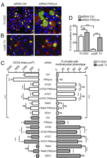
PIKfyve Inhibition Favors the Recruitment of the Autophagy Machinery to CCVs to Mediate Their Homotypic Fusion.Our obser-vations raised the interesting hypothesis that CvpB perturbs PIKfyve activity to enrich CCVs in PI(3)P for optimal vacuole biogenesis. To test whether PIKfyve inhibition favors CCV bio-genesis, U2OS cells were transfected with nontargeting or with three independent PIKfyve-targeting siRNAs (SI Appendix, Fig. S8A) before challenge with the Coxiella control transposon mu-tant Tn1832 or the cvpB::Tn mumu-tant. Five days postinfection, cells were fixed and the morphology of CCVs labeled by LAMP1 was investigated. CCV development was remarkably enhanced in PIKfyve-depleted cells, with LAMP1-positive vacuoles being
Fig. 5. Ectopic expression of CvpB or PIKfyve inhibition counters the effect of PI 3-kinase inhibitors. U2OS GFP-2xFYVE cells were incubated with DMSO (Top) or the PI3-kinase inhibitor LY294002 (Bottom) for 4 h and transfected with pLVX-mCherry (A) or pLVX-CvpB-mCherry (B) for 12 h. The redistribution of the GFP-2xFYVE probe into the cytoplasm was measured as the vesicle-to-background signal ratio of GFP-2xFYVE from 20 treated cells for each condition (C) (SI Appendix, Fig. S7). (D) The PI3-kinase activity of CvpB at 780 nM (light red bars) or 1,560 nM (dark red bars) was measured at room temperature (RT) or 37 °C and compared with no-lipid (white bars) or no-enzyme (gray bars) con-ditions as negative controls and purified Vps34 at 780 nM (light blue bars) or 1,560 nM (dark blue bars) as positive controls. (E) U2OS GFP-2xFYVE cells were incubated with DMSO (control) or YM201636 for 4 h or transfected with pLVX-CvpB-mCherry for 12 h. (F) The size of GFP-2xFYVE–positive vesicles in U2OS GFP-2xFYVE cells treated as in D was calculated. (G) U2OS GFP-2xFYVE cells were incubated with DMSO or the PI3-kinase inhibitor LY294002 for 4 h. The PIKfyve inhibitor YM201636 was then added to cells treated with LY294002, and cells were incubated for 4 h. Values in C and D are means± SEM of triplicate experiments (ns, nonsignificant; ***P< 0.0001, two-way ANOVA, Bonferroni’s multiple comparison test). Values in F are calculated from 500 vesicles measured for each condition (***P< 0.0001, one-way ANOVA). (Scale bars: 10 μm.)
Martinez et al. PNAS | Published online May 25, 2016 | E3265
MIC
ROBIOLOGY
PNAS
significantly larger than those developed by control cells infected by Coxiella Tn1832 (Fig. 7 A and C). More importantly, the number of cells infected with Coxiella cvpB::Tn presenting a multivacuolar phenotype was largely reduced in PIKfyve-depleted cells, with Coxiella cvpB::Tn replicating in large CCVs in-distinguishable from those generated by Coxiella Tn1832 (Fig. 7 B and C). As expected, the multivacuolar phenotype was still ob-served in cells treated with scrambled siRNA sequences (Fig. 7 B and C). PIKfyve depletion also restored LC3 labeling on the single CCVs generated by the cvpB::Tn mutants (Fig. 7D), indicating that PIKfyve inhibition and the concomitant increase in the levels of PI(3)P are functional to the recruitment of the autophagosomal machinery to CCVs. Next, we investigated whether the inhibition of PIKfyve is capable of restoring the single CCV phenotype in-dependently of the autophagy machinery. U2OS cells treated with siRNAs targeting ATG5 or ATG7 (SI Appendix, Fig. S8B), alone or in combination with siRNAs targeting PIKfyve, were chal-lenged with Coxiella Tn1832 or the cvpB::Tn mutant and analyzed 5 d post infection for the presence of multiple CCVs per cell. Inhibition of autophagy triggered the appearance of a multi-vacuolar phenotype in cells infected with Coxiella Tn1832, as previously described (6) (Fig. 7C). Importantly, the combined inhibition of ATG proteins and PIKfyve failed to rescue the multivacuolar phenotype triggered by the inhibition of autophagy (Fig. 7C); however, the multiple CCVs per cell were larger, compared with cells interfered for ATG proteins alone (Fig. 7C). Accordingly, the same phenotype was observed in cells challenged with the cvpB::Tn mutant, where PIKfyve depletion largely failed to rescue the multivacuolar phenotype in absence of a functional autophagy machinery (Fig. 7C). In these cells, the inhibition of ATG5 or ATG7 alone had no impact on the multivacuolar phe-notype triggered by the cvpB::Tn mutant (Fig. 7C), suggesting that the CvpB-mediated inhibition of PIKfyve is a prerequisite for CCV expansion and to prime CCV membranes for the recruitment
of the autophagy machinery. In turn, autophagy controls homotypic fusion between CCVs. To further define the relative contribution of the endocytic and autophagy pathway to CCV biogenesis with re-spect to CvpB, U2OS cells treated with siRNAs targeting EEA1 or Rab5 (SI Appendix, Fig. S8B), alone or in combination with PIK-fyve, were challenged with Coxiella Tn1832 or the cvpB::Tn mutant and analyzed 5 d post infection for the presence of multiple CCVs per cell. The inhibition of EEA1 or Rab5 in cells infected with Coxiella Tn1832 or the cvpB::Tn mutant significantly reduced the size of CCVs, but had no effect on the number of CCVs per cell, compared with cells treated with control siRNA sequences (Fig. 7C). The combined inhibition of PIKfyve and Rab5 failed to rescue the size of CCVs; however, it partially rescued the multivacuolar phenotype in cells challenged with the cvpB::Tn mutant (Fig. 7C).
Fig. 6. CvpB perturbs PIKfyve activity by interfering with its recruitment to PI(3)P-positive membranes. U2OS cells were transfected with pGFP-PIKfyve (PIKfyve-GFP) in combination with pLVX-mCherry (A), pLVX-CvpB-mCherry (B), or pLVX-CvpB100–809-mCherry, pLVX-CvpB1–400-mCherry, and pLVX-CvpB1–500
-mCherry (C). pLVX-CvpB--mCherry(1–500)(red) and PIKfyve (green) are merged
in C to illustrate colocalization (arrows and Inset). (D) The number of PIK-fyve-positive puncta (arrows) per cell was calculated by using ICY from 42 cells for each condition. Red bars indicate medians (***P< 0.0001; *P < 0.05; ns, not significant, one-way ANOVA, Dunnett’s multiple comparison test). (Scale bars: 10μm.)
Fig. 7. CvpB-mediated inhibition of PIKfyve promotes homotypic fusion of CCVs via autophagy. U2OS cells were treated with control siRNA (Left) or siRNA targeting PIKfyve (Right) for 24 h before being challenged with Coxiella Tn1832 (A) or the cvpB::Tn mutant (B) for 5 d. Cells were then fixed, and LAMP1 and DNA were labeled by using an anti-LAMP1 antibody cou-pled to Alexa Fluor 555 (red) or Hoechst 33258 (blue), respectively. Coxiella colonies were detected by using green Fluorescence (green). (C) U2OS cells transfected with the indicated siRNA sequences were challenged with Coxiella Tn1832 (white bars) or the cvpB::Tn mutant (gray bars). The median area of CCVs and the percentage of cells presenting a multivacuolar phe-notype (CCVs per cell > 3) were calculated for each condition by using CellProfiler. (D) The percentage of CCVs positive for LC3 in cells treated as in A and B was assessed for each condition. Values are means± SEM of trip-licate experiments in which 200 CCVs or 200 cells were analyzed for each condition (***P< 0.0001; **P < 0.01; *P < 0.05; ns, not significant, one-way ANOVA, Bonferroni’s multiple comparison test). (Scale bars: 10 μm.)
Unfortunately, cells did not survive the combined inhibition of EEA1 and PIKfyve. These observations indicate that the endocytic pathway is mainly involved in the initial expansion of CCVs, whereas the autophagy pathway mediates CCV homotypic fusion as previously reported (6).
Discussion
One of the key features of CCVs is their remarkable fusogenicity, which allows these compartments to intercept and merge with the endocytic, autophagy, secretory, and recycling pathways (28–30). To date, however, little is known about the mechanisms responsible for CCV biogenesis as well as the protein and lipid composition of CCVs. In this study, we addressed the fusogenicity of Coxiella CCVs, first by describing the multivacuolar phenotype associated with a transposon insertion in the Dot/Icm chaperone IcmS. By using image-based supervised machine learning, we identify Coxiella candidate effectors regulating homotypic fusion between CCVs. We have thus identified six independent transposon insertions in cvpB, which conferred a multivacuolar phenotype, rescuable by gene complementation. CvpB has been recently identified as a Dot/ Icm effector that localizes at CCVs and plays a role in the in-tracellular development of Coxiella (8, 9, 14). A parallel screen of a Coxiella transposon library also highlighted the emergence of a multivacuolar phenotype triggered by mutations in cvpB (8). The absence of detectable amounts of LC3 on CCVs generated by the cvpB mutant suggested a link between CvpB, autophagy, and homotypic fusion between CCVs (8).
By using live imaging in combination with an inducible com-plementation system, we demonstrate that the multivacuolar phenotype associated with cvpB mutations is indeed a result of impaired fusion between CCVs. Importantly, by using the insect model G. mellonella, we show that, regardless of the lack of in-tracellular replication phenotypes observed in cells infected with the Coxiella cvpB::Tn mutants, defective CCV biogenesis has in vivo relevance. Intrigued by the remarkable colocalization be-tween CvpB and FYVE domain, a well-established probe for PI (3)P, we used PIP Strip and in vitro cosedimentation with LUV assays to show that the N-terminal domain encompassed be-tween amino acids 1 and 500 allows CvpB binding to PI(3)P- and PS-containing membranes, which is essential to target CvpB to early endosomes. The reported intracellular distribution of PI(3)P on early endosomal membranes and that of PS at the plasma membrane and early endosomes correlated with our results on the subcellular localization of CvpB. Interestingly, ectopically expressed CvpB showed a differential localization in infected vs. noninfected cells. In the absence of infection, CvpB localizes at early endosomes and is excluded from LAMP1-positive lyso-somes and autophagolyso-somes, despite the latter sharing a similar lipid composition with early endosomes. On the contrary, in in-fected cells, the majority of CvpB is recruited to the LAMP1-positive CCV, suggestive of the presence of PI(3)P and/or PS at CCVs. Indeed, we report an unexpected PI(3)P enrichment of Coxiella CCVs, which were thus far believed to have mainly ly-sosomal characteristics. Other early endosomal markers such as EEA1 and Rab5 remain largely undetected at CCVs.
PIs are key players in the regulation of eukaryotic signal trans-duction, cytoskeleton architecture, and membrane trafficking (10). Specific kinases and phosphatases control their spatial and temporal distribution, allowing a precise and local modulation of essential processes including endocytosis and phagocytosis, membrane traffic, and autophagy (31–33). These processes are commonly manipulated by bacterial pathogens to invade, survive, and rep-licate within host cells. It is therefore not surprising that PIs and their metabolism have emerged as the target of a growing number of bacterial pathogens (11–13). Bacterial effectors can bind PIs for membrane targeting (34) or acting on PI metabo-lism, either by means of eukaryotic-like kinases and phospha-tases secreted by the pathogen or by modulating the recruitment
of host PI-metabolizing enzymes (11, 12). Intravacuolar patho-gens subvert PI metabolism to manipulate the lipid profile of their host-derived replicative niche, thus controlling the inter-actions with the endosomal maturation pathway according to their needs. Mycobacterium tuberculosis (35, 36) and Legionella pneumophila (37, 38) use effectors to deplete PI(3)P at the surface of their vacuoles to block phagosomal maturation and avoid fusion with degradative compartments. On the contrary, Salmonella enterica serovar Typhimurium uses the SPI-1 sub-strate SopB to maintain elevated levels of PI(3)P at the surface of Salmonella-containing vacuoles, favoring their biogenesis (39–41). Based on previous reports, the main source of PI(3)P-positive membranes for CCVs are early endosomes and autophagosomes (4). However, PI(3)P at late endosomes/lysosomes is expected to be further phosphorylated to PI(3,5)P2, by the activity of the PI
5-kinase PIKfyve. Similarly to Legionella effectors, CvpB uses PIs for membrane anchoring, apparently using a nonconventional lipid-binding domain as previously reported for the Legionella effectors SidC and SidM (42, 43). Similarly to the Salmonella effector SopB, Coxiella CvpB triggers an increase in PI(3)P levels of infected cells to favor the biogenesis of a spacious replicative compartment (40). CvpB, however, does not possess PI3-kinase activity. Instead, it uses its C-terminal domain to perturb the recruitment of the PI 5-kinase PIKfyve at PI(3)P-positive membranes, thereby interfering with its function and preserving PI(3)P at CCVs. Whether this occurs by steric hindrance or by a yet unidentified catalytic activity of the C-terminal domain of CvpB remains to be elucidated. Accordingly, it has been reported that the genetic, siRNA, or pharmacological-mediated inhibition of PIKfyve is accompanied by an increase in PI(3)P (23–25). PIKfyve inhibition has an important impact on endosomal mat-uration (23, 24, 44, 45), retrograde traffic (46), and autophagy (47, 48), which collectively result in the formation of large, late endosomal compartments in which the autophagosomal marker LC3 accumulates as a result of the defective fusion with ly-sosomes. These features are also shared by Coxiella CCVs. Accordingly, inhibition of PIKfyve phenocopied the ectopic expression of CvpB, and the depletion of PIKfyve rescued the multivacuolar phenotype in cells infected with the Coxiella cvpB::Tn mutant, restoring the formation of large CCVs positive for LAMP1 and LC3.
PI(3)P generated by the autophagy-associated class III PI3-kinase Vps34 is a master regulator of autophagy (49–51), and the PI3-kinase inhibitors used in this study are also commonly used autophagy inhibitors. A significant proportion of CCVs are positive for LC3, indicating that the Coxiella replicative com-partment intersects the autophagy pathway during maturation. Rather than stimulating autophagy, however, Coxiella infections seem to perturb its flux, similarly to what has been described for PIKfyve inhibition (8, 47, 52). The emergence of multivacuolar phenotypes when Coxiella-infected cells are silenced for ATG5, ATG12, and the autophagy SNARE Syntaxin-17 (6, 8) demon-strates an important role of autophagy in the homotypic fusion between CCVs. As previously reported (8), we confirm here that Coxiella cvpB mutants generate LC3-deficient CCVs. However, the connection between CvpB and the autophagy machinery remained to be defined. CvpB and LC3 localization at distinct intracellular compartments in noninfected cells seems to rule out a direct interaction between CvpB and autophagosomes. In-terestingly, however, the presence of LC3 on CCVs generated by the cvpB::Tn mutant is rescued, together with the single CCV morphology, by gene complementation or by PIKfyve silencing, indicating that the CvpB-mediated inhibition of PIKfyve is the link between the Coxiella effector and the autophagy machinery during CCV biogenesis. PIKfyve inhibition by CvpB, and the concomitant presence of PI(3)P at CCVs, may contribute to stabilize preexisting LC3 at the surface of Coxiella vacuoles.
Martinez et al. PNAS | Published online May 25, 2016 | E3267
MIC
ROBIOLOGY
PNAS
Alternatively, other Coxiella effectors may act in response to CvpB to recruit LC3 to CCVs.
In summary, here we show that, similarly to other intracellular bacterial pathogens, Coxiella uses effectors to manipulate PI metabolism during infection. Upon translocation, CvpB asso-ciates with CCVs and early endosomes by interacting with PI(3)P and PS. There, CvpB exerts a dual function by interfering with the activity of the PI 5-kinase PIKfyve to trigger an increase in the levels of PI(3)P at specific subcellular compartments. At early endosomes, this triggers vacuolation and clustering, which may enhance the delivery of membranes, proteins, and lipids to CCVs for optimal expansion. At CCVs, CvpB perturbation of PIKfyve activity stabilizes the autophagy machinery that medi-ates homotypic fusion between independent CCVs. Solving the crystal structure of CvpB will provide important insights into the precise molecular mechanism of this remarkable Coxiella effec-tor. Four-dimensional microscopy revealed interactions between PI(3)P-positive endosomes and forming CCVs, which become enriched in PI(3)P. The unexpected observation of PI(3)P at CCVs, revealed by the specific probe FYVE, stresses the im-portance of characterizing the protein as well as lipid composi-tion of the Coxiella replicative niche to better understand its biogenesis.
Materials and Methods
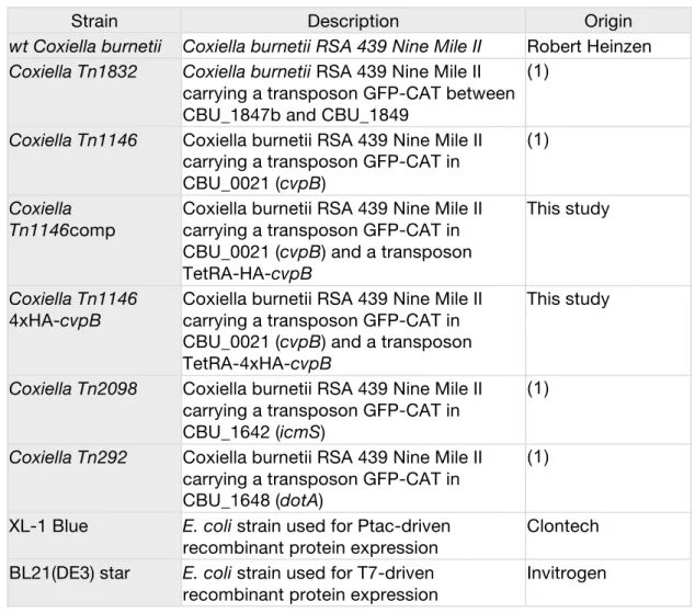
Bacterial Strains, Cell Lines, and Growth Conditions. Strains used in this study are listed inSI Appendix, Table S1. Escherichia coli strains were grown in Luria–Bertani medium supplemented with ampicillin (100 μg/mL), kanamycin (50μg/mL), or chloramphenicol (30 μg/mL) as appropriate. C. burnetii NMII and transposon mutants were grown in ACCM-2 supplemented with kana-mycin (340μg/mL) or chloramphenicol (3 μg/mL) as appropriate in a hu-midified atmosphere of 5% CO2and 2.5% O2at 37 °C. U2OS GFP-2xFYVE
cells were provided by Tassula Proikas-Cezanne (Eberhard Karls University Tübingen, Tuebingen, Germany). Cells (Vero, HeLa, A431, U2OS, U2OS GFP-2xFYVE, U2OS mCherry-2xFYVE) were routinely maintained in DMEM con-taining 10% (vol/vol) FCS in a humidified atmosphere of 5% CO2at 37 °C.
Cell growth medium was supplemented with 600μg/mL of Geneticin G418 (Gibco) as appropriate. For PI3-kinase inhibition assay, U2OS GFP-2xFYVE cells were incubated at 37 °C, 5% CO2, with 50μM LY294002, 10 mM 3-MA,
or 10μM Spautin-1 for a minimum of 4 h. Delocalization of GFP-2xFYVE to the cytoplasm was monitored over time by using an inverted EVOS fluo-rescence microscope. Where appropriate, after 4 h of treatment with the aforementioned inhibitors, cells were further incubated with 1μM YM201636 (always in the presence of the PI3-kinase inhibitors) for 4 h to block PIKfyve activity. Alternatively, U2OS GFP-2xFYVE cells were incubated with 1 μM YM201636 alone for 4 h at 37 °C, 5% CO2, to monitor the effects of PIKfyve
inhibition.
Cell Transfection. For the ectopic expression of proteins in mammalian cells, cells were grown to 60% confluence and transfected with JetPEI cationic polymer transfection reagent (Polyplus Transfection) according to the manufacturer’s recommendations. Cells were assayed 12–24 h post trans-fection. For the generation of U2OS mCherry-2xFYVE, cells were grown to 60–80% confluence before being transfected as described earlier. Cells were then incubated at 37 °C and 5% CO2for 24–48 h. For enrichment of U2OS
mCherry-2xFYVE cells, these were maintained in DMEM supplemented with 10% (vol/vol) FCS and containing 600μg/mL of Geneticin G418. For gene silencing, U2OS cells were seeded at 2,000 cells per well in black, clear-bot-tomed, 96-well plates in triplicate and transfected with siRNA oligonucleo-tides 24 h later by using the RNAiMAX transfection reagent (Thermo Fisher Scientific) according to the manufacturer’s recommendations. At 24 h post transfection, cells were challenged with C. burnetii transformants (MOI of 100) and further incubated for 5 d. Cells are then fixed and processed for immunofluorescence. In parallel, U2OS cells were cultured in six-well plates and transfected as described earlier. Cell lysates were collected at 24-h in-tervals during the time course of the experiment, and efficient gene si-lencing was monitored by Western blot.
Transformation and Axenic Growth of C. burnetii. C. burnetii strains were transformed with their respective plasmids as described by Martinez et al (14). For routine culture of C. burnetii strains, 106GE/mL of bacteria were
inoculated in 4 mL ACCM-2 and allowed to grow for 8 d in a humidified
atmosphere of 5% CO2and 2.5% O2at 37 °C. Where needed, 340μg/mL
kanamycin and/or 3μg/mL chloramphenicol were added as appropriate to bacterial cultures. At the indicated time points, bacterial concentrations were evaluated from 100μL of cultures by using the PicoGreen (Invitrogen) assay according to the manufacturer’s instructions.
Immunofluorescence Staining and Microscopy. Cells were fixed in 3% (wt/vol) paraformaldehyde in PBS solution at room temperature for 30 min or in methanol/acetone (1:1) at−20 °C for 5 min. Samples were then rinsed in PBS solution and incubated in blocking solution (0.5% BSA, 50 mM NH4Cl in PBS
solution, pH 7.4). When appropriate, 0.05% saponin was added to the blocking solution for cell permeabilization. Cells were then incubated with the primary antibodies diluted in blocking solution for 30 min at room temperature, rinsed five times in PBS solution, and further incubated for 30 min with the secondary antibodies diluted in the blocking solution. Fluorescent phalloidin was added to the secondary antibodies to label actin when needed. To visualize translocated 4xHA-tagged CvpB, cells were fixed as previously described in 3% (wt/vol) paraformaldehyde in PBS solution. Then, cells were permeabilized with 0.5% Triton X-100 in PBS solution for 3 min at room temperature. Sample were then rinsed in PBS solution and incubated with blocking solution [0.1% Triton X-100, 5% (wt/vol) milk in PBS solution] for 1 h at room temperature. Cells were then incubated with the anti-HA antibody diluted in the blocking solution for 1h at 37 °C, rinsed five times in PBS solution, and incubated with the secondary antibody for 1 h at 37 °C. For all conditions, coverslips were mounted by using Prolong Gold antifade mounting medium supplemented with Hoechst 33258 for DNA staining. Samples were imaged with a Zeiss Axio Imager Z1 epifluorescence microscope (Carl Zeiss) connected to a CoolSNAP HQ2CCD camera
(Photo-metrics). Images were acquired alternatively with 63× or 40× oil immersion objectives and processed with MetaMorph (Universal Imaging). Alterna-tively, samples were imaged with an ArrayScan VTI Live epifluorescence automated microscope (Cellomics) equipped with an ORCA-ER CCD camera (Hamamatsu). In this case, 25 fields per well were acquired for image anal-ysis. ImageJ, ICY, and CellProfiler software were used for image analysis and quantifications.
PI3K Activity Assay. For the analysis of PI3-kinase activity of CvpB, the class III PI3-kinase kit was used according to the manufacturer’s recommendations (K-3000; Echelon Biosciences). Recombinant VPS34 (Sigma) was used as positive control and conditions without enzyme or PI substrate were used as negative controls. The kinase reaction was performed in 10 mM Tris·HCl, pH 7.6, 100 mM NaCl, 1 mM EDTA, 10 mM MnCl2, and 50μM ATP using 780 nM or
1.56μM of recombinant protein.
Protein–Lipid Overlay Assay. Purified GST or GST-CvpB was incubated with PIP Strips and PIP Arrays (Molecular Probes) following the manufacturer’s recommendations. Briefly, TBS-T/BSA [10 mM Tris·HCl, pH 8, 150 mM NaCl, 0.1% Tween 20, 3% (wt/vol) BSA] was used throughout the assay. PIP Strips and PIP Arrays were first blocked for 1 h in TBS-T/BSA before being in-cubated with 1μg/mL of GST or GST-CvpB at 4 °C overnight. Membranes were washed three times with TBS-T/BSA and immunoblotted with anti-GST HRP-conjugated antibodies for 1 h at room temperature. Following three washes with TBS-T/BSA, the membranes were probed with ECL for signal detection.
In Vitro Cosedimentation Assays with LUVs. Binding of histidine-tagged CvpB, CvpB1–500, and CvpB500–809to LUVs was determined by cosedimentation
as-says. LUVs were made with a mixture of egg PC, brain PS, and PI(3)P at different molar ratios (100:0:0, 70:30:0, 98:0:2, or 68:30:2). Lipid mixtures were solubilized in chloroform, dried by evaporation, and resuspended overnight in a solubilizing buffer (150 mM KCl, 0.5 mM EDTA, 20 mM Hepes, pH 7.4) and extruded to obtain 200-nm-diameter LUVs. A constant amount of proteins (8 pmol), full-length CvpB, CvpB1–500, or CvpB500–809was incubated for
30 min at room temperature with LUVs in a final volume of 100μL. Samples were ultracentrifuged for 30 min at 71,000 rpm in a Beckman TLA 100 rotor at 4 °C. Each sample was then divided into a supernatant (SN; 90μL), con-taining unbound proteins, and a pellet (P; 10μL) containing LUV-bound proteins. The pellet P was resuspended in 80μL of solubilizing buffer. A total of 20μL of SN or P was analyzed on a 12% SDS/PAGE gel, and proteins were detected by Western blotting by using an anti-histidine antibody.
Antibodies and reagents, plasmids, and primers used in this study are listed inSI Appendix. Procedures for plasmid construction, image-based supervised machine learning,β-lactamase translocation assay, G. mellonella survival assay, transmission EM, live imaging, protein expression and purification, and cell fractionation are detailed inSI Appendix.
ACKNOWLEDGMENTS. The authors thank Dr. Tassula Proikas-Cezanne (In-ternational Max Planck Research School, Eberhard Karls University Tuebingen); Prof. Assia Shisheva (Wayne State University); Dr. Kai Wengelnik (CNRS, UMR 5235, DIMNP); Dr. Gunnar Schroeder (Imperial College London); and Bruno Beaumelle, Martine Biard, Lucile Espert, and Fabien Blanchet (CNRS, FRE 3689, CPBS) for providing cell lines, plasmids, and constructs and for scientific discussions. We thank Virginie Georget, Simon Lachambre, and Sylvain
DeRossi (Montpellier RIO Imaging-MRI) and Chantal Cazevieille (COMET EM Platform, Montpellier RIO Imaging) for their technical assistance and data analysis and Dr. Mariella Lomma (CHU, Nimes) for critical reading of the manuscript. This work was supported by Agence Nationale de la Recherche (ANR) Grant ANR-14-CE14-0012-01, project AttaQ, ERA-NET Infect-ERA ANR-13-IFEC-0003, project EUGENPATH, and the ATIP-AVENIR programme.
1. van Schaik EJ, Chen C, Mertens K, Weber MM, Samuel JE (2013) Molecular patho-genesis of the obligate intracellular bacterium Coxiella burnetii. Nat Rev Microbiol 11(8):561–573.
2. Madariaga MG, Rezai K, Trenholme GM, Weinstein RA (2003) Q fever: A biological weapon in your backyard. Lancet Infect Dis 3(11):709–721.
3. Newton HJ, McDonough JA, Roy CR (2013) Effector protein translocation by the Coxiella burnetii Dot/Icm type IV secretion system requires endocytic maturation of the pathogen-occupied vacuole. PLoS One 8(1):e54566–e54569.
4. Moffatt JH, Newton P, Newton HJ (2015) Coxiella burnetii: Turning hostility into a home. Cell Microbiol 17(5):621–631.
5. Campoy EM, Mansilla ME, Colombo MI (2013) Endocytic SNAREs are involved in op-timal Coxiella burnetii vacuole development. Cell Microbiol 15(6):922–941. 6. McDonough JA, et al. (2012) Host pathways important for Coxiella burnetii infection
revealed by genome-wide RNA interference screening. mBio 4(1):e00606–12. 7. Kohler LJ, Roy CR (2015) Biogenesis of the lysosome-derived vacuole containing
Coxiella burnetii. Microbes Infect 17(11-12):766–771, 10.1016/j.micinf.2015.08.006. 8. Newton HJ, et al. (2014) A screen of Coxiella burnetii mutants reveals important roles
for Dot/Icm effectors and host autophagy in vacuole biogenesis. PLoS Pathog 10(7): e1004286–e16.
9. Larson CL, et al. (2015) Coxiella burnetii effector proteins that localize to the para-sitophorous vacuole membrane promote intracellular replication. Infect Immun 83(2): 661–670.
10. De Matteis MA, Godi A (2004) PI-loting membrane traffic. Nat Cell Biol 6(6):487–492. 11. Weber SS, Ragaz C, Hilbi H (2009) Pathogen trafficking pathways and host
phos-phoinositide metabolism. Mol Microbiol 71(6):1341–1352.
12. Pizarro-Cerdá J, Kühbacher A, Cossart P (2015) Phosphoinositides and host-pathogen interactions. Biochim Biophys Acta 1851(6):911–918.
13. Pizarro-Cerdá J, Cossart P (2004) Subversion of phosphoinositide metabolism by in-tracellular bacterial pathogens. Nat Cell Biol 6(11):1026–1033.
14. Martinez E, Cantet F, Fava L, Norville I, Bonazzi M (2014) Identification of OmpA, a Coxiella burnetii protein involved in host cell invasion, by multi-phenotypic high-content screening. PLoS Pathog 10(3):e1004013–e1004022.
15. Martinez E, Cantet F, Bonazzi M (2015) Generation and multi-phenotypic high-con-tent screening of Coxiella burnetii transposon mutants. J Vis Exp (99):e52851–e52851. 16. Zusman T, Yerushalmi G, Segal G (2003) Functional similarities between the icm/dot pathogenesis systems of Coxiella burnetii and Legionella pneumophila. Infect Immun 71(7):3714–3723.
17. Ninio S, Zuckman-Cholon DM, Cambronne ED, Roy CR (2005) The Legionella IcmS-IcmW protein complex is important for Dot/Icm-mediated protein translocation. Mol Microbiol 55(3):912–926.
18. Beare PA, et al. (2014) Essential role for the response regulator PmrA in Coxiella burnetii type 4B secretion and colonization of mammalian host cells. J Bacteriol 196(11):1925–1940.
19. Lifshitz Z, et al. (2013) Computational modeling and experimental validation of the Legionella and Coxiella virulence-related type-IVB secretion signal. Proc Natl Acad Sci USA 110(8):E707–E715.
20. Norville IH, et al. (2014) Galleria mellonella as an alternative model of Coxiella bur-netii infection. Microbiology 160(Pt 6):1175–1181.
21. van Meer G, Voelker DR, Feigenson GW (2008) Membrane lipids: Where they are and how they behave. Nat Rev Mol Cell Biol 9(2):112–124.
22. Uchida Y, et al. (2011) Intracellular phosphatidylserine is essential for retrograde membrane traffic through endosomes. Proc Natl Acad Sci USA 108(38):15846–15851. 23. Ikonomov OC, Sbrissa D, Shisheva A (2001) Mammalian cell morphology and endo-cytic membrane homeostasis require enzymatically active phosphoinositide 5-kinase PIKfyve. J Biol Chem 276(28):26141–26147.
24. Ikonomov OC, Sbrissa D, Shisheva A (2006) Localized PtdIns 3,5-P2 synthesis to regulate early endosome dynamics and fusion. Am J Physiol Cell Physiol 291(2): C393–C404.
25. Jefferies HBJ, et al. (2008) A selective PIKfyve inhibitor blocks PtdIns(3,5)P(2) pro-duction and disrupts endomembrane transport and retroviral budding. EMBO Rep 9(2):164–170.
26. Sbrissa D, Ikonomov OC, Shisheva A (2002) Phosphatidylinositol 3-phosphate-inter-acting domains in PIKfyve. Binding specificity and role in PIKfyve.Endomenbrane lo-calization. J Biol Chem 277(8):6073–6079.
27. Cabezas A, Pattni K, Stenmak H (2006) Cloning and subcellular localization of a hu-man phosphatidylinositol 3-phosphate 5-kinase, PIKfyve/Fab1. Gene 371(1):34–41. 28. Larson CL, Beare PA, Howe D (2013) Coxiella burnetii effector protein subverts
cla-thrin-mediated vesicular trafficking for pathogen vacuole biogenesis. Proc Natl Acad Sci USA 110(49):E4770–E4779, 10.1073/pnas.1309195110/-/DCSupplemental. 29. Berón W, Gutierrez MG, Rabinovitch M, Colombo MI (2002) Coxiella burnetii localizes
in a Rab7-labeled compartment with autophagic characteristics. Infect Immun 70(10): 5816–5821.
30. Campoy EM, Zoppino FCM, Colombo MI (2011) The early secretory pathway con-tributes to the growth of the Coxiella-replicative niche. Infect Immun 79(1):402–413. 31. Levin R, Grinstein S, Schlam D (2015) Phosphoinositides in phagocytosis and
macro-pinocytosis. Biochim Biophys Acta 1851(6):805–823.
32. Simonsen A, Wurmser AE, Emr SD, Stenmark H (2001) The role of phosphoinositides in membrane transport. Curr Opin Cell Biol 13(4):485–492.
33. Vicinanza M, Rubinsztein DC (2016) Mirror image phosphoinositides regulate auto-phagy. Mol Cell Oncol 3(2):e1019974, 10.1080/23723556.2015.1019974.
34. Hilbi H, Weber S, Finsel I (2011) Anchors for effectors: Subversion of phosphoinositide lipids by legionella. Front Microbiol 2:91.
35. Fratti RA, Chua J, Vergne I, Deretic V (2003) Mycobacterium tuberculosis glycosylated phosphatidylinositol causes phagosome maturation arrest. Proc Natl Acad Sci USA 100(9):5437–5442.
36. Vergne I, et al. (2005) Mechanism of phagolysosome biogenesis block by viable My-cobacterium tuberculosis. Proc Natl Acad Sci USA 102(11):4033–4038.
37. Weber S, Wagner M, Hilbi H (2013) Live-cell imaging of phosphoinositide dynamics and membrane architecture during Legionella infection. mBio 5(1):e00839–13. 38. Toulabi L, Wu X, Cheng Y, Mao Y (2013) Identification and structural characterization
of a Legionella phosphoinositide phosphatase. J Biol Chem 288(34):24518–24527. 39. Mallo GV, et al. (2008) SopB promotes phosphatidylinositol 3-phosphate formation
on Salmonella vacuoles by recruiting Rab5 and Vps34. J Cell Biol 182(4):741–752. 40. Hernandez LD, Hueffer K, Wenk MR, Galán JE (2004) Salmonella modulates vesicular
traffic by altering phosphoinositide metabolism. Science 304(5678):1805–1807. 41. Scott CC, Cuellar-Mata P, Matsuo T, Davidson HW, Grinstein S (2002) Role of
3-phosphoinositides in the maturation of Salmonella-containing vacuoles within host cells. J Biol Chem 277(15):12770–12776.
42. Ragaz C, et al. (2008) The Legionella pneumophila phosphatidylinositol-4 phosphate-binding type IV substrate SidC recruits endoplasmic reticulum vesicles to a replication-permissive vacuole. Cell Microbiol 10(12):2416–2433.
43. Brombacher E, et al. (2009) Rab1 guanine nucleotide exchange factor SidM is a major phosphatidylinositol 4-phosphate-binding effector protein of Legionella pneumo-phila. J Biol Chem 284(8):4846–4856.
44. de Lartigue J, et al. (2009) PIKfyve regulation of endosome-linked pathways. Traffic 10(7):883–893.
45. Kim GHE, Dayam RM, Prashar A, Terebiznik M, Botelho RJ (2014) PIKfyve inhibition interferes with phagosome and endosome maturation in macrophages. Traffic 15(10):1143–1163.
46. Rutherford AC, et al. (2006) The mammalian phosphatidylinositol 3-phosphate 5-kinase (PIKfyve) regulates endosome-to-TGN retrograde transport. J Cell Sci 119(pt 19): 3944–3957.
47. Martin S, et al. (2013) Inhibition of PIKfyve by YM-201636 dysregulates autophagy and leads to apoptosis-independent neuronal cell death. PLoS One 8(3):e60152–e14. 48. Vicinanza M, et al. (2015) PI(5)P regulates autophagosome biogenesis. Mol Cell 57(2):
219–234.
49. Simonsen A, Tooze SA (2009) Coordination of membrane events during autophagy by multiple class III PI3-kinase complexes. J Cell Biol 186(6):773–782.
50. Vergne I, Deretic V (2010) The role of PI3P phosphatases in the regulation of auto-phagy. FEBS Lett 584(7):1313–1318.
51. Burman C, Ktistakis NT (2010) Regulation of autophagy by phosphatidylinositol 3-phosphate. FEBS Lett 584(7):1302–1312.
52. Winchell CG, Graham JG, Kurten RC, Voth DE (2014) Coxiella burnetii type IV secre-tion-dependent recruitment of macrophage autophagosomes. Infect Immun 82(6): 2229–2238.
Martinez et al. PNAS | Published online May 25, 2016 | E3269
MIC
ROBIOLOGY
PNAS
Supporting Information
Martinez et al. 10.1073/pnas.1522811113
Movie S1. U2OS cells were challenged with the Coxiella control transposon mutant Tn1832 (Left), the cvpB::Tn mutant (Middle), or the cvpB::Tn com-plemented strain (in the presence of aTc; Right) for 3 d before imaging. Images were acquired every 10 min for 32 h in the phase-contrast and GFP channels.
Movie S1
Movie S2. U2OS cells were challenged with the Coxiella cvpB::Tn complemented strain for 3 d in the absence of aTc. Complementation induction was triggered at the moment of image acquisition. Images were acquired every 10 min for 32 h in the phase-contrast and GFP channels.
Movie S2
Movie S3. U2OS cells expressing mCherry-2xFYVE were challenged with the Coxiella control transposon mutant Tn1832 for 3 d before imaging using a spinning-disk confocal microscope. Image stacks were acquired every 2 min in the mCherry (white) and GFP channels (green) for 3 h (Left). Surface rendering of mCherry-2xFYVE–positive compartments and Coxiella colonies was performed with IMARIS software on two ROIs. Surface rendering was superimposed to the image sequence (Middle) or extracted for easier visualization (Right).
Movie S3