HAL Id: hal-01931278
https://hal.uca.fr/hal-01931278
Submitted on 1 Jul 2019
HAL is a multi-disciplinary open access archive for the deposit and dissemination of sci-entific research documents, whether they are pub-lished or not. The documents may come from teaching and research institutions in France or abroad, or from public or private research centers.
L’archive ouverte pluridisciplinaire HAL, est destinée au dépôt et à la diffusion de documents scientifiques de niveau recherche, publiés ou non, émanant des établissements d’enseignement et de recherche français ou étrangers, des laboratoires publics ou privés.
with Blastocystis spp.
Manon Defaye, Céline Nourrisson, Elodie Baudu, Ivan Warwzyniak, Virginie
Bonnin, Mathilde Bonnet, Nicolas Barnich, Denis Ardid, Frédéric Delbac,
Frédéric Antonio Carvalho, et al.
To cite this version:
Manon Defaye, Céline Nourrisson, Elodie Baudu, Ivan Warwzyniak, Virginie Bonnin, et al.. Efficient and reproducible experimental infections of rats with Blastocystis spp.. PLoS ONE, Public Library of Science, 2018, 13 (11), pp.1-15. �10.1371/journal.pone.0207669�. �hal-01931278�
Efficient and reproducible experimental
infections of rats with Blastocystis spp.
Manon Defaye1,2☯
, Ce´line Nourrisson1,3☯
, Elodie Baudu2,4, Ivan Warwzyniak1,
Virginie Bonnin4, Mathilde BonnetID4, Nicolas Barnich4, Denis Ardid2, Fre´de´ric Delbac1,
Fre´de´ric Antonio Carvalho2‡
*, Philippe PoirierID1,3‡*
1 Universite´ Clermont Auvergne, 3iHP, CNRS, Laboratoire Microorganismes: Ge´nome et Environnement, Clermont-Ferrand, France, 2 Universite´ Clermont Auvergne, 3iHP, Inserm U1107, NeuroDol, Clermont-Ferrand, France, 3 Universite´ Clermont Auvergne, CHU, 3iHP, CNRS, Laboratoire Microorganismes: Ge´nome et Environnement, Clermont-Ferrand, France, 4 Universite´ Clermont Auvergne, 3iHP, Inserm U1071, USC INRA 2018, Microbes, Intestin, Inflammation et Susceptibilite´ de l’Hoˆte, Clermont-Ferrand, France
☯These authors contributed equally to this work. ‡ These authors also contributed equally to this work.
*ppoirier@chu-clermontferrand.fr(PP);frederic.carvalho@uca.fr(FAC)
Abstract
Although Blastocystis spp. infect probably more than 1 billion people worldwide, their clinical significance is still controversial and their pathophysiology remains poorly understood. In this study, we describe a protocol for an efficient and reproducible model of chronic infection in rats, laying the groundwork for future work to evaluate the pathogenic potential of this par-asite. In our experimental conditions, we were unable to infect rats using vacuolar forms of an axenically cultivated ST4 isolate, but we successfully established chronic infections of 4 week-old rats after oral administration of both ST3 and ST4 purified cysts isolated from human stool samples. The infection protocol was also applied to 4 week-old C57BL/9, BALB/C and C3H mice, but any mouse was found to be infected by Blastocystis. Minimal cyst inoculum required for rat infection was higher with ST3 (105) than with ST4 (102). These results were confirmed by co-housing experiments highlighting a higher contagious potential of ST4 in rats compared to ST3. Finally, experiments mimicking fecal microbiota transfer from infected to healthy animals showed that Blastocystis spp. could easily infect a new host, even though its intestinal microbiota is not disturbed. In conclusion, our results provide a well-documented and robust rat model of Blastocystis chronic infection, reproducing “natu-ral” infection. This model will be of great interest to study host parasite interactions and to better evaluate clinical significance of Blastocystis.
Introduction
Blastocystis spp. are anaerobic enteric protist found in the intestinal tract of a wide range of
animals, and are probably the most prevalent human parasites [1]. Four morphological stages ofBlastocystis spp. have been described including amoeboid, granular, vacuolar and cystic
stages. Both vacuolar and cystic forms are commonly found in human fecal samples and cyst is
a1111111111 a1111111111 a1111111111 a1111111111 a1111111111 OPEN ACCESS
Citation: Defaye M, Nourrisson C, Baudu E,
Warwzyniak I, Bonnin V, Bonnet M, et al. (2018) Efficient and reproducible experimental infections of rats with Blastocystis spp.. PLoS ONE 13(11): e0207669.https://doi.org/10.1371/journal. pone.0207669
Editor: Adriana Calderaro, Universita degli Studi di
Parma, ITALY
Received: July 13, 2018 Accepted: November 5, 2018 Published: November 19, 2018
Copyright:© 2018 Defaye et al. This is an open access article distributed under the terms of the Creative Commons Attribution License, which permits unrestricted use, distribution, and reproduction in any medium, provided the original author and source are credited.
Data Availability Statement: All relevant data are
within the paper and its Supporting Information files.
Funding: This work was achieved by obtaining a
co-financing Region Auvergne-Rhoˆne-Alpes and FEDER in 2015 (“The´matiques e´mergentes”). MD and EB were supported by grants from the Region Auvergne-Rhoˆne-Alpes. This work was supported by the Ministère de la Recherche et de la Technologie, Inserm and Universite´ Clermont Auvergne [UMR1107, UMR1071]; INRA
[USC-considered to be the infectious stage [1–3].Blastocystis spp. have been classified into 17
sub-types (ST) based on nuclear small subunit (SSU) ribosomal RNA-encoding gene, the ST1 to ST9 and ST12 being recovered from human [4–7]. ST3 is the most frequent subtype in human, followed by ST1, ST2 and ST4 [5]. However, this distribution depends on geographic areas, and a higher prevalence of ST4 was reported from the north of Europe [8–10].
Blastocystis spp. infection occurs in both immunocompetent and immunocompromised
hosts and recent studies highlighted higher prevalence of these parasites in patients with Irrita-ble Bowel Syndrome (IBS) and colorectal cancer [11–14]. However, the involvement of Blasto-cystis spp. in human diseases is highly debated and well-designed epidemiological studies still
need to be performed. Animal experimental models forBlastocystis infection have been
pur-posed for proving Koch’s postulates, but few animal-based studies have been published so far, suggesting difficulties in the establishment and reproducibility of these models [15]. To date various animal models were used forBlastocystis infection as rodents (rats or mice), guinea
pigs or chickens. Moreover, studies have shown that both vacuolar and cystic stages of differ-ent STs can efficidiffer-ently infect animals [16–19]. The heterogeneity of infection methods with various animal models and/or different STs inoculation confirm that the establishment of a standardized model is necessary.
In this study, we evaluated several methods to inoculateBlastocystis spp. into laboratory
animals (mice and rats) using different parasitic stages (vacuolar and cystic forms) isolated fromin vitro cultures or from human or animal feces. Interestingly, we succeeded in the
devel-opment of a reproducible model of chronic infection in juvenile and adult Wistar rats using purified cysts isolated from human stools.
Materials and methods
Animals
All animals were housed in animal biosafety level 2 (21–22˚C, 12:12 h light-dark cycle) with access to food and waterad libitum. Three- and ten-week-old specific-pathogen-free (SPF)
Wistar male rats and three-week-old SPF C57BL/9, BALB/C and C3H mice were purchased from Charles River Lab (Saint Germain Nuelles, France). All experiments were performed according to the ethical guidelines set out in the Guide for the Care and Use of Laboratory Ani-mals and with approval of the “Comite´ d’Ethique pour l’Expe´rimentation Animale Auvergne" (C2E2A), the local ethics committee (Reference number: EU0116-3003). After experimental infections withBlastocystis, fecal samples were collected every week until sacrifice.
Blastocystis ST4 axenic cultures
Blastocystis ST4 WR1 strain isolated from Wistar rats and axenized by Chen et al. [20] was cul-tured anaerobically in Iscove’s Modified Dulbecco’s Medium (IMDM) (Gibco, Life Technolo-gies, Carlsbad, CA, USA) with 10% horse serum (PAA, Pasching, Austria) and 1% penicillin/ streptomycin (Gibco, Life Technologies, Carlsbad, CA, USA) at 37˚C. Vacuolar forms were collected from 48h-old cultures by centrifugation at 1000 g for 10 min and suspended in sterile PBS to reach a concentration of 107parasites per milliliter and were used for experimental infections.
Oral administration of vacuolar forms isolated from axenic
Blastocystis
ST4 strain
In a first experiment, four-week-old Wistar male rats were orally inoculated with 107ST4 vac-uolar forms per rat fromin vitro axenic culture (n = 14). Eight rats were used to evaluate
2018]; CNRS [UMR6023]. The funders had no role in study design, data collection and analysis, decision to publish, or preparation of the manuscript.
Competing interests: The authors have declared
kinetic of ST4 route through the gastrointestinal tract and six rats were kept to monitor the progression of infection over time. To reduce gastric acidity, rats received 3 oral administra-tions every 2 hours of cimetidine (50 mg/kg, Mylan, Canonsburg, PA, USA) (n = 4) or 0.2 M sodium bicarbonate (Cooper, Melun, France) (n = 4) before oral inoculation with 107vacuolar forms of ST4 in a second experiment. Similarly as before, four rats were used to evaluate kinetic of ST4 route through the gastrointestinal tract and four rats were kept to monitor the progression of infection over time.
Kinetic of ST4 route through the gastrointestinal tract of
experimentally-infected rats
After oral administration of ST4, rats were euthanized at 3 (n = 2), 6 (n = 2), 12 (n = 2) or 24 h (n = 2) post-inoculation (PI). Stomach content was collected at 3 h PI, stomach and duodenum contents at 6 h PI, ileum and caecum contents at 12 h PI, and caecum, proximal and distal colon contents at 24 h PI.
For the experiments with animals pretreated with cimetidine or sodium bicarbonate, eutha-nasia of rats was done at 3 or 6 h (n = 2 for each time and condition). Stomach contents were collected at 3 h PI and caecum contents at 6 h PI.
Intra-caecal inoculation of vacuolar forms isolated from axenic
Blastocystis
ST4
Four-week-old Wistar male rats were intra-caecally inoculated with 107ST4 vacuolar forms per rat from axenic culture (n = 6). Briefly, after 24 h of fasting, rats were anesthetized (Keta-mine 75 mg/kg + Xylazine 10 mg/kg), followed by an abdomen incision, caecum externaliza-tion and injecexternaliza-tion of 107ST4 vacuolar forms. Then, caecum was repositioned and abdomen sutured.
Blastocystis isolates collected from human stools
Four asymptomatic human carriers ofBlastocystis ST2, ST3, ST4 and ST7 were identified at
the medical laboratory of Parasitology of the Clermont-Ferrand teaching hospital (France). BothBlastocystis cysts and vacuolar forms were observed in stools for patients with ST2, ST3
and ST4, whereas only vacuolar forms were observed for the ST7 carrier. Fresh stools were col-lected for animal infections (approved by the research ethics committees of the Clermont-Fer-rand Hospital, “Comite´ de Protection des Personnes Sud-Est 6”, France, agreement ref: 2014/ CE29). Subtyping was performed by SSU rDNA sequencing as previously described [10,21]. Sequences were analyzed using the Basic Local Alignment Search Tool (BLAST;http://blast. ncbi.nlm.nih.gov/).
Purification of cysts isolated from human stools
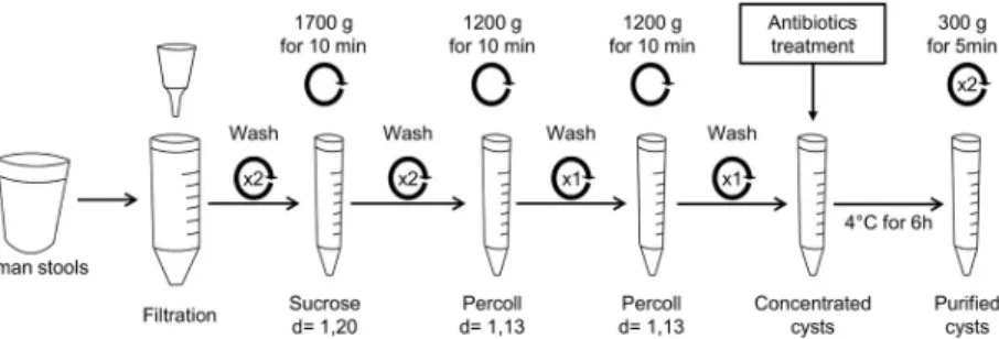
Cysts were purified from human stools of ST2-, ST3- or ST4-infected patients (one patient for each subtype) by an optimized method (Fig 1) adapted from Yoshikawa’s protocol [18]. Almost 50 g of fresh stools were suspended in distilled water and larger debris were removed by filtration (funnel with filter disc 1mm, Theradiag, Marne La Vallee, France). The fecal sus-pension was washed twice in distilled water by centrifugation at 1700 g for 10 min. The super-natant was discarded and pellet was suspended in 40 ml of sucrose solution (d = 1.20) (Sigma-Aldrich, Saint-Louis, MO, USA) and was divided into 15 ml tubes. Then, 0.5 ml of distilled water was added in each tube followed by a centrifugation at 1700 g for 10 min. The interface between sucrose and distilled water was collected and suspended in distilled water. Two
washes in distilled water were performed by centrifugation at 1700 g for 10 min. The pellet was suspended in 0.5 ml of distilled water and mixed with 7 ml Percoll solution (d = 1.13) (MP Bio-medicals, Illkirch, France). Then, 6 ml of distilled water were added, followed by a centrifuga-tion at 1200 g for 10 min. The interface between distilled water and Percoll solucentrifuga-tion was collected, adjusted to 15 ml with distilled water and centrifuged at 1700 g for 10 minutes. This step using Percoll gradients was performed twice. Pellets containing purified cysts were sus-pended in 0.5 ml of an antibiotic cocktail containing 0.01 g/L Vancomycin (Sandoz, Levallois-Perret, France), 0.1 g/L Amoxicillin-clavulanic acid (Sandoz, Levallois-Levallois-Perret, France) and 0.1 g/L Cefotaxim (SteriMax, Oakville, ON, Canada) diluted in sterile PBS (Gibco, Life Technolo-gies, Carlsbad, CA, USA) and incubated for 6 h at 4˚C. Cysts were washed twice in sterile PBS by centrifugation at 300 g for 5 min and finally suspended in sterile PBS. Ten microliters of the suspension were plated on LB media and incubated at 37˚C for 24 h in order to quantify the number of Colony Forming Units (CFU). Cysts were counted in Malassez chamber and sus-pension was diluted in sterile PBS to obtain concentrations of 102, 103,104or 105cysts per milliliter.
Oral administration of purified cysts isolated from human stools
Wistar male rats (four or twelve-week-old) were orally inoculated with 102/rat to 105/rat ST2, ST3 or ST4 purified cysts isolated from human stools (n = 5 for each condition).Blastocystis
isolated from rat feces after infection were subtyped by sequencing the SSU rRNA encoding gene as described above. For mice infections, 105ST4 purified cysts per animal were orally inoculated to four-week-old C57BL/6 (n = 3), BALB/C (n = 3) or C3H mice (n = 3).
Co-housing experiments
Four-week-old Wistar male rats were orally inoculated with 105ST3 (n = 2) or ST4 (n = 2) purified cysts per animal, as described above. Four-weeks PI, these animals were co-housed for 6 weeks with eight-week-old non-infected rats (n = 4 per ST).
Oral administration of rat feces containing
Blastocystis cysts
Fresh feces from experimentally-infected rats, containing ST3 or ST4 cysts, were collected. Almost 300 mg of feces samples were pooled and suspended in 3 ml of sterile PBS. Then, eight-week-old rats (n = 3, for each ST) were orally inoculated with 105cysts.
Fig 1. Method of cyst purification from human stools. About 50 g of stools were suspended in 200 ml of distilled
water and filtered (1mm) to remove larger particles. After washing by centrifugation in distilled water (1700 g for 10 min), cysts were concentrated by sucrose gradient (d = 1.20) followed by two Percoll gradients (d = 1.13). Then, cysts were incubated with an antibiotic cocktail composed of Vancomycin, Amoxicilin-clavunalic acid and Cefotaxim to eliminate bacteria. After washing to remove antibiotics, purified cysts were suspended in sterile PBS and quantified. This method was adapted from Yoshikawa’s protocol [18].
Fecal microbiota transplantation (FMT) using
Blastocystis-positive human
stools
Fresh stools from infected humans, containing ST2 or ST3 or ST4 cysts, or ST7 vacuolar forms, were collected. Almost 30 g of each fecal samples were suspended in 90 ml of sterile PBS. Moreover, almost 30 g of ST4 fecal sample were cryopreserved in 90 ml of PBS/glycerol 10% and stored at -80˚C for two months. Four-week-old Wistar male rats were orally inocu-lated with 1 ml (corresponding to 300 mg of Human stools) of the suspension containing either ST2 (n = 3), ST3 (n = 3), ST4 with or without cryopreservation (n = 3 for each condi-tion) cysts or ST7 (n = 3) vacuolar forms.
Xenic cultures of
Blastocystis-positive animal or human feces
Human or rat feces were cultured in Jone’s medium at 37˚C in anaerobic chambers as previ-ously described [22]. After 48 h of incubation, the culture was observed by standard light microscopy (x 400). Identification of vacuolar forms revealed the presence of viable parasites.
Detection of
Blastocystis by quantitative PCR (qPCR)
DNA was extracted from gastrointestinal tract contents or fecal samples of rats using the NucleoSpin Soil kit protocol (Macherey-Nagel SARL, Hoerdt, France). DNA was amplified with LC-FastStart DNA Master SYBR green kit (Roche Diagnostics, France) and specific prim-ers BL18SPPF1 (5’-AGTAGTCATACGCTCGTCTCAAA-3’) and BL18SR2PP (5’-TCTTCG TTACCCGTTACTGC-3’) using a Rotor-Gene 6000 system (Corbett Life Science, France) as described by Poirieret al [10].
Immunofluorescence labeling of
Blastocystis cysts
Purified cyst smears were incubated with 5% milk in PBS for 1 h and washed in PBS/Triton-X100 0.1%. Then, cysts were incubated with anti-Blastocystis mouse polyclonal antibodies
(diluted 1:200 in PBS) for 1 h at room temperature (RT), followed by three washes, and incu-bated with AlexaFluor 488-conjugated secondary antibody (anti-mouse IgG, 2μg/ml in PBS) (Invitrogen Carlsbad, CA, USA) for 1 h at RT. Preparations were washed twice with PBS and stained with 4’,6-diamidino-2-phenylindole (DAPI).
Immunofluorescence staining of intestinal sections
Four weeks PI, ST4- or ST3-infected rats were euthanized and the intestine was prepared as “Swiss rolls” of 4 cm and fixed by incubating in PFA 4% during 24 h. Then, the rolls were trans-ferred in sucrose 30% during 24–48 h at 4˚C. Finally, the rolls were included in OCT compound (CellPath, Newton, United Kingdom). Sections of OCT-embedded intestine were incubated with 5% milk in PBS for 1 h, and then washed in 0.1% Triton-X100 in PBS. Anti-Blastocystis
mouse polyclonal antibodies (diluted 1:200) were added for 1 h at RT, washed, and incubated further 1 h at RT with AlexaFluor 546-conjugated secondary antibody (diluted 1:1000) (anti-mouse IgG, Thermo Fisher Scientific, Waltham, MS, USA). Sections were washed and then incubated with fluorescein phalloidin (Thermo Fisher Scientific, Waltham, MS, USA) diluted 1:100 in PBS during 20 min. Finally, sections were washed and stained with DAPI. For each ST, six sections of duodenum, jejunum, ileum, caecum and colon from three rats were observed.
Transmission electron microscopy (TEM)
Samples were washed in 0.2 M Sodium Cacodylate buffer and fixed overnight at 4˚C with a mixture of 2% glutaraldehyde and 0,5% PAF in 0,2 M Sodium Cacodylate buffer. Specimens
were then washed three-times in 0.2 M Sodium Cacodylate buffer, post-fixed 1 h with 1% OsO4 in 0.2 M Sodium Cacodylate buffer, and washed three-times (10 min) in 0.2 M Sodium Cacodylate buffer. Specimens were then dehydrated in a graded ethanol and acetone solution. Subsequently, they were infiltrated with acetone and EPON resin mixture (2:1) for 1 h, with acetone and EPON resin mixture (1:1) for 1 h, and with acetone and EPON resin mixture (1:2) for 1 h. Specimens were embedded in resin overnight at RT, and cured for 2 days in a 60˚C oven. Thin sections (70 nm) were cut using a UC6 ultramicrotome (Leica, Wetzlar, Germany) and stained with uranyl acetate and lead citrate. Carbone was evaporated using CE6500 unit. Specimens sections were observed at 80 kV with a Hitachi H-7650 TEM and a camera Hama-matsu AMT40.
Electron microscopy preparations were all performed by the “Centre d’Imagerie Cellulaire Sante´” (Clermont-Ferrand, France). All chemical products were from Electron Microscopy Science, and distributed in France by Delta Microscopies.
Results
Inoculation of rats with vacuolar forms of
in vitro-cultivated Blastocystis
ST4
In all our tested conditions, oral infections with vacuolar forms ofBlastocystis ST4 axenic
cul-tures failed (Table 1) even in animals pretreated with molecules which increase intragastric pH (cimetidine 50 mg/kg or sodium bicarbonate 0.2 M). To confirm the absence of parasites, we performed a follow-up of the parasite course through the intestinal tract of rats from 3 h to 24 h post-infection (PI) by xenic culture and qPCR. Even though high parasite loads were inocu-lated (107per animal), xenic culure and qPCR were all negative suggesting that vacuolar forms are not able to pass through the stomach alive (Table 2). After treatment with cimetidine or sodium bicarbonate, qPCR analysis revealed the presence ofBlastocystis DNA in the caecum
of rats 6 h PI but no viable parasitic form was observed after xenic cultures from collected cae-cum contents (Table 2). To avoid stomach passage, ST4 vacuolar forms were directly inocu-lated into the caecum of rats, but here again all animals remained uninfected (Table 1). These data suggest that vacuolar forms are not able to infect rats in our experimental conditions.
Infection of mice and rats with purified cysts isolated from human stool
samples
We optimized a purification protocol described previously by adding another Percoll gradient step and a treatment with an antibiotic cocktail (Fig 1). This protocol decreased drastically the number of bacteria below 250 CFUs inoculated per animal. From 50 g of human stool about 4 million cysts were purified for each subtype (Fig 2A). The size of purified cysts ranged from 2 to 7μm and cysts were characterized by the presence of two or four nuclei (Fig 2B). Both juve-nile (four-week-old) and adult (twelve-week-old) rats were successfully infected by oral inocu-lation of 105cysts ofBlastocystis ST2, ST3 and ST4 per animal (Table 1). Cultures of feces collected 8 weeks after oral inoculation of cysts were positive forBlastocystis, confirming a
chronic infection by the parasite.
We then evaluated the minimal inoculum for both ST3 and ST4 by using infectious dose ranging from 102to 105purified cysts per animal. Interestingly, rats were chronically infected with the lowest cyst inoculum of ST4 (102), whereas higher inoculum of ST3 (105) was required for the establishment of infection (Table 1).
During all experiments, only cystic forms were observed in feces from infected rats for both
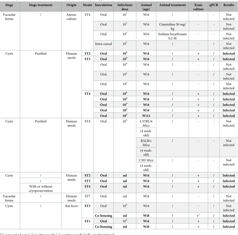
Table 1. Experimental infections of rats and mice withBlastocystis spp.
Stage Stage treatment Origin Strain Inoculation Infectious dose
Animal (age)
Animal treatment Xenic culture qPCR Results Vacuolar forms / Axenic culture
ST4 Oral 107 Wi4 / - - Not
infected
Oral 107 Wi4 Cimetidine 50 mg/
kg
- - Not
infected
Oral 107 Wi4 Sodium bicarbonate
0.2 M
- - Not
infected
Intra-caecal 107 Wi4 / - / Not
infected
Cysts Purified Human
stools
ST2 Oral 105 Wi4 / + / Infected
ST3 Oral 105 Wi4 / + / Infected
Oral 104 Wi4 / - / Not
infected
Oral 103 Wi4 / - / Not
infected
Oral 102 Wi4 / - / Not
infected
ST4 Oral 105 Wi4 / + / Infected
Oral 104 Wi4 / + / Infected
Oral 103 Wi4 / + / Infected
Oral 102 Wi4 / + / Infected
Oral 105 Wi12 / + / Infected
Cysts Purified Human
stools ST4 Oral 105 C57BL/6 Mice / - - Not infected (4 week-old) BALB/c Mice / - - Not infected (4 week-old) C3H Mice / - - Not infected (4 week-old) Cysts / Human stools
ST2 Oral nd Wi4 / + / Infected
/ ST3 Oral nd Wi4 / + / Infected
With or without cryopreservation
ST4 Oral nd Wi4 / + / Infected
Vacuolar forms
/ Human
stools
ST7 Oral nd Wi4 / - / Not
infected
Cysts / Rat feces ST3 Oral 105 Wi4 / - / Not
infected
Co-housing nd Wi8 / +� / Infected
ST4 Oral 105 Wi4 / + / Infected
Co-housing nd Wi8 / + / Infected
“/”: not carried out; “+”: positive result; “-“: negative result; “nd”: not determined;
“�”: only 1/4 naive rat became infected with ST3; Wi4: Wistar rats 4-week-old; Wi8: Wistar 8-week-old; Wi12: Wistar rats 12-week-old. https://doi.org/10.1371/journal.pone.0207669.t001
and immunofluorescence labeling using polyclonal antibodies. Cysts from ST3 and ST4 infected rats were similar in size to those isolated from human stools and were also character-ized by the presence of two to four nuclei (S1 Fig).
The same infection protocol was further applied to 3 different genetic mice backgrounds, C57BL/6, BALB/c and C3H mice (Table 1). However, we were not able to obtain any infection, suggesting that immunocompetent mice could be resistant to infection by these ST4 Blastocys-tis strains.
Transmission between animals by co-housing experiments or oral
inoculation
Eight-week-old ST3 (n = 2) or ST4 (n = 2) infected rats were co-housed with eight-week-old naive rats (n = 4 per subtype) during six weeks. Infections were monitored by microscopy
Table 2. Kinetic ofBlastocystis ST4 route through the intestinal tract.
Animal treatment PI Time Part of the gastrointestinal tract qPCR Xenic culture
NA 3h Stomach - -NA 6h Stomach - -Duodenum NA 12h Ileum - -Caecum NA 24h Caecum - -Proximal colon Distal colon Cimetidine 50 mg/kg 3h Stomach + -6h Caecum +
-Sodium bicarbonate 0.2 M 3h Stomach +
-6h Caecum +
-Four-week-old Wistar rats orally inoculated with 107vacuolar forms per animal. “NA”: not applied; “+”: positive result; “-“: negative result https://doi.org/10.1371/journal.pone.0207669.t002
Fig 2.Blastocystis ST4 cysts purified from human stools. (A) Purified cysts from human stools observed by light
microscopy. Numerous cysts can be observed (asterisk). More than 4 million of cysts were counted after purification from 50 g of human stool. Cysts size ranged from 2 to 7μm. (B) Purified cysts from human stools observed by immunofluorescence after labeling with polyclonal anti-Blastocystis ST4 antibodies (Green) and DAPI staining (Blue).
Cysts contained two to four nuclei (white arrows, two on this picture). https://doi.org/10.1371/journal.pone.0207669.g002
observation and xenic cultures of fecal samples. For ST4 co-housing experiments, all naive rats became infected only two weeks after the beginning of the experiment, whereas only 1/4 naive rat became infected with ST3, 6 weeks after the beginning of the experiment (Table 1).
Naive eight-week-old rats (n = 3 for each subtype) were also orally inoculated with feces from ST3 or ST4 infected rats. Only ST4-inoculated rats were infected (Table 1).
Fecal microbiota transplantation from human to rats
Four-week-old rats were orally inoculated with fresh human stools containingBlastocystis
ST2, ST3, ST4 or ST7, mimicking fecal microbiota transplantation. Both cystic and vacuolar forms were present in ST2, ST3 and ST4 human positive stools. In contrast, only vacuolar forms were observed in ST7-positive stools. Infection monitoring was done following FMT by xenic cultures of rat feces. Vacuolar forms were detected in culture feces from ST2, ST3 and ST4 infected rats but no viable parasite was identified in ST7-inoculated rat feces (Table 1). Interestingly, we also performed FMT from Human to rat after cryopreservation ofBlastocystis
ST4 stools, by following the recommended protocol for Human to Human FMT [23]. After 2 months at -80˚C, stools were thaw out and transferred to naive rats. All animals were infected, suggesting that the procedure of cryopreservation use for Human to Human FMT also main-tainBlastocystis alive.
Colonization of the intestinal tract by
Blastocystis
Both ST3- and ST4-infected rats by purified cysts from human stools were sacrificed four weeks PI. Full gastrointestinal tract was collected and “Swiss rolls” were performed on duode-num, jejuduode-num, ileum, caecum, proximal and distal colon.Blastocystis vacuolar forms were
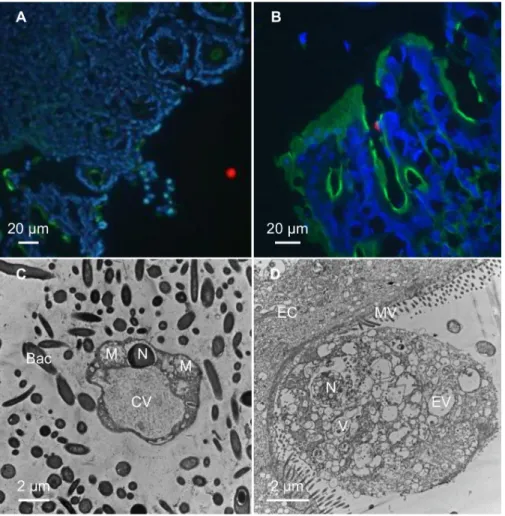
observed from duodenum to distal colon for both ST3 and ST4 (S2 Fig). Immunofluorescence labeling revealed that parasites were mainly localized in the intestine lumen (Fig 3A), and sometime in close contact with epithelial cells (Fig 3B). No invasive form was observed. Trans-mission electron microscopy performed on colonic sections confirmed the presence of para-sites in the lumen (Fig 3C) and in close contact with epithelial cells (Fig 3D).
Discussion
Animal models are essential for a better understanding of the pathogenic potential of Blasto-cystis spp which still remains controversial. Rats (Wistar strain being the most common)
[18,24–28], mice [16,17,29–31], guinea pigs [32] and chickens [25] were previously demon-strated as potential models ofBlastocystis infection. However, to date [33] there is no standard-ized animal infection model forBlastocystis, suggesting difficulties in the establishment or
reproducibility of these models.
In the present study, we aimed to provide a robust and well-described protocol to obtain efficient and reproducible experimental infection withBlastocystis spp. Four different human Blastocystis STs were used in our experiments, including ST3, the most common ST found in
human, followed by ST2 and ST4, the latter reported to be highly prevalent in Europe [8–10], and ST7 whose whole genome was the first sequenced [33,34]. ST4 was also used because it is the most prevalent ST found in rodents and its whole genome was recently sequenced [35]. Parasites used in our study originated from axenic cultures (ST4-WR1, first isolated from a laboratory rat) [20] or were isolated from human stools by a highly efficient purification pro-cess (ST2, ST3 and ST4) adapted from Yoshikawaet al [18].
Based on the literature and the easiest models to handle in an animal facility, we focused on common rodent models with rats and mice. We were not able to infect rats with the axenic ST4 WR1 strain. In ourin vitro culture conditions that strain only produced vacuolar forms.
Then, failures in animal infections could be explained by sensitivity of vacuolar forms to gas-tric pH, but also to oxygen exposure during inoculum preparation [1,36]. Indeed, studies have shown that rat stomach pH ranged from 3.2–3.9 [37]. However, we also failed to infect animals with ST4 WR1 by increasing the intragastric pH or after intracaecal injection of parasites. Then, we hypothesized that long-term culture and/or axenization of vacuolar forms may have attenuated the infectious potential of the ST4 WR1 strain. Indeed, the maintenance of the par-asites asTrypanosoma cruzi, Entamoeba histolytica or Leishamania infantum in laboratory
cul-tures led to gene expression changes and decreased infectivity [38–41]. However, previous studies have reported successful infections of rats using axenicBlastocystis strains [16,17,19]. We cannot exclude that some axenic strains may conserve their infectious potential, or may produce cysts in axenic culture conditions. Indeed, cyst is considered to be the main infectious stage, responsible for feco-oral contamination between hosts [18,24,25,27,28,42]. Different protocols have been described for cyst isolation from human stool samples [17,18]. We opti-mized Yoshikawa’s protocol in order to decrease cyst-associated bacteria [18]. Then, an
Fig 3. Colonic sections fromBlastocystis ST4-infected rats. Colonic sections were stained with fluorescein phalloidin
(Green), DAPI (Blue) and polyclonal anti-Blastocystis ST4 antibodies (Red). Vacuolar forms were detected in the
intestinal lumen (A) and in close contact with the intestinal epithelium (B). Transmission electron micrographs of colonic sections from experimentally-infected ratsBlastocystis vacuolar form surrounding by numerous bacteria
localized in the intestinal lumen (C) and a granular form in close contact with an epithelial cell (D). M, mitochondrion-like organelle; N, nucleus, CV, central vacuole; EV, empty vacuole; V, vacuole; Bac, bacteria; EC, epithelial cell; MV, microvilli.
additional Percoll step was performed and purified cysts were incubated with a wide spectrum antibiotic cocktail targeting both aerobic and anaerobic bacteria. Moreover, successful infec-tions following this treatment demonstrated low impact of this antibiotic treatment on Blasto-cystis cyst viability.
We were able to infect rats with ST2, ST3 and ST4 cysts by using high parasite load (105 cysts/animal). Infected animals were followed as long as 8 weeks post-infection. Animals excretedBlastocystis cysts until the end of experiments, confirming the establishment of a
chronic infection in our model.
Our results reinforced that ST4 infection failures were not related to a resistance mecha-nism, but more likely to the stage used or long-termin vitro cultivation. The susceptibility of
animals toBlastocystis was reported to be age-dependent by Moe et al., but these authors
didn’t mentioned theBlastocystis ST used in their experiments [17]. In our study, we were also able to infect twelve-old-week rats withBlastocystis ST4 cysts, suggesting that at least for this
ST, age is not a limiting factor. However, the effects of aging on susceptibility to ST3 infection remain to be investigated. Interestingly, rats were infected with as low as 102ST4 cysts per ani-mal, whereas the minimal inoculum dose required for infection with ST3 reached 105cysts suggesting host adaptation. Transmission between animal by co-housing experiments and oral inoculations confirm our hypothesis. Transmission between animal by co-housing experi-ments and oral inoculations confirm our hypothesis. These results may explain why ST4 is the more prevalent than ST3 in rodents [43]. However, oral inoculation with less than 102ST4 cysts per rat remains to be investigated, but a previous work has shown that infection efficiency with 10 cysts of ST4 RN94-9 or ST4 NIH:1295:1 strains varies between 20–100% [18].
Our results suggest that even though host barrier is not critical forBlastocystis infection,
some STs are more adapted to particular hosts. Then, we applied our purification protocol to infect 3 strains of mice. The first one was juvenile BALB/c mice that have been shown to be more susceptible to parasite infections [44,45] such asBlastocystis [17,19]. We also used C3H mice that have been described to be susceptible toEntamoeba histolytica [46] andGiardia intestinalis [47]. Finally, C57BL/6 mice, being the most used genetic background for transgenic mice, were also used. However, we were not able to infect mice even with high ST4 cyst inocu-lum (105/animal), suggesting that mice would be resistant to the ST4 strain used in our study. A recent study has shown that the Dextran Sodium Sulfate (DSS) treatment result in biophysi-cal changes in mucus layer (increased penetrability to microorganism), increasing susceptibil-ity of mice toBlastocystis ST7 colonization for at least 3 days after intra-ceacal injection [48]. These results suggest that mucus layer play in important role in the resistance of mice for Blas-tocystis persistence.
Immunofluorescence labelling revealed the presence of the parasites (ST4 or ST3) all along the intestinal tract of infected animals.Blastocystis were detected in the intestinal lumen and in
close contact with epithelial cells. Granular forms presenting empty vacuoles or vacuoles con-taining electron-dense granules as described previously by Tan [1], were observed in direct contact with epithelial cells.
Moreover, FMT experiments highlighted the capacity ofBlastocystis to overcome barrier
microflora and infect a new host, even though its microbiota is not altered. We also demon-strated that cryopreservation procedure used for the stool storage before Human to Human FMT keepBlastocystis alive and able to infect a new host [23]. These data support the recent recommendations for the screening of fecal donors by confirming the ability ofBlastocystis to
be directly transmitted through stools [23].
In conclusion, our work provides a well-documented and reproducible animal model of
interest to decipher the host-parasite-microbiota interactions, and to better evaluate clinical significance ofBlastocystis.
Supporting information
S1 Fig.Blastocystis ST4 cysts from experimentally infected rats. (A) Cysts (red arrow) from rats observed by light microscopy. As purified cysts from human stools, size ranged from 2 to 7μm. (B) Cysts from rats observed by immunofluorescence after labeling with mouse poly-clonal anti-Blastocystis ST4 antibodies (Green) and DAPI staining (Blue). Cysts contained two
to four nuclei (white arrows, two on this picture). (TIF)
S2 Fig. Intestinal sections fromBlastocystis ST4-infected rats. Sections of the intestinal tract were stained with fluorescein phalloidin (Green), DAPI (Blue) and mouse polyclonal
anti-Blastocystis ST4 antibodies (Red). Parasites were detected in small intestine (A), in caecum (B)
and colon (C) in the lumen or in close contact with the intestinal epithelium. (TIF)
Acknowledgments
This work was achieved by obtaining a co-financing Region Auvergne-Rhoˆne-Alpes and FEDER in 2015 (“The´matiques e´mergentes”). MD and EB were supported by grants from the Region Auvergne-Rhoˆne-Alpes.
This work was supported by the Ministère de la Recherche et de la Technologie, Inserm and Universite´ Clermont Auvergne [UMR1107, UMR1071]; INRA [USC-2018]; CNRS [UMR6023].
The funders had no role in study design, data collection and analysis, decision to publish, or preparation of the manuscript.
Electron microscopy preparations were all performed by the “Centre d’Imagerie Cellulaire Sante´” (CICS, Clermont-Ferrand, France).
Author Contributions
Conceptualization: Fre´de´ric Delbac, Fre´de´ric Antonio Carvalho, Philippe Poirier. Investigation: Manon Defaye, Ce´line Nourrisson.
Methodology: Ivan Warwzyniak, Virginie Bonnin.
Project administration: Manon Defaye, Denis Ardid, Fre´de´ric Delbac, Fre´de´ric Antonio
Car-valho, Philippe Poirier.
Supervision: Fre´de´ric Delbac.
Writing – original draft: Manon Defaye.
Writing – review & editing: Manon Defaye, Ce´line Nourrisson, Elodie Baudu, Ivan
Warwzy-niak, Mathilde Bonnet, Nicolas Barnich, Denis Ardid, Fre´de´ric Delbac, Fre´de´ric Antonio Carvalho, Philippe Poirier.
References
1. Tan KSW. New Insights on Classification, Identification, and Clinical Relevance of Blastocystis spp. Clin Microbiol Rev. 2008 Oct; 21(4):639–65.https://doi.org/10.1128/CMR.00022-08PMID:18854485
2. Stenzel DJ, Boreham PF. Blastocystis hominis revisited. Clin Microbiol Rev. 1996 Oct; 9(4):563–84. PMID:8894352
3. Wawrzyniak I, Poirier P, Viscogliosi E, Dionigia M, Texier C, Delbac F, et al. Blastocystis, an unrecog-nized parasite: an overview of pathogenesis and diagnosis. Ther Adv Infect Dis. 2013; 1(5):167–78. https://doi.org/10.1177/2049936113504754PMID:25165551
4. Alfellani MA, Stensvold CR, Vidal-Lapiedra A, Onuoha ESU, Fagbenro-Beyioku AF, Clark CG. Variable geographic distribution of Blastocystis subtypes and its potential implications. Acta Trop. 2013 Apr; 126 (1):11–8.https://doi.org/10.1016/j.actatropica.2012.12.011PMID:23290980
5. Stensvold CR, Alfellani MA, Nørskov-Lauritsen S, Prip K, Victory EL, Maddox C, et al. Subtype distribu-tion of Blastocystis isolates from synanthropic and zoo animals and identificadistribu-tion of a new subtype. Int J Parasitol. 2009 Mar; 39(4):473–9.https://doi.org/10.1016/j.ijpara.2008.07.006PMID:18755193 6. Stensvold CR, Clark CG. Current status of Blastocystis: A personal view. Parasitol Int. 2016; 65
(6):763–71.
7. Ramı´rez JD, Sa´nchez A, Herna´ndez C, Flo´rez C, Bernal MC, Giraldo JC, et al. Geographic distribution of human Blastocystis subtypes in South America. Infect Genet Evol. 2016 Jul; 41:32–5.https://doi.org/ 10.1016/j.meegid.2016.03.017PMID:27034056
8. Domı´nguez-Ma´ rquez MV, Guna R, Muñoz C, Go´mez-Muñoz MT, Borra´s R. High prevalence of subtype 4 among isolates of Blastocystis hominis from symptomatic patients of a health district of Valencia (Spain). Parasitol Res. 2009 Oct 27; 105(4):949–55.https://doi.org/10.1007/s00436-009-1485-yPMID: 19471964
9. Olsen KEP, Christiansen DB, Nielsen HV, Stensvold CR. Blastocystis sp. Subtype 4 is Common in Dan-ish Blastocystis-Positive Patients Presenting with Acute Diarrhea. Am J Trop Med Hyg. 2011 Jun 1; 84 (6):883–5.https://doi.org/10.4269/ajtmh.2011.11-0005PMID:21633023
10. Poirier P, Wawrzyniak I, Albert A, El Alaoui H, Delbac F, Livrelli V. Development and Evaluation of a Real-Time PCR Assay for Detection and Quantification of Blastocystis Parasites in Human Stool Sam-ples: Prospective Study of Patients with Hematological Malignancies. J Clin Microbiol. 2011 Mar 1; 49 (3):975–83.https://doi.org/10.1128/JCM.01392-10PMID:21177897
11. Nourrisson C, Scanzi J, Pereira B, NkoudMongo C, Wawrzyniak I, Cian A, et al. Blastocystis is associ-ated with decrease of fecal microbiota protective bacteria: Comparative analysis between patients with irritable bowel syndrome and control subjects. PLoS One. 2014; 9(11).
12. Padukone S, Mandal J, Parija SC. Severe Blastocystis subtype 3 infection in a patient with colorectal cancer. Trop Parasitol. 2017; 7(2):122–4.
13. Rostami A, Riahi SM, Haghighi A, Saber V, Armon B, Seyyedtabaei SJ. Erratum to: the role of
Blasto-cystis sp. and Dientamoeba fragilis in irritable bowel syndrome: a systematic review and meta-analysis.
Parasitol Res. 2017 Sep 18; 116(9):2611–2.https://doi.org/10.1007/s00436-017-5556-1PMID: 28725935
14. Mohamed AM, Ahmed MA, Ahmed SA, Al-Semany SA, Alghamdi SS, Zaglool DA. Predominance and association risk of Blastocystis hominis subtype I in colorectal cancer: a case control study. Infect Agent Cancer. 2017 Dec 12; 12(1):21.
15. Ajjampur SSR, Tan KSW. Pathogenic mechanisms in Blastocystis spp.—Interpreting results from in vitro and in vivo studies. Parasitol Int. 2016 Dec; 65(6):772–9.
16. Pavanelli MF, Kaneshima EN, Uda CF, Colli CM, Falavigna-Guilherm AL, Gomes ML. PATHOGENIC-ITY OF Blastocystis sp. TO THE GASTROINTESTINAL TRACT OF MICE: RELATIONSHIP BETWEEN INOCULUM SIZE AND PERIOD OF INFECTION. Rev Inst Med Trop Sao Paulo. 2015 Dec; 57(6):467–72.https://doi.org/10.1590/S0036-46652015000600002PMID:27049699
17. Moe KT, Singh M, Howe J, Ho LC, Tan SW, Chen XQ, et al. Experimental Blastocystis hominis infection in laboratory mice. Parasitol Res. 1997; 83(4):319–25. PMID:9134552
18. Yoshikawa H, Yoshida K, Nakajima A, Yamanari K, Iwatani S, Kimata I. Fecal-oral transmission of the cyst form of Blastocystis hominis in rats. Parasitol Res. 2004; 94(6):391–6.https://doi.org/10.1007/ s00436-004-1230-5PMID:15480786
19. Santos HJ, Rivera WL. Kinetic analysis of antibody responses to Blastocystis hominis in sera and intes-tinal secretions of orally infected mice. Parasitol Res. 2009 Oct 14; 105(5):1303–10.https://doi.org/10. 1007/s00436-009-1556-0PMID:19597843
20. Chen XQ, Singh M, Ho LC, Tan SW, Ng GC, Moe KT, et al. Description of a Blastocystis species from Rattus norvegicus. Parasitol Res. 1997; 83(4):313–8. PMID:9134551
21. Scicluna SM, Tawari B, Clark CG. DNA barcoding of Blastocystis. Protist. 2006 Feb; 157(1):77–85. https://doi.org/10.1016/j.protis.2005.12.001PMID:16431158
22. Clark CG, Stensvold CR. Blastocystis: Isolation, Xenic Cultivation, and Cryopreservation. In: Current Protocols in Microbiology. Hoboken, NJ, USA: John Wiley & Sons, Inc.; 2016. p. 20A.1.1–20A.1.8.
23. Cammarota G, Ianiro G, Tilg H, Rajilić-StojanovićM, Kump P, Satokari R, et al. European consensus conference on faecal microbiota transplantation in clinical practice. Gut. 2017 Apr; 66(4):569–80. https://doi.org/10.1136/gutjnl-2016-313017PMID:28087657
24. Suresh K, Ng GC, Ramachandran NP, Ho LC, Yap EH, Singh M. Parasitology. 1993;456–60.
25. Iguchi A, Ebisu A, Nagata S, Saitou Y, Yoshikawa H, Iwatani S, et al. Infectivity of different genotypes of human Blastocystis hominis isolates in chickens and rats. Parasitol Int. 2007; 56(2):107–12.https://doi. org/10.1016/j.parint.2006.12.004PMID:17251054
26. Iguchi A, Yoshikawa H, Yamada M, Kimata I, Arizono N. Expression of interferon gamma and proinflam-matory cytokines in the cecal mucosa of rats experimentally infected with Blastocystis sp. strain RN94-9. Parasitol Res. 2009; 105(1):135–40.https://doi.org/10.1007/s00436-009-1373-5PMID:19255785 27. Hussein EM, Hussein AM, Eida MM, Atwa MM. Pathophysiological variability of different genotypes of
human Blastocystis hominis Egyptian isolates in experimentally infected rats. Parasitol Res. 2008; 102 (5):853–60.https://doi.org/10.1007/s00436-007-0833-zPMID:18193282
28. Chandramathi S, Suresh KG, Mahmood AA, Kuppusamy UR. Urinary hyaluronidase activity in rats infected with Blastocystis hominis-evidence for invasion? Parasitol Res. 2010; 106(6):1459–63.https:// doi.org/10.1007/s00436-010-1825-yPMID:20358228
29. Moe KT, Singh M, Gopalakrishnakone P, Ho LC, Tan SW, Chen XQ, et al. Cytopathic effect of
Blasto-cystis hominis after intramuscular inoculation into laboratory mice. Parasitol Res. 1998 Jun; 84(6):450–
4. PMID:9660133
30. Abou El Naga IF, Negm AY. Morphology, histochemistry and infectivity of Blastocystis hominis cyst. J Egypt Soc Parasitol. 2001 Aug; 31(2):627–35. PMID:11478461
31. Yao F, Qiao J, Zhao Y, Zhang X, Yang J, Li X. [Experimental infection of mice with Blastocystis homi-nis]. Zhongguo Ji Sheng Chong Xue Yu Ji Sheng Chong Bing Za Zhi. 2005 Dec 30; 23(6):444–8. PMID: 16566218
32. Phillips BP, Zierdt CH. Blastocystis hominis: pathogenic potential in human patients and in gnotobiotes. Exp Parasitol. 1976 Jun; 39(3):358–64. PMID:1269579
33. Denoeud F, Roussel M, Noel B, Wawrzyniak I, Da Silva C, Diogon M, et al. Genome sequence of the stramenopile Blastocystis, a human anaerobic parasite. Genome Biol. 2011; 12(3):R29.https://doi.org/ 10.1186/gb-2011-12-3-r29PMID:21439036
34. Wawrzyniak I, Roussel M, Diogon M, Couloux A, Texier C, Tan KSW, et al. Complete circular DNA in the mitochondria-like organelles of Blastocystis hominis. Int J Parasitol. 2008 Oct; 38(12):1377–82. https://doi.org/10.1016/j.ijpara.2008.06.001PMID:18694756
35. Wawrzyniak I, Courtine D, Osman M, Hubans-Pierlot C, Cian A, Nourrisson C, et al. Draft genome sequence of the intestinal parasite Blastocystis subtype 4-isolate WR1. Genomics Data. 2015 Jun; 4:22–3.https://doi.org/10.1016/j.gdata.2015.01.009PMID:26484170
36. Zierdt CH. Blastocystis hominis—past and future. Clin Microbiol Rev. 1991 Jan; 4(1):61–79. PMID: 2004348
37. McConnell EL, Basit AW, Murdan S. Measurements of rat and mouse gastrointestinal pH, fluid and lym-phoid tissue, and implications for in-vivo experiments. J Pharm Pharmacol. 2008 Jan; 60(1):63–70. https://doi.org/10.1211/jpp.60.1.0008PMID:18088506
38. Contreras VT, De Lima AR, Zorrilla G. Trypanosoma cruzi: maintenance in culture modify gene and antigenic expression of metacyclic trypomastigotes. Mem Inst Oswaldo Cruz. 93(6):753–60. PMID: 9921298
39. Villalta F, Kierszenbaum F. Insect-borne and culture-derived metacyclic Trypanosoma cruzi: differ-ences in infectivity and virulence. Am J Trop Med Hyg. 1987 May; 36(3):529–32. PMID:3107410 40. Phillips BP. Entamoeba histolytica: concurrent irreversible loss of infectivity-pathogenicity and
encyst-ment potential after prolonged maintenance in axenic culture in vitro. Exp Parasitol. 1973 Oct; 34 (2):163–7. PMID:4355413
41. Santare´ m N, Cunha J, Silvestre R, Silva C, Moreira D, Ouellette M, et al. The impact of distinct culture media in Leishmania infantum biology and infectivity. Parasitology. 2014 Feb 6; 141(2):192–205. https://doi.org/10.1017/S0031182013001388PMID:24007671
42. Li J, Deng T, Li X, Cao G, Li X, Yan Y. A rat model to study Blastocytis subtype 1 infections. Parasitol Res. 2013; 112(10):3537–41.https://doi.org/10.1007/s00436-013-3536-7PMID:23892480
43. Yoshikawa H, Tokoro M, Nagamoto T, Arayama S, Asih PBS, Rozi IE, et al. Molecular survey of
Blasto-cystis sp. from humans and associated animals in an Indonesian community with poor hygiene.
Parasi-tol Int. 2016 Dec 1; 65(6):780–4.
44. Sherwood D, Angus KW, Snodgrass DR, Tzipori S. Experimental cryptosporidiosis in laboratory mice. Infect Immun. 1982 Nov; 38(2):471–5. PMID:7141705
45. Laurent F, McCole D, Eckmann L, Kagnoff MF. Pathogenesis of Cryptosporidium parvum infection. Microbes Infect. 1999 Feb; 1(2):141–8. PMID:10594978
46. Ivory C, Kammanadiminti S, Chadee K. Innate resistance to Entamoeba histolytica in murine models. Trends Parasitol. 2007 Feb; 23(2):46–8.https://doi.org/10.1016/j.pt.2006.12.006PMID:17185037 47. Sharma AW, Mayrhofer G. A comparative study of infections with rodent isolates of Giardia duodenalis
in inbred strains of rats and mice and in hypothymic nude rats. Parasite Immunol. 1988 Mar; 10(2):169– 79. PMID:2967456
48. Ajjampur SSR, Png CW, Chia WN, Zhang Y, Tan KSW. Ex Vivo and In Vivo Mice Models to Study
Blas-tocystis spp. Adhesion, Colonization and Pathology: Closer to Proving Koch’s Postulates. Renia L,
edi-tor. PLoS One. 2016 Aug 10; 11(8):e0160458.https://doi.org/10.1371/journal.pone.0160458PMID: 27508942