HAL Id: hal-02075132
https://hal.umontpellier.fr/hal-02075132
Submitted on 27 May 2021
HAL is a multi-disciplinary open access
archive for the deposit and dissemination of
sci-entific research documents, whether they are
pub-lished or not. The documents may come from
teaching and research institutions in France or
abroad, or from public or private research centers.
L’archive ouverte pluridisciplinaire HAL, est
destinée au dépôt et à la diffusion de documents
scientifiques de niveau recherche, publiés ou non,
émanant des établissements d’enseignement et de
recherche français ou étrangers, des laboratoires
publics ou privés.
Distributed under a Creative Commons Attribution| 4.0 International License
phosphoprotein using small angle x-ray scattering
Max Renner, Guido Paesen, Claire Grison, Sébastien Granier, Jonathan
Grimes, Cedric Leyrat
To cite this version:
Max Renner, Guido Paesen, Claire Grison, Sébastien Granier, Jonathan Grimes, et al.. Structural
dis-section of human metapneumovirus phosphoprotein using small angle x-ray scattering. Scientific
Re-ports, Nature Publishing Group, 2017, 7 (1), pp.14865. �10.1038/s41598-017-14448-z�. �hal-02075132�
www.nature.com/scientificreports
Structural dissection of human
metapneumovirus phosphoprotein
using small angle x-ray scattering
Max Renner
1, Guido C. Paesen
1, Claire M. Grison
2, Sébastien Granier
2, Jonathan M. Grimes
1,3& Cédric Leyrat
2The phosphoprotein (P) is the main and essential cofactor of the RNA polymerase (L) of non-segmented, negative-strand RNA viruses. P positions the viral polymerase onto its nucleoprotein– RNA template and acts as a chaperone of the nucleoprotein (N), thereby preventing nonspecific encapsidation of cellular RNAs. The phosphoprotein of human metapneumovirus (HMPV) forms homotetramers composed of a stable oligomerization domain (Pcore) flanked by large intrinsically disordered regions (IDRs). Here we combined x-ray crystallography of Pcore with small angle x-ray scattering (SAXS)-based ensemble modeling of the full-length P protein and several of its fragments to provide a structural description of P that captures its dynamic character, and highlights the presence of varyingly stable structural elements within the IDRs. We discuss the implications of the structural properties of HMPV P for the assembly and functioning of the viral transcription/replication machinery.
Acute respiratory tract infections constitute an important cause of mortality in children under five, with ~1.5
million fatalities reported in 20081. Human metapneumovirus (HMPV), first described in the Netherlands in
2001, is a major agent of viral respiratory illness and pneumonia worldwide2. Although most often
asympto-matic in healthy adults, HMPV can be severe in immunocompromised and elderly populations3,4. After the
closely related respiratory syncytial virus (RSV), HMPV is recognized to be the second most important cause of
viral bronchiolitis and pneumonia in young children4,5. It has been posited that HMPV evolved from an avian
metapneumovirus-like ancestor and that there has been a zoonotic cross-species transmission event from birds
to humans around 200 years ago6. Both HMPV and RSV are classified as members of the Pneumoviridae, recently
elevated to family status within the viral order Mononegavirales7. The Mononegavirales order harbours numerous
significant human pathogens such as Ebola virus (family Filoviridae), measles virus (family Paramyxoviridae), and rabies virus (family Rhabdoviridae).
All mononegaviruses possess common transcription/replication strategies and a similar genome
organisa-tion8. In members of Mononegavirales the single-stranded (ss), negative-sense (−) RNA genome is protected by a
sheath of oligomerised viral nucleoproteins N (NP in Filoviridae) which form the nucleocapsid9–11. This packaged
form of the RNA genome prevents antiviral signalling and degradation by host nucleases, but also serves as the
template for transcription and replication by the L polymerase8,9. The HMPV genome (~13 kB) encodes 9 genes,
N-P-M-F-M2-SH-G-L, ordered from 3′- to 5′-end of the RNA which are transcribed sequentially by L12,13 within
cytoplasmic viral inclusion bodies that serve as viral transcription/replication factories14–16. To transcribe viral
mRNAs from the genome and to replicate progeny genomes L requires the essential ancillary factor P17,18. In
addi-tion, processive transcription into full-length and polycistronic mRNAs also requires the antitermination factor M2–119,20. Components of the replicase/transcriptase complexes, consisting of the N, L, P, and M2-1 proteins, are
important targets for the development of antiviral therapeutics21.
The polymerase cofactor P (VP35 in Filoviridae) serves as a central hub of the replicase/transcriptase by bringing together their various elements, but also performs key functions throughout the viral life cycle. In the case of HMPV, the phosphoprotein was shown to play an important role in direct cell-to-cell viral spread by co-localizing with actin
and inducing membrane deformations in bronchial airway cells22. A role in manipulating the host immune response
1Division of Structural Biology, Wellcome Trust Centre for Human Genetics, Oxford University, Roosevelt Drive,
Oxford, OX3 7BN, UK. 2Institut de Génomique Fonctionnelle, CNRS UMR-5203 INSERM U1191, University of
Montpellier, Montpellier, France. 3Science Division, Diamond Light Source Ltd., Diamond House, Harwell Science
and Innovation Campus, Didcot, Oxfordshire, OX11 0DE, United Kingdom. Correspondence and requests for materials should be addressed to J.M.G. (email: jonathan@strubi.ox.ac.uk) or C.L. (email: cedric.leyrat@igf.cnrs.fr) Received: 24 July 2017
Accepted: 10 October 2017 Published: xx xx xxxx
by preventing RIG-I-mediated sensing of HMPV viral 5′ triphosphate RNA has also been demonstrated for the
phosphoprotein of the HMPV B1 strain23. Pneumoviral P interacts with both L and the nucleocapsid, thereby
teth-ering the polymerase to its template24–27. During transcription, P also recruits the M2-1 antiterminator to the
poly-merase, where M2-1 is thought to bind nascent viral mRNA emerging from L28–30. In replication, P supplies growing
progeny nucleocapsids with naive, RNA-free nucleoproteins (N0) for immediate, co-transcriptional packaging. This
is achieved via an N-terminal region of P which binds to N0, thereby preventing premature RNA encapsidation
and oligomerisation31–34. A hallmark of mononegavirus P proteins is their propensity to form distinct functional
oligomers. In HMPV and RSV a central oligomerization domain facilitates the formation of P tetramers35–37.
Polymerase cofactors from members of Mononegavirales feature conditionally-folded molecular recognition ele-ments (MoREs) and extended intrinsically disordered regions (IDRs) which cannot be described by a single unique conformation in solution26,37–41. This characteristic seriously hampers the structural understanding of full-length
P proteins by x-ray crystallography or cryo-electron microscopy and, consequently, to date these techniques have not delivered a structural description of any full-length mononegavirus polymerase cofactor. However, the use of structural information from isolated stable domains combined with small angle x-ray scattering (SAXS) and molec-ular dynamics simulations (MDS) can reveal an ensemble description of the entire protein. Here we have used this integrated approach to structurally dissect the functional regions of HMPV P.
Results
Structures of P
corefrom two new crystal forms.
We have previously reported the crystallographicstruc-ture of the HMPV Pcore domain (P residues ~169–194) at an intermediate resolution of 3.1 Å37. Serendipitously (see
Materials and Methods), we obtained two additional crystal forms of Pcore with improved resolution, allowing us to
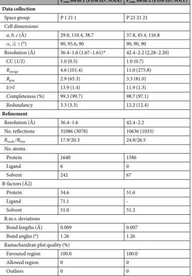
build a higher quality model (100% Ramachandran favoured, see Table 1). The first crystal (form 1, space group P21)
diffracted to a resolution of 1.6 Å, while the second crystal (form 2, space group P212121) gave rise to diffraction data
up to 2.2 Å. In all of the crystal forms, including the original crystal (PDB ID: 4BXT)37, the asymmetric unit is
com-posed of two tetrameric, helical coiled-coils packing against each other in varying orientations (Fig. 1A–C). Sample electron density maps of the higher resolution Pcore structures are shown in Supplementary Fig. S1.
Pcore form 1 (PDB ID: 5OIX) Pcore form 2 (PDB ID: 5OIY)
Data collection Space group P 1 21 1 P 21 21 21 Cell dimensions a, b, c (Å) 29.0, 110.4, 38.7 37.8, 45.4, 116.8 α, β, γ (°) 90, 95.6, 90 90, 90, 90 Resolution (Å) 36.4–1.6 (1.67–1.61)* 42.4–2.2 (2.28–2.20) CC (1/2) 1.0 (0.5) 1.0 (0.7) Rmerge 4.6 (101.4) 11.0 (275.8) Rpim 2.9 (65.3) 3.3 (81.0) I/σI 13.9 (1.4) 11.9 (1.3) Completeness (%) 99.3 (99.7) 98.7 (97.1) Redundancy 3.3 (3.3) 12.2 (12.4) Refinement Resolution (Å) 36.4–1.6 42.4–2.2 No. reflections 31086 (3078) 10636 (1033) Rwork /Rfree 17.9/20.3 24.8/26.5 No. atoms Protein 1640 1586 Ligand 6 0 Solvent 242 67 B-factors (Å2) Protein 34.6 51.6 Ligand 71.1 - Solvent 51.0 51.2 R.m.s. deviations Bond lengths (Å) 0.009 0.007 Bond angles (°) 1.26 1.26
Ramachandran plot quality (%)
Favoured region 100.0 100.0
Allowed region 0 0
Outliers 0 0
www.nature.com/scientificreports/
Mapping of the sequence conservation within the Pneumoviridae onto Pcore reveals the strict conservation of
the hydrophobic amino acids (Leu and Ile residues) lining the interior of the coiled-coil, indicating that all family
members share a similar P oligomerization region (Fig. 1D, strictly conserved residues in purple). Furthermore,
Arg175 is strictly conserved in all Pneumoviridae and plays a role in stabilizing the quaternary structure of the tetramer by forming a salt bridge with either an Asp or Glu on the neighbouring helix. Notably, residues Ser184 and Glu181 are also invariable despite being exposed to the solvent and not directly involved in oligomerization. It is tempting to speculate that these residues may instead be involved in interactions with other viral compo-nents, for instance the L polymerase42.
In one of the tetramers within crystal form 2 we observed a single, solvent-exposed helix which possessed markedly blurred electron density and, as a result, significantly higher crystallographic B-factors than the three
other protomers of the coiled-coil (Fig. 1E, white arrow). This suggests that, in absence of packing constraints
Figure 1. Structures of HMPV Pcore from different crystal forms. A, B and C. Asymmetric units of two different
Pcore crystal forms shown in side view and top view orientations. In both cases the asymmetric unit contained
two coiled-coil Pcore tetramers (one depicted in grey and one coloured by chain). The structure in (A) is
derived from the P21 crystal which diffracted to 1.6 Å, while the structure in (B) is derived from the P212121
crystal at 2.2 Å resolution. The previously published structure (PDB ID: 4BXT) is shown for comparison in (C) highlighting the distinct packing arrangements of each crystal. (D) The sequence conservation within
Pneumoviridae members was mapped onto the Pcore structure and amino acids are coloured by conservation as
indicated. For clarity, the side chains of only one of the four protomers are shown. Strictly conserved residues (coloured in deep purple) are explicitly labelled. E. B-factor putty representation of a single Pcore tetramer from
crystal form 2. Red and thick regions of the putty represent high B-factors (~50 Å2), whilst thin and blue regions
indicate low B-factors (~20 Å2). A solvent accessible helix displayed significantly higher B-factors that the rest of
the coiled-coil (indicated by arrow). F. Structural superposition of six Pcore tetramers taken from crystal forms 1
within the crystal (as is the case for this solvent-exposed helix), the protomers of the Pcore region are somewhat
flexible and there is a certain degree of malleability of the tetramer interface. Alignment of all six tetramers from crystal forms 1 and 2 and the original crystal confirmed that there are small-scale breathing motions within the
coiled-coil (Fig. 1F). This is fully consistent with the overall lower buried surface area (~4800 Å) and predicted
dissociation energy (~21 kcal/mol) for HMPV P than in the more extended paramyxoviral coiled-coil oligomer-ization domains. For comparison, the buried surface area and predicted dissociation energy of the Nipah virus P
oligomerization domain have been reported to lie between 15000–20000 Å and 140–200 kcal/mol43. These data
indicate a flexibly associated tetramer of HMPV P protomers.
SAXS characterization of the HMPV P constructs.
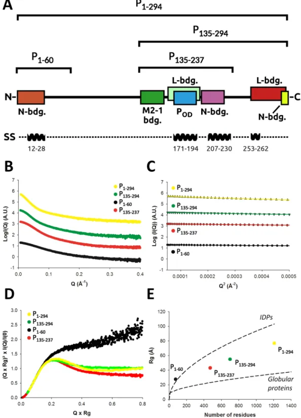
Next, we turned to SAXS in order to obtain struc-tural information in the solution state for the different functional regions of P (Fig. 2). In total 4 P constructs werecharacterized by SAXS: 1) the N-terminal region (P1–60) which encompasses the N0 binding site, 2) the central
region (P135–237) composed of the M2-1 binding site, the tetramerization domain Pcore and a putative
nucleopro-tein binding region, 3) the P135–294 construct, which includes potential L and N binding regions at its C-terminus,
and 4) the full-length P protein (P1–294) (domain annotations are shown in Fig. 2A and Supplementary Fig. S2).
Our approach of splitting the protein into different fragments enables more accurate X-ray scattering profiles to be obtained compared to focussing on the full-length protein only, thus increasing the overall structural infor-mation content that can be extracted from the SAXS data. For each construct, high-quality SAXS profiles could
be obtained (Fig. 2B), and samples were free from aggregates as evidenced by the linearity of the Guinier region
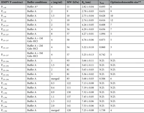
(Fig. 2C). Parameters extracted from the SAXS data are summarized in Table 2. Molecular weights were estimated
based on calculation of the concentration-independent volume of correlation VC, as defined in44, and were found
to be consistent with the theoretical molecular weight expected for each construct. In the case of the full-length P, a SEC-MALLS profile was also recorded, which confirmed the sample monodispersity, oligomerization state,
and molecular weight (Supplementary Fig. S3, MWMALLS = 125 kDa ± 12 kDa and MWtheor. = 134.8 kDa). The
SAXS-derived radii of gyration (Rg) were independent of concentration for P1–60 and P135–237. On the contrary
P135–294 and P1–294, respectively, were observed to significantly compact or expand with increasing protein
con-centration indicating the presence of a structure factor contribution to the data, which was treated through data merging of different protein concentrations (Table 2).
We used normalized Kratky plots to obtain a semi-quantitative view of the level of intrinsic disorder present
in each construct. As can be seen in Fig. 2D, all constructs showed a mixed profile indicating a balance between
order and disorder, with the exception of P1–60 which displayed a typical intrinsically disordered protein (IDP)
Kratky plot. This is likely due to the absence of the tetramerization domain, the most ordered region in P. A
similar behaviour was observed when the experimental Rg of each construct was placed on a Rg versus number
of residues plot and compared with the empirical laws available for both globular45 and intrinsically disordered
proteins46 (Fig. 2E). P
1–60 appeared as a canonical IDP while the tetrameric constructs showed an intermediate
behavior between fully folded proteins and IDPs. Of note, the full-length P1–294 was slightly offset towards the
IDP curve compared to P135–237 and P135–294, consistent with an increase in intrinsic disorder upon inclusion of the
N-terminal region. We then used computational modeling in combination with ensemble optimization to derive SAXS-validated ensembles of atomic models for each construct.
The N-terminal region of P shows α-helical propensity and is in equilibrium between extended
and more compact conformers.
All-atom models of P1–60 were generated using the state-of-the-artpro-gram flexible-meccano47 and used for ensemble optimization of the available experimental SAXS curves,
result-ing in a high-quality fit to the data (χexp ~ 0.6 - Table 2, Fig. 3A). Because no significant concentration dependence
of the Rg was observed, an average Rg distribution was calculated and is shown in Fig. 3B, revealing an equilibrium
between two populations of different sizes centered around 2.2 nm and 4.0 nm. Representative models of these
two populations are shown in Fig. 3C. A significant fraction of the selected models (~20%) displayed α-helical
structure in the previously reported N0 binding region (Fig. 3C) indicating that this MoRE is able to partially fold
as an α-helix in the absence of its binding partner.
Ensemble analysis of the central region of P indicates the presence of loose tertiary structure
located C-terminally of P
core.
Ensembles of atomic models of P135–237 were generated by combiningclassi-cal and coarse-grained atomistic molecular dynamics simulations and using the available SAXS-validated models of P158–237 as templates (see material and methods)37. The two constructs differ only by the presence of the
puta-tive M2–1 binding region approximately located between residues 135 and 158, which mainly adopts extended conformations within the selected ensembles, although residual α-helical structure between residues 138–145 is present in a small fraction of the models. The ensemble optimization results show that the selected ensembles (Fig. 4A) successfully reproduced the experimental data (0.6 < χexp < 1.1 - Table 2, Fig. 4C). The Rg
distribu-tion highlights the equilibrium between two main populadistribu-tions (Fig. 4B) which differ in the conformation of
their C-terminal regions (Fig. 4A, inset). Residues 204–219 (which overlap with a recently proposed secondary
N-binding site in RSV26) tend to form an α-helix (α
204–219) that packs against the Pcore mainly through
hydropho-bic interactions. This relatively unstable structural motif is present in about 30% of the selected models and gives
rise to a somewhat more compact population, shifting down the Rg values by about 1 nm compared to the other,
more extended population. Interestingly, these ordered conformations of residues 204–219 have originally been
observed when modeling P158–237 and were found to be relatively stable for several hundreds of nanoseconds of
classical MDS both in our previous work37 and in this study (not shown). It is worth noting that when
perform-ing the ensemble optimization usperform-ing a pool in which the compact population of models was excluded, we could
www.nature.com/scientificreports/
Figure 2. HMPV P constructs and small angle x-ray scattering experiments (SAXS). (A) Functional regions
and HMPV P constructs used in this study. The N0 binding region (residues 1–28) is shown as a brown
box and the crystallographically observed tetramerization domain Pcore in blue. The putative M2-1, L and
N-RNA binding regions are indicated based on homology with human/bovine respiratory syncytial viruses (RSV)16,26,27,42,60,66,99,100. The location of predicted α-helices is indicated on a separate bar below. (B) Measured
SAXS profiles of P1–60 (black), P135–237 (red), P135–294 (green) and P1–294 (yellow). (C) Corresponding Guinier
plots, showing linear behavior in the low q range. (D) Normalized Kratky plots. (E) The radius of gyration versus number of residues for the different HMPV constructs is shown as colored spheres, and is compared with empirical laws for globular45 and intrinsically disordered proteins (IDPs)46.
indicating that the presence of a packed α204–219 in the ensemble models is important to correctly reproduce the
experimental SAXS profile.
To further assess the validity of our ensemble analysis procedure, SAXS profiles of P135–237 were recorded
at increasing concentrations of guanidinium hydrochloride (Gdn-HCl) (Fig. 5 and Supplementary Fig. S4).
Addition of 1, 2 and 3 M Gdn-HCl resulted in the progressive disappearance of the most compact population
and a concomitant increase in the proportion of extended models (Fig. 5A and B). As can be seen in Fig. 5A,
the population of models where α204–219 packs against the neighboring Pcore is completely absent in the 1 M Gdn
HCl selected ensemble, while residual α-helical structure in this region is retained. At higher Gdn-HCl
concen-trations, these secondary structure elements are also lost. Interestingly, Pcore was found to remain stable at all
Gdn-HCl concentrations indicating a highly stable tetrameric core.
The C-terminal region of P may stabilize the loose tertiary structure present in the central
region.
Models of P135–294 were obtained using coarse-grained atomistic MDS based on the P135–237 modelingresults. Again, the quality of the fit to the experimental data was very good (χexp = 0.5 - Table 2, Fig. 6A). The
Rg distribution is dominated by a main population centered around 5–6 nm in equilibrium with more extended
models (Fig. 6C). At high protein concentrations, we observed a significant decrease of the measured Rg from
5.66 to 5.36 nm (Table 2). This concentration-dependent Rg decrease might be linked to the presence of a highly
negatively charged patch present in the C-terminal part of the molecule (Supplementary Fig. S5), leading to long
range intermolecular repulsion. About 55% of selected models appear to have the packed α204–219, suggesting that
the C-terminal region of the protein might stabilize the cap of helices located C-terminally to the tetramerization
domain (and also observed with the P135–237 construct). The 75 C-terminal amino acids adopt various disordered
conformations, with a significant fraction of the selected models displaying a short α-helical motif near their
C-terminus in the predicted region (residues 253–262, at the beginning of the putative L-binding site) (Fig. 6B).
It is however not possible to ascertain the presence of this motif based on SAXS data alone for such a large con-struct, as only a modest fraction of the scattering arises from this region of the molecule.
Ensemble structure of the full-length phosphoprotein.
Finally, we generated an ensemble of atomicmodels of P1–294 using coarse-grained MDS based on the results of the P60 and P135–294 modeling and performed
ensemble optimization against the experimental SAXS data. We were able to adequately fit the data with χexp = 1.7
(Fig. 7A and Table 2). A representative ensemble of models optimized using the merged SAXS curve is shown
in Fig. 7C. The full-length phosphoprotein appears as a large, tentacular molecule with maximal intramolecular
distances (Dmax) roughly comprised between 25 and 35 nm, and a partitioning of the N-terminal and C-terminal
intrinsically disordered extensions on each side of Pcore. The Rg distribution shows a main population centered
HMPV P construct Buffer conditions c (mg/ml) MW (kDa) Rg (nm) χexp Optimizedensemble size**
P1–60 Buffer A* 4 11 2.82 ± 0.04 0.693 6 P1–60 Buffer A 2 9 2.78 ± 0.03 0.631 9 P1–60 Buffer A 1.5 10 2.75 ± 0.04 0.628 10 P1–60 Buffer A 1 10 2.74 ± 0.05 0.616 12 P135–237 Buffer A 2 55 4.26 ± 0.05 0.608 7 P135–237 Buffer A 4 54 4.29 ± 0.03 0.656 7 P135–237 Buffer A 8 57 4.27 ± 0.01 1.094 5 P135–237 Buffer A + 1M Gdn-HCl 4 50 4.76 ± 0.06 0.875 5 P135–237 Buffer A + 2M Gdn-HCl 4 54 5.22 ± 0.10 0.860 3 P135–237 Buffer A + 3M Gdn-HCl 4 57 5.25 ± 0.13 0.742 5 P135–294 Buffer A 1 93 5.66 ± 0.11 N.D. N.D. P135–294 Buffer A 1.5 82 5.63 ± 0.11 N.D. N.D. P135–294 Buffer A 2 81 5.65 ± 0.03 N.D. N.D. P135–294 Buffer A 3 81 5.36 ± 0.02 N.D. N.D. P135–294 Buffer A merged 83 5.68 ± 0.03 0.506 6 P1–294 Buffer A 0.3 122 7.13 ± 0.08 N.D. N.D. P1–294 Buffer A 0.4 111 7.19 ± 0.08 N.D. N.D. P1–294 Buffer A 0.9 130 7.32 ± 0.08 N.D. N.D. P1–294 Buffer A 1.1 137 7.45 ± 0.05 N.D. N.D. P1–294 Buffer A 1.5 112 7.49 ± 0.06 N.D. N.D. P1–294 Buffer A 2.0 141 7.53 ± 0.06 N.D. N.D. P1–294 Buffer A merged 128 7.25 ± 0.08 1.738 4
Table 2. SAXS-derived parameters. *Buffer A: 20 mM Tris pH 7.5 150 mM NaCl. **Optimized ensemble size: The optimum selected ensemble size and relative weights of the models were determined automatically by GAJOE using default parameters.
www.nature.com/scientificreports/
around 7–8 nm (Fig. 7B) and about 60% of selected models displayed a packed α204–219. Contrary to P135–294,
higher protein concentrations lead to higher apparent Rg values that increase from 7.1 to 7.5 nm in the
(rela-tively narrow) concentration range used (Table 2). This concentration-dependent increase in Rg indicates
attrac-tive interparticle interference which possibly results from the presence of both highly negaattrac-tively and posiattrac-tively
charged patches located in the N-terminal and C-terminal regions (Supplementary Fig. S5).
Discussion
The essential polymerase cofactor P from Pneumoviridae is a multifunctional hub which interacts with numerous components of the viral RNA-synthesis machinery. It also possesses large IDRs and can therefore not easily be characterized by classical structural biology methods, such as X-ray crystallography. NMR studies of the RSV P
protein have shown that IDRs account for around 80% of the protein26. Analysis of whole eukaryotic genomes
has suggested that as much as 41% of protein sequences contain IDRs of significant length48 and research on IDPs
indicates that these regions are frequently involved in protein-protein interactions (reviewed in49). Pneumoviridae
P is commonly described as possessing an N-terminal IDR (mainly associated with binding RNA-free N) and a C-terminal IDR (mainly associated with binding N-RNA), which are separated by a central tetramerization module, Pcore24,32,37,50. In this study we present a description of the structure and dynamics of HMPV P, using a
combination of crystallography of stable/bound domains and SAXS ensemble analysis. We show that full-length phosphoprotein exists in solution as an ensemble of inter-converting conformers with a tentacular architecture. The four long N-terminal IDRs and the four somewhat shorter C-terminal IDRs are partitioned by the central tetramerization domain owing to the parallel orientation of Pcore α-helices. The P protein tetramer (1176 residues)
has huge dimensions, with an average Rg of 7.5 nm and an average Dmax of about 30 nm. For comparison, the
entire L polymerase (2109 residues) of vesicular stomatitis virus (VSV), which is likely similar in size as HMPV L polymerase, displays a Rg of 3.9 nm and a Dmax of about 13 nm, based on its cryo-electron microscopy structure51.
Undoubtedly, the large dimensions and flexibility of the P protein reflect its functions in viral transcription and
replication which require the simultaneous binding and adequate positioning of the N0, the M2-1 anti-terminator,
the L polymerase and the nucleocapsid. In addition, simultaneous binding of P to multiple N-RNA subunits may influence the conformation and curvature of the nucleocapsid.
Figure 3. SAXS-based ensemble analysis of P1–60. (A) Fitted SAXS profiles of P1–60 for 4 different
concentrations. Experimental scattering curves are shown as black spheres with optimized ensembles (OEs)
fits shown as red lines. The curves are ordered by increasing protein concentrations (Table 2) from bottom to
top. (B) Radius of gyration distributions for the initial pool ensemble (gray area) and for the OEs (black line), averaged over all protein concentrations. (C) Families of models extracted from the OEs were classified into “collapsed” or “extended” based on their radius of gyration. About 20% of models with α-helical structure are also present in the OEs.
Conformation of the N-terminal region.
The disordered N-terminal region of P has been implicated in chaperoning the N protein, keeping it in an RNA-free state prior to nucleocapsid assembly, for a range of neg-ative strand viruses32,33,39,52,53. This is important as N may otherwise unproductively bind to host nucleic acids.N-chaperoning is facilitated by a MoRE located towards the very N-terminus of the P-protein, which folds upon
binding nascent and RNA-free nucleoproteins31,39. Kratky analysis of the first 60 N-terminal residues of HMPV
P (P60) confirmed the disordered character of this region. However, in our ensemble analysis 20% of the models
exhibited helical secondary structure within residues 13–28, corresponding to the same helix that is observed
in the crystal structure of HMPV P bound to N032. Interestingly, a recent NMR study similarly observed a 20%
helical propensity in the equivalent residues (amino acids 12–24) of RSV P26. Such pre-formed secondary
struc-ture elements in the apo form have been suggested to lead to increased association rates to the binding partner
via conformational selection49. Rapid on-rates may be especially important in the case of N-chaperoning as N0
needs to be captured by P before unspecific RNA-uptake and polymerization can take place, leading to a dead-end state for N. Following initial binding, the pre-formed helix may act as a nucleus from which the N-P binding interface is extended to what is observed in the crystallographic structure (encompassing P residues 1–28) via a
dock-and-coalesce mechanism54.
Conformation of the M2-1 binding region.
Pneumoviral P proteins recruit the additional processivityfactor M2-1 to the viral transcription machinery, forming an extended non-globular complex28,29,55. HMPV M2-1
is a highly dynamic, tetrameric, RNA-binding protein featuring a CCCH-type zinc-finger on each protomer, which is involved in recognition of nucleic acids30,56. The M2-1 protein is thought to bind to nascent RNA
emerg-ing from the viral polymerase, thus inhibitemerg-ing premature termination of transcripts via an as-of-yet undetermined mechanism19,20,57–59. Mutational studies have mapped RSV P residues 100–120 as the M2-1 interaction site60,
which roughly corresponds to the region around residues 135–158 in HMPV. In contrast to the significant
frac-tion of pre-formed N0-binding region described above, our ensembles display only marginal residual secondary
Figure 4. SAXS-based ensemble analysis of P135–237. (A) Models extracted from the OEs were categorized based on the presence (left) or absence (right) of C-terminal α-helical elements (residues 202–219) packed against the coiled coil region. A zoom of the C-terminal α-helical elements is shown in inset and highlighted by an arrow. (B) Radius of gyration distributions for the initial pool ensemble (gray area) and for the OEs (black line), averaged over all protein concentrations. (C) Fitted SAXS profiles of P135–237. Experimental scattering curves are
shown as black spheres with optimized ensembles (OEs) fits shown as red lines. The three curves are ordered by
www.nature.com/scientificreports/
structure within the M2-1 binding site of apo P. In-line with this observation, the NMR study of RSV P could detect a small segment of five residues (RSV amino acids 98–103) with only 8% helical propensity at the M2-1
binding region26. In this case, recruitment of M2-1 by P may involve folding-upon-binding of an otherwise highly
unstructured region. M2-1 and P may thus form a so-called fuzzy complex, in which substantial flexibility is
retained even within the bound form61.
Conformation of the C-terminal IDR.
Previous crystallographic and solution scattering studies of Nipah virus43,62 and Sendai virus P63 have highlighted a cap-like structure of helices folding back onto the N-terminalpart of the oligomerization domain. However, the cap was not observed in the crystal structures of the helical
coiled-coil oligomerization domains of Measles virus P64, Mumps virus P65, or HMPV P37. Although not present
in the crystal, in our HMPV P solution ensembles we find a compact subpopulation of conformers which con-sistently features a cap of helices (amino acids 204–219) located C-terminally of the tetramerization domain. It is quite possible that the cap is a specific feature of pneumoviral P proteins that exists in solution but is insufficiently stable, at least in HMPV, to be observed in the crystalline state. Deletion studies for bovine respiratory syncytial virus (BRSV) P have suggested that the region corresponding to the cap in HMPV may represent a secondary
nucleoprotein binding site, besides the primary one at the very C-terminus of P66. In addition, a recent NMR
study of RSV P has identified a helical region partially overlapping with the corresponding cap-forming
resi-dues in HMPV26. The authors observed line broadening in this helix upon adding the N-terminal domain of the
nucleoprotein, strongly indicating that these do indeed interact. It is worth noting that the directly adjacent
oli-gomerization region has been suggested to interact with the large L polymerase, at least in BRSV42. The secondary
N-binding site of the cap-helices may thus bring L and N into close vicinity and may facilitate the interplay of N with L during the viral RNA-synthesis cycle. This is in line with mutational studies in RSV which have revealed that mutation of a conserved charged cluster within the cap to alanines (corresponding HMPV residues R215/
E216/E217) abrogated all reporter gene activity in a minigenome assay67. The R215/E216/E217 cluster of the
Figure 5. Effect of guanidinium hydrochloride on the solution structure of P135–237. (A) Models from the OEs obtained in the presence of increasing concentrations of guanidinium hydrochloride (Gdn-HCl) showing the progressive disappearance of the α-helical elements outside of Pcore. (B) Radius of gyration distributions for the
initial pool ensemble (gray area) and for the OEs obtained in the presence of 1 M Gdn HCl (black line), 2 M
Gdn HCl (red line) and 3 M Gdn HCl (green line). (C) Fitted SAXS profiles of P135–237 measured in the presence
of increasing concentrations of guanidinium hydrochloride (1, 2 and 3 M, from bottom to top). Experimental scattering curves are shown as black spheres with optimized ensembles (OEs) fits shown as red lines.
cap-structure in HMPV P is solvent accessible in our ensembles and may thus be positioned ideally to interact with further viral components.
Our structural ensembles of P135–294 and P1–294 feature a C-terminal α-helix in the predicted region (residues
253–262), which is part of a putative L-binding site that was mapped by mutagenesis studies in HRSV27. Although
it is difficult to validate the presence of this secondary structure element on the basis of SAXS data alone due to the weak contribution to the scattering signal arising from this region of the molecule (within the experimental SAXS profiles), it is worth mentioning that this region was suggested to fold upon binding to the L polymerase
based on the effect of point mutations on HRSV RNA synthesis27. However, this region of the P protein is poorly
conserved between RSV and HMPV owing to the insertion of a highly acidic stretch of about 18 residues in the
middle of the L binding site in the HMPV P sequence (Supplementary Fig. S2), and no residual α-helical
struc-ture could be detected in the equivalent region of RSV P by NMR26.
Long range effects modulating P conformational ensembles.
The structural characterization ofP135–294 and P1–294 also revealed the ability of specific IDRs of the P protein to influence the conformation of other
parts of the protein. In particular it was observed that a significantly higher proportion of models featuring the α204–219 cap-like structure is present in P135–294 and P1–294 structural ensembles compared to P135–237 (55–60%
ver-sus 30%). The ability of the C-terminal region of P to stabilize this α-helical structure might explain its absence
from the crystal structures of Pcore (as the P constructs used for crystallization required C-terminal degradation
in order to yield diffracting crystals, which in turn destabilizes the cap-like structure). We hypothesize that this property might arise from the highly acidic nature of the C-terminal region of P which contains a large number of negatively charged residues arranged in repetitive blocks (for example residues 140–170 and residues 263–294,
Supplementary Figs S2 and S5), leading to intramolecular electrostatic repulsion. Interestingly, the full-length
P protein SAXS profiles display higher apparent Rg values at high protein concentrations, indicating attractive
interparticle interference. Based on the distribution of charged residues along the P sequence, it is tempting to
speculate that the presence of basic patches of residues within the N-terminal IDR (Supplementary Fig. S5)
com-bined with the acidic C-terminal IDR is responsible for this attraction.
Possible implications for viral inclusion bodies formation.
The presence of a structure factor contri-bution in the SAXS profiles of P within the relatively small concentration range used for measurements (0.3 to 2 mg/ml) shows that the protein has a propensity to affect the structure of the liquid itself when present at high concentrations. This property might be relevant to viral replication in physiological conditions as pneumoviralFigure 6. SAXS-based ensemble analysis of P135–294. (A) SAXS profiles of P135–294. Experimental scattering
curves are shown as black spheres with the fit to the optimized ensemble (OE) shown as a red line for the
merged data curve. The curves are ordered by increasing protein concentrations (Table 2) from bottom
to top. The top curve corresponds to a merging of high and low concentrations to remove structure factor contributions. (B) Models extracted from the OE obtained using the merged SAXS profile. The short α-helical motif near the C-terminus (residues 253–262) is indicated by an arrow. (C) Radius of gyration distributions for the initial pool ensemble (gray area) and for the OE (black line).
www.nature.com/scientificreports/
replication was shown to occur in segregated cytoplasmic inclusion bodies which harbour the components of the
replication machinery and concentrate viral proteins68,69. Furthermore, expression of N and P was necessary and
sufficient to induce the formation of cytoplasmic inclusions in HMPV and HRSV15,16,70, but also in more distantly
related viruses such as human parainfluenza virus type 371 or rabies virus72,73. Recently, there is great interest in
the ability of proteins with IDRs to form phase-separated micro-compartments without the need of lipid bilayers,
especially in conjunction with molecular crowding74,75 (cellular examples of such membrane-less compartments
are Cajal bodies or cytoplasmic stress granules). These phase-separated organelles feature higher local concen-trations of a subset of factors which are often relevant for a functionally related pathway. Typically, proteins with low-complexity regions, low sequence diversity, multivalent binding properties, and blocks of oppositely-charged residues are potentially able to form these biomolecular condensates under specific conditions. The amino-acid compositions of many mononegavirus P proteins satisfy these prerequisites, with HMPV P possessing low-complexity regions, and being composed of over 35% Lys, Arg, Glu, and Asp, often arranged in repetitive blocks. Because each N molecule displays 2 P binding sites, it is tempting to speculate that binding of P proteins to N-RNA and N0 in cells would result in very high local concentrations of P. This, in turn might facilitate the
forma-tion of phase-separated microenvironments, constituting highly specialized viral RNA-synthesis and replicaforma-tion micro-factories. Phosphorylation of constituent proteins has previously been shown to control phase-separating behaviour76,77. Similarly, phosphorylation of P-proteins might modulate their general phase-separating behaviour,
instead of or along with specific protein-protein interactions, thus controlling inclusion body formation. In summary, we have shown that HMPV P is a highly dynamic protein that is composed of a stable helical tetramerization domain flanked by large N-terminal and C-terminal IDRs that sample a large volume in
solu-tion and display varying degrees of preformed structural elements. We found that the N-terminal N0 binding
site contained a significant proportion of α-helical structure and that the region C-terminally adjacent to the tetramerization domain had a tendency to form a cap-like helical structure that mapped to a putative N- RNA binding site. Our results further suggested that this cap-like structure might be stabilized by the presence of the full C-terminal IDR, and that the full-length phosphoprotein’s basic and acidic patches of residues may play a role in viral inclusion body formation by inducing long range intermolecular attraction and facilitating the formation of phase-separated microenvironments.
Material and Methods
Protein cloning, expression and purification.
The regions of the HMPV P gene (strain NL1-00, A1, GenBank: AAK62966.1) encompassing phosphoprotein residues 1–60, 135–237, 135–294, and 1–294 were ampli-fied by polymerase chain reaction and cloned into pOPINF (P 1–60, 135–237 and 135–294) or pOPINE (P1-294)plasmids78 using the In-Fusion system (TAKARA CLONTECH) following the instructions of the manufacturer.
Figure 7. SAXS-based ensemble analysis of P1–294. SAXS profiles of P1–294. Experimental scattering curves are
shown as black spheres with the fit of the optimized ensemble (OE) shown as a red line. The curves are ordered
by increasing protein concentrations (Table 2) from bottom to top. The top curve corresponds to a merging of
high and low concentrations to remove structure factor contributions. (B) Radius of gyration distributions for the initial pool ensemble (gray area) and for the OE (black line). (C) Models extracted from the OE obtained by fitting the merged data curve.
Recombinant proteins expressed from pOPINF feature a N-terminal (His)6-tag followed by a 3C protease site,
while pOPINE has a C-terminal (His)6-tag. All constructs were verified by nucleotide sequencing.
The (His)6-tagged constructs were transformed into Rosetta2 E. coli cells for recombinant expression. E. coli
were grown at 37 °C in terrific broth (TB) in presence of appropriate antibiotics to an OD600 of ~0.8 and
expres-sion was induced by addition of 1 mM β-D-1-thiogalactopyranoside (IPTG). Following induction, the cells were incubated at 18 °C overnight while shaking and were subsequently harvested by centrifugation (18 °C, 20 min, 4000×g). Cell pellets were resuspended in 20 mM Tris, pH 7.5, 500 mM NaCl and then lysed by sonication. The lysate was centrifuged for 45 min at 4 °C and 50000×g. The cleared lysate was syringe-filtered (0.45 μm pore
size, MILLIPORE) and transferred onto a column packed with pre-equilibrated Ni2+-NTA Agarose (QIAGEN).
Following several washes, the (His)6-tagged samples were eluted from the beads with 20 mM Tris, pH 7.5, 150 mM
NaCl, 300 mM imidazole. Finally, the samples were subjected to size exclusion chromatography and buffer exchanged into 20 mM Tris, pH 7.5, 150 mM NaCl. For small-angle X-ray scattering experiments, proteins were concentrated on-site with centrifugal filter units (MILLIPORE).
Small angle X-ray scattering experiments.
SAXS measurements were performed on beamline BM29(P1–60, P135–237 and P135–294) and former beamline ID14-3 (P1–294) at the European Synchrotron Radiation Facility
(ESRF), Grenoble, France. Samples were kept at 20 °C and data were collected at a wavelength of 0.0995 nm and a sample-to-detector distance of 1 m. 1D scattering profiles were generated and buffer subtraction was carried out by the automated data processing pipeline available at BM29 (ID14-3). The radius of gyration was
deter-mined with the program PRIMUS79 according to the Guinier approximation at low Q values, and molecular
weights were estimated based on44. In the case of P
135–294 and P1–294, which displayed concentration dependent
changes in the SAXS profile at low Q values, a merged curve was produced by combining the low Q region of data measured at low concentration with the high Q region of the high concentration SAXS profiles. These particle interference-free merged curves were used for subsequent analysis.
Structural modelling and molecular dynamics simulations.
Different strategies were used to obtainall atom models for each construct. 1000 models of the backbone of monomeric P1–60 were obtained using the
program flexible-meccano47, with an α-helical propensity of 50% for residues 14–26 which were observed to form
an α-helix when complexed with the HMPV N protein32. Protein side chains were then added using the program
SCCOMP80.
Initial models of P135–237 were built based on three families of models extracted from our previous HMPV P
modeling study that were found to accurately reproduce SAXS data for this P158–23737. All three models are
com-posed of a tetrameric coiled coil ranging from residues 168 to 198, with disordered residues at the N-terminus that have residual α-helical structure. In the first model, residues 202–219 adopt α-helical structures that pack laterally against the C-terminal part of the coiled coil region, while residues 220–237 are extended. In the second model, residues 208–237 form an α-helix consistently with secondary structure predictions, and in the third model, residues 168–237 are in an extended conformation with no secondary or tertiary structure. The region composed of residues 135–158 (including the N-terminal His6-3C site) was obtained based on a LOMETS
model81 adopting a relatively extended conformation which was grafted onto the three initial models of P
158–237.
Two additional starting models were generated by shortening the coiled coil by one helix turn at its C-terminus in the third model (to match the x-ray structure) and by further removing residual helical structure in the N-terminal region (that had been carried out from previous classical M.D. simulations). All five starting models of P158–237 were then simulated in GROMACS82 using either an atomistic coarse-grained structure-based model
(SBM)83,84, or explicit solvent classical molecular dynamics simulations (MDS).
In the case of the SBM MDS, a timestep of 0.0005 time units was used and the simulation was coupled to a temperature bath via Langevin dynamics. A single 100 ns trajectory was obtained for each starting model, and snapshots were extracted every 50 ps leading to an ensemble of 5000 models. In the case of classical MDS, we generated multiple trajectories for an aggregated simulation time of ~660 ns. MDS was performed using the
amber99SBws forcefield85 which has been developed to reproduce the properties of intrinsically disordered
pro-teins. At the beginning of each simulation, the protein was immersed in a box of SPC/E water, with a minimum distance of 0.9 nm between protein atoms and the edges of the box. 150 mM of NaCl were then added using
genion. Long range electrostatics were treated with the particle-mesh Ewald summation86. Bond lengths were
constrained using the P-LINCS algorithm. The integration time step was 5 fs. The v-rescale thermostat and the Parrinello–Rahman barostat were used to maintain a temperature of 300 K and a pressure of 1 atm. Each system was energy minimized using 1,000 steps of steepest descent and equilibrated for 500 ps with restrained protein heavy atoms prior to production simulations. Snapshots were extracted every 200 ps from each trajectory, leading to the generation of ~3300 additional models of P135–237.
In order to generate models of P135–294 and of P1–294, we adopted a similar strategy in which residues 238–
294 and residues 1–134 were grafted onto the existing P135–237 models with or without the α-helical secondary
structure elements (residues 14–26 and residues 251–262). Model types that were not selected through ensemble
optimization of P135–237 were not considered for this procedure. We then used the SBM approach to generate
ensembles for P135–294 and P1–294, yielding ~5000 P135–294 and ~8000 P1–294 models.
Ensemble optimization.
For each model from each ensemble, theoretical SAXS patterns were calculatedwith the program CRYSOL87 and ensemble optimization fitting was performed with GAJOE88,89. GAJOE uses a
genetic algorithm to select from a large pool of conformers optimized sub-ensembles that minimize the
www.nature.com/scientificreports/
( )
( )
( )
K I Q I Q Q exp 1 1j (1) K j exp j j 2 1 2∑
χ µ σ = − − =where K is the number of points in the experimental curve, σ is the standard deviation and µ is a scaling fac-tor. The optimum selected ensemble size and relative weights of the models were determined automatically by GAJOE. For each curve, the ensemble optimization procedure was repeated for a minimum of 20 times, from which the Rg distributions of the optimized ensembles were built.
Crystallization and data collection.
Our initial goal was the crystallization of a complex of the HMPV M2-1 protein bound to a P construct including the putative M2-1 binding region. The expression and purificationof HMPV M2-1 has been described previously30. In an attempt to crystallize the M2-1 – P
135–237 complex, vapour
diffusion crystallization trials of a 1:1 mixture of these two proteins at 7 mg/ml in 20 mM Tris, pH 7.5, 150 mM
NaCl were set up using a Cartesian Technologies pipetting system90. Although we were not able to grow crystals
of a complex, crystals which later proved to harbour only the P oligomerization region could be obtained after
extended time periods. The P21 crystal (form 1) of HMPV Pcore grew at 20 °C after 291–344 days with mother
liquor containing 20% polyethylene glycol (PEG) 6000, 200 mM NaCl, and 100 mM Tris, pH 8.0. The P212121
crystal (form 2) grew at 20 °C after between 132 and 185 days with mother liquor containing 25% PEG 3350, 200 mM MgCl, and 100 mM Tris, pH 8.5. Crystals were frozen in liquid nitrogen after being cryoprotected with 25% glycerol. Diffraction data were recorded on beamlines I03 (P21 crystal) and I04 (P212121 crystal) at Diamond
Light Source, Didcot, UK. Data reduction was carried out automatically with XIA291.
Structure determination and refinement.
The HMPV Pcore data sets were phased by molecularreplace-ment with PHASER92 using the previously published structure (pdbID:4BXT). The structures from both crystal
forms were subjected to multiple rounds of manual building in COOT93 and refinement in PHENIX94. We made
use of translation-libration-screw (TLS) parameters and 8-fold torsion-angle non-crystallographic symmetry
(NCS) restraints as implemented in PHENIX94. For the 1.6 Å data of crystal form 1 we additionally carried out
anisotropic atomic displacement parameter (ADP) refinement. The structures were validated with the wwPDB Validation Service (https://validate-rcsb-1.wwpdb.org/). Refinement statistics are given in Table 1. The final coor-dinates and structure factors have been deposited in the PDB with accession codes 5OIX and 5OIY.
Structure and sequence analyses.
Structure-related figures were prepared with the PyMOL MolecularGraphics System (DeLano Scientific LLC). Protein interfaces were analysed with the PISA webserver95. Mapping
of sequence conservation onto the Pcore structure was carried out with the ConSurf server96 using P sequences
from the Pneumoviridae family members human metapneumovirus (HMPV), avian metapneumovirus (AMPV), canine pneumonia virus (CPV), murine pneumonia virus (MPV), bovine respiratory syncytial virus (BRSV), and
human respiratory syncytial virus (HRSV). Sequences were aligned using using PROMALS3D97 and Jalview98
in order to analyse the conservation of the protein binding sites that were identified in RSV. The putative func-tional regions have been assigned based on homology and previously reported studies16,26,27,42,60,66,99,100. The linear
net charge per residue (NCPR) for HMPV P was calculated using the Classification of Intrinsically Disordered
Ensemble Regions (CIDER) webserver101.
Data availability.
Coordinates and structure factors have been deposited in the Protein Data Bank with accession numbers 5OIX and 5OIY. The SAXS datasets generated during the current study are available from the corresponding author on reasonable request.References
1. Black, R. E. et al. Global, regional, and national causes of child mortality in 2008: a systematic analysis. Lancet 375, 1969–1987, https://doi.org/10.1016/S0140-6736(10)60549-1 (2010).
2. van den Hoogen, B. G. et al. A newly discovered human pneumovirus isolated from young children with respiratory tract disease. Nature medicine 7, 719–724, https://doi.org/10.1038/89098 (2001).
3. van den Hoogen, B. G. et al. Prevalence and clinical symptoms of human metapneumovirus infection in hospitalized patients. The Journal of infectious diseases 188, 1571–1577, https://doi.org/10.1086/379200 (2003).
4. Schildgen, V. et al. Human Metapneumovirus: lessons learned over the first decade. Clin Microbiol Rev 24, 734–754, https://doi. org/10.1128/CMR.00015-11 (2011).
5. Esposito, S. & Mastrolia, M. V. Metapneumovirus Infections and Respiratory Complications. Semin Respir Crit Care Med 37, 512–521, https://doi.org/10.1055/s-0036-1584800 (2016).
6. de Graaf, M., Osterhaus, A. D., Fouchier, R. A. & Holmes, E. C. Evolutionary dynamics of human and avian metapneumoviruses. J Gen Virol 89, 2933–2942, https://doi.org/10.1099/vir.0.2008/006957-0 (2008).
7. Afonso, C. L. et al. Taxonomy of the order Mononegavirales: update 2016. Arch Virol 161, 2351–2360, https://doi.org/10.1007/ s00705-016-2880-1 (2016).
8. Ortin, J. & Martin-Benito, J. The RNA synthesis machinery of negative-stranded RNA viruses. Virology 479-480, 532–544, https:// doi.org/10.1016/j.virol.2015.03.018 (2015).
9. Ruigrok, R. W., Crepin, T. & Kolakofsky, D. Nucleoproteins and nucleocapsids of negative-strand RNA viruses. Curr Opin Microbiol
14, 504–510, https://doi.org/10.1016/j.mib.2011.07.011 (2011).
10. Gutsche, I. et al. Structural virology. Near-atomic cryo-EM structure of the helical measles virus nucleocapsid. Science 348, 704–707, https://doi.org/10.1126/science.aaa5137 (2015).
11. Tawar, R. G. et al. Crystal structure of a nucleocapsid-like nucleoprotein-RNA complex of respiratory syncytial virus. Science 326, 1279–1283, https://doi.org/10.1126/science.1177634 (2009).
12. Sutherland, K. A., Collins, P. L. & Peeples, M. E. Synergistic effects of gene-end signal mutations and the M2-1 protein on transcription termination by respiratory syncytial virus. Virology 288, 295–307, https://doi.org/10.1006/viro.2001.1105 (2001). 13. Noton, S. L. & Fearns, R. Initiation and regulation of paramyxovirus transcription and replication. Virology 479-480, 545–554,
14. Norrby, E., Marusyk, H. & Orvell, C. Morphogenesis of respiratory syncytial virus in a green monkey kidney cell line (Vero). J Virol
6, 237–242 (1970).
15. Derdowski, A. et al. Human metapneumovirus nucleoprotein and phosphoprotein interact and provide the minimal requirements for inclusion body formation. J Gen Virol 89, 2698–2708, https://doi.org/10.1099/vir.0.2008/004051-0 (2008).
16. Garcia-Barreno, B., Delgado, T. & Melero, J. A. Identification of protein regions involved in the interaction of human respiratory syncytial virus phosphoprotein and nucleoprotein: significance for nucleocapsid assembly and formation of cytoplasmic inclusions. J Virol 70, 801–808 (1996).
17. Yu, Q., Hardy, R. W. & Wertz, G. W. Functional cDNA clones of the human respiratory syncytial (RS) virus N, P, and L proteins support replication of RS virus genomic RNA analogs and define minimal trans-acting requirements for RNA replication. J Virol
69, 2412–2419 (1995).
18. Fearns, R. & Plemper, R. K. Polymerases of paramyxoviruses and pneumoviruses. Virus Res, https://doi.org/10.1016/j. virusres.2017.01.008 (2017).
19. Collins, P. L., Hill, M. G., Cristina, J. & Grosfeld, H. Transcription elongation factor of respiratory syncytial virus, a nonsegmented negative-strand RNA virus. Proc Natl Acad Sci USA 93, 81–85 (1996).
20. Fearns, R. & Collins, P. L. Role of the M2-1 transcription antitermination protein of respiratory syncytial virus in sequential transcription. J Virol 73, 5852–5864 (1999).
21. Cox, R. & Plemper, R. K. The paramyxovirus polymerase complex as a target for next-generation anti-paramyxovirus therapeutics. Front Microbiol 6, 459, https://doi.org/10.3389/fmicb.2015.00459 (2015).
22. El Najjar, F. et al. Human metapneumovirus Induces Reorganization of the Actin Cytoskeleton for Direct Cell-to-Cell Spread. PLoS pathogens 12, e1005922, https://doi.org/10.1371/journal.ppat.1005922 (2016).
23. Goutagny, N. et al. Cell type-specific recognition of human metapneumoviruses (HMPVs) by retinoic acid-inducible gene I (RIG-I) and TLR7 and viral interference of RIG-I ligand recognition by HMPV-B1 phosphoprotein. J Immunol 184, 1168–1179, https://doi.org/10.4049/jimmunol.0902750 (2010).
24. Tran, T. L. et al. The nine C-terminal amino acids of the respiratory syncytial virus protein P are necessary and sufficient for binding to ribonucleoprotein complexes in which six ribonucleotides are contacted per N protein protomer. J Gen Virol 88, 196–206, https://doi.org/10.1099/vir.0.82282-0 (2007).
25. Galloux, M. et al. Characterization of a viral phosphoprotein binding site on the surface of the respiratory syncytial nucleoprotein. J Virol 86, 8375–8387, https://doi.org/10.1128/JVI.00058-12 (2012).
26. Pereira, N. et al. New Insights into Structural Disorder in Human Respiratory Syncytial Virus Phosphoprotein and Implications for Binding of Protein Partners. J Biol Chem 292, 2120–2131, https://doi.org/10.1074/jbc.M116.765958 (2017).
27. Sourimant, J. et al. Fine mapping and characterization of the L-polymerase-binding domain of the respiratory syncytial virus phosphoprotein. J Virol 89, 4421–4433, https://doi.org/10.1128/JVI.03619-14 (2015).
28. Tran, T. L. et al. The respiratory syncytial virus M2-1 protein forms tetramers and interacts with RNA and P in a competitive manner. J Virol 83, 6363–6374, https://doi.org/10.1128/JVI.00335-09 (2009).
29. Blondot, M. L. et al. Structure and functional analysis of the RNA- and viral phosphoprotein-binding domain of respiratory syncytial virus M2-1 protein. PLoS pathogens 8, e1002734, https://doi.org/10.1371/journal.ppat.1002734 (2012).
30. Leyrat, C., Renner, M., Harlos, K., Huiskonen, J. T. & Grimes, J. M. Drastic changes in conformational dynamics of the antiterminator M2-1 regulate transcription efficiency in Pneumovirinae. Elife 3, e02674, https://doi.org/10.7554/eLife.02674 (2014).
31. Leyrat, C. et al. Structure of the vesicular stomatitis virus N(0)-P complex. PLoS pathogens 7, e1002248, https://doi.org/10.1371/ journal.ppat.1002248 (2011).
32. Renner, M. et al. Nucleocapsid assembly in pneumoviruses is regulated by conformational switching of the N protein. Elife 5, e12627, https://doi.org/10.7554/eLife.12627 (2016).
33. Yabukarski, F. et al. Structure of Nipah virus unassembled nucleoprotein in complex with its viral chaperone. Nat Struct Mol Biol
21, 754–759, https://doi.org/10.1038/nsmb.2868 (2014).
34. Galloux, M. et al. Identification and characterization of the binding site of the respiratory syncytial virus phosphoprotein to RNA-free nucleoprotein. J Virol 89, 3484–3496, https://doi.org/10.1128/JVI.03666-14 (2015).
35. Llorente, M. T. et al. Structural properties of the human respiratory syncytial virus P protein: evidence for an elongated homotetrameric molecule that is the smallest orthologue within the family of paramyxovirus polymerase cofactors. Proteins 72, 946–958, https://doi.org/10.1002/prot.21988 (2008).
36. Llorente, M. T. et al. Structural analysis of the human respiratory syncytial virus phosphoprotein: characterization of an alpha-helical domain involved in oligomerization. J Gen Virol 87, 159–169, https://doi.org/10.1099/vir.0.81430-0 (2006).
37. Leyrat, C., Renner, M., Harlos, K. & Grimes, J. M. Solution and crystallographic structures of the central region of the phosphoprotein from human metapneumovirus. PLoS One 8, e80371, https://doi.org/10.1371/journal.pone.0080371 (2013). 38. Jensen, M. R. et al. Structural disorder within sendai virus nucleoprotein and phosphoprotein: insight into the structural basis of
molecular recognition. Protein Pept Lett 17, 952–960 (2010).
39. Leyrat, C. et al. The N(0)-binding region of the vesicular stomatitis virus phosphoprotein is globally disordered but contains transient alpha-helices. Protein Sci 20, 542–556, https://doi.org/10.1002/pro.587 (2011).
40. Leyrat, C. et al. Ensemble structure of the modular and flexible full-length vesicular stomatitis virus phosphoprotein. J Mol Biol
423, 182–197, https://doi.org/10.1016/j.jmb.2012.07.003 (2012).
41. Erales, J., Beltrandi, M., Roche, J., Mate, M. & Longhi, S. Insights into the Hendra virus NTAIL-XD complex: Evidence for a parallel organization of the helical MoRE at the XD surface stabilized by a combination of hydrophobic and polar interactions. Biochim Biophys Acta 1854, 1038–1053, https://doi.org/10.1016/j.bbapap.2015.04.031 (2015).
42. Khattar, S. K., Yunus, A. S. & Samal, S. K. Mapping the domains on the phosphoprotein of bovine respiratory syncytial virus required for N-P and P-L interactions using a minigenome system. J Gen Virol 82, 775–779, https://doi.org/10.1099/0022-1317-82-4-775 (2001).
43. Bruhn, J. F. et al. Crystal structure of the nipah virus phosphoprotein tetramerization domain. J Virol 88, 758–762, https://doi. org/10.1128/JVI.02294-13 (2014).
44. Rambo, R. P. & Tainer, J. A. Accurate assessment of mass, models and resolution by small-angle scattering. Nature 496, 477–481, https://doi.org/10.1038/nature12070 (2013).
45. Narang, P., Bhushan, K., Bose, S. & Jayaram, B. A computational pathway for bracketing native-like structures fo small alpha helical globular proteins. Physical chemistry chemical physics: PCCP 7, 2364–2375 (2005).
46. Bernado, P. & Svergun, D. I. Structural analysis of intrinsically disordered proteins by small-angle X-ray scattering. Mol Biosyst 8, 151–167, https://doi.org/10.1039/c1mb05275f (2012).
47. Ozenne, V. et al. Flexible-meccano: a tool for the generation of explicit ensemble descriptions of intrinsically disordered proteins and their associated experimental observables. Bioinformatics 28, 1463–1470, https://doi.org/10.1093/bioinformatics/bts172 (2012).
48. Dunker, A. K., Obradovic, Z., Romero, P., Garner, E. C. & Brown, C. J. Intrinsic protein disorder in complete genomes. Genome Inform Ser Workshop Genome Inform 11, 161–171 (2000).
49. Mollica, L. et al. Binding Mechanisms of Intrinsically DisorderedProteins: Theory, Simulation, and Experiment. Front Mol Biosci