HAL Id: hal-01630281
https://hal.sorbonne-universite.fr/hal-01630281
Submitted on 7 Nov 2017
HAL is a multi-disciplinary open access
archive for the deposit and dissemination of
sci-entific research documents, whether they are
pub-lished or not. The documents may come from
teaching and research institutions in France or
abroad, or from public or private research centers.
L’archive ouverte pluridisciplinaire HAL, est
destinée au dépôt et à la diffusion de documents
scientifiques de niveau recherche, publiés ou non,
émanant des établissements d’enseignement et de
recherche français ou étrangers, des laboratoires
publics ou privés.
Gut microbiota and obesity: Concepts relevant to
clinical care
Maria Carlota Dao, Karine Clément
To cite this version:
Maria Carlota Dao, Karine Clément. Gut microbiota and obesity: Concepts relevant to clinical care.
European Journal of Internal Medicine, Elsevier, 2017, �10.1016/j.ejim.2017.10.005�. �hal-01630281�
Gut microbiota and obesity: Concepts relevant to clinical care
Maria Carlota Dao
⁎,1, Karine Clément
⁎Institute of Cardiometabolism and Nutrition, ICAN, Assistance Publique Hôpitaux de Paris, Pitié-Salpêtrière Hospital, Paris, France INSERM, UMR S U1166, Nutriomics Team, Paris, France
Sorbonne Universités, UPMC University Paris 06, UMR_S 1166 I, Nutriomics Team, Paris, France
A R T I C L E I N F O
Keywords: Obesity Metabolic disease Gut microbiotaA B S T R A C T
The composition and function of gut microbiota play a role in obesity and metabolic disease, yet the mechanisms have not been fully described. As new discoveries and advances in thefield have occurred, the relevance of gut microbiota in clinical care has become more substantial. There is promising potential for manipulation of the gut microbiota as treatment of obesity and associated health complications, both as a standalone therapy and as part of interventions such as weight loss. In this review we have compiled knowledge and concepts that are important in the consideration of gut microbiota for clinical care.
1. Introduction
Even though there have been notable scientific advances in the study of gut microbiota and obesity, a causal relationship between the two remains undefined[1]. Although promising mechanistic links have been uncovered in rodents, the myriad factors underlying human obe-sity and related-metabolic dysfunction (including genetics/epigenetics and lifestyle) make it difficult to demonstrate an independent role for gut dysbiosis. Studies have measured composition, functional potential, metabolomics, and ecologic dynamics of the gut microbiota, but we still do not know their relative contribution to complex disease pathophy-siology and their concrete applicability to clinical care.
We, here summarize key discoveries made thus far that could have relevance in the management of obesity and its co-morbidities (Fig. 1). 2. Cross-talk between microbiota and host in metabolic disorders Composition and function of the microbiota differ between healthy lean and obese subjects[2]. Gut microbiota is modified in obesity per se and related-comorbidities, including type 2 diabetes (T2D)[3–7], non-alcoholic steatohepatitis[8], and cardiovascular diseases[9]. The me-chanisms believed to link the gut microbiota with obesity, at least in animals, include energy extraction capacity from food, influence on the integrity of the gut barrier, modulation of chronic inflammation and the immune system, and production of specific metabolites that, besides having a local effect on the gut-associated immune system and in-testinal barrier, also signal to other tissues and organs including the brain, liver and adipose tissue.
2.1. Factors influencing gut microbiota and metabolic diseases
Metabolic diseases stem from a combination of factors, including host intrinsic characteristics, lifestyle and environment, genetic/epi-genetic factors and gut microbiota composition and function. Diet has been widely studied in connection with the gut microbiota in obesity. For example, microbiota enterotypes, which have been used to group people according to their dominant phyla, are associated with long-term dietary habits[10]. Fermented foods andfiber consumption are associated with a healthier and more diverse microbiota[11]. As shall be described below, people living in more industrialized environments tend to have lower microbial diversity than people living in a more traditional manner.
Exchange of microbiota between individuals is another factor that shapes the microbial ecosystem. Adults consuming Western or re-stricted diets had distinct gut microbiota compositions, and lower richness was found in Western diet consumers. The microbes from these individuals were transplanted onto mice. Upon co-housing and enabling the transfer of gut microbiota (i.e. mice are coprophagic) recipients of the Western diet microbiota acquired traits of the restricted diet[12]. Similar results were seen in Ridaura et al.[13], showing the phenotypic transmissibility of some microbiota properties from humans to mice.
Pharmacology has an important effect on gut microbiota composi-tion. Antibiotic treatment leads to profound and long-lasting mod-ifications in the gut ecosystem [14]. Metformin, a key antidiabetic agent, has been identified as a confounder of microbiota observations in diabetes studies. These studies have suggested that the effect of met-formin on the host may be partially induced through the gut microbiota
⁎Corresponding authors.
1Present address: Jean Mayer USDA Human Nutrition Research Center on Aging at Tufts University, Boston, MA.
[15–17]. It is not excluded, however, that microbial composition may modify the pharmacology of drug compounds frequently used in me-tabolic disease leading in some circumstances to differential clinical effect as it was shown for example for digoxin, a well-known antiar-rhythmic agent[18].
2.2. The gut microbiota influences host intestinal barrier and immune response
There is an association between gut dysbiosis and disruption of the intestinal barrier's integrity, specifically mucus production and layer thickness, tight junctions, insulin sensitivity, and inflammation. Disruption of the intestinal architecture may lead to leaking of gut-derived compounds that would otherwise stay in the gut lumen. Cani et al. termed the detection of lipopolysaccharide (LPS) in circulation ‘metabolic endotoxemia,’ and found it to be associated with chronic inflammation and disruption of metabolic homeostasis, particularly insulin sensitivity, in mice. LPS acts by activating Toll-like receptor 4 (TLR4) and inducing an inflammatory cascade. LPS from certain bac-terial groups is more inflammatory than others, and it may be trans-located in chylomicrons or by leaking through a permeable gut. High fat diets are associated with endotoxemia[19,20].
There is a complex interplay between microbiota, intestinal epi-thelium and the gastrointestinal immune system, with many metabo-lites and microbial components having a direct influence on the host's immunity. The production of metabolites from nutrients or modifica-tion of host-produced metabolites has a direct effect on immune cells and on both the integrity and permeability of the intestinal epithelium. The enteric immune system is constantly assessing and responding to the gut microbiota. A healthy gut ecosystem is needed in the develop-ment of immune tolerance, for example by promoting regulatory T cell (Treg) differentiation and expansion [21], and prevention of
auto-immune disease or chronic inflammation.
The most studied metabolites in connection with microbiota and host are short chain fatty acids (SCFA). They are synthesized fromfiber metabolism by certain bacterial groups. SCFA act on the host in dif-ferent ways. They serve as a source of energy for colonocytes, they have a critical influence on glucose homeostasis by inducing gluconeogenesis on colonocytes, as histone deacetylase (HDAC) inhibitors they impact epigenetic modifications, and they influence incretin secretion, speci-fically glucagon-like peptide 1 (GLP-1), through activation of G protein-coupled receptors GPR41 and GPR43 [22,23]. SCFA are elevated in obesity [24,25], where it is believed microbiota is more efficient at extracting energy from otherwise indigestiblefibers, although this has not been fully demonstrated. Acetate may also have a role in central
signaling of hunger and satiety [26,27]. SCFA have an anti-in-flammatory effect through different pathways via both innate and adaptive immunity; they may inhibit inflammatory cytokine pro-duction and promote Tregexpansion. They also maintain the integrity of
the intestinal epithelial barrier[21]. Since differences were found in
immune cells of the jejunum layer in severe obesity[28], it would be of major importance to examine the interaction between obesity-related immune dysfunction, the intestinal tract and gut microbiota. One ex-ample pertains to lymphocyte subtypes known to be modified in obese condition[29]. Mucosal-associated invariant T (MAIT) cells are innate-like T cells that recognize bacterial ligands. They are present in blood and enriched in mucosal and inflamed tissues[30]. We showed a de-pletion of circulating MAIT cells in obese and diabetic subjects[108]. MAIT cells in metabolic disorders have an exacerbated pro-in-flammatory phenotype (increased IL-17). Furthermore, MAIT cell acti-vation is directly influenced by metabolites synthesized from vitamin B2 and B9 by gut bacteria[30].
There are various other examples of microbiota metabolites and co-metabolites that have been implicated in metabolic disease. For ex-ample, trimethylamine (TMA) is generated from dietary choline and carnitine by certain bacterial taxa, and converted in the liver to tri-methylamine N-oxide (TMAO). This compound has been consistently associated with increased risk of cardiovascular disease and mortality in humans and found to promote atherosclerosis in mice [31–33], though the mechanism remains unknown. Importantly, certain micro-biomes (e.g. vegans and vegetarians) are unable to produce TMA. An-other example is the production of branched chain amino acids (BCAA) by microbiota. A microbiome with a higher potential to produce BCAA has been associated with obesity[13], and insulin resistance[34]. This is relevant because high circulating concentrations of BCAA may dis-rupt glucose homeostasis and have been associated with T2D and obesity[35]. These findings call for detailed studies not only of mi-crobiota composition but also of functional potential and metabo-lomics.
2.3. Gut microbiota diversity is decreased in metabolic diseases
A lower microbial diversity has been shown in populations where the burden of obesity and metabolic disease is greater[36–39]. When comparing fecal microbiota between groups from urban areas in the United States, rural areas in Malawi, and Amerindians from the Vene-zuelan Amazon it was found that subjects from the United States had the least diverse microbiota and the Amerindians had the highest di-versity[36], suggesting a link between urbanization, lowfiber content of Western diets, microbiota and metabolic diseases.
PATIENT ► Lifestyle Diet Exercise ► Intrinsic factors Age, Gender Genetics ► Health status Obesity Co-morbidities Pharmacology Immune response Endotoxemia ► Weight loss (CR, BS) ► Prebiotic / Probiotic
► Next generation probiotic (i.e. Akk) ► Dietary intervention
► Fecal microbiota transplant
MICROBIOTA
Shift in microbial ecosystem Change in diversity
Change in abundance of beneficial microbes Change in metabolite concentration
PATIENT
Decreased endotoxemia
Improvement in metabolism, e.g. insulin sensitivity Improvement in immune response
Reduction in disease burden MICROBIOTA
► Microbial richness
► Abundance of certain species, e.g. Akk ► Metabolomics, e.g. SCFA
CONSIDER INTERVENTIONS THAT MODULATE GUT
MICROBIOTA
EFFECTS
(To be demonstrated in humans)
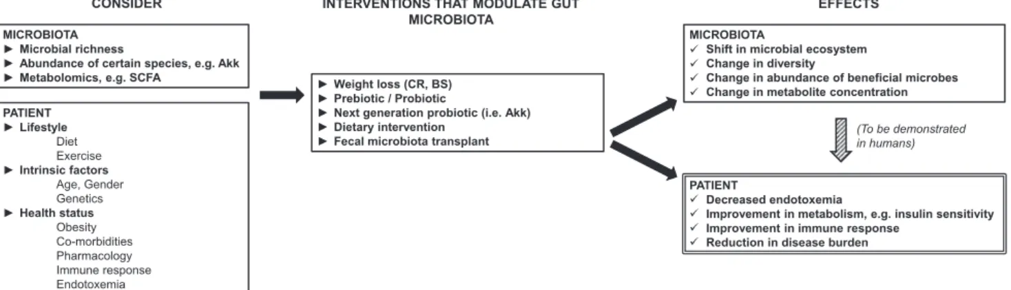
Fig. 1. Potential of gut microbiota in clinical care. Several aspects of gut microbiota composition and function have been implicated in metabolic diseases. Taking into consideration intrinsic patient characteristics, health status and environmental factors, manipulation of the gut microbiota could eventually be used in a wide array of treatments for metabolic disease, such as calorie restriction, bariatric surgery, prebiotic/probiotic intake and fecal microbiota transplantation. These treatments have shown changes in gut microbiota, which have been in turn associated with positive health outcomes, although causation remains to be demonstrated in humans. CR = calorie restriction, BS = bariatric surgery, Akk = Akkermansia muci-niphila.
In a French group of overweight and obese adults (MICRO-Obes study) a lower microbial diversity, quantified using metagenomic se-quencing, was associated with higher inflammation, dyslipidemia, adiposity and insulin resistance[41]. Individuals with higher diversity had a healthier dietary pattern[11]. Similarly, in a Danish group of lean and obese adults diversity was inversely associated with corpu-lence, and individuals with lower diversity had lower abundance of butyrate-producing bacteria such as Faecalobacterium prausnitzii. Moreover, these subjects had a lower Akkermansia muciniphila to Ru-minoccocus gnavus ratio potentially resulting in higher mucus degrada-tion, and a microbial functional potential less capable of handling oxidative stress[4]. As diversity appears to be an important phenotype, it remains to be determined whether lower microbial diversity is a consequence or one of the causes for the deterioration of metabolic health in obese individuals. One possible mechanism could be that a greater microbial diversity may lead to a complete and complex func-tional repertoire that is able to metabolize complex carbohydrates and other substrates more readily[22].
Analysis using 16S rRNA sequencing has yielded consistent results with metagenomics[42,43]. In a subset from the TwinsUK cohort lower diversity was associated with greater abdominal adiposity[42]. There were associations between host genetic variants and adiposity-asso-ciated OTUs, corroborating the existence of a link between host genetics and gut microbiota. In the same cohort lower diversity was associated with greater weight gain over 9 years of follow up[44]. Although si-milar observations have been obtained with different methodologies, metagenomics provides greater insight to both the composition and functional potential of the gut microbiome. A recent meta-analysis compared studies that had reported richness and Bacteroidetes-to-Fir-micutes ratio using 16S rRNA sequencing[45]. There was consistent yet narrowly lower richness in obesity. This was partly attributed to low statistical power and large inter-individual differences in microbiota composition. Human microbiota studies usually lack statistical power to detect the mild effect sizes of the microbiota. Future studies should be carefully designed and presented in a way that allows harmonization with previous reports in an effort to find consistencies in the field. 2.4. Gut bacteria species and host health: the example of A. muciniphila
Akkermansia muciniphila (A. muciniphila) is one of most widely stu-died gut bacterial species in relation to obesity and glucose homeostasis
[46]. A. muciniphila is a gram negative bacterium that can use mucin glycans of the intestinal mucus layer as its sole source of energy. In overweight adults we showed that A. muciniphila was associated with insulin sensitivity, smaller adipocyte size, and in general better meta-bolic health [47]. The mechanistic link between A. muciniphila and human health remains unknown. There is, however, compelling evi-dence in mice on how A. muciniphila may impact the host. Everard et al. showed that A. muciniphila abundance was lower in obese mice, and that increasing its intestinal abundance either with oligofructose or live culture gavage led to decreased endotoxemia, body fat, improved in-sulin sensitivity, and protected integrity of the gut barrier[48]. More recently, an outer membrane protein of A. muciniphila, Amuc_1100, has been identified. It is involved in pilus formation and stimulates the Toll-like receptor 2 (TLR2) system thereby possibly participating in cross-talk with the host and maintaining epithelial layer integrity. When given to mice, this protein had similar beneficial effects on the host than live A. muciniphila with respect to body composition, insulin sensitivity and protection of intestinal barrier integrity [49]. However, the me-chanisms of action of Amuc_1100 and A. muciniphila may only partially overlap. This study also showed that A. muciniphila could be grown in synthetic medium and that Amuc_1100 remains active after pasteur-ization, making it an attractive therapeutic target. In fact, protocols for its preparation and preservation for therapeutic applications have al-ready been developed[50]. Currently Dr. Cani's group is conducting a clinical trial of A. muciniphila supplementation in overweight and obese
adults, hypothesizing that it will improve metabolic health (NCT02637115).
A. muciniphila abundance may increase through dietary changes. In mice, diets enriched with oligofructose[48], fruit-derived polyphenols
[51,52],fish oil[53], or afiber-free diet[54]have all led to increased abundance in gut A. muciniphila. However, we did notfind an asso-ciation between food or nutrient intake, or diet quality and A. mucini-phila abundance in humans [47]. The modulation of A. muciniphila through prebiotic intake requires further investigation.
While the potential of A. muciniphila as an individual species in clinical applications is clear, the effect of its microbial ecosystem and intestinal environment must be considered. For example, we showed that the best clinical status was seen in individuals with both higher abundance of A. muciniphila and microbial diversity[47]. A. muciniphila is a producer of acetate and propionate[55], which may be used as sources of energy by other bacterial species. Future research should study the impact of increasing A. muciniphila abundance on gut ecology, functional potential, and metabolite output.
A. muciniphila combines a series of unique qualities. As a mucin degrader it resides in close proximity to the epithelial barrier; it has been shown to interact with human epithelial cell lines in vitro[56]. This bacterium has an attenuating and wide-ranging effect on the im-mune system[57,58]: animal studies show that it mediates the negative effects of interferon gamma (IFNɣ) on glucose homeostasis[59], has lower capacity to stimulate interleukin 8 (IL-8) production and TLR4 response than certain pro-inflammatory species[56], and induces the TLR2 pathway[49]which may have a protective effect on the epithelial
layer. Therefore, it may be through strengthening of the epithelial barrier, reduction of gut permeability and endotoxemia, attenuation of the immune system, and improvement of glucose homeostasis that A. muciniphila impacts the host. Furthermore, as a SCFA producer an effect on the gut-brain axis is expected.
3. Approaches to modulate gut microbiota and improve metabolic disease
3.1. Weight loss, prebiotic and probiotic interventions
Dietary interventions lead to compositional and functional mod-ifications in the gut microbiota (reviewed in [60]) that have been correlated with improvements in various health outcomes. Thefield is currently trying to go beyond correlations and discern the role that microbiota plays in outcomes from these interventions (Fig. 1). 3.1.1. Calorie restriction and dietary interventions
To gain a better understanding of how gut microbiota could impact the host, one approach would be to study the traits that have been consistently associated with better health and determine how they change with a dietary intervention, and their relationship with clinical outcomes. In the MICRO-Obes study, mentioned above, 49 overweight and obese adults underwent CR for 6 weeks followed by a weight maintenance regime for 6 additional weeks. The individuals that had low gene richness at baseline experienced a significant increase after CR
[41]. Conversely, richness did not change for those with higher baseline levels. This suggests that there may be a diversity ceiling in each in-dividual that, once reached, cannot be overcome by a dietary inter-vention alone[22]. The reversibility of microbial diversity was studied in mice consuming high or low amounts of microbiota-associated car-bohydrates. For the mice whose diversity was lowered due to low consumption of the carbohydrates, it took supplementation of both the carbohydrates and replacement of lost microbial groups to restore di-versity[61]. This study has implications in the consequences of Wes-tern-style diets on gut microbiota and health.
Additionalfindings from the MICRO-Obes study showed that A. muciniphila actually decreased over the weight loss period in subjects that had the highest baseline abundance, and only moderately
increased for subjects with low baseline abundance[47]. Throughout the intervention, subjects with higher baseline A. muciniphila retained an abundance 100-fold greater than those with low baseline levels.
Clinical outcomes and microbial compositional shifts in response to dietary interventions are variable among individuals (i.e. as responders and non-responders) [62]. This raises the question of whether perso-nalized interventions are the next step in unveiling how the microbiota influences health outcomes, and how response to interventions can be optimized. Using metadata from 800 healthy or prediabetic individuals, which included microbiota, lifestyle and clinical parameters, together with machine learning, researchers recently were able to device an algorithm that predicts a person's glycemic response to a given meal
[63]. They demonstrated in both the main and validation cohorts that these personalized interventions led to improved postprandial glycemic responses. Another approach has been developed by Shoaie et al. whereby studying the complex interactions between host, diet and microbiota composition, a dietary intervention that would in theory increase microbial richness was designed[64]. Future research should determine whether a beneficial outcome could be induced in non-re-sponders through the use of personalized interventions.
While even short term dietary interventions lead to compositional changes in the gut microbiota [65], it is becoming increasingly clear that long term dietary habits and microbiota composition prior to a dietary or CR intervention impact the individual's response[10,41,47]. For example, a 10-day dietary intervention induced a change in mi-crobiota composition as early as 24-h after baseline, but not enough for individuals to change their enterotype[10]. Associations between en-terotypes and diet were also stronger when studying habitual diet through FFQ than recent dietary intake through 24-h recall. Deeper understanding of the complex interaction between dietary profiles and gut microbiota is mandatory.
3.1.2. Bariatric surgery
Bariatric surgery (BS) is increasingly being used and an effective treatment for T2D [66]. T2D remission occurs in an overwhelming number of patients shortly after surgery. The reconfiguration of the gut architecture and change in gut microbiota are believed to play a role in metabolic ameliorations (reviewed in[60,67,68]). Studies of gut mi-crobiota after BS in humans have had for the most part small sample sizes. Some consistencies between human and animal studies have been found. For example, abundance of A. muciniphila and Proteobacteria increased after BS [69–73]. Richness has been found to increase 3 months after Roux-en-Y gastric bypass (RYGB) [73,74] but to de-crease 6 months after bilio-intestinal bypass, as measured with 16S rRNA sequencing[75]. Changes in F. prausnitzii abundance have been inconsistent, with increases being reported in some instances[76,77], and decreases in others[71,73].
The functional potential of the microbiome warrants further study in bariatric interventions. Interestingly, the microbial functional po-tential of 13 patients undergoing RYGB changed to a greater extent than the abundance of individual species[73], highlighting the importance of not only studying compositional changes but also function and even metabolomics output. In this study, the functional potential reflected a reaction by the gut microbiota to the changes in oxygen levels and nutrient availability in the gut after the surgery.
Strong evidence for involvement of microbiota in clinical outcomes long term after surgery has been shown [72]. Varying degrees of overweight were replicated in germ free mice receiving microbiota from obese women or women that had undergone RYGB, or vertical banded gastroplasty. Differences in microbiota composition, function, and metabolomic output were seen between the different groups. The type of BS certainly has an effect on compositional modifications of the gut microbiota because the intestinal architecture, pH, incretin and metabolite secretion, changes in bile production [67,78], and even post-intervention diet[79]differ across surgeries. In fact, findings in humans generally differ across the different types of BS and follow-up
time[80–82].
Microbiota may mediate changes in the host after BS in various ways. Secondary bile acids generated by microbes may play a role in the beneficial effects after BS. Higher levels of both primary and sec-ondary bile acids have been observed after RYGB in humans[80,83–85] and in mice[86]. In humans, although not directly linked with clinical outcomes, an early postoperative rise in total bile acids was attributed to a surge in bacterially-derived secondary bile acids. Mouse studies have unveiled the potential mechanisms linking secondary bile acids and shifts in gut microbial communities to metabolic outcomes after BS, suggesting pathways through the activation of farnesoid X receptor (FXR) and G-protein-coupled bile acid receptor (TGR5)[40,87].
Patient characteristics prior to an intervention have an impact on gut microbiota changes and clinical response to the surgery. In the fu-ture one could envision a system whereby clinical background, lifestyle and gut microbiota analysis of the patient prior to surgery are sys-tematically analyzed and taken into account in the prediction and op-timization of their response[60].
3.1.3. Do prebiotics and probiotics aid in weight loss interventions? There is compelling evidence that pre and probiotics may have a positive impact on metabolic health in animal models and in humans (reviewed in[60,88–90]). Here we focus on human studies that have added pre or probiotic supplementation to weight loss interventions to determine if they synergize with the treatment and improve response. Few weight loss studies with pro or prebiotic supplementation have been conducted. There has been a tendency towards greater weight loss and metabolic improvement when probiotics are taken in combination with CR [91–93]. Among these, an RCT compared outcomes after 12 weeks of CR in overweight and obese women that were given either a probiotic yogurt or a low-fat yogurt. Although there was no difference in weight loss, the group taking the probiotic yogurt experienced an improvement in blood lipid profile and glucose homeostasis[93]. Few strains and doses have been tested and the gut microbiota has not been characterized in most of these studies. Future studies should include these elements, probably combining different bacterial strains.
There have also been few BS interventions measuring the impact of probiotics supplementation post-surgery. One study found that RYGB patients taking 2.4 billion Lactobacillus daily for 6 months after surgery experienced lower bacterial overgrowth, greater short term weight loss, and improved vitamin B12 status over patients not taking the probiotic
[94]. However, two other studies with different design found no added benefit of probiotic supplementation over placebo in measured out-comes [95,96]. Similarly to CR interventions, more studies testing varying doses and strains of bacteria are needed to clarify whether there is an added benefit of probiotic supplementation after BS.
3.2. Fecal microbiota transplantation
Fecal microbiota transplantation (FMT) not only offers great po-tential for the treatment of a wide array of diseases, but is also a good model to study causality in the relationship between gut microbiota and human metabolic disorders. Animal microbiota transplantation studies have demonstrated that gut microbiota may modulate obesity and re-lated disruptions in host metabolism such as insulin resistance
[24,97,98]. Ridaura et al. showed that higher weight gain could be transferred through gut microbiota by fecal transplants from human twins discordant for obesity to germ free mice[13]. When co-housed lean and obese recipient mice received a low fat highfiber diet, mi-crobiota from the lean mice colonized the obese recipient mice thereby transferring the phenotype. Even though it is uncertain whether FMT would be an effective therapy against metabolic syndrome, it is a good proof of concept approach to study the causal relationship between microbiota and obesity.
FMT is an effective therapy to treat enteric infections, particularly Clostridium difficile, and perhaps also intestinal chronic inflammatory
diseases [99,100]. The prospect of using FMT for the treatment of metabolic diseases has also been contemplated but results have not been as definitive as in previous applications. This question has been best explored in a study where obese men with metabolic syndrome received either autologous or allogenic fecal microbiota from lean healthy donors [101]. Fecal microbiota transplants were done by duodenal tube into the small intestine. Median insulin sensitivity, as measured by glucose disappearance rate during euglycemic hyper-insulinemic clamps, tended to improve in the allogenic group after 6 weeks of transplantation. This effect however was variable between individuals, with some responders and non-responders. While microbial diversity was lower in the overweight group, it increased after allogenic transplantation. The abundance of butyrate producing bacteria in-creased in both fecal and small intestine microbiota samples.
These patients were further followed for a total of 3 months post FMT to study the resilience of the microbiota. New species were found to coexist with pre-existing ones in the recipients especially if they were phylogenetically related[102]. In fact, strain replacement was more marked than uptake of donor species, calling for future studies to ex-amine microbial composition at the strain level. There were different degrees of engraftment and resistance to donor colonization. The mi-crobiota of one of the healthy donors stood out as having greater ability to invade several recipients. Resilience of certain strains was detected up to at least 3 months after transplantation, but the changes in mi-crobiota were not associated with clinical outcomes. The lack of a more marked effect of FMT on insulin sensitivity may be partly explained by the selection and characterization of recipients. Insulin resistance as a complication of metabolic disruption may manifest similarly across patients, but some of these pathologies may be more dependent on the gut microbiota than others.
For FMT to be used in a clinical setting in the treatment of metabolic disorders future studies should consider: analysis of gut microbiota composition at the strain level, background intra-individual variation of the gut microbiota so that it is not confused with treatment-specific changes, and immune response of the recipient[102–104]. To go be-yond FMT, perhaps sets of strains identified as beneficial should be tested as supplements. This would circumvent identification of com-patible donors and the risks associated with transplantation of fecal matter. There is also the question of resilience. Even though microbiota composition may be transplantable, using this as a treatment would require repeated inoculations to the patient. Studying what changes in lifestyle factors are required in order to better maintain donor micro-biota composition is warranted.
There has been a lack of standardization in the procedures of FMT, which makes comparisons across studies difficult. Standardization should include guidelines in donor selection, route of delivery, pre-treatment preparation of recipient, and collection and processing of the fecal sample [103]. The impact of host microbiota composition and genetics on the effectiveness of FMT to treat obesity-associated mor-bidities also needs to be studied.
A better understanding of what is being transferred is also needed. As explained by Bojanova and Bordenstein, not only bacteria but also colonocytes, metabolites, and other microorganisms such as viruses, phages, fungi, archaea, and protists are also transferred[105]. Focus has usually been given to bacteria but the other components are likely also having a biologically significant impact.
4. Conclusions
We have summarized current knowledge on gut microbiota in re-lation to obesity and clinical care. In order to broaden our under-standing in thisfield and move onto established clinical applications, which may include personalized interventions, careful consideration should be given to study design, statistical power and method selection. Various confounders of gut microbiota observations, such as stool consistency and pharmacology [106] should be routinely measured.
Furthermore, elements from both host and environment that influence gut microbiota composition and function should be studied through data integration analytical methods. This kind of approach is being applied in the METACARDIS study, where extensive phenotyping is being gathered from individuals representing the different stages of metabolic disease (NCT02059538). Finally, investigation of gut mi-crobes in relation to metabolic disorders needs to include bacterial differences at the strain level, as well as other members of the microbial community, such as the enteric virome[107]. The gut microbiota is an important player in metabolic health and even though the mechanisms are not fully understood, further advances will be made through methodologic harmonization, deep phenotyping, and integration of knowledge.
Conflicts of interest None.
Acknowledgements
The studies of the microbiota conducted by the authors' team are funded by the FFP7 European Metacardis project (HEALTH-F4_2012_305312), the “Heart and Artery” Association (Fondacœur), clinical research contracts (AP/HP Microbaria) and the National Research Agency (ANR).
References
[1] Zhao L. The gut microbiota and obesity: from correlation to causality. Nat Rev Microbiol 2013;11:639–47.http://dx.doi.org/10.1038/nrmicro3089.
[2] Tremaroli V, Bäckhed F. Functional interactions between the gut microbiota and host metabolism. Nature 2012;489:242–9.http://dx.doi.org/10.1038/ nature11552.
[3] Khan MT, Nieuwdorp M, Bäckhed F. Microbial modulation of insulin sensitivity. Cell Metab 2014;20:753–60.http://dx.doi.org/10.1016/j.cmet.2014.07.006. [4] Le Chatelier E, Nielsen T, Qin J, Prifti E, Hildebrand F, Falony G, et al. Richness of
human gut microbiome correlates with metabolic markers. Nature 2013;500:541–6.http://dx.doi.org/10.1038/nature12506.
[5] Karlsson FH, Tremaroli V, Nookaew I, Bergström G, Behre CJ, Fagerberg B, et al. Gut metagenome in European women with normal, impaired and diabetic glucose control. Nature 2013;498:99–103.http://dx.doi.org/10.1038/nature12198. [6] Qin J, Li Y, Cai Z, Li S, Zhu J, Zhang F, et al. A metagenome-wide association study
of gut microbiota in type 2 diabetes. Nature 2012;490:55–60.http://dx.doi.org/ 10.1038/nature11450.
[7] Zhang X, Shen D, Fang Z, Jie Z, Qiu X, Zhang C, et al. Human gut microbiota changes reveal the progression of glucose intolerance. PLoS ONE
2013;8:e71108http://dx.doi.org/10.1371/journal.pone.0071108.
[8] Kirpich IA, Marsano LS, McClain CJ. Gut-liver axis, nutrition, and non-alcoholic fatty liver disease. Clin Biochem 2015;48:923–30.http://dx.doi.org/10.1016/j. clinbiochem.2015.06.023.
[9] Karlsson FH, Fåk F, Nookaew I, Tremaroli V, Fagerberg B, Petranovic D, et al. Symptomatic atherosclerosis is associated with an altered gut metagenome. Nat Commun 2012;3:1245.http://dx.doi.org/10.1038/ncomms2266.
[10] GD Wu, Chen J, Hoffmann C, Bittinger K, Chen Y-Y, Keilbaugh SA, et al. Linking long-term dietary patterns with gut microbial enterotypes. Science
2011;334:105–8.http://dx.doi.org/10.1126/science.1208344.
[11] Kong LC, Holmes BA, Cotillard A, Habi-Rachedi F, Brazeilles R, Gougis S, et al. Dietary patterns differently associate with inflammation and gut microbiota in overweight and obese subjects. PLoS ONE 2014;9:e109434http://dx.doi.org/10. 1371/journal.pone.0109434.
[12] Griffin NW, Ahern PP, Cheng J, Heath AC, Ilkayeva O, Newgard CB, et al. Prior dietary practices and connections to a human gut microbial metacommunity alter responses to diet interventions. Cell Host Microbe 2017;21:84–96.http://dx.doi. org/10.1016/j.chom.2016.12.006.
[13] Ridaura VK, Faith JJ, Rey FE, Cheng J, Duncan AE, Kau AL, et al. Gut microbiota from twins discordant for obesity modulate metabolism in mice. Science 2013;341:1241214.http://dx.doi.org/10.1126/science.1241214.
[14] Cho I, Yamanishi S, Cox L, Methé BA, Zavadil J, Li K, et al. Antibiotics in early life alter the murine colonic microbiome and adiposity. Nature 2012;488:621–6.
http://dx.doi.org/10.1038/nature11400.
[15] de la Cuesta-Zuluaga J, Mueller NT, Corrales-Agudelo V, Velásquez-Mejía EP, Carmona JA, Abad JM, et al. Metformin is associated with higher relative abun-dance of mucin-degrading Akkermansia muciniphila and several short-chain fatty acid-producing microbiota in the gut. Diabetes Care 2017;40:54–62.http://dx.doi. org/10.2337/dc16-1324.
[16] Shin N-R, Lee J-C, Lee H-Y, Kim M-S, Whon TW, Lee M-S, et al. An increase in the Akkermansia spp. population induced by metformin treatment improves glucose
homeostasis in diet-induced obese mice. Gut 2013;63:727–35.http://dx.doi.org/ 10.1136/gutjnl-2012-303839.
[17] Forslund K, Hildebrand F, Nielsen T, Falony G, Le Chatelier E, Sunagawa S, et al. Disentangling type 2 diabetes and metformin treatment signatures in the human gut microbiota. Nature 2015;528:262–6.http://dx.doi.org/10.1038/nature15766. [18] Klaassen CD, Cui JY. Review: mechanisms of how the intestinal microbiota alters the effects of drugs and bile acids. Drug Metab Dispos 2015;43:1505–21.http://dx. doi.org/10.1124/dmd.115.065698.
[19] Cani PD, Amar J, Iglesias MA, Poggi M, Knauf C, Bastelica D, et al. Metabolic endotoxemia initiates obesity and insulin resistance. Diabetes 2007;56:1761–72.
http://dx.doi.org/10.2337/db06-1491.
[20] Erridge C, Attina T, Spickett CM, Webb DJ. A high-fat meal induces low-grade endotoxemia: evidence of a novel mechanism of postprandial inflammation. Am J Clin Nutr 2007;86:1286–92.
[21] Postler TS, Ghosh S. Understanding the holobiont: how microbial metabolites af-fect human health and shape the immune system. Cell Metab 2017;26:110–30.
http://dx.doi.org/10.1016/j.cmet.2017.05.008.
[22] Sonnenburg JL, Bäckhed F. Diet-microbiota interactions as moderators of human metabolism. Nature 2016;535:56–64.http://dx.doi.org/10.1038/nature18846. [23] Morrison DJ, Preston T. Formation of short chain fatty acids by the gut microbiota
and their impact on human metabolism. Gut Microbes 2016;7:189–200.http://dx. doi.org/10.1080/19490976.2015.1134082.
[24] Turnbaugh PJ, Ley RE, Mahowald MA, Magrini V, Mardis ER, Gordon JI. An obesity-associated gut microbiome with increased capacity for energy harvest. Nature 2006;444:1027–131.http://dx.doi.org/10.1038/nature05414. [25] Schwiertz A, Taras D, Schäfer K, Beijer S, Bos NA, Donus C, et al. Microbiota and
SCFA in lean and overweight healthy subjects. Obesity 2010;18:190–5.http://dx. doi.org/10.1038/oby.2009.167.
[26] Canfora EE, Jocken JW, Blaak EE. Short-chain fatty acids in control of body weight and insulin sensitivity. Nat Rev Endocrinol 2015;11:577–91.http://dx.doi.org/10. 1038/nrendo.2015.128.
[27] Frost G, Sleeth ML, Sahuri-Arisoylu M, Lizarbe B, Cerdan S, Brody L, et al. The short-chain fatty acid acetate reduces appetite via a central homeostatic me-chanism. Nat Commun 2014;5:3611http://dx.doi.org/10.1038/ncomms4611. [28] Monteiro-Sepulveda M, Touch S, Mendes-Sá C, André S, Poitou C, Allatif O, et al.
Jejunal T cell inflammation in human obesity correlates with decreased enterocyte insulin signaling. Cell Metab 2015;22:113–24.http://dx.doi.org/10.1016/j.cmet. 2015.05.020.
[29] Touch S, Clément K, André S. T cell populations and functions are altered in human obesity and type 2 diabetes. Curr Diab Rep 2017;17:81http://dx.doi.org/ 10.1007/s11892-017-0900-5.
[30] Eckle SBG, Corbett AJ, Keller AN, Chen Z, Godfrey DI, Liu L, et al. Recognition of vitamin B precursors and byproducts by mucosal associated invariant T cells. J Biol Chem 2015;290:30204–11.http://dx.doi.org/10.1074/jbc.R115.685990. [31] Koeth RA, Wang Z, Levison BS, Buffa JA, Org E, Sheehy BT, et al. Intestinal
mi-crobiota metabolism ofL-carnitine, a nutrient in red meat, promotes athero-sclerosis. Nat Med 2013;19:576–85.http://dx.doi.org/10.1038/nm.3145. [32] Tang WHW, Hazen SL. The contributory role of gut microbiota in cardiovascular
disease. J Clin Invest 2014;124:4204–11.http://dx.doi.org/10.1172/JCI72331. [33] Tang WHW, Wang Z, Levison BS, Koeth RA, Britt EB, Fu X, et al. Intestinal mi-crobial metabolism of phosphatidylcholine and cardiovascular risk. N Engl J Med 2013;368:1575–84.http://dx.doi.org/10.1056/NEJMoa1109400.
[34] Pedersen HK, Gudmundsdottir V, Nielsen HB, Hyotylainen T, Nielsen T, Jensen BAH, et al. Human gut microbes impact host serum metabolome and insulin sensitivity. Nature 2016;535:376–81.http://dx.doi.org/10.1038/nature18646. [35] Newgard CB, An J, Bain JR, Muehlbauer MJ, Stevens RD, Lien LF, et al. A
bran-ched-chain amino acid-related metabolic signature that differentiates obese and lean humans and contributes to insulin resistance. Cell Metab 2009;9:311–26.
http://dx.doi.org/10.1016/j.cmet.2009.02.002.
[36] Yatsunenko T, Rey FE, Manary MJ, Trehan I, Dominguez-Bello MG, Contreras M, et al. Human gut microbiome viewed across age and geography. Nature 2012;486:222–7.http://dx.doi.org/10.1038/nature11053.
[37] De Filippo C, Cavalieri D, Di Paola M, Ramazzotti M, Poullet JB, Massart S, et al. Impact of diet in shaping gut microbiota revealed by a comparative study in children from Europe and rural Africa. Proc Natl Acad Sci U S A
2010;107:14691–6.http://dx.doi.org/10.1073/pnas.1005963107.
[38] Schnorr SL, Candela M, Rampelli S, Centanni M, Consolandi C, Basaglia G, et al. Gut microbiome of the Hadza hunter-gatherers. Nat Commun 2014;5:3654http:// dx.doi.org/10.1038/ncomms4654.
[39] Clemente JC, Pehrsson EC, Blaser MJ, Sandhu K, Gao Z, Wang B, et al. The mi-crobiome of uncontacted Amerindians. Sci Adv 2015;1.http://dx.doi.org/10. 1126/sciadv.1500183.
[40] Liu H, Hu C, Zhang X, Jia W. Role of gut microbiota, bile acids and their cross-talk in the effects of bariatric surgery on obesity and type 2 diabetes. J Diabetes Investig 2017.http://dx.doi.org/10.1111/jdi.12687.
[41] Cotillard A, Kennedy SP, Kong LC, Prifti E, Pons N, Le Chatelier E, et al. Dietary intervention impact on gut microbial gene richness. Nature 2013;500:585–8.
http://dx.doi.org/10.1038/nature12480.
[42] Beaumont M, Goodrich JK, Jackson MA, Yet I, Davenport ER, Vieira-Silva S, et al. Heritable components of the human fecal microbiome are associated with visceral fat. Genome Biol 2016;17:189http://dx.doi.org/10.1186/s13059-016-1052-7. [43] Turnbaugh PJ, Hamady M, Yatsunenko T, Cantarel BL, Duncan A, Ley RE, et al. A
core gut microbiome in obese and lean twins. Nature 2008;457:480–4.http://dx. doi.org/10.1038/nature07540.
[44] Menni C, Jackson MA, Pallister T, Steves CJ, Spector TD, Valdes AM. Gut micro-biome diversity and high-fibre intake are related to lower long-term weight gain.
Int J Obes 2017;2005.http://dx.doi.org/10.1038/ijo.2017.66. (41:1099–105). [45] Sze MA, Schloss PD. Looking for a signal in the noise: revisiting obesity and the
microbiome. MBio 2016;7.http://dx.doi.org/10.1128/mBio.01018-16. [46] Derrien M, Belzer C, de Vos WM. Akkermansia muciniphila and its role in regulating
host functions. Microb Pathog 2017;106:171–81.http://dx.doi.org/10.1016/j. micpath.2016.02.005.
[47] Dao MC, Everard A, Aron-Wisnewsky J, Sokolovska N, Prifti E, Verger EO, et al. Akkermansia muciniphila and improved metabolic health during a dietary inter-vention in obesity: relationship with gut microbiome richness and ecology. Gut 2015;65:426–36.http://dx.doi.org/10.1136/gutjnl-2014-308778.
[48] Everard A, Belzer C, Geurts L, Ouwerkerk JP, Druart C, Bindels LB, et al. Cross-talk between Akkermansia muciniphila and intestinal epithelium controls diet-induced obesity. Proc Natl Acad Sci 2013;110:9066–71.http://dx.doi.org/10.1073/pnas. 1219451110.
[49] Plovier H, Everard A, Druart C, Depommier C, Van Hul M, Geurts L, et al. A purified membrane protein from Akkermansia muciniphila or the pasteurized bac-terium improves metabolism in obese and diabetic mice. Nat Med
2017;23:107–13.http://dx.doi.org/10.1038/nm.4236.
[50] Ouwerkerk JP, Aalvink S, Belzer C, De Vos WM. Preparation and preservation of viable Akkermansia muciniphila cells for therapeutic interventions. Benefic Microbes 2017;8:163–9.http://dx.doi.org/10.3920/BM2016.0096.
[51] Anhê FF, Roy D, Pilon G, Dudonné S, Matamoros S, Varin TV, et al. A polyphenol-rich cranberry extract protects from diet-induced obesity, insulin resistance and intestinal inflammation in association with increased Akkermansia spp. population in the gut microbiota of mice. Gut 2014;64:872–83.http://dx.doi.org/10.1136/ gutjnl-2014-307142.
[52] Roopchand DE, Carmody RN, Kuhn P, Moskal K, Rojas-Silva P, Turnbaugh PJ, et al. Dietary polyphenols promote growth of the gut bacterium Akkermansia muciniphila and attenuate high-fat diet-induced metabolic syndrome. Diabetes 2015;64:2847–58.http://dx.doi.org/10.2337/db14-1916.
[53] Caesar R, Tremaroli V, Kovatcheva-Datchary P, Cani PD, Bäckhed F. Crosstalk between gut microbiota and dietary lipids aggravates WAT inflammation through TLR signaling. Cell Metab 2015;22:658–68.http://dx.doi.org/10.1016/j.cmet. 2015.07.026.
[54] Jakobsdottir G, Xu J, Molin G, Ahrné S, Nyman M. High-fat diet reduces the for-mation of butyrate, but increases succinate, inflamfor-mation, liver fat and cholesterol in rats, while dietaryfibre counteracts these effects. PLoS ONE
2013;8:e80476http://dx.doi.org/10.1371/journal.pone.0080476.
[55] Derrien M, Vaughan EE, Plugge CM, de Vos WM. Akkermansia muciniphila gen. nov., sp. nov., a human intestinal mucin-degrading bacterium. Int J Syst Evol Microbiol 2004;54:1469–76.http://dx.doi.org/10.1099/ijs.0.02873-0. [56] Reunanen J, Kainulainen V, Huuskonen L, Ottman N, Belzer C, Huhtinen H, et al.
Akkermansia muciniphila adheres to enterocytes and strengthens the integrity of epithelial cell layer. Appl Environ Microbiol 2015.http://dx.doi.org/10.1128/ AEM.04050-14.
[57] Derrien M, Van Baarlen P, Hooiveld G, Norin E, Müller M, de Vos WM. Modulation of mucosal immune response, tolerance, and proliferation in mice colonized by the mucin-degrader Akkermansia muciniphila. Front Microbiol 2011;2:166http://dx. doi.org/10.3389/fmicb.2011.00166.
[58] Ottman N, Reunanen J, Meijerink M, Pietilä TE, Kainulainen V, Klievink J, et al. Pili-like proteins of Akkermansia muciniphila modulate host immune responses and gut barrier function. PloS One 2017;12:e0173004http://dx.doi.org/10.1371/ journal.pone.0173004.
[59] Greer RL, Dong X, Moraes ACF, Zielke RA, Fernandes GR, Peremyslova E, et al. Akkermansia muciniphila mediates negative effects of IFNγ on glucose metabolism. Nat Commun 2016;7:13329http://dx.doi.org/10.1038/ncomms13329. [60] Dao MC, Everard A, Clément K, Cani PD. Losing weight for a better health: role for
the gut microbiota. Clin Nutr Exp 2016;6:39–58.http://dx.doi.org/10.1016/j. yclnex.2015.12.001.
[61] Sonnenburg ED, Smits SA, Tikhonov M, Higginbottom SK, Wingreen NS, Sonnenburg JL. Diet-induced extinctions in the gut microbiota compound over generations. Nature 2016;529:212–5.http://dx.doi.org/10.1038/nature16504. [62] Kovatcheva-Datchary P, Nilsson A, Akrami R, Lee YS, De Vadder F, Arora T, et al.
Dietaryfiber-induced improvement in glucose metabolism is associated with in-creased abundance of Prevotella. Cell Metab 2015;22:971–82.http://dx.doi.org/ 10.1016/j.cmet.2015.10.001.
[63] Zeevi D, Korem T, Zmora N, Israeli D, Rothschild D, Weinberger A, et al. Personalized nutrition by prediction of glycemic responses. Cell 2015;163:1079–94.http://dx.doi.org/10.1016/j.cell.2015.11.001.
[64] Shoaie S, Ghaffari P, Kovatcheva-Datchary P, Mardinoglu A, Sen P, Pujos-Guillot E, et al. Quantifying diet-induced metabolic changes of the human gut microbiome. Cell Metab 2015;22:320–31.http://dx.doi.org/10.1016/j.cmet.2015.07.001. [65] David LA, Materna AC, Friedman J, Campos-Baptista MI, Blackburn MC, Perrotta
A, et al. Host lifestyle affects human microbiota on daily timescales. Genome Biol 2014;15:R89.http://dx.doi.org/10.1186/gb-2014-15-7-r89.
[66] Nguyen NT, Varela JE. Bariatric surgery for obesity and metabolic disorders: state of the art. Nat Rev Gastroenterol Hepatol 2017;14:160–9.http://dx.doi.org/10. 1038/nrgastro.2016.170.
[67] Aron-Wisnewsky J, Doré J, Clement K. The importance of the gut microbiota after bariatric surgery. Nat Rev Gastroenterol Hepatol 2012;9:590–8.http://dx.doi.org/ 10.1038/nrgastro.2012.161.
[68] Madsbad S, Dirksen C, Holst JJ. Mechanisms of changes in glucose metabolism and bodyweight after bariatric surgery. Lancet Diabetes Endocrinol 2014;2:152–64.
http://dx.doi.org/10.1016/s2213-8587(13)70218-3.
[69] Liou AP, Paziuk M, Luevano J-M, Machineni S, Turnbaugh PJ, Kaplan LM. Conserved shifts in the gut microbiota due to gastric bypass reduce host weight
and adiposity. Sci Transl Med 2013;5.http://dx.doi.org/10.1126/scitranslmed. 3005687. (178ra41-178ra41).
[70] Zhang H, DiBaise JK, Zuccolo A, Kudrna D, Braidotti M, Yu Y, et al. Human gut microbiota in obesity and after gastric bypass. Proc Natl Acad Sci
2009;106:2365–70.http://dx.doi.org/10.1073/pnas.0812600106.
[71] Graessler J, Qin Y, Zhong H, Zhang J, Licinio J, Wong M-L, et al. Metagenomic sequencing of the human gut microbiome before and after bariatric surgery in obese patients with type 2 diabetes: correlation with inflammatory and metabolic parameters. Pharmacogenomics J 2012;13:514–22.http://dx.doi.org/10.1038/ tpj.2012.43.
[72] Tremaroli V, Karlsson F, Werling M, Ståhlman M, Kovatcheva-Datchary P, Olbers T, et al. Roux-en-Y gastric bypass and vertical banded gastroplasty induce long-term changes on the human gut microbiome contributing to fat mass regulation. Cell Metab 2015;22:228–38.http://dx.doi.org/10.1016/j.cmet.2015.07.009. [73] Palleja A, Kashani A, Allin KH, Nielsen T, Zhang C, Li Y, et al. Roux-en-Y gastric
bypass surgery of morbidly obese patients induces swift and persistent changes of the individual gut microbiota. Genome Med 2016;8:67http://dx.doi.org/10.1186/ s13073-016-0312-1.
[74] Kong L-C, Tap J, Aron-Wisnewsky J, Pelloux V, Basdevant A, Bouillot J-L, et al. Gut microbiota after gastric bypass in human obesity: increased richness and associa-tions of bacterial genera with adipose tissue genes. Am J Clin Nutr 2013;98:16–24.
http://dx.doi.org/10.3945/ajcn.113.058743.
[75] Patrone V, Vajana E, Minuti A, Callegari ML, Federico A, Loguercio C, et al. Postoperative changes in fecal bacterial communities and fermentation products in obese patients undergoing bilio-intestinal bypass. Front Microbiol
2016;7:200http://dx.doi.org/10.3389/fmicb.2016.00200.
[76] Furet J-P, Kong L-C, Tap J, Poitou C, Basdevant A, Bouillot J-L, et al. Differential adaptation of human gut microbiota to bariatric surgery-induced weight loss: links with metabolic and low-grade inflammation markers. Diabetes 2010;59:3049–57.
http://dx.doi.org/10.2337/db10-0253.
[77] Damms-Machado A, Mitra S, Schollenberger AE, Kramer KM, Meile T, Königsrainer A, et al. Effects of surgical and dietary weight loss therapy for obesity on gut microbiota composition and nutrient absorption. Biomed Res Int 2015;2015:1–12.http://dx.doi.org/10.1155/2015/806248.
[78] Aron-Wisnewsky J, Clement K. The effects of gastrointestinal surgery on gut mi-crobiota: potential contribution to improved insulin sensitivity. Curr Atheroscler Rep 2014;16.http://dx.doi.org/10.1007/s11883-014-0454-9.
[79] Verger EO, Aron-Wisnewsky J, Dao MC, Kayser BD, Oppert J-M, Bouillot J-L, et al. Micronutrient and protein deficiencies after gastric bypass and sleeve gastrectomy: a 1-year follow-up. Obes Surg 2015;26:785–96.http://dx.doi.org/10.1007/ s11695-015-1803-7.
[80] Kohli R, Bradley D, Setchell KD, Eagon JC, Abumrad N, Klein S. Weight loss in-duced by Roux-en-Y gastric bypass but not laparoscopic adjustable gastric banding increases circulating bile acids. J Clin Endocrinol Metab 2013;98:E708–12.http:// dx.doi.org/10.1210/jc.2012-3736.
[81] Ilhan ZE, DiBaise JK, Isern NG, Hoyt DW, Marcus AK, Kang D-W, et al. Distinctive microbiomes and metabolites linked with weight loss after gastric bypass, but not gastric banding. ISME J 2017;11:2047–58.http://dx.doi.org/10.1038/ismej. 2017.71.
[82] Murphy R, Tsai P, Jüllig M, Liu A, Plank L, Booth M. Differential changes in gut microbiota after gastric bypass and sleeve gastrectomy bariatric surgery vary ac-cording to diabetes remission. Obes Surg 2017;27:917–25.http://dx.doi.org/10. 1007/s11695-016-2399-2.
[83] Albaugh VL, Flynn CR, Cai S, Xiao Y, Tamboli RA, Abumrad NN. Early increases in bile acids post Roux-en-Y gastric bypass are driven by insulin-sensitizing, sec-ondary bile acids. J Clin Endocrinol Metab 2015;100:E1225–33.http://dx.doi. org/10.1210/jc.2015-2467.
[84] Patti M-E, Houten SM, Bianco AC, Bernier R, Larsen PR, Holst JJ, et al. Serum bile acids are higher in humans with prior gastric bypass: potential contribution to improved glucose and lipid metabolism. Obes Silver Spring Md 2009;17:1671–7.
http://dx.doi.org/10.1038/oby.2009.102.
[85] Ahmad NN, Pfalzer A, Kaplan LM. Roux-en-Y gastric bypass normalizes the blunted postprandial bile acid excursion associated with obesity. Int J Obes 2013;2005.
http://dx.doi.org/10.1038/ijo.2013.38. (37:1553–9).
[86] Ryan KK, Tremaroli V, Clemmensen C, Kovatcheva-Datchary P, Myronovych A, Karns R, et al. FXR is a molecular target for the effects of vertical sleeve gas-trectomy. Nature 2014;509:183–8.http://dx.doi.org/10.1038/nature13135. [87] Kaska L, Sledzinski T, Chomiczewska A, Dettlaff-Pokora A, Swierczynski J.
Improved glucose metabolism following bariatric surgery is associated with in-creased circulating bile acid concentrations and remodeling of the gut microbiome. World J Gastroenterol 2016;22:8698–719.http://dx.doi.org/10.3748/wjg.v22.
i39.8698.
[88] Cani PD, Van Hul M. Novel opportunities for next-generation probiotics targeting metabolic syndrome. Curr Opin Biotechnol 2015;32:21–7.http://dx.doi.org/10. 1016/j.copbio.2014.10.006.
[89] Park S, Bae J-H. Probiotics for weight loss: a systematic review and meta-analysis. Nutr Res 2015;35:566–75.http://dx.doi.org/10.1016/j.nutres.2015.05.008. [90] Delzenne NM, Neyrinck AM, Bäckhed F, Cani PD. Targeting gut microbiota in
obesity: effects of prebiotics and probiotics. Nat Rev Endocrinol 2011;7:639–46.
http://dx.doi.org/10.1038/nrendo.2011.126.
[91] Sanchez M, Darimont C, Panahi S, Drapeau V, Marette A, Taylor VH, et al. Effects of a diet-based weight-reducing program with probiotic supplementation on sa-tiety efficiency, eating behaviour traits, and psychosocial behaviours in obese in-dividuals. Forum Nutr 2017;9.http://dx.doi.org/10.3390/nu9030284. [92] Sanchez M, Darimont C, Drapeau V, Emady-Azar S, Lepage M, Rezzonico E, et al.
Effect of Lactobacillus rhamnosus CGMCC1.3724 supplementation on weight loss and maintenance in obese men and women. Br J Nutr 2014;111:1507–19.http:// dx.doi.org/10.1017/S0007114513003875.
[93] Madjd A, Taylor MA, Mousavi N, Delavari A, Malekzadeh R, Macdonald IA, et al. Comparison of the effect of daily consumption of probiotic compared with low-fat conventional yogurt on weight loss in healthy obese women following an energy-restricted diet: a randomized controlled trial. Am J Clin Nutr 2016;103:323–9.
http://dx.doi.org/10.3945/ajcn.115.120170.
[94] Woodard GA, Encarnacion B, Downey JR, Peraza J, Chong K, Hernandez-Boussard T, et al. Probiotics improve outcomes after Roux-en-Y gastric bypass surgery: a prospective randomized trial. J Gastrointest Surg Off J Soc Surg Aliment Tract 2009;13:1198–204.http://dx.doi.org/10.1007/s11605-009-0891-x. [95] Chen JJ, Wang R, Li X, Wang R. Bifidobacterium longum supplementation
im-proved high-fat-fed-induced metabolic syndrome and promoted intestinal Reg I gene expression. Exp Biol Med 2011;236:823–31.http://dx.doi.org/10.1258/ebm. 2011.010399.
[96] Sherf-Dagan S, Zelber-Sagi S, Zilberman-Schapira G, Webb M, Buch A, Keidar A, et al. Probiotics administration following sleeve gastrectomy surgery: a rando-mized double-blind trial. Int J Obes (Lond) 2005;2017.http://dx.doi.org/10. 1038/ijo.2017.210.
[97] Backhed F, Ding H, Wang T, Hooper LV, Koh GY, Nagy A, et al. The gut microbiota as an environmental factor that regulates fat storage. Proc Natl Acad Sci 2004;101:15718–23.http://dx.doi.org/10.1073/pnas.0407076101.
[98] Bäckhed F, Manchester JK, Semenkovich CF, Gordon JI. Mechanisms underlying the resistance to diet-induced obesity in germ-free mice. Proc Natl Acad Sci 2007;104:979–84.http://dx.doi.org/10.1073/pnas.0605374104.
[99] Khoruts A, Sadowsky MJ. Understanding the mechanisms of faecal microbiota transplantation. Nat Rev Gastroenterol Hepatol 2016;13:508–16.http://dx.doi. org/10.1038/nrgastro.2016.98.
[100] Singh R, Nieuwdorp M, ten Berge IJM, Bemelman FJ, Geerlings SE. The potential beneficial role of faecal microbiota transplantation in diseases other than Clostridium difficile infection. Clin Microbiol Infect Off Publ Eur Soc Clin Microbiol Infect Dis 2014;20:1119–25.http://dx.doi.org/10.1111/1469-0691.12799. [101] Vrieze A, Van Nood E, Holleman F, Salojärvi J, Kootte RS, Bartelsman JFWM, et al.
Transfer of intestinal microbiota from lean donors increases insulin sensitivity in individuals with metabolic syndrome. Gastroenterology 2012;143:913–916.e7.
http://dx.doi.org/10.1053/j.gastro.2012.06.031.
[102] Li SS, Zhu A, Benes V, Costea PI, Hercog R, Hildebrand F, et al. Durable coex-istence of donor and recipient strains after fecal microbiota transplantation. Science 2016;352:586–9.http://dx.doi.org/10.1126/science.aad8852. [103] Marotz CA, Zarrinpar A. Treating obesity and metabolic syndrome with fecal
microbiota transplantation. Yale J Biol Med 2016;89:383–8.
[104] van Nood E, Speelman P, Nieuwdorp M, Keller J. Fecal microbiota transplantation: facts and controversies. Curr Opin Gastroenterol 2014;30:34–9.http://dx.doi.org/ 10.1097/MOG.0000000000000024.
[105] Bojanova DP, Bordenstein SR. Fecal transplants: what is being transferred? PLoS Biol 2016;14:e1002503http://dx.doi.org/10.1371/journal.pbio.1002503. [106] Falony G, Joossens M, Vieira-Silva S, Wang J, Darzi Y, Faust K, et al.
Population-level analysis of gut microbiome variation. Science 2016;352:560–4.http://dx. doi.org/10.1126/science.aad3503.
[107] Pfeiffer JK, Virgin HW. Viral immunity. Transkingdom control of viral infection and immunity in the mammalian intestine. Science 2016;351.http://dx.doi.org/ 10.1126/science.aad5872.
[108] Magalhaes I, Pingris K, Poitou C, Bessoles S, Venteclef N, Kiaf B, et al. Mucosal-associated invariant T cell alterations in obese and type 2 diabetic patients. J Clin Invest 2015;125:1752–62.http://dx.doi.org/10.1172/JCI78941.