HAL Id: hal-02110114
https://hal.sorbonne-universite.fr/hal-02110114
Submitted on 25 Apr 2019
HAL is a multi-disciplinary open access
archive for the deposit and dissemination of
sci-entific research documents, whether they are
pub-lished or not. The documents may come from
teaching and research institutions in France or
abroad, or from public or private research centers.
L’archive ouverte pluridisciplinaire HAL, est
destinée au dépôt et à la diffusion de documents
scientifiques de niveau recherche, publiés ou non,
émanant des établissements d’enseignement et de
recherche français ou étrangers, des laboratoires
publics ou privés.
role in hypertrophied cardiomyocyte by promoting
TNFR2- and Orai3- dependent signaling
Mathilde Keck, Mathilde Flamant, Nathalie Mougenot, Sophie Favier, Fabrice
Atassi, Camille Barbier, Sophie Nadaud, Anne-Marie Lompré, Jean-Sébastien
Hulot, Catherine Pavoine
To cite this version:
Mathilde Keck, Mathilde Flamant, Nathalie Mougenot, Sophie Favier, Fabrice Atassi, et al.. Cardiac
inflammatory CD11b/c cells exert a protective role in hypertrophied cardiomyocyte by promoting
TNFR2- and Orai3- dependent signaling. Scientific Reports, Nature Publishing Group, 2019, 9,
pp.6047. �10.1038/s41598-019-42452-y�. �hal-02110114�
Cardiac inflammatory CD11b/c
cells exert a protective role in
hypertrophied cardiomyocyte
by promoting TNFR
2
- and Orai3-
dependent signaling
Mathilde Keck
1, Mathilde Flamant
1, Nathalie Mougenot
1,2, Sophie Favier
1, Fabrice Atassi
1,
Camille Barbier
1, Sophie Nadaud
1, Anne-Marie Lompré
1, Jean-Sébastien Hulot
1&
Catherine Pavoine
1Early adaptive cardiac hypertrophy (EACH) is initially a compensatory process to optimize pump function. We reported the emergence of Orai3 activity during EACH. This study aimed to characterize how inflammation regulates store-independent activation of Orai3-calcium influx and to evaluate the functional role of this influx. Isoproterenol infusion or abdominal aortic banding triggered EACH. TNFα or conditioned medium from cardiac CD11b/c cells activated either in vivo [isolated from rats displaying EACH], or in vitro [isolated from normal rats and activated with lipopolysaccharide], were added to adult cardiomyocytes before measuring calcium entry, cell hypertrophy and cell injury. Using intramyocardial injection of siRNA, Orai3 was in vivo knockdown during EACH to evaluate its protective activity in heart failure. Inflammatory CD11b/c cells trigger a store-independent calcium influx in hypertrophied cardiomyocytes, that is mimicked by TNFα. Pharmacological or molecular (siRNA) approaches demonstrate that this calcium influx, depends on TNFR2, is Orai3-driven, and
elicits cardiomyocyte hypertrophy and resistance to oxidative stress. Neutralization of Orai3 inhibits protective GSK3β phosphorylation, impairs EACH and accelerates heart failure. Orai3 exerts a pathophysiological protective impact in EACH promoting hypertrophy and resistance to oxidative stress. We highlight inflammation arising from CD11b/c cells as a potential trigger of TNFR2- and
Orai3-dependent signaling pathways.
Cardiac hypertrophy (CH) is initially a compensatory process to optimize cardiac pump function1. However, CH
is progressively associated with structural changes that become pathogenic, with cardiomyocyte death, induction of exacerbated inflammatory responses and interstitial fibrosis. These harmful changes ultimately lead to transi-tion to heart failure (HF). Activatransi-tion of the sympathetic nervous system plays a determinant role in the inductransi-tion of early adaptive CH (EACH) and further progression to pathological remodeling2,3. HF is a major health issue4
and a better understanding of cellular mechanisms elicited during EACH is needed to prevent the progression to HF or favor recovery5,6.
Altered myocardial calcium (Ca2+) cycling is a hallmark of HF underlying perturbation in excitation-
contraction coupling7. Voltage-gated ion channels, the sarcoplasmic reticulum Ca2+ ATPase, the Na+-Ca2+
exchanger, the ryanodine receptor and t-tubule structure became promising targets for therapeutical inter-vention. In addition, Ca2+ handling remodeling also drives hypertrophic and apoptotic responses. In this
con-text, TRPCs (canonical transient receptor potential channels)-, STIM1 (stromal interaction molecule 1)-, and Orai1-dependent Ca2+ entry are instrumental for pathological left ventricular hypertrophy development8–13. As
1Sorbonne Universités, UPMC Univ Paris 06, INSERM, Institute of Cardiometabolism and Nutrition (ICAN), Team 3,
F-75013, Paris, France. 2UMS28, plateforme PECMV, F-75013, Paris, France. Mathilde Keck and Mathilde Flamant
contributed equally. Correspondence and requests for materials should be addressed to C.P. (email: catherine. pavoine@inserm.fr)
Received: 12 December 2018 Accepted: 29 March 2019 Published: xx xx xxxx
described in the non-excitable cells, TRPCs, STIM1 and Orai1 molecules drive store-operated Ca2+ entry10,11,13–15.
An alternative Ca2+ entry pathway, independent of store-depletion, involves the key participation of the Orai3
molecule14–20. Orai3-driven store-independent Ca2+ entry relies on initial arachidonic acid (AA) production, and
is selectively activated by AA itself (ARC channels) or its leukotriene C4 (LTC4) metabolite (LRC channels), in all cell types examined to date. Knowledge regarding Orai3 contribution to cardiac remodeling remains scarce. We recently demonstrated the emergence of an Orai3-dependent pathway that drives an AA-dependent Ca2+ influx
in hypertrophied cardiomyocytes from rats subjected to abdominal aortic banding12. This study documented
the essential role of constitutive Orai3-dependent activity to initiate and maintain early adaptive hypertrophy in response to pressure overload. But pathophysiological triggers and mechanisms leading to Orai3 activation dur-ing EACH remained unknown, as well as its direct impact on cardiomyocytes and its functional relevance in HF. Cardiac remodeling is a complex inflammatory syndrome5, and beneficial or detrimental role of inflammatory
signaling during EACH is not fully understood. Growing evidence indicates that inflammatory responses emerg-ing in EACH and HF are different, displayemerg-ing divergent cytokine profilemerg-ing21. The pro-inflammatory cytokine
TNFα is upregulated in CH and HF. In the 1990’s, the “cytokine hypothesis” argued for the detrimental contribu-tion of an excessive produccontribu-tion of TNFα to the pathogenesis of HF22, via binding to the TNFR
1 receptor subtype,
suggesting that TNFα neutralization would be beneficial. Surprisingly, large clinical trials failed to demonstrate a benefit of anti-TNFα strategies23,24. There is now evidence that TNFα can also improve remodeling and
hypertro-phy and alleviate inflammation and fibrosis upon binding to the TNFR2 receptor subtype or regulation of TNFR1
signaling, in cardiomyocytes, or indirectly after induction of GM-CSF secretion by endothelial renal cells, or influencing cardiac immune cell phenotypes2,25–29. In this context, we have previously shown that AA mediates
dual effect of TNFα on Ca2+ transients and contraction of adult rat myocytes30 and identified TNFR
2-dependent
activation of the cytosolic phospholipase A2 (cPLA2) activity as a pathway leading to AA production and
confer-ring resistance of adult cardiomyocytes to H2O226. Recent studies suggested the potential adaptive role of TNFα
in early cardiac remodeling showing that myocardial gene expression of TNFα is significantly higher in patients with well compensated aortic stenosis than in patients with decompensated stenosis31 and the association of
cir-culating TNFα with concentric left ventricular remodeling32. The present study aimed to investigate the potential
regulation of the AA-dependent Orai3 influx by TNFα in early adaptive cardiac remodeling, identify the poten-tial cellular source of such an inflammatory signal, evaluate the impact of TNFα-induced Orai3 regulation on cardiomyocyte hypertrophy and resistance to H2O2, and assess the functional relevance of Orai3 activity in HF.
Our study points out a novel TNFR2-dependent signaling pathway in cardiomyocytes that triggers
Orai3-driven Ca2+ influx enhancing hypertrophy and promoting an increased resistance to oxidative stress.
Cardiac CD11b/c cells arise as a potential source of this protective inflammatory stimulus. Neutralization of Orai3 during EACH fosters evolution towards HF.
Results
TNFα triggers activation of Orai3-Ca
2+influx in hypertrophied cardiomyocytes.
To investigatethe regulation of the Orai3-Ca2+ influx by TNFα, we first used adult cardiomyocytes isolated from normal rats
and incubated or not with isoproterenol (iso) (100 nM overnight) to elicit in vitro hypertrophy, as demonstrated by an increased cell area (2256 ± 37 vs. 2541 ± 53 µm2, n = 308 control vs. 313 Iso cells, p < 0.0001, Mann Whitney
U test). We performed Ca2+-imaging experiments in Fura
2-loaded cardiomyocytes to directly evaluate the impact
of inflammation on Orai-dependent Ca2+ influx. After electrical stimulation as a quality test, cells were placed in
a medium appropriate for the measurement of voltage- and store-independent Ca2+ influx containing diltiazem
and ryanodine where Na+ was replaced by the large organic ion N-methyl-D-glucamine, as previously reported12
(Fig. S1). After equilibration in the absence of Ca2+, 1 mmol/L Ca2+ was added into the extracellular medium and
the resultant initial increase in Fura2 fluorescence (1st slope) was taken as an index of initial rate of Ca2+ influx.
This protocol was routinely applied a second time (2nd slope) to allow paired comparison, either between two
identical perfusion conditions and to ascertain reproducibility of measurements (Fig. 1A,C), or between two different perfusion conditions (Fig. 1B,D). Two successive applications of the same “basal” medium gave rise to similar rates of Ca2+ influx, either in control or hypertrophied cardiomyocytes (Fig. 1A,C). In contrast, addition
of TNFα to the second incubation medium induced a significant increase in the rate of Ca2+ influx (2nd slope) as
compared to basal (1st slope). Importantly, this effect was selectively detected in hypertrophied cardiomyocytes
(Fig. 1B) but not in control ones (Fig. 1D). These data show that TNFα selectively induces Ca2+ influx in
hyper-trophied cardiomyocytes.
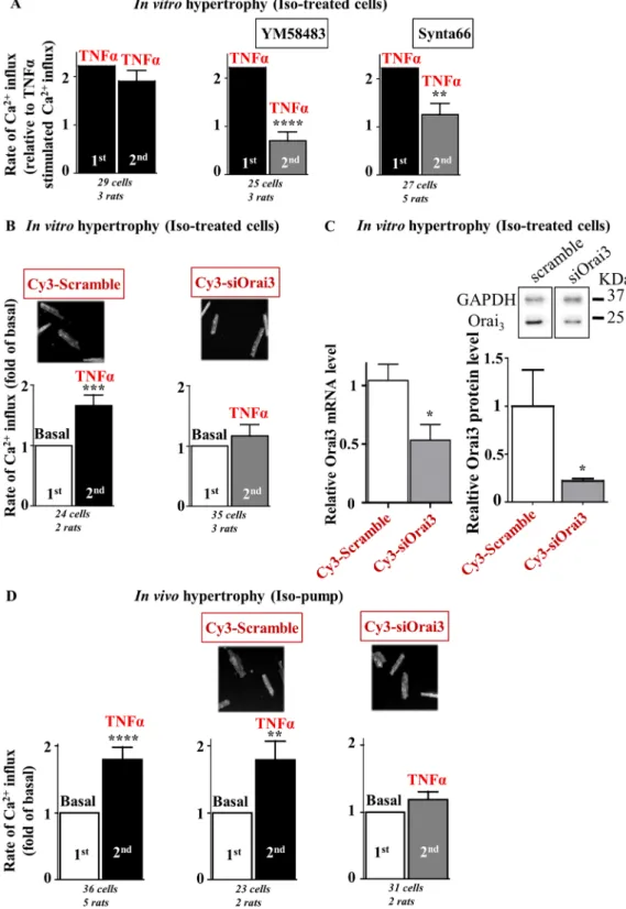
We then asked whether Orai3 drives this TNFα-activated store- and voltage-independent Ca2+ influx. In in
vitro iso-treated hypertrophied cells, two successive applications of identical TNFα containing medium gave
rise to similar rates of Ca2+ influx (Fig. 2A left). However, preincubation for 10 min with Orai pharmacological
inhibitors, YM58483 or Synta66, reduced the rate of Ca2+ influx observed in response to a second challenge
with TNFα, to a value similar to basal (Fig. 2A middle and right). Moreover, in vivo molecular knockdown of Orai3, via intramyocardial injection with Cy3-tagged Orai3 siRNA, also blunted the TNFα effect on Ca2+ influx
that was still observed in cells isolated from scramble siRNA-injected hearts (Fig. 2B). Orai3 neutralization was performed as previously reported12 and demonstrated by quantitative RT-PCR and Western-blot (Fig. 2C) and
by detection of Cy3 positive cells (Fig. 2B). Of note, knockdown of Orai3 by siRNA injection in normal rats did not modify the cardiomyocyte size (Table S1). In contrast, both siScramble and siOrai3 cardiomyocytes had a tendency to be bigger after in vitro post-treatment with iso and displayed similar sizes. This suggested that the in
vitro iso-hypertrophic response was not altered in siOrai3 cardiomyocyte, in contrast to TNFα/Orai3 signaling.
Complementary experiments were performed in hearts from rats subjected to chronic iso-infusion for 14 days (1.5 mg/kg/day) to elicit in vivo hypertrophy (Table 1). Iso-induced EACH remodeling was confirmed by an increase in end-diastolic and end-systolic interventricular septum (IVSd and IVSs), posterior wall thicknesses (PWd and PWs) and concentric hypertrophy (h/r, diastolic wall thickness to radius ratio) in iso-treated rats,
relative to control rats, associated with an increased heart rate (HR) and a better cardiac function assessed by fractional shortening measurements (FS).
Iso-induced hypertrophy was also attested by an increased heart weight (HW) to body weight (BW) ratio (0.84 ± 0.12 vs. 0.62 ± 0.11, n = 8 Iso vs. 8 control rats, p < 0.01, Mann Whitney U test) and cell area (3128 ± 63 vs. 2538 ± 54 µm2, n = 214 Iso vs. 223 control cells, p < 0.0001, Mann Whitney U test).
Ca2+-imaging experiments confirmed that TNFα also activated Orai3-dependent Ca2+ influx in in vivo
hyper-trophied cardiomyocytes isolated from rats implanted with iso-pump and injected or not with siRNAs three days before cardiomyocyte isolation (Fig. 2D). Of note, efficient Orai3 knockdown in in vivo hypertrophied cardiomy-ocytes was attested by a 40.0 ± 14.6% decrease in Orai3 mRNA expression as compared to cardiomycardiomy-ocytes iso-lated from scramble siRNA injected rats (p < 0.05, Mann-Whithney U test, n = 3 Orai3 and n = 6 Scramble siRNA injected rats). Moreover, in vivo reduction in Orai3 expression in iso-infused rats decreased the mean cardiomy-ocyte area (3384 ± 81 vs. 4311 ± 135 µm2, n = 116 vs. 120 cells from Orai3 vs. Scramble siRNA injected rat hearts
at day 3 post injection, p < 0.0001, Mann Whitney U test). These results highlighted the role of Orai3-dependent Ca2+ influx as a target of TNFα in in vitro and in vivo iso-hypertrophied cardiomyocytes.
Overall, these experiments identified TNFα as an activator of the Orai3-dependent Ca2+ influx in
hypertro-phied cardiomyocytes.
Activation of Orai3-Ca
2+influx by TNFα relies on binding to TNFR
2
, stimulation of cPLA
2and
potential production of AA metabolites via the lipoxygenase pathway.
Next we aimed to evaluate the role of TNFα receptors 1 and 2 and of the cPLA2 pathways in TNFα signaling. In in vitro iso-treatedhypertro-phied cells, stimulation of Ca2+ influx by TNFα was unaffected by the preincubation for 1 hour either with control
IGg2A or anti-TNFR1-antibodies (Ab) but was impaired in the presence of neutralizing TNFR2-Ab (Fig. 3A).
Preincubation with the cPLA2 inhibitor, methyl arachidonyl fluorophosphonate (MAFP), suppressed TNFα
sign-aling whereas addition of the phospholipase A2 activating peptide (PLAP) mimicked TNFα effect, and stimulated
Ca2+ influx (Fig. 3B). TNFα-induced activation was unaffected by the pretreatment with the cyclo-oxygenase
inhibitor indomethacin but impaired in the presence of a lipoxygenase inhibitor nordihydroguaiaretic acid (NDGA), suggesting the potential requirement of AA metabolism into leukotrienes in this signal transduction (Fig. 3C). TNFα signaling persisted in the presence of the antagonist of leukotriene receptors, montelukast, sug-gesting an effect independent of binding to external receptors (Fig. 3C). These results indicate that TNFα signals
Figure 1. TNFα activates a store- and voltage-independent Ca2+ influx in hypertrophied but not in control
cardiomyocytes. Representative recordings of Fura2 fluorescence ratio (F340/F380) in iso-treated hypertrophied
(A,B) or control (C,D) cardiomyocytes subjected to 2 successive measurements of the rate of voltage- and store-independent Ca2+ entry and quantification of rates of Ca2+ entry. Two successive applications of the same 1 mM
Ca2+ basal perfusion medium gave rise to similar rates of Ca2+ entry, both in hypertrophied (A) and normal
cells (C). Addition of TNFα to the second 1 mM Ca2+ perfusion medium enhanced the rate of Ca2+ entry (2nd
slope as compared to 1st slope) in hypertrophied cardiomyocytes (B) but not in normal cardiomyocytes (D).
Number of cells analyzed and number of cell isolations (rats) as indicated. Mean ± SEM of cells, Wilcoxon matched-paired tests to examine if the mean of the 2nd rate differs from the 1st one, arbitrarily set as 1,
Figure 2. TNFα activates a store- and voltage-independent Ca2+ influx in hypertrophied cardiomyocytes
further identified as Orai3 dependent. Hypertrophied cardiomyocytes were loaded with Fura2 before
measurement of voltage- and store-independent Ca2+ influx. (A) Two successive applications of the same TNFα
perfusion medium on iso-treated hypertrophied cardiomyocytes gave rise to similar rates of Ca2+ entry. The
first TNFα-induced Ca2+ influx was arbitrarily set to 2.2 to allow comparisons with the control conditions
(see Fig. 1B). Preincubation with Orai inhibitors, YM58483 or Synta66, prior the second application of TNFα perfusion medium blunted activation of Ca2+ entry by TNFα. (B) Hypertrophied cardiomyocytes isolated
from hearts injected with Cy3-tagged scramble or Orai3 siRNAs three days before isolation and iso-treatment. Typical images show Cy3-fluorescence in cardiomyocytes. TNFα activates Ca2+ influx in scramble
siRNA-transfected cells but not in siOrai3-siRNA-transfected cardiomyocytes. Number of cells analyzed and number of cell isolations (rats) as indicated, mean ± SEM of cells, Wilcoxon matched-paired tests to examine if the mean of the 2nd rate differs from the 1st one, arbitrarily set as 2.2 (A) or 1 (B), **p < 0.01, ***p < 0.001, ****p < 0.0001.
in iso-hypertrophied cardiomyocytes through TNFR2 to activate cPLA2 and produce AA potentially leading to
increased leukotriene levels which in turn activate Orai3.
Of note, we recently demonstrated the emergence of an Orai3-dependent pathway that drives an AA-dependent Ca2+ influx in hypertrophied cardiomyocytes from rats subjected to abdominal aortic banding
(AAB) for 28 days12. Complementary experiments were performed to examine the regulation of Orai3 by TNFα
in this model (protocol in Fig. 4A). EACH remodeling was confirmed in AAB rats, relative to Sham rats, by an increase in IVSd, IVSs, PWd and PWs and concentric hypertrophy (h/r) (Table 2). AAB-induced hypertrophy was also attested by an increased cell area (4718 ± 128 vs. 3311 ± 124 µm2, n = 129 vs. 117 cells, p < 0.0001, Mann
Whitney U test). A slight decrease of FS was observed in this model (Table 2).
TNFα also induced activation of Orai3-dependent Ca2+ influx in AAB-induced hypertrophied
cardiomy-ocytes (Fig. 4B,C) that relied on binding to TNFR2 (Fig. 4D) and potential production of AA metabolites via
activation of the lipoxygenase pathway (Fig. 4E).
Thus, emergence of a TNFR2-dependent Orai3-driven Ca2+ influx characterized cardiomyocyte hypertrophy
triggered by either iso-treatment or pressure overload.
Inflammatory CD11b/c cells trigger TNFR
2-dependent activation of Orai3-Ca
2+influx in
hyper-trophied cardiomyocytes.
Next experiments aimed to evaluate the potential cellular source of inflamma-tion in EACH hearts. Immunohistological examinainflamma-tion of cardiac secinflamma-tions from iso-induced EACH rats indicated an increased number of TNFα-positive cells as compared to control and 66 ± 4% of TNFα-positive cells were identified as myeloid CD11b/c-positive cells (Fig. S2).Rat hearts (obtained from rats implanted with iso-pump for 14 days, Table 1) were used to isolate cardiomy-ocytes, fibroblasts and myeloid CD11b/c cells (denominated as in vivo cell activation) (Fig. 5A,B). Conditioned medium (Cmed) obtained from cardiomyocytes or cardiac fibroblasts were without effect on voltage- and store-independent Ca2+ influx in in vitro hypertrophied cardiomyocytes in contrast to the Cmed obtained from
their cardiac CD11b/c counterparts (Fig. 5C). This pointed out the CD11b/c cells as the potential source of the Orai3 inflammatory trigger in EACH hearts. Of note, levels of TNFα detected in Cmed from CD11b/c cells (1.15 ± 0.3 pg/ml, n = 6) were 10 to 20 fold higher than levels measured in Cmed from their cardiac counterparts. CD11b/c-Cmed-induced activation was sensitive to neutralizing TNFR2 antibodies and Orai inhibitor YM58483
(Fig. 5D).
A similar effect was triggered using in vitro CD11b/c-Cmed (obtained from CD11b/c cells isolated from normal rat hearts and in vitro stimulated with lipopolysaccharide (LPS) (10 ng/ml for 2 hours) to induce pro-inflammatory activation) (Fig. 5B,E). When CD11b/c cells were pretreated with the anti-inflammatory drug semapimod (Sema) before LPS stimulation, in vitro CD11b/c-Cmed Sema was without effect on Ca2+ influx
(Fig. 5B,E). Anti-inflammatory impact of semapimod was indicated by a reduction in TNFα-positive staining of CD11b/c cells (Fig. S3) and a limited TNFα content in Cmed Sema as compared to Cmed LPS (0.01 ± 0.004 vs. 0.24 ± 0.06 pg/ml, n = 5 vs. 8, p < 0.01, Mann Whitney U test).
These results highlighted the cardiac inflammatory CD11b/c cells as the potential triggers of Orai3-dependent Ca2+ influx in hypertrophied cardiomyocytes.
TNFα or inflammatory cardiac CD11b/c cells trigger TNFR
2and Orai signaling pathways,
enhancing hypertrophy and promoting resistance to oxidative stress in hypertrophied
cardi-omyocytes.
We next investigated the potential impact of inflammation on cardiomyocyte hypertrophy and evaluated the role of TNFR2 and Orai signaling pathways. Cardiomyocyte hypertrophy was initially induced bythe iso stimulation (100 nM) for 1.5 hours. Cells were then challenged with TNFα or in vitro CD11b/c-Cmed. Cell hypertrophy was measured after 18 hours and attested by an increase in cell area (Fig. 6A). Addition of iso alone triggered a mean 14 ± 2% hypertrophy (Fig. 6B). TNFα enhanced iso-induced hypertrophy (up to 28 ± 3%), in a TNRF2- and YM58483-sensitive manner (hypertrophy reduced to 11 ± 3% and 7 ± 3%, by preincubation with
TNFR2-Ab or YM58483, respectively) (Fig. 6B). CD11b/c-Cmed also increased iso-induced hypertrophy (up
to 35 ± 2%) (Fig. 6B). Pretreatment of CD11b/c cells with anti-inflammatory semapimod blunted the prohy-pertrophic effect of Cmed (reduced to 5 ± 3%), as well as treatment with neutralizing TNFR2-Ab or YM58483
(reduced to 14 ± 4% and 7 ± 2%, respectively) (Fig. 6B).
These results indicate that inflammation (TNFα or Cmed from cardiac inflammatory CD11b/c cells) enhances cardiomyocyte hypertrophy via TNFR2- and Orai-dependent signaling pathways.
We also evaluated the potential impact of inflammation on the resistance to oxidative stress using in vitro hyper-trophied cardiomyocytes and demonstrated the role of TNFR2 and Orai signaling pathways. Iso-hypertrophied
(C) Efficient knockdown of Orai3 mRNA and protein in cardiomyocytes isolated from Wistar rats at day 3 following injection with Cy3-tagged scramble or Orai3 siRNAs. Histograms representing relative transcript levels normalized to the RPL32 mRNA or relative protein levels normalized to Glyceraldehyde 3-phosphate dehydrogenase (GAPDH) protein. Mean ± SEM of cardiomyocytes from 2–6 rats/group, Mann-Whitney U test, *p < 0.05. Full length blots were included in SI. (D) Hypertrophied cardiomyocytes isolated from rats after fourteen days of chronic iso-infusion injected with Cy3-tagged scramble or Orai3 siRNAs three days before isolation. TNFα activates Ca2+ influx in hypertrophied cardiomyocytes isolated from hearts injected or not with
tagged scramble siRNA, but not in hypertrophied cardiomyocytes isolated from hearts injected with Cy3-tagged Orai3 siRNA, three days before isolation. Number of cells analyzed and number of cell isolations (rats) as indicated, mean ± SEM of cells, Wilcoxon matched-paired tests to examine if the mean of the 2nd rate differs
d14 control
rats (n = 7) d14 iso-treated rats (n = 21) Mann-Whitney U test
HR (bpm) 390 ± 7 491 ± 4 p < 0.0001 IVSd (mm) 1.47 ± 0.04 1.93 ± 0.03 p < 0.0001 LVd (mm) 7.67 ± 0.16 7.37 ± 0.07 ns PWd (mm) 1.89 ± 0.02 2.3 ± 0.05 p < 0.0001 IVSs (mm) 2.44 ± 0.08 3.11 ± 0.07 p < 0.0001 LVs (mm) 4.31 ± 0.07 3.51 ± 0.0001 p < 0.0001 PWs (mm) 2.7 ± 0.06 3.18 ± 0.06 p < 0.0001 h/r 0.44 ± 0.01 0.57 ± 0.01 p < 0.0001 EF (%) 79.98 ± 0.45 87.2 ± 0.65 p < 0.0001 FS (%) 43.6 ± 0.5 51.3 ± 1.1 p < 0.001
Table 1. echocardiography parameters at day 14 in control or iso-infused rats. HR, heart rate; IVSd,
end-diastolic interventricular septum thickness; LVd, end-end-diastolic left ventricular diameter; PWd, end-end-diastolic posterior wall thickness; IVSs, end-systolic interventricular septum thickness; LVs, end-systolic left ventricular diameter; PWs, end-systolic posterior wall thickness; h/r, diastolic wall thickness to radius ratio; EF, ejection fraction; FS, fractional shortening.
Figure 3. Activation by TNFα relies on binding to TNFR2, stimulation of cPLA2 and potential production of
AA metabolites via the lipoxygenase pathway. (A–C) Iso-treated hypertrophied cardiomyocytes were loaded with Fura2 before measurement of voltage- and store-independent Ca2+ influx. (A) Rates of Ca2+ entry in
response to TNFα after a 1 hour preincubation with control IGg2A, neutralizing TNFR1-Ab or TNFR2-Ab.
(B) TNFα effect is sensitive to the cPLA2 inhibitor, MAFP, but mimicked by cPLA2 activating peptide PLAP.
(C) Activation of Ca2+ entry by TNFα is blunted by preincubation with NDGA (lipoxygenase inhibitor), but
unaffected by indomethacin (cyclooxygenase inhibitor) or montelukast (leukotriene receptor antagonist) pretreatments. Number of cells analyzed and number of cell isolations (rats) as indicated. Mean ± SEM of cells, Wilcoxon matched-paired tests to examine if the mean of the 2nd rate differs from the 1st one, arbitrarily set as 1,
cardiomyocytes (100 nM for 18 hours) were preincubated with TNFα or in vitro CD11b/c-Cmed, and cell resist-ance was estimated by the number of rod-shaped cells after H2O2-oxidative stress (100 µM H2O2 for 2.5 hours)
(Fig. 7A). Counting of resistant cells indicated 21 ± 2% vs. 100% in the presence and in the absence of H2O2,
respectively (Fig. 7B). Treatment with TNFα increased resistance of hypertrophied cardiomyocytes from 21 ± 2% to 36 ± 3%, in a TNFR2- and YM58483-sensitive manner (rod-shaped cells reduced to 23 ± 1% and 26 ± 4%, after
preincubation with the TNFR2-Ab or YM58483, respectively) (Fig. 7B). CD11b/c-Cmed alleviated the deleterious
impact of H2O2, improving the yield of resistant cells to 50 ± 6% (Fig. 7B). Pretreatment of CD11b/c cells with
anti-inflammatory semapimod blunted the beneficial impact of Cmed resulting in only 21 ± 4% resistant cells, as well as pretreatment with the TNFR2-Ab or YM58483 (36 ± 3% and 38 ± 2% resistant cells, respectively) (Fig. 7B).
These results demonstrate that inflammation (TNFα or Cmed from cardiac inflammatory CD11b/c cells) improves resistance to H2O2 in hypertrophied cardiomyocytes via TNFR2- and Orai-dependent protective
sign-aling pathways.
Taken together our in vitro results argue for a protective Orai3-driven signal emerging during the phase of EACH and promoting adaptive cardiomyocyte hypertrophy and beneficial resistance to oxidative stress. In order
Figure 4. TNFα activates Orai-Ca2+ influx after binding to TNFR
2 and potential production of AA metabolites
via the lipoxygenase pathway in hypertrophied cardiomyocytes from rats with AAB-induced EACH. (A) Schematic representation of the protocol where rats with AAB-induced EACH for 28 days were subjected to echographic analyses. (B–E) Hypertrophied cardiomyocytes isolated from AAB rats were loaded with Fura2
before measurement of voltage- and store-independent Ca2+ influx. TNFα activates Ca2+ entry (B) in a manner
sensitive to Orai inhibitor YM58483 (C), neutralizing TNFR2-Ab (D) or lipoxygenase inhibitor NDGA (E).
Number of cells analyzed and number of cell isolations (rats) as indicated, mean ± SEM of cells, Wilcoxon matched-paired tests to examine if the mean of the 2nd rate differs from the 1st one, arbitrarily set as 1 (B,D,E) or
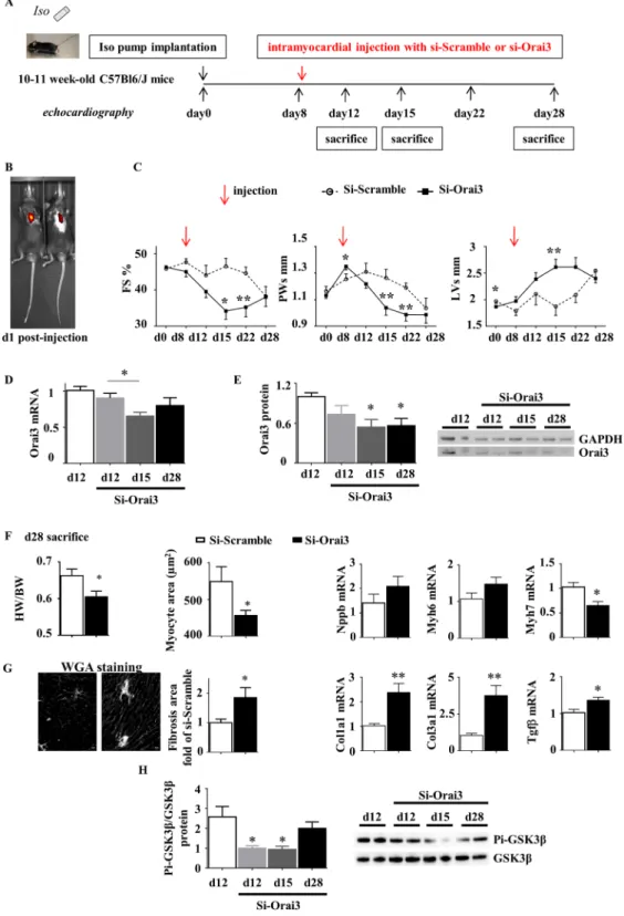
to evaluate their relevance in the pathophysiology of HF, we performed a kinetic analysis of echocardiographic parameters and evaluated tissue remodeling in response to a unique intramyocardial Orai3 siRNA injection applied at the onset of EACH.
Orai3 neutralization during EACH impairs cardiac hypertrophy, fosters alteration of function
and dilation and deleterious tissue remodeling.
To validate in vivo the protective role of Orai3 activ-ity on EACH, we studied the effect of cardiac Orai3 knockdown during iso infusion in mice. First, as shown in Fig. S4, as compared to previous results obtained in rats (Table 1 and12), we checked that this model displayedsimilar iso-induced EACH remodeling after a 14 days infusion period in control mice. EACH was characterized by evolution of echocardiographic parameters, increase in heart weight to body weight ratio and cardiomyocyte size, and associated with an increase in Nppa and Tnfα mRNA expressions, but no change in Orai3 mRNA level (Fig. S4 and Table S2). This model was chosen to develop a novel approach of percutaneous intramyocardial injection of siRNA under echographic guidance validated in mice by other groups (i.e.33) in order to avoid
poten-tial artefactual consequences of the surgical thoracotomy on tissue remodeling that are susceptible to affect the post-operative echographic surveillance and evolution of cardiac parameters.
Iso-infused mice were subjected to a unique intramyocardial injection of either Scramble or Orai3 siRNA at day 8 following pump implantation (Fig. 8A). Efficient knockdown of Orai3 during EACH was attested at the mRNA and protein levels (Fig. 8D,E). At day 15, hearts from siOrai3-injected mice displayed a decreased hyper-trophy (lower PWs), an increased dilation (higher LVs) and an altered cardiac function (lower FS) as compared to hearts from siScramble-injected mice (Fig. 8C and Table 3). At day 28, siOrai3-injected mice still presented with a lower heart weight to body weight ratio, a smaller cardiomyocyte area, a decrease in Myh7 mRNA level, as compared to siScramble-injected mice (Fig. 8F). Orai3 knockdown during EACH also fostered fibrosis attested by histological analysis and increased Col1a1, Col3a1 and Tgfβ mRNA levels (Fig. 8G). Mechanistically, a decrease in the ratio phospho-GSK3β/GSK3β was correlated with the reduction of Orai3 expression (Fig. 8H).
Importantly, the intramyocardial injection of Orai3 siRNA in control mice showed that knockdown of Orai3 was without functional impact (Fig. S5 and Table S3).
Taken together, these results demonstrated the emergence of a functional protective role of Orai3 during EACH.
Discussion
Our results highlight the emergence of a protective role for Orai3 in the hypertrophied cardiomyocytes. We iden-tified TNFα as a mechanistic trigger of the TNFR2-dependent activation of Orai3-Ca2+ influx and showed that
CD11b/c cells are a potential driving source of this signaling in EACH hearts. This paracrine signaling enhances hypertrophy and promotes resistance to oxidative stress of hypertrophied cardiomyocytes. Furthermore, we show that Orai3 knockdown during EACH fosters HF (Fig. 9).
Cardiac Orai3-dependent Ca2+ influx was previously identified as a prohypertrophic stimulus in AAB-induced
CH12. We confirm these results in a model of iso-induced EACH, a model of reproducible progressive concentric
hypertrophy. In our study, we found that in vivo reduction in Orai3 expression in iso-infused rats decreased the mean cardiomyocyte area. In keeping with this atrophic impact of Orai3 siRNA, neutralization of Orai3 during EACH in iso-infused mice triggers a rapid and significant decrease in hypertrophic parameters still detectable at day 20 post injection. Furthermore, our in vitro experiments document a direct prohypertrophic effect of Orai3 in isolated iso-treated cardiomyocytes.
Interestingly, our results highlight novel protective properties of Orai3-dependent Ca2+ influx. In line with
the reported resistance of Orai3 channel to redox regulation34, we show that Orai3 activation confers resistance to
oxidative stress in isolated hypertrophied cardiomyocytes. Furthermore, our in vivo results indicate that efficient cardiac knockdown of Orai3 during EACH inhibits adaptive hypertrophy, alters cardiac function and promotes fibrosis. Mechanistically, recent results from our laboratory indicate that the Orai3-interacting protein STIM1 is essential to tune the Akt/GSK3β prosurvival signaling8–10,35,36. Our in vivo results show that neutralization of
d28 Sham rats (n = 7) d28 AAB rats (n = 6) Mann-Whitney U test
HR (bpm) 386 ± 12 383 ± 18 ns IVSd (mm) 1.31 ± 0.03 2.08 ± 0.05 p < 0.001 LVd (mm) 7.43 ± 0.15 8.5 ± 0.4 p < 0.05 PWd (mm) 1.67 ± 0.03 2.38 ± 0.15 p < 0.01 IVSs (mm) 2.23 ± 0.1 2.7 ± 0.08 p < 0.05 LVs (mm) 4.07 ± 0.1 5.53 ± 0.3 p < 0.01 PWs (mm) 2.6 ± 0.05 3.2 ± 0.17 p < 0.05 h/r 0.41 ± 0.01 0.53 ± 0.04 p < 0.05 EF (%) 81.1 ± 0.6 70 ± 2.1 p < 0.001 FS (%) 44.7 ± 0.7 35.4 ± 1.5 p < 0.01
Table 2. echocardiography parameters at day 28 in rats after AAB. HR, heart rate; IVSd, end-diastolic
interventricular septum thickness; LVd, end-diastolic left ventricular diameter; PWd, end-diastolic posterior wall thickness; IVSs, end-systolic interventricular septum thickness; LVs, end-systolic left ventricular diameter; PWs, end-systolic posterior wall thickness; h/r, diastolic wall thickness to radius ratio; EF, ejection fraction; FS, fractional shortening.
Figure 5. Conditioned medium from CD11b/c cells isolated from EACH heart activates Ca2+ influx in a
manner sensitive to TNFR2-Ab and Orai inhibitor YM58483. (A) Schematic representation of the protocol
where rats implanted with an iso-pump for fourteen days were subjected to echographic analyses and developed EACH. (B) CD11b/c cells, cardiomyocytes and fibroblasts were isolated from EACH hearts and cultured for 18 hours before recovery and concentration of conditioned media. CD11b/c cells pre-incubated with/without the anti-inflammatory drug, semapimod, prior in vitro LPS application to induce pro-inflammatory activation, were isolated from normal hearts and cultured for 18 hours before recovery and concentration of conditioned media. Iso-treated hypertrophied cardiomyocytes were loaded with Fura2 before measurement of voltage- and
store-independent Ca2+ influx, 1st in the absence and 2nd in the presence of these conditioned media (Cmed).
(C) Only Cmed from CD11b/c cells isolated from EACH hearts activates a voltage- and store-independent Ca2+ influx (D) in a manner sensitive to TNFR
2-Ab and Orai inhibitor YM58483. (E) Cmed from CD11b/c
cells isolated from normal hearts and in vitro stimulated with LPS activates a voltage- and store-independent Ca2+ influx in a manner sensitive to neutralizing TNFR
2-Ab, Orai inhibitor YM58483 and anti-inflammatory
pretreatment with semapimod. Number of cells analyzed, number of cell isolations (rats) and number of Cmed tested as indicated, mean ± SEM of cells, Wilcoxon matched-paired tests to examine if the mean of the 2nd rate
Figure 6. TNFR2 and Orai signaling pathways enhance hypertrophy in cardiomyocytes after inflammatory
stimulation with TNFα or Cmed from in vitro LPS-activated cardiac CD11b/c cells. (A) CD11b/c cells were isolated from normal hearts and further subjected to in vitro activation by LPS incubation following or not semapimod pretreatment. Conditioned media (recovered after 18 hrs in culture) or TNFα were applied on rat cardiomyocytes after 1.5 hours iso treatment and cell hypertrophy analyzed after 18 hours. (B) TNFα or Cmed
in vitro enhance hypertrophy of iso-treated cardiomyocytes in a manner sensitive to semapimod, TNFR2-Ab or
YM58483. Mean ± SEM of 3–4 experiments, cardiomyocytes from 2–4 rats, 2 Cmed. Kruskal-Wallis followed by Dunn’s post-hoc test vs. the Iso-treated control (white column), *p < 0.05.
Figure 7. TNFR2 and Orai signaling pathways promote resistance to oxidative stress of cardiomyocytes after
inflammatory stimulation with TNFα or Cmed from in vitro LPS-activated cardiac CD11b/c cells. (A) CD11b/c cells were isolated from normal hearts and further subjected to in vitro activation by LPS incubation following or not semapimod pretreatment. Conditioned media (recovered after 18 hrs in culture) or TNFα were applied on in vitro iso hypertrophied rat cardiomyocytes before H2O2 treatment and analyses of cell resistance. (B)
TNFα or Cmed in vitro increase resistance of hypertrophied cardiomyocytes to H2O2 stress in a manner
sensitive to semapimod, TNFR2-Ab or YM58483. Mean ± SEM of 3–5 experiments, cardiomyocytes from 3–5
rats, 2–4 Cmed. Kruskal-Wallis followed by Dunn’s post-hoc test vs. the Iso-treated control (white column), **p < 0.01.
Figure 8. Orai3 knockdown at the onset of EACH limits cardiac hypertrophy, accelerates alteration of function
and dilation and promotes fibrosis. (A) Schematic representation of the protocol where mice implanted with an iso-pump at day 0 were subjected to a unique ultrasound-guided intramyocardial transthoracic injection of Scramble or Orai3 siRNA at day 8 and submitted to an echocardiographic follow-up. (B) In some experiments, siRNA was added with sulforhodamine: typical in-vivo visualization of intracardiac localization of sulforhodamine at day 1 post-injection using an IVIS spectrum in-vivo imaging system. (C) Echocardiographic parameters (mean ± SEM of mice, n = 8–22 mice/group, see Table 3, ANOVA for repeated measures followed by Dunn-Sidak post-hoc tests). (D,E) Efficient knockdown of Orai3 mRNA and protein levels in cardiac homogenates at d12, d15 and d28 following injection (mean ± SEM of mice, n = 7 mice/group, Kruskal-Wallis followed by Dunn’s post-hoc tests). Full length blots were included in SI. (F,G) SiOrai3 injection induced a decrease in Heart Weight/Body Weight ratio, myocyte area and Myh7 mRNA level, an increase in fibrosis, and an elevation of Col1a1, Col3a1 and Tgfβ mRNA levels (mean ± SEM of mice, n = 7 mice/group, Mann-Whitney
Orai3 during EACH is associated with a decrease in phospho-GSK3β/GSK3 ratio. In the current absence of pub-lished knockout data for Orai3, our study is the first to address its pathophysiological relevance and indicate its essential protective role during EACH.
We have identified the first pathophysiological trigger of Orai3-driven store-independent Ca2+ influx in
car-diomyocytes, namely a TNFα/TNFR2-dependent signaling. Interestingly, the regulation of Orai3 by TNFα is
detected in hypertrophied cardiomyocytes (both in response to iso treatment or in the AAB-model), but not in normal cardiomyocytes. Of note, iso-hypertrophied and normal cardiomyocytes display similar TNFR2
expression (2.4 ± 0.4 vs. 2.4 ± 0.3 pg TNFR2/mg, respectively, n = 4). However, and as previously reported in
AAB-induced CH hearts12, co-immunoprecipitation experiments using STIM1 antibodies indicate enhanced
Orai3 recruitment to STIM1 in the iso-induced EACH (Fig. S6). Since Orai3-STIM1 interaction is a prerequisite for Orai3-dependent Ca2+ influx activation14,17, its absence in normal cells and its enhancement in hypertrophied
cells constitute a major difference likely to explain the lack of impact of Orai3-dependent Ca2+-influx as well as
the absence of TNFα regulation in normal cells. Accordingly, our in vivo and in vitro experiments in normal mice show that the knockdown of Orai3 is without impact on echocardiographic parameters as well as heart or cardi-omyocyte size. They argue for a pathophysiological impact of Orai3 exerted during EACH but not under control conditions. Dominant impact of inflammation is currently reported as deleterious in normal hearts, as it is asso-ciated with reactive oxygen species production, proapoptotic signaling and a dominant role of TNFR1 pathways
overwhelming TNFR2 signaling26. Using TNFR1 and TNFR2 knockout mice implanted with iso-pumps, Prabhu et
al. demonstrated that TNFR2 but not TNFR1 signaling prevents the detrimental long-term effects of β-adrenergic
receptor stimulation in the heart37. In another study, the group of Prasad proposed that the beneficial effects
of TNFR2 signaling in presence of sympathetic overdrive could act through the preferential TNFR2-mediated
recruitment of GRK2 to mediate βAR desensitization thus reducing deleterious cardiac signaling and remodeling. In agreement with these studies, our study further suggests the emergence of a protective TNFR2 pathway during
EACH development via the stimulation of a novel downstream effector Orai3.
We show here that cardiac CD11b/c cells are a potential source of inflammatory TNFα/TNFR2-dependent
signaling leading to Orai3-dependent Ca2+ channel activation. Both Cmed from in vitro and in vivo activated
CD11b/c cells stimulate a store-independent Ca2+ influx in hypertrophied cardiomyocytes in a TNFR
2-Ab and
YM58483 sensitive manner. This suggests the presence of activated inflammatory CD11b/c cells in EACH hearts. We demonstrated that LPS-activated CD11b/c cells enhance hypertrophy and promote resistance of hypertro-phied cardiomyocytes to oxidative stress, in a TNFR2-Ab and YM58483 sensitive manner. This protective impact
is blunted by preincubation of CD11b/c cells with the anti-inflammatory drug semapimod. Our data strengthen the concept that inflammation arising from cardiac myeloid cells may exert paracrine beneficial impact on cardio-myocytes38,39. Of note, cardiac macrophages are an emerging focus for therapeutic strategies aimed at minimizing
cardiomyocyte death, ameliorating pathological cardiac remodeling and for treating HF40.
We observed that TNFα-dependent activation of Orai3-Ca2+ influx relies on cPLA
2 activation and is
mim-icked by a cPLA2 activator. This is in accordance with the reported presence of the lipid interaction site located
in the NH2 terminal intracytosolic sequence of Orai320. TNFα-dependent effect is sensitive to NDGA treatment
which suggests the potential requirement of a lipoxygenase-dependent AA metabolism to activate Orai3-Ca2+
influx. Accordingly, Trebak et al. previously described LTC4, a lipoxygenase AA-metabolite, as an activator of Orai3 in the vascular smooth muscle cells41. However, this proposal needs to be tempered since NDGA, in
addition to inhibit lipoxygenase activity may also exert several off target effects (i.e., PKC inhibition, overall anti-oxidant, ER-Golgi protein shuttling inhibition). Our results further illustrate the dual role of cPLA2-AA
signaling in mediating TNFα effects in adult cardiac myocytes. We have identified Orai3 as a novel protective TNFα-cPLA2-AA pathway. Accordingly, the beneficial impact of TNFα-cPLA2-AA pathways has been
previ-ously reported on the cardiomyocyte calcium transients and contraction (i.e. by the group of Oceandy42 and our
previous results26,30) and on the survival to oxidative stress26. In contrast, the group of Gugiyama reported the
deleterious impact of a TNFα-cPLA2 cardiac signaling in a model of ischemia-reperfusion43.
In conclusion, our in vitro and in vivo studies characterize the Orai3 signaling pathway that exerts a direct protective role against HF in hypertrophied cardiomyocytes. Mechanistically, we have identified Orai3 as a novel driver of TNFR2-dependent inflammation instrumental in this protection. Furthermore, we showed a protective
Orai3-dependent paracrine role of cardiac myeloid cells leading to adaptive hypertrophy and improved resistance to oxidative stress. In summary, our results are the first to address the functional role of Orai3 signaling in HF that may open new perspectives for patients’ treatments.
Methods
Ethics.
Care of the animals and surgical procedures were performed according to the Directive 2010/63/ EU of the European Parliament, which had been approved by the Ministry of Agriculture, France, (authori-zation for surgery C-75-665-R). The project was submitted to the French Ethic Committee CEEA (Comitéd’Ethique en Expérimentation Animale) and obtained the authorization Ce5/2012/050 and
APAFIS#1729-2015-083114195840v8. All experiments were performed in accordance with relevant named guidelines and regulations.
Animals.
6 week-old male Wistar rats and 10-11 week-old male C57BL/6JRj mice were purchased from Janvier Labs.U tests). (H) SiOrai3 injection induced a decrease in the ratio phospho-GSK3β/GSK3β at day 12 and day 15 following injection (mean ± SEM of mice, n = 7 mice/group, Kruskal-Wallis followed by Dunn’s post-hoc test). Full length blots were included in SI. *p < 0.05, **p < 0.01.
In vivo chronic isoproterenol infusion.
Rats and mice anesthetized under isoflurane (Iso-vet®
, Piramal, UK) (1–3%) were implanted subcutaneously with an osmotic minipump (Alzet, Charles River) containing either isoproterenol (1.5 mg/kg/day for rat or 30 mg/kg/day for mouse) (iso-pump) or vehicle for 14 or 28 days to develop either EACH or HF, respectively, or as otherwise stated.Abdominal aortic banding.
Rats were anaesthetized by intra-peritoneal injection of ketamine (Imalgene®
, Merial, Germany) and xylazine (Rompun®
, Bayer, Germany) (75 and 10 mg/kg, respectively). Medial abdominal laparotomy was performed and a clip with an internal opening of 0.58 mm was placed, as previously reported12.Sham-operated rats served as controls.
Measurement of cardiac parameters.
Echocardiography was performed on lightly anesthetized animals under isoflurane (0.5–1%) with a probe emitting ultrasounds from 8- to 14-MHz frequency (Vivid7 PRO appara-tus; GE Medical System Co). The two-dimensionally guided Time Motion mode recording (parasternal long-axis view) of the left ventricle (LV) provided the following measurements: end-diastolic and end-systolic interven-tricular septum (IVSd and IVSs), posterior wall thicknesses (PWd and PWs), internal diameter (LVEDD and LVESD), and heart rate (HR). Each set of measurements was obtained from the same cardiac cycle. At least three sets of measurements were registered from three different cardiac cycles. Fractional shortening (FS): [(LVEDD − LVESD)/LVEDD] × 100 and h/r: [left ventricle diastolic wall thickness/radius] were calculated.Iso mice (iso pump implantation at d0) Time of
echocardio-graphy d0 d8 d12 d15 d22 d28
Injection
at d8 Scr (n = 11) Orai3 (n = 22) p Scr (n = 11) Orai3 (n = 22) p Scr (n = 11) (n = 22)Orai3 p Scr (n = 11) Orai3 (n = 15) p Scr (n = 8) Orai3 (n = 8) p Scr (n = 8) Orai3 (n = 8) p
HR (bpm) 624 ± 7 634 ± 3 ns 668 ± 11 652 ± 9 ns 606 ± 15 619 ± 9 ns 622 ± 11 632 ± 12 ns 630 ± 6 609 ± 8 ns 594 ± 29 618 ± 11 ns IVSd (mm) 0.6 ± 0.01 0.7 ± 0.01 ns 1 ± 0.02 1 ± 0.02 ns 1 ± 0.06 0.9 ± 0.03 ns 1 ± 0.04 0.8 ± 0.03 <0.05 0.9 ± 0.04 0.8 ± 0.03 ns 0.8 ± 0.04 0.7 ± 0.04 ns LVd (mm) 3.6 ± 0.05 3.5 ± 0.04 <0.05 3.4 ± 0.11 3.6 ± 0.07 ns 3.7 ± 0.19 3.9 ± 0.09 ns 3.4 ± 0.17 4.0 ± 0.10 <0.05 3.8 ± 0.18 4.0 ± 0.14 ns 4.1 ± 0.24 3.8 ± 0.14 ns PWd (mm) 0.7 ± 0.02 0.7 ± 0.01 ns 0.9 ± 0.03 0.9 ± 0.02 ns 0.9 ± 0.05 0.8 ± 0.02 <0.05 0.9 ± 0.05 0.7 ± 0.04 <0.01 0.8 ± 0.02 0.7 ± 0.03 ns 0.7 ± 0.05 0.6 ± 0.03 ns IVSs (mm) 1.1 ± 0.02 1.1 ± 0.02 ns 1.5 ± 0.03 1.5 ± 0.04 ns 1.4 ± 0.07 1.4 ± 0.04 ns 1.5 ± 0.06 1.1 ± 0.05 <0.05 1.4 ± 0.06 1.3 ± 0.04 ns 1.3 ± 0.06 1.3 ± 0.07 ns LVs (mm) 2.0 ± 0.04 1.9 ± 0.02 <0.05 1.8 ± 0.08 2.0 ± 0.08 ns 2.1 ± 0.19 2.4 ± 0.12 ns 1.9 ± 0.16 2.6 ± 0.14 <0.01 2.2 ± 0.17 2.6 ± 0.17 ns 2.6 ± 0.29 2.4 ± 0.16 ns PWs (mm) 1.2 ± 0.03 1.1 ± 0.02 ns 1.3 ± 0.04 1.4 ± 0.02 <0.05 1.3 ± 0.06 1.2 ± 0.03 ns 1.3 ± 0.06 1.0 ± 0.05 <0.01 1.2 ± 0.03 1.0 ± 0.04 <0.01 1.0 ± 0.07 1.0 ± 0.06 ns h/r 0.3 ±0.007 0.3 ±0.004 ns 0.56 ±0.018 0.5 ±0.017 ns 0.54 ±0.044 0.4 ±0.016 <0.05 0.55 ±0.041 0.40 ±0.021 <0.01 0.44 ±0.024 0.39 ±0.019 ns 0.38 ±0.042 0.36 ±0.019 ns EF (%) 83.1 ± 0.5 83.5 ± 0.2 ns 84.6 ± 0.8 81.7 ± 1.6 ns 80.0 ± 2.6 75.7 ± 2.1 ns 82.8 ± 2.2 68.5 ± 3.2 <0.05 79.4 ± 1.9 70.6 ± 3 <0.01 72.7 ± 4.8 74.1 ± 2.7 ns FS (%) 45.9 ± 0.5 46.3 ± 0.2 ns 47.7 ± 0.9 45.1 ± 1.4 ns 44.1 ± 2.6 39.6 ± 1.6 ns 46.5 ± 2.1 34.2 ± 2.2 <0.05 44.6 ± 1.6 35.3 ± 2.4 <0.01 38.3 ± 2.6 38.1 ± 2.4 ns
Table 3. kinetics of echocardiography parameters in mice implanted with Iso pump after Scramble (Scr) or
Orai3 siRNA intramyocardial injection. Two-way ANOVA followed by Sidak’s post-hoc tests. HR, heart rate; IVSd, diastolic interventricular septum thickness; LVd, diastolic left ventricular diameter; PWd, end-diastolic posterior wall thickness; IVSs, end-systolic interventricular septum thickness; LVs, end-systolic left ventricular diameter; PWs, end-systolic posterior wall thickness; h/r, diastolic wall thickness to radius ratio; EF, ejection fraction; FS, fractional shortening.
Figure 9. Orai3 enhances hypertrophy, promotes resistance to oxidative stress in adult hypertrophied
cardiomyocytes and during EACH limits evolution towards HF. Mechanistically, a TNFα paracrine signaling potentially mediated by the cardiac inflammatory CD11b/c cells promotes TNFR2-dependent activation of
store-independent arachidonic acid-dependent Orai3-related Ca2+ influx enhancing hypertrophy and inducing
resistance to H2O2. Knockdown of Orai3 at the onset of adaptive hypertrophy suppresses the EACH response
In vivo ultrasound mediated Cy3-tagged siRNA delivery in rats.
After an intra-peritoneal injec-tion of ketamine and xylazine (75 and 10 mg/kg, respectively), an incision was made in the fourth inter-costal space to expose the heart and Cy3-tagged siRNAs were delivered as previously described44 two weeks afteriso-pump implantation and three days before cardiac cell isolation. The siRNA for scramble (Life Technologies) and Orai3, were chosen from12,45,46 (FWsiOrai3: 5′-GUUUAUGGCCUUUGCCCUATT-3′; RVsiOrai3:
5′-UAGGGCAAAGGCCAUAAACTT-3′).
In vivo intramyocardial ultrasound-guided transthoracic siRNA delivery in mice.
On-targetplus Scramble and Orai3 siRNA (Dharmacon GE healthcare) were injected by ultrasound-guided transthoracic intramyocardial injection, as described in33, at day 8 after iso-pump implantation. Echocardiographic parameters
were measured regularly as stated.
Cardiac cell isolation and culture.
Rat cardiac myocytes were isolated either from chronically iso-infused hearts or from normal hearts, after injection or not with Cy3-tagged siRNA, three days before, when stated. Rats were administered a sodium pentobarbital (Ceva Sante Animale, France) intra-peritoneal injection (200 mg/ kg). Hearts were harvested and kept in ice-cold Krebs-Henseleit (KH) solution supplemented with 10 mmol/L taurine and 0.5 mmol/L EGTA, then rapidly canulated and mounted on the Langendorff apparatus. The hearts were retrogradely perfused through the aorta, first with a Krebs-Henseleit (KH) solution supplemented with 10 mmol/L taurine for 5 minutes, then with enzymatic solution for 20 minutes. The KH solution contained (in mmol/L): 100 NaCl, 4 KCl, 5.5 NaHCO3, 1 KH2PO4, 1.7 MgCl2, 10 D-glucose, 15 2,3-butanedione monoxime,22 Hepes, pH = 7.4 with NaOH. The enzyme solution was supplemented with 1 mg/ml collagenase A (Roche Applied Science, Meylan, France) and 5 mg/ml bovine serum albumin BSA (Sigma, Lyon, France). All chemicals were from Sigma (Lyon, France. Cell suspension was then used to differentially isolate ventricular cardiomyo-cytes, cardiac fibroblasts and cardiac CD11b/c cells.
Ventricular cardiomyocytes from iso-pump rats (in vivo hypertrophy) were plated onto laminin-coated glasses and maintained overnight in M199 medium (Life technologies). Cells isolated from normal hearts or from saline-pump rats were added with 100 nM isoproterenol plus 100 µM ascorbic acid (Sigma), when stated, to induce in vitro hypertrophy, three hours after plating, and let overnight in M199 medium.
Cardiac fibroblasts were isolated by centrifugation, plated onto 12-well plates and maintained in DMEM medium (Life technologies).
Cardiac CD11b/c cells were isolated by centrifugation, enriched using an anti-CD11b/c antibody coupled to magnetic beads (MiltenyiBiotec) and maintained overnight in RPMI medium (Life technologies) supplemented with 10 mmol/L Hepes. When stated, CD11b/c cells were in vitro polarized towards a pro-inflammatory pheno-type upon incubation for 2 hours with 10 ng/ml lipopolysaccharide (LPS) before overnight incubation with a new LPS-free medium.
All cardiac cells were cultured for 18 hours following plating. Conditioned media from cardiac cells were con-centrated on Amicon 10 kDa centrifugal filter.
Fura-2 AM calcium imaging.
Isolated rat ventricular myocytes were loaded with Fura2-AM (MolecularProbes, Life Technologies) as reported26. Transfected cells (detected by fluorescence imaging as cells positive for
Cy3-tagged siRNA) or non-transfected cells, rhythmically beating in response to electrical stimulation (square waves, 0.5 Hz, as previously described26), were analyzed. Measurements were recorded on a Zeiss Platform
equipped with an Axio Observer Z1 microscope, a DGA plus illuminator and a camera Coolsnap HQ2 (work-station Carl Zeiss).
Cells were first paced for few cycles and Ca2+ transients were recorded to ensure viability and functionality of
the cell. Cells were incubated in tyrode buffer (1.8 mmol/L Ca2+) to check the stability of basal cytosolic calcium
level and then switched to appropriate store- and voltage-independent Ca2+-free buffer. Store-independent Ca2+
entry was then measured upon readdition of 1 mmol/L Ca2+. Ca2+off/Ca2+on protocols repeated twice allowed
paired comparison between two similar (for reproducibility assessment) or distinct perfusion conditions. Tyrode buffer contained (in mmol/L): 135 NaCl, 4 KCl, 1 MgCl2, 10 D-glucose, 20 Hepes, pH = 7.4 with
NaOH. Store- and voltage-independent buffer contained 1 µM ryanodine, 20 µM diltiazem and 135 mmol/L N-methyl D-glucamine (NMDG) instead of NaCl. All chemicals, excepted ryanodine (Tocris), were from Sigma.
Data analysis was performed using the Zen Software (2012, blue edition). The rates of Ca2+ entry were
esti-mated by the slope of the first minute of initial increase in Fura-2 fluorescence ratios in response to the re-addition of Ca2+. 10–100 myocytes isolated from 2–16 animals were analyzed per experimental condition (as stated in
Figs).
Measurement of cell hypertrophy.
Cardiomyocyte hypertrophy was estimated after 18 hours in culture in the presence of isoproterenol. After an initial incubation with iso alone (100 nM) for 1.5 hours, cardiomyocytes were then treated or not for 1 hour with TNFR2-Ab or YM58483 before addition of control medium, TNFα orCD11b/c Cmed. Cardiomyocytes were visualized using brightfield at x20 magnification and cell area was meas-ured in at least 300 cells per condition per experiment. Results were the mean of at least three different experi-ments performed on two cell isolations (using at least two different in vitro CD11b/c-Cmed).
Measurement of cell resistance to H
2o
2.
Cell resistance experiments were performed as previouslydescribed26. In vitro hypertrophied cardiomyocytes were preincubated for 1 hour with or without TNFR
2-Ab
or for 10 minutes with or without YM58483. Then, TNFα or Cmed from in vitro activated CD11b/c cells, or control medium were added for 10 minutes before subsequent treatment or not with H2O2 (100 µM, (Sigma)) for
2.5 hours, that was the time corresponding to a mean 75% injury in response to H2O2 for control. Cardiomyocytes
were visualized using brightfield at x100 magnification, and resistance was estimated by counting rod-shaped cells in 12 random microscopic fields. At least 300 cells were counted in each dish, and results were the mean of at least three different experiments performed on three different cell isolations (using at least two different in vitro CD11b/c-Cmed).
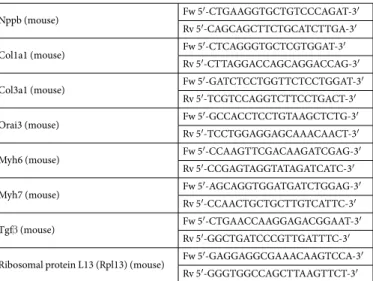
Quantitative RT-PCR.
Rat total RNA was isolated with the RNeasy Mini kit (Qiagen). RNA reverse transcriptase-PCR analysis was performed using the Absolute QPCR SYBR green mix (ABgene) on an MX3005P QPCR system (Stratagene, Agilent Technologies). Primer sequences are listed below in Table 4. Transcript levels were normalized to the RPL32 (rat) mRNA levels.Mice total RNA was isolated using TRIzol (invitrogen). RNA reverse transcriptase-PCR analysis was per-formed using Brilliant III Ultra-Fast SYBR
®
Green QPCR Master Mix (Agilent Technologies) on an LightCycler®
480 Real-Time PCR System (Roche). Primer sequences are listed below in Table 5. Transcript levels were normal-ized to the RPL13 (mice) mRNA levels.Western-blot.
Cardiac myocytes were lysed in 150 mM NaCl, 50 mM Tris pH = 7.5, EDTA 5 mM, 0.5% NP40, 1% triton, protease and phosphatase inhibitors cocktail (Sigma, Lyon, France) and samples isolated by centrifugation. Forty µg of proteins in a final volume of thirty µL of Laemmli loading buffer were heated to 70 °C for 10 minutes. Samples were run on a 4–12% Nu-PAGE gel (Life technologies), transferred to Hybond-C PVDF membrane according to the manufacturer protocol (Amersham Biosciences, GE Healthcare). Membrane was incubated with rabbit anti-Orai3 (1/500, ProsciInc 4117), or rabbit anti-GAPDH (1/2500, Cell Signaling 2118) followed by anti-rabbit HRP (1/5000, Amersham Biosciences, GE Healthcare). Detection was performed using the ECL Western Blotting Substrate (Pierce) and signals were recorded using a Camera LAS 4000.Cardiac tissue was lysed in 150 mM NaCl, 50 mM Tris pH = 7.5, EDTA 5 mM, 0.5% NP40, 1% triton, protease and phosphatase inhibitors cocktail (Sigma, Lyon, France) and samples isolated by centrifugation. Eighty µg of proteins in a final volume of fifteen µL of Laemmli loading buffer were heated to 70 °C for 10 minutes. Samples were run on a 4–12% Nu-PAGE gel (Life technologies), transferred to Trans-Blot Turbo Mini-size nitrocellulose membrane according to the manufacturer protocol (Bio-Rad). Membrane was incubated with rabbit anti-Orai3 (1/500, ProsciInc 4117), rabbit anti-Pi-GSK3β (1/1000, Cell signaling mAb#9323), rabbit anti-GSK3β (1/1000, Cell signaling mAb#9315), or mouse-GAPDH (1/1000, Santa Cruz sc-365062) followed by anti-rabbit HRP (1/10000, Abcam 6721), or anti-mouse HRP (1/5000, NA931 GE Healthcare). Detection was performed using the Clarity Western ECl Substrate (Bio-Rad) and signals were recorded using a Camera LAS 4000.
Quantification of TNFα and TNFR
2expression.
TNFα and TNFR2 protein expression was quantifiedas previously reported using Elisa R&D kits26.
Orai3 (rat) Fw 5′-CTGTCCACCAGTCACCACAC-3′
Rv 5′-CCACCAAGGATCGGTAGAAA-3′ Ribosomal protein L32
(RPL32) (rat) Fw 5′-CCAGAGGCATCGACAACA-3′Rv 5′-GCACTTCCAGCTCCTTGACAT-3′ Table 4. Sequences of the primers used (5′ to 3′) for rat.
Nppb (mouse) Fw 5′-CTGAAGGTGCTGTCCCAGAT-3′
Rv 5′-CAGCAGCTTCTGCATCTTGA-3′
Col1a1 (mouse) Fw 5′-CTCAGGGTGCTCGTGGAT-3′
Rv 5′-CTTAGGACCAGCAGGACCAG-3′
Col3a1 (mouse) Fw 5′-GATCTCCTGGTTCTCCTGGAT-3′
Rv 5′-TCGTCCAGGTCTTCCTGACT-3′
Orai3 (mouse) Fw 5′-GCCACCTCCTGTAAGCTCTG-3′
Rv 5′-TCCTGGAGGAGCAAACAACT-3′
Myh6 (mouse) Fw 5′-CCAAGTTCGACAAGATCGAG-3′
Rv 5′-CCGAGTAGGTATAGATCATC-3′
Myh7 (mouse) Fw 5′-AGCAGGTGGATGATCTGGAG-3′
Rv 5′-CCAACTGCTGCTTGTCATTC-3′
Tgfβ (mouse) Fw 5′-CTGAACCAAGGAGACGGAAT-3′
Rv 5′-GGCTGATCCCGTTGATTTC-3′
Ribosomal protein L13 (Rpl13) (mouse) Fw 5′-GAGGAGGCGAAACAAGTCCA-3′
Rv 5′-GGGTGGCCAGCTTAAGTTCT-3′ Table 5. Sequences of the primers used (5′ to 3′) for mouse.
Quantification of cardiomyocyte area and tissue fibrosis.
Frozen sections fixed in paraformaldehyde were labeled with Wheat Germ Agglutinin (WGA)-Alexa 647 (1/500 dilution, Molecular Probes). Tissue sections were analyzed with a Zeiss Axio Observer Z1 microscope using ImageJ software. A low vs. high threshold allowed quantification of cardiomyocyte area or tissue fibrosis, respectively, as previously reported47. Results werequanti-fied from 7 mice/group (12 images/animal).
Drugs.
Isoproterenol (Sigma) and TNFα (R&D) were respectively used at 100 nM and 50 ng/mL, as previously reported26. Neutralizing anti-TNFR1 or anti-TNFR2 antibodies (R&D) were preincubated 1 hour at 37 °C at a final
concentration of 2.5 µg/mL. All pharmacological inhibitors were preincubated for 10 min. Orai pharmacological inhibitors YM58483 (Tocris) and Synta66 (Servier) were added at 1 µM. PLA2 activating peptide (R&D) and
the cPLA2 inhibitor, methyl arachidonylfluorophosphonate (MAFP) (Sigma), were respectively used at 20 µg/mL
and 4 µg/mL. Lipoxygenase and cycloxygenase inhibitors, nordihydroguaiaretic acid (NDGA) and indomethacin (Sigma), were used at 1 µM. Montelukast (Sigma) was used at 2 µM. To explore ventricular myocyte resistance to oxidative stress, we applied 100 µM of H2O2 (Sigma). Semapimod (MedKoo) was used in vitro on CD11b/c cells
at 10 µM.
Statistical analysis.
Quantitative data are represented using means and error bars indicating standard error of the mean (SEM) and were analyzed using XLStat 2014 (Addinsoft, New York, USA). Non-parametric Mann-Whitney U or Wilcoxon matched-paired tests were used for comparisons between two groups, when appropriate. Non-parametric Kruskal-Wallis test was used for comparisons between more than two groups. ANOVA for repeated measures followed by Dunn-Sidak post-hoc test was used to analyze differences in echocar-diographic parameters over time. All values with p < 0.05 were considered significant.References
1. van Bilsen, M., van der Vusse, G. J. & Reneman, R. S. Transcriptional regulation of metabolic processes: implications for cardiac metabolism. Pflugers Archiv: European journal of physiology 437, 2–14 (1998).
2. Fujiu, K. et al. A heart-brain-kidney network controls adaptation to cardiac stress through tissue macrophage activation. Nature
medicine 23, 611–622, https://doi.org/10.1038/nm.4326 (2017).
3. Sillje, H. H. W. & de Boer, R. A. Heart failure: Macrophages take centre stage in the heart-brain-kidney axis. Nature reviews.
Nephrology 13, 388–390, https://doi.org/10.1038/nrneph.2017.73 (2017).
4. Mozaffarian, D. et al. Heart disease and stroke statistics–2015 update: a report from the American Heart Association. Circulation
131, e29–322, https://doi.org/10.1161/CIR.0000000000000152 (2015).
5. Balakumar, P. & Jagadeesh, G. Multifarious molecular signaling cascades of cardiac hypertrophy: can the muddy waters be cleared?
Pharmacol Res 62, 365–383, https://doi.org/10.1016/j.phrs.2010.07.003 (2010).
6. Crozatier, B. & Ventura-Clapier, R. Inhibition of hypertrophy, per se, may not be a good therapeutic strategy in ventricular pressure overload: other approaches could be more beneficial. Circulation 131, 1448–1457, https://doi.org/10.1161/ CIRCULATIONAHA.114.013895 (2015).
7. Roe, A. T., Frisk, M. & Louch, W. E. Targeting cardiomyocyte Ca2+ homeostasis in heart failure. Current pharmaceutical design 21, 431–448 (2015).
8. Benard, L. et al. Cardiac Stim1 Silencing Impairs Adaptive Hypertrophy and Promotes Heart Failure Through Inactivation of mTORC2/Akt Signaling. Circulation 133, 1458–1471; discussion 1471, https://doi.org/10.1161/CIRCULATIONAHA.115.020678
(2016).
9. Hulot, J. S. et al. Critical role for stromal interaction molecule 1 in cardiac hypertrophy. Circulation 124, 796–805, https://doi. org/10.1161/CIRCULATIONAHA.111.031229 (2011).
10. Luo, X. et al. STIM1-dependent store-operated Ca(2)(+) entry is required for pathological cardiac hypertrophy. Journal of molecular
and cellular cardiology 52, 136–147, https://doi.org/10.1016/j.yjmcc.2011.11.003 (2012).
11. Sabourin, J. et al. Ca(2+) handling remodeling and STIM1L/Orai1/TRPC1/TRPC4 upregulation in monocrotaline-induced right ventricular hypertrophy. Journal of molecular and cellular cardiology 118, 208–224, https://doi.org/10.1016/j.yjmcc.2018.04.003
(2018).
12. Saliba, Y. et al. Emergence of Orai3 activity during cardiac hypertrophy. Cardiovascular research 105, 248–259, https://doi. org/10.1093/cvr/cvu207 (2015).
13. Zhao, G., Li, T., Brochet, D. X., Rosenberg, P. B. & Lederer, W. J. STIM1 enhances SR Ca2+ content through binding phospholamban in rat ventricular myocytes. Proceedings of the National Academy of Sciences of the United States of America 112, E4792–4801, https:// doi.org/10.1073/pnas.1423295112 (2015).
14. Shuttleworth, T. J. Selective activation of distinct Orai channels by STIM1. Cell calcium 63, 40–42, https://doi.org/10.1016/j. ceca.2016.11.001 (2017).
15. Trebak, M. & Putney, J. W. Jr. ORAI Calcium Channels. Physiology 32, 332–342, https://doi.org/10.1152/physiol.00011.2017 (2017). 16. Ruhle, B. & Trebak, M. Emerging roles for native Orai Ca2+ channels in cardiovascular disease. Current topics in membranes 71,
209–235, https://doi.org/10.1016/B978-0-12-407870-3.00009-3 (2013).
17. Shuttleworth, T. J., Thompson, J. L. & Mignen, O. STIM1 and the noncapacitative ARC channels. Cell calcium 42, 183–191, https:// doi.org/10.1016/j.ceca.2007.01.012 (2007).
18. Tanwar, J., Trebak, M. & Motiani, R. K. Cardiovascular and Hemostatic Disorders: Role of STIM and Orai Proteins in Vascular Disorders. Advances in experimental medicine and biology 993, 425–452, https://doi.org/10.1007/978-3-319-57732-6_22 (2017). 19. Zhang, X., Gueguinou, M. & Trebak, M. In Calcium Entry Channels in Non-Excitable Cells (eds Kozak, J. A. & Putney, J. W. Jr.)
197–214 (2018).
20. Thompson, J., Mignen, O. & Shuttleworth, T. J. The N-terminal domain of Orai3 determines selectivity for activation of the store-independent ARC channel by arachidonic acid. Channels (Austin) 4, 398–410, https://doi.org/10.4161/chan.4.5.13226 (2010). 21. Grisanti, L. A. et al. Temporal and gefitinib-sensitive regulation of cardiac cytokine expression via chronic beta-adrenergic receptor
stimulation. American journal of physiology. Heart and circulatory physiology 308, H316–330, https://doi.org/10.1152/ ajpheart.00635.2014 (2015).
22. Lecour, S. & James, R. W. When are pro-inflammatory cytokines SAFE in heart failure? Eur Heart J 32, 680–685, https://doi. org/10.1093/eurheartj/ehq484 (2011).
23. Mann, D. L. Inflammatory mediators and the failing heart: past, present, and the foreseeable future. Circulation research 91, 988–998 (2002).
24. Mann, D. L. et al. Targeted anticytokine therapy in patients with chronic heart failure: results of the Randomized Etanercept Worldwide Evaluation (RENEWAL). Circulation 109, 1594–1602, https://doi.org/10.1161/01.CIR.0000124490.27666.B2 (2004).