Blastocystis, an unrecognized parasite: an
overview of pathogenesis and diagnosis
Ivan Wawrzyniak, Philippe Poirier, Eric Viscogliosi, Meloni Dionigia, Catherine Texier, Fre´de´ric Delbac and Hicham El AlaouiAbstract: Blastocystis sp. is among the few enteric parasites with a prevalence that often
exceeds 5% in the general population of industrialized countries and can reach 3060% in
developing countries. This parasite is frequently found in people who are immunocompromised
(patients with human immunodeficiency virus/acquired immunodeficiency syndrome or cancer)
and a higher risk of Blastocystis sp. infection has been found in people with close animal
contact. Such prevalence in the human population and the zoonotic potential naturally raise
questions about the impact of these parasites on public health and has increased interest in
this area. Recent in vitro and in vivo studies have shed new light on the pathogenic power of this
parasite, suggesting that Blastocystis sp. infection is associated with a variety of
gastrointes-tinal disorders, may play a significant role in irritable bowel syndrome, and may be linked with
cutaneous lesions (urticaria). Despite recent significant advances in the knowledge of the
extensive genetic diversity of this species, the identification of extracellular proteases as
virulence factors and the publication of one isolate genome, many aspects of the biology of
Blastocystis sp. remain poorly investigated. In this review, we investigate several biological
aspects of Blastocystis sp. (diversity and epidemiology, diagnosis tools and pathophysiology).
These data pave the way for the following challenges concerning Blastocystis sp. research:
deciphering key biological mechanisms and pathways of this parasite and clarification of its
clinical impact in humans.
Keywords: Blastocystis, pathogenesis, diagnosis, subtypes, gut
Blastocystis sp. is an anaerobic intestinal parasite of humans and a wide range of animals [Stenzel and Boreham, 1996; Tan, 2004, 2008]. This parasite belongs to the stramenopiles, a complex and heterogeneous evolutionary assemblage of
heterotrophic and photosynthetic protozoa
[Silberman et al. 1996; Arisue et al. 2002; Riisberg et al. 2009]. Interestingly, Blastocystis sp. is the only stramenopiles known to cause infection in humans. Four major morphological forms of Blastocystis sp. were described in stools or in vitro cultures: vacuolar (Figure 1), granular, amoeboid and cyst forms [Stenzel and Boreham, 1996; Tan, 2008; Suresh et al. 2009]. The two former forms are the most easily recognizable and frequently observed in laboratory culture and stool samples. Although rarely reported, the irregular amoeboid form was postulated to play a role in pathogenesis [Tan et al. 2006, Katsarou-Katsari et al. 2008 but this hypothesis was
contradicted [Souppart et al. 2009].
Experimental infectivity studies in animals with the cyst form demonstrated that the water-resist-ant and environmentally resistwater-resist-ant infective cysts represented the transmissible stage of the parasite [Suresh et al. 1993, 2005; Moe et al. 1996; Yoshikawa et al. 2004]. Taking into account these observations and those of in vitro encyst-ations studies [Suresh et al. 1993; Villar et al. 1998; Chen et al. 1999], a life cycle for Blastocystis sp. was proposed with the cyst as the infectious stage [Tan, 2008]. Upon ingestion of cysts, the parasite undergoes excystation in the large intestine and develops into vacuolar forms. These vacuolar forms divide by binary fission and may develop into amoeboid or granular forms. Then, encystation may occur while crossing along the colon before cyst excretion in the faeces [Tan, 2008]. Therefore, Blastocystis sp.
lives in oxygen-poor environments and is
Ther Adv Infect Dis (2013) 1(5) 167178 DOI: 10.1177/ 2049936113504754 !The Author(s), 2013. Reprints and permissions: http://www.sagepub.co.uk/ journalsPermissions.nav
Correspondence to:
Hicham El Alaoui, PhD
CNRS, UMR 6023, LMGE, F-63177 Aubie`re, France Email:
hicham.el_alaoui@ univ-bpclermont.fr
Ivan Wawrzyniak, PhD Clermont Universite´, Universite´ Blaise Pascal, Laboratoire Microorganismes, Ge´nome et Environnement, Clermont-Ferrand and CNRS, UMR 6023, LMGE, Aubie`re, France Philippe Poirier, PhD Clermont Universite´, Universite´ Blaise Pascal, Laboratoire
Microorganismes, Ge´nome et Environnement, Clermont-Ferrand, CNRS, UMR 6023, LMGE, Aubie`re, Clermont Universite´, Universite´ d’Auvergne, JE 2526, Evolution des
characterized by the presence of some double-membrane surrounded organelles called mito-chondria-like organelles (MLOs) (Figure 1) [Nasirudeen and Tan, 2004]. Sequencing of complete circular DNA in the MLO of these parasites by different authors [Perez-Brocal and Clark, 2008; Stechmann et al. 2008; Wawrzyniak et al. 2008] showed that the cellular compart-ments have the metabolic properties of both
aerobic and anaerobic mitochondria. The
nuclear genome of Blastocystis sp. was recently sequenced and revealed a compact nature and
an intriguing architecture [Denoeud et al.
2011]. This genome has also provided clues to decipher the genetic diversity and pathogenesis of this parasite.
Summary of diagnosis tools
The most common approaches for the detection of Blastocystis sp. [Stenzel and Boreham, 1996; Stensvold et al. 2007a; Tan, 2008] consist of direct smear examination by light microscopic or xenic in vitro culture. However, given the occurrence of different forms of Blastocystis sp. (especially the hardly recognizable cystic form), deterioration caused by environmental conditions or drug treatment and the fact that Blastocystis sp. can be confused with other microorganisms, this method seems to have largely underestimated this parasite in the context of enteric parasite diagno-sis. Moreover, culturing this parasite is time con-suming and can bias subsequent genotyping due
to the different ability of isolates to grow in select-ive medium [Roberts et al. 2011]. Therefore, to overcome these limitations, several molecular polymerase chain reaction (PCR)-based diagnos-tic approaches using faeces directly or after cul-ture of faecal specimens have been described [Santin et al. 2011; Abe et al. 2003a; 2003b; Yoshikawa et al. 2004; Scicluna et al. 2006; Stensvold et al. 2006; Roberts et al. 2011]. Studies comparing the relative performances of these various diagnostic methods [Suresh and Smith, 2004; Parkar et al. 2007; Stensvold et al. 2007a] showed that the PCR approach was as sensitive as the culture approach. More recently, Poirier and colleagues reported a highly sensitive real-time quantitative PCR (qPCR) assay devel-oped to detect Blastocystis sp. in stool samples [Poirier et al. 2011]. This assay targets a region of the small subunit rRNA gene (SSU-rDNA) and allowed subtyping of isolates by direct
sequencing of qPCR products. Moreover,
Stensvold and colleagues developed a qPCR on stool samples using the SSU-rDNA marker, including an internal process control enabling
the evaluation of potential PCR inhibitors
[Stensvold et al. 2012]. This approach had the advantage of increasing the specificity and avoid-ing the amplification of false positives. Therefore, currently, SSU-rDNA genotyping is the method of choice for diagnosis [Poirier et al. 2011; Stensvold et al. 2012].
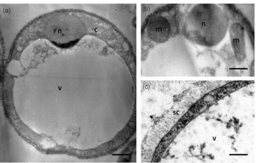
Figure 1. Vacuolar form of in vitro cultivated Blastocystis sp. viewed under transmission electron microscopy (a). This form is spherical with a large central vacuole (v) and a thin peripheral band of cytoplasm (c) around the vacuole. (b) The cytoplasm contains the nucleus (n) and mitochondrion-like organelles (m). (c) Blastocystis sp. cell is surrounded by a surface coat (sc). Bars, 2 mm for (a) and 500 nm for (b) and (c).
bacte´ries pathoge`nes et susceptibilite´ de l’hoˆte, Clermont-Ferrand and CHU Clermont-Ferrand, Service Parasitologie Mycologie, Clermont-Ferrand, France Eric Viscogliosi, PhD Institut Pasteur de Lille, Centre d’Infection et d’Immunite´ de Lille, Inserm U1019, CNRS UMR 8204, Universite´ Lille Nord de France, Biology and Diversity of Emerging Eukaryotic Pathogens, Lille cedex, France Meloni Dionigia, PhD Institut Pasteur de Lille, Centre d’Infection et d’Immunite´ de Lille, Inserm U1019, CNRS UMR 8204, Universite´ Lille Nord de France, Biology and Diversity of Emerging Eukaryotic Pathogens, Lille cedex, France and Microbe Division/Japan Collection of Microorganisms, RIKEN BioResource Center 3-1-1 Koyadai, Tsukuba-shi, Ibaraki, Japan Catherine Texier, PhD Fre´de´ric Delbac, PhD Clermont Universite´, Universite´ Blaise Pascal, Laboratoire Microorganismes, Ge´nome et Environnement, Clermont-Ferrand and CNRS, UMR 6023, LMGE, Aubie`re, France
Blastocystis sp., a highly prevalent and divergent parasite
Numerous epidemiological surveys carried out in different countries identify Blastocystis sp. as the most common eukaryotic parasite reported in human faecal samples [Tan, 2008]. Overall the prevalence of Blastocystis sp. is higher than those of other intestinal protozoan parasites such as
Giardia, Entamoeba and Cryptosporidium, as
observed in France [The ANOFEL
Cryptosporidium National Network, 2010] and the United States [Boorom et al. 2008]. An increasing trend in identification of Blastocystis sp. suggests that it is an emerging parasite with a worldwide distribution [WHO, 2008]. Prevalence varies widely from country to country and within communities of the same country [Tan, 2008; Souppart et al. 2009; Alfellani et al. 2013]. Developing countries have a higher prevalence of the parasite than industrialized countries. This difference can be explained by poor hygiene practices, close animal contact and consumption of contaminated food or water [Li et al. 2007; Leelayoova et al. 2008; Eroglu and Koltas, 2010; Baldursson and Karanis, 2011; Ithoi et al. 2011; Lee et al. 2012; Nagel et al. 2012] since the faecaloral route is considered to be the main mode of transmission of this parasite [Yoshikawa et al. 2004]. Prevalence is low in developed coun-tries such as Japan (0.51%) [Hirata et al. 2007] and Singapore (3.3%) [Wong et al. 2008] and high in developing nations including Brazil (40.9%) [Aguiar et al. 2007], Egypt (33.3%) [Rayan et al. 2007] and Indonesia (60%) [Pegelow et al. 1997]. In some countries, the prevalence can be rather variable and ranges from 1.9% to 32.6% in China [Li et al. 2007]
and from 0.9% to 45.2% in Thailand
[Saksirisampant et al. 2003, 2006], depending on the subpopulation studied. Such variations within the same country could reflect true differ-ences between communities or the use of different diagnostic approaches [Stensvold, 2013].
Blastocystis sp. isolates from humans and other animals have been reported to be morphologic-ally indistinguishable. However, extensive genetic variation among numerous Blastocystis sp. isolates from both humans and animals was mainly observed by PCR restriction fragment length polymorphism [Hoevers et al. 2000; Kaneda et al. 2001; Abe et al. 2003b; Rivera and Tan, 2005] and PCR using sequenced-tagged site pri-mers [Yoshikawa et al. 2004; Yan et al. 2006; Li
et al. 2007; Yoshikawa et al. 2009].
This considerable genetic divergence among iso-lates was subsequently confirmed by molecular phylogenies, mainly inferred from SSU-rDNA sequences [Arisue et al. 2003; Abe, 2004; Noe¨l et al. 2005; Scicluna et al.2006; Jones et al. 2008; Ozyurt et al. 2008; Souppart et al. 2009, 2010; Stensvold et al. 2009; Parkar et al. 2010; Whipps et al. 2010]. From these molecular analyses, a consensus on Blastocystis sp. terminology was proposed in an international collaborative project [Stensvold et al. 2007b]. In this new
classifica-tion, all isolates should be designated
Blastocystis sp. and assigned to one of the described subtypes (STs). Each of the STs exhibiting sufficient genetic diversity should be classified as separate species. Thereafter, a new ST was identified from primates and artiodactyls
and designated as Blastocystis sp. ST10
[Stensvold et al. 2009]. More recently, seven add-itional STs (ST1117) were identified from zoo animals [Parkar et al. 2010; Fayer et al. 2012; Alfellani et al. 2013; Roberts et al. 2013]. Most of the samples included in published epi-demiological surveys represented simple infec-tions. Mixed infections (i.e. infections by at least two different STs) probably result from multiple sources of infection. The prevalence of these infections is similar in different countries and roughly comprised between 2.6% and 14.3% [Yan et al. 2006; Li et al. 2007; Dogruman-Al et al. 2008; Souppart et al. 2009; Meloni et al. 2012]. However, the true distribu-tion of mixed infecdistribu-tions remains difficult to ascer-tain in a particular individual and is likely underestimated as this depends on the method employed for subtyping [Meloni et al. 2012]. In almost all the studies reported so far, including those in Europe [Souppart et al. 2009; Meloni et al. 2011; Forsell et al. 2012; Alfellani et al. 2013], Africa [Souppart et al. 2010; Alfellani et al. 2013], Oceania [Roberts et al. 2013], Asia [Jantermtor et al. 2013] and the Middle East [Moosavi et al. 2012], a large majority of human infections with Blastocystis sp. were attrib-utable to ST3 isolates. Only a few exceptions showed a higher prevalence of ST4 in Spain [Dominguez-Marquez et al. 2009], Denmark [Stensvold et al. 2011] and in a region of France [Poirier et al. 2011], and of ST1 in
Thailand [Thathaisong et al. 2013].
Collectively, these studies suggest that the dom-inant ST3 was the only ST of human origin as was first proposed by Noe¨l and colleagues [Noe¨l et al. 2005], even if it can also be found in some
animals. Consequently, the predominance of this ST might be mainly explained by large-scale human to human transmission [Yoshikawa et al. 2000]. Proportions of ST1 to ST4 differ between locations. ST6 and ST7 are common in Asia but rarely observed in European countries. ST5, ST8 and ST9 are found episodically in humans [Yan et al. 2007; Tan, 2008; Stensvold et al. 2009; Tan et al. 2010; Moosavi et al. 2012].
Comparison of SSU-rDNA gene sequences, cross-transmission experiments and respective prevalence of different STs in the human popu-lation indicate that almost all of the known STs of supposed animal origin are likely zoonotic and able to infect human (Figure 2) [Noe¨l et al. 2005; Parkar et al. 2007, 2010; Yan et al. 2007; Yoshikawa et al. 2009]. Therefore, a higher risk of Blastocystis sp. infection was found in people with close animal contact, including zoo keepers [Parkar et al. 2010] and abattoir workers [Rajah Salim et al. 1999; Parkar et al. 2010], indicating that animals may represent a significant zoonotic source of this parasite for humans.
Insights into Blastocystis sp. pathogenesis
The pathogenic status of Blastocystis sp. was widely debated in the literature to determine whether this microorganism was a truly patho-genic or commensal organism [Stenzel and Boreham, 1996; Boorom et al. 2008; Tan, 2008; Tan et al. 2010], although an increasingly number of recent studies cited Blastocystis sp. as an emerging pathogen [Tan, 2008, Tan et al. 2010; Poirier et al. 2012; Scanlan, 2012]. This is mainly due to the fact that Blastocystis sp. can be found in both symptomatic and asymptomatic patients [Dogruman-Al et al. 2008; Eroglu et al. 2009; Souppart et al. 2009]. However, recent in vitro and in vivo studies show that Blastocystis sp. infection is associated with a variety of gastro-intestinal disorders (called blastocystosis), espe-cially in irritable bowel syndrome (IBS) and cutaneous lesions.
Blastocystis sp. is associated with various clinical symptoms in humans
In humans, blastocystosis is mainly characterized by nonspecific gastrointestinal symptoms, like Figure 2. Blastocystis sp. subtypes (STs 19) with various host specificities. Humans can be infected by nine STs, some being mainly found in humans (ST3 and ST9). ST1, 2, 5 and 8 are found both in human and mam-malian isolates (primate, pig, human, cattle and pig), while ST4 is also present among rodent isolates, and ST6, 7 and 8 among avian isolates. Some STs are exclusively found in animals (ST1017). ST10 and 15 are present among Artiodactyla and nonhuman primates, ST11 among Proboscidea, ST12 among Artiodactyla and marsu-pials, ST13 among nonhuman primates and marsumarsu-pials, ST14 among Artiodactyla, ST16 among marsupials and ST17 among rodents.
diarrhoea, abdominal pain, flatulence, nausea, vomiting, constipation, weight loss or fatigue [Stenzel and Boreham, 1996; Boorom et al. 2008; Tan, 2008; Tan et al. 2010]. The severity of these diseases is variable and ranges from acute to chronic infections [Tan, 2008]. A hypothesis to explain differences in the disease caused by Blastocystis sp. is its genetic diversity [Hussein et al. 2008; Tan, 2008; Tan et al. 2010; Scanlan, 2012], although no association was detected between symptoms and Blastocystis subtypes in several studies [Ozyurt et al. 2008; Dogruman-Al et al. 2009; Souppart et al. 2009; Jantermtor et al. 2013]. However, ST4 isolates are more common in symptomatic patients in Sweden, Denmark and Spain [Dominguez-Marquez et al. 2009; Stensvold et al. 2011; Forsell et al. 2012], arguing for an important role of this subtype that needs to be investigated further.
In addition to aspecific gastrointestinal symp-toms, studies associated the parasite with cutane-ous disorders and chronic or acute urticaria [Vogelberg et al. 2010; Hameed et al. 2011; Zuel-Fakkar et al. 2011; Verma and Delfanian, 2013]. These diseases were correlated with the presence of Blastocystis sp. belonging to the ST2 [Vogelberg et al. 2010] or ST3 [Zuel-Fakkar et al. 2011] in the patient stools. An association was also found between urticaria and amoeboid forms of a ST3 isolate [Katsarou-Katsari et al. 2008]. It was suggested that the amoeboid form adheres efficiently to the intestinal epithelium, affecting gut immune homeostasis and causing an inflammatory response against the parasite that led to urticaria [Valsecchi et al. 2004]. Blastocystis sp. is also suspected to be involved in IBS [Tan et al. 2010; Poirier et al. 2012; Scanlan, 2012]. Indeed several studies reported a higher incidence of the parasite in patients with IBS
compared with healthy populations [Poirier
et al. 2012]. IBS is a common gastrointestinal disorder characterized by abdominal pain and discomfort associated with changes in bowel habits [Longstreth et al. 2006]. Studies on the IBS population showed a higher prevalence of ST1 and ST3 isolates of Blastocystis sp. [Yakoob et al. 2010; Fouad et al. 2011; Jimenez-Gonzalez et al. 2012]. However, these studies did not con-clude that Blastocystis sp. was the sole etiologic agent. Moreover, a recent study demonstrated that even though Blastocystis sp. was more fre-quent in symptomatic patients with IBS, the
dif-ferences compared with controls were not
significant [Cekin et al. 2012]. The presence of Blastocystis sp. in symptomatic patients can also indicate that this parasite could be involved with other factors in this disease pathophysiology [Poirier et al. 2012]. It is possible that the alter-ation of the intestinal environment, provoked by pathogens (bacteria), genetic or environmental factors promotes its development. Furthermore, studies showing the concomitant eradication of Blastocystis sp. with the disappearance of symp-toms in patients with IBS are needed to clarify the role of this parasite.
High-risk populations
As in many parasitic infections, some populations are more susceptible to Blastocystis sp. infection. Therefore, this parasite is frequently found in immunocompromised individuals such as those with human immunodeficiency virus/acquired
immunodeficiency syndrome or cancer
[Kurniawan et al. 2009; Tan et al. 2010]. Children from developing countries and those who are immunocompromised are also more sus-ceptible [Calik et al. 2011; Canete et al. 2012; Daryani et al. 2012]. The socioeconomic status, the quality of drinking water, the consumption of contaminated food and the personal hygiene habits are the major risks explaining contamin-ation in children [Abdulsalam et al. 2012; Canete et al. 2012]. A higher risk of Blastocystis sp. infection was also found in people who are in close contact with animals, which increases exposure to the parasite [Yoshikawa et al. 2009; Parkar et al. 2010], reinforcing the zoonotic nature of Blastocystis sp.
Pathophysiology of blastocystosis
One of the major obstacles to the study of the pathogenesis of Blastocystis sp. is the lack of
animal models to test Koch’s postulate.
However, a variety of experimental infections involving different animals have been described, including rats, mice, guinea pigs or chickens [Tan, 2008; Tan et al. 2010]. From these, it was deduced that laboratory mice are not suitable as animal models. Infections were generally self lim-iting, although some mice showed weight loss and lethargy. However, histological examination of the cecum and colon revealed intense inflamma-tory cell infiltration, oedematous lamina propria and mucosal sloughing [Moe et al. 1997]. These authors also showed that there was an age-related susceptibility to Blastocystis sp. in mice. Indeed, juvenile BALB/c mice were more susceptible
than adult mice and 8-week-old adult BALB/c mice were totally resistant to Blastocystis sp. Studies on rat models suggest that this species is more suitable for developing an animal model [Tan, 2008]. Ten to a hundred cysts were able to establish an infection via intracecal or oral inoculations in 3-week-old Wistar rats and all contents from the cecum and large intestine
were positive for Blastocystis sp. infection
[Yoshikawa et al. 2004]. Hussein and colleagues tested the infectivity of human ST14 isolates obtained from both asymptomatic and symptom-atic patients in 4-week-old male Wister rats orally inoculated with 4-day-old cultures of Blastocystis
sp. negative for Cryptosoridium, Cyclospora,
Isospora, Microsporidium and common bacterial pathogens [Hussein et al. 2008]. Interestingly, the moderate and severe degrees of pathological changes observed 6 weeks post infection were found only with symptomatic isolates while mild changes were found only with asymptomatic isolates. Interestingly, ST1 symptomatic isolates induced 25% mortality in rats, indicating its pathogenesis. In parallel, the authors described
intense inflammatory reaction and sloughing mucosa, oedema and precancerous polyps in cecum and proximal colon tissues in symptom-atic infected rats. It was suggested that the inflammation induced by Blastocystis sp. had the ability to alter tight junctions between the intes-tinal epithelial cells and the intesintes-tinal content, which led to disturbances of the barrier function and permeability [Hussein et al. 2008]. However, the possibility that these pathologies could be of bacterial or viral origin was not excluded. Some in vitro studies were conducted to investi-gate the mechanisms of physiopathology by studying the cytopathic effects of Blastocystis sp. on mammalian cell cultures. A first study [Long et al. 2001] showed that 24 h incubation with Blastocystis sp. ST1 cells or culture filtrates induced the production of proinflammatory cyto-kines interleukin (IL)-8 and granulocytemacro-phage colony-stimulating factor, suggesting that the parasite was able to modulate the host immune response (Figure 3). The induction of IL-8 production from human colonic epithelial cells (HT84) was demonstrated to be activated
Figure 3. Mechanisms of blastocystosis physiopathology. Blastocystis sp. may release cysteine protease that may participate in the attack of intestinal epithelium with other hydrolases and may cause the increase in paracellular permeability that is observed in several digestive pathologies such as irritable bowel syndrome (IBS). Blastocystis sp. is able to induce physiological disturbances linked to IBS: host cell apoptosis, the modulation of host immune response and a microinflammation. Some as yet uncharacterized secondary metabolites produced by the polyketide synthase or nonribosomal peptide synthases could participate in host intestinal symptoms by inducing changes in the host microbiota, another feature of IBS. Finally, drug-resistant isolates of the parasite could be explained by the presence of multidrug resistance proteins that could eject active drugs. GM-CSF, granulocytemacrophage colony-stimulating factor; Ig, immunoglobulin; IL, interleukin; INF, interferon; TNF, tumour necrosis factor.
by cysteine proteases from Blastocystis sp. ST4 in a nuclear factorkB dependent manner [Puthia et al. 2008]. The same authors also showed that coin-cubation of intestinal epithelial cells of rat inter-stitial IEC6 with either Blastocystis sp. ST4 isolates or parasite lysates induced the apoptosis of host cells [Puthia et al. 2006] in a contact-independent manner. A decrease in transepithe-lial resistance and an increase in epithetransepithe-lial permeability were also observed and could be explained by the rearrangement of actin filaments [Tan et al. 2010; Mirza et al. 2012]. These data suggest that Blastocystis sp. is able to disturb host gut homeostasis (Figure 3). As observed in other parasitic protozoa [Sajid and McKerrow, 2002], cysteine proteases of Blastocystis sp. should be involved in parasite survival in vivo and represent virulence factors [Tan et al. 2010; Scanlan, 2012]. Variations in cysteine protease activity were observed between ST4 and ST7 isolates, which may be attributable to differences in virulence [Mirza and Tan, 2009]. Protease activities were identified in Blastocystis sp. secretory products
and were able to cleave human secretory
immunoglobulin A, the prevalent immunoglobu-lin defence at the mucosal surface [Puthia et al. 2005]. Another study revealed that cysteine pro-teases increased permeability of human epithe-lium by reorganization of the tight junction complex and modulation of the rho associated kinase/phosphorylation of myosin light chain pathway [Mirza et al. 2012]. The sequence of the Blastocystis sp. ST7 complete genome pro-vides molecular candidates that could be involved in pathogenesis [Denoeud et al. 2011]. Twenty-two proteases were predicted to be secreted [Denoeud et al. 2011] and could therefore play a role at the hostparasite interface. Among them, 20 cysteine proteases, 1 serine protease and 1 aspartic protease were characterized and 2 cysteine proteases were experimentally identified and characterized in the secretory products by mass spectrometry analysis [Wawrzyniak et al. 2012]. These secreted proteases are serious
can-didates to explain gut function disruption
observed in intestinal pathologies [Poirier et al. 2012]. Apart from proteases, hydrolases and pro-tease inhibitors were predicted to be secreted and could participate in the blastocystosis physio-pathology. Moreover, genes coding a polyketide synthase and nonribosomal peptide synthases were identified [Denoeud et al. 2011]. These enzymes may produce molecules with interesting pharmacological activities like antibiotics and toxins and may be implicated in dysbiosis
(Figure 3). The next challenge is to understand the role of Blastocystis sp. in gut dysfunctions [Poirier et al. 2012].
Treatment of blastocystosis
This treatment is usually considered if diarrhoea is persistent and no other pathogen apart from Blastocystis sp. is identified in faecal specimens [Coyle et al. 2012]. In this case, metronidazole is considered as the first-line therapy for Blastocystis sp. infection [Nigro et al. 2003; Cassano et al. 2005; Moghaddam et al. 2005; Stensvold et al. 2010]. In a first evaluation of this drug efficacy, Nigro and colleagues showed that immunocom-petent individuals with Blastocystis sp. infection as the only evident cause of diarrhoea responded to metronidazole treatment and consequently they suggested that the parasite induced intestinal dis-ease [Nigro et al. 2003]. However, there were accumulating reports of treatment failure, par-ticularly in patients with severe Blastocystis sp. infections [Haresh et al. 1999; Moghaddam et al. 2005; Stensvold et al. 2008, 2010], suggesting the existence of extensive variations in drug suscepti-bility [Mirza et al. 2011]. Accordingly, some genes coding for multidrug resistance pump proteins (ATP-binding cassette transporters) were identi-fied in the Blastocystis sp. ST7 genome (Figure 3) [Denoeud et al. 2011]. Several standard antimicro-bials (cotrimoxazole, ornidazole, nitazoxanide,
paromomycin, chloroquine,
trimethoprim-sulfamethoxazole, iodoquinol, tinidazole, emetine, pentamidine, iodochlorhydroxyquin and furazoli-done) may be considered as second-choice drugs [Ok et al. 1999; Cimerman et al. 2003; Diaz et al. 2003; Rossignol et al. 2005; Stensvold et al. 2008; Mirza et al. 2011; Coyle et al. 2012]. Even if some of these drugs were shown to be globally effective against Blastocystis sp., treatment failures were also largely reported [Haresh et al. 1999; Nigro et al. 2003; Mirza et al. 2011].
Conclusion
Blastocystis sp. was included in the Water
Sanitation and Health programmes of the
World Health Organization [WHO, 2008].
Increasing interest of the scientific and medical community in Blastocystis sp. was coupled with new data about epidemiology, pathogeny and
more recently the first whole genome of
Blastocystis ST7. Accumulating in vivo, in vitro and in silico data assessed the importance of Blastocystis sp. in human health, with probably the association with major host and environmen-tal factors to be determined. To answer most of
the crucial questions regarding Blastocystis sp., we need to develop and standardize axenization of new STs and diagnosis tools, transfection and animal models, microarrays and genotyping mar-kers. Sequencing of genomes from different STs and comparative genomic projects are in progress and will be useful to identify specific virulence factors. Multicentre studies are also required to further our comprehension of the clinical impli-cations of Blastocystis sp. in IBS and other pathol-ogies. To conclude, the interaction of Blastocystis sp. with gut microbiota needs to be studied because of the increasing interest in microbiota disturbances in the genesis of various gastrointes-tinal dysfunctions.
Funding
This research received no specific grant from any funding agency in the public, commercial, or not-for-profit sectors.
Conflict of interest statement
The authors declare no conflicts of interest in preparing this article.
References
Abdulsalam, A., Ithoi, I., Al-Mekhlafi, H., Ahmed, A., Surin, J. and Mak, J. (2012) Drinking water is a sig-nificant predictor of Blastocystis infection among rural Malaysian primary schoolchildren. Parasitology 139: 10141020.
Abe, N. (2004) Molecular and phylogenetic analysis of Blastocystis isolates from various hosts. Vet Parasitol 120: 235242.
Abe, N., Wu, Z. and Yoshikawa, H. (2003a) Molecular characterization of Blastocystis isolates from birds by PCR with diagnostic primers and restriction fragment length polymorphism analysis of the small subunit ribosomal RNA gene. Parasitol Res 89: 393396. Abe, N., Wu, Z. and Yoshikawa, H. (2003b) Zoonotic genotypes of Blastocystis hominis detected in cattle and pigs by PCR with diagnostic primers and restriction fragment length polymorphism analysis of the small subunit ribosomal RNA gene. Parasitol Res
90: 124128.
Aguiar, J., Goncalves, A., Sodre, F., Pereira Sdos, R., Boia, M., De Lemos, E. et al. (2007) Intestinal protozoa and helminths among Terena Indians in the State of Mato Grosso do Sul: high prevalence of Blastocystis hominis. Rev Soc Bras Med Trop 40: 631634.
Alfellani, M., Stensvold, C., Vidal-Lapiedra, A., Onuoha, E., Fagbenro-Beyioku, A. and Clark, C. (2013) Variable geographic distribution of Blastocystis subtypes and its potential implications. Acta Trop 126: 1118.
Alfellani, M., Taner-Mulla, D., Jacob, A., Imeede, C., Yoshikawa, H., Stensvold, C. et al. (2013) Genetic diversity of Blastocystis in livestock and zoo animals. Acta Trop 126: 1118.
Arisue, N., Hashimoto, T. and Yoshikawa, H. (2003) Sequence heterogeneity of the small subunit ribosomal RNA genes among Blastocystis isolates. Protist 164: 497509.
Arisue, N., Hashimoto, T., Yoshikawa, H., Nakamura, Y., Nakamura, G., Nakamura, F. et al. (2002) Phylogenetic position of Blastocystis
hominis and of stramenopiles inferred from multiple molecular sequence data. J Eukaryot Microbiol 49: 4253.
Baldursson, S. and Karanis, P. (2011) Waterborne transmission of protozoan parasites: review of world-wide outbreaks an update 20042010. Water Res 45: 66036614.
Boorom, K., Smith, H., Nimri, L., Viscogliosi, E., Spanakos, G., Parkar, U. et al. (2008) Oh my aching gut: irritable bowel syndrome, Blastocystis, and asymptomatic infection. Parasit Vectors 1: 40. Calik, S., Karaman, U. and Colak, C. (2011) Prevalence of microsporidium and other intestinal parasites in children from Malatya, Turkey. Indian J Microbiol 51: 345349.
Canete, R., Diaz, M., Avalos Garcia, R., Laud Martinez, P. and Manuel Ponce, F. (2012) Intestinal parasites in children from a day care centre in Matanzas City, Cuba. PLoS One 7: e51394.
Cassano, N., Scoppio, B., Loviglio, M. and Vena, G. (2005) Remission of delayed pressure urticaria after eradication of Blastocystis hominis. Acta Derm Venereol 85: 357358.
Cekin, A., Cekin, Y., Adakan, Y., Tasdemir, E., Koclar, F. and Yolcular, B. (2012) Blastocystosis in patients with gastrointestinal symptoms: a case-control study. BMC Gastroenterol 12: 122.
Chen, X., Singh, M., Howe, J., Ho, L., Tan, S. and Yap, E. (1999) In vitro encystation and excystation of Blastocystis ratti. Parasitology 118: 151160.
Cimerman, S., Ladeira, M. and Iuliano, W. (2003) Blastocystosis: nitazoxanide as a new therapeutic option. Rev Soc Bras Med Trop 36: 415417. Coyle, C., Varughese, J., Weiss, L. and Tanowitz, H. (2012) Blastocystis: to treat or not to treat. Clin Infect Dis 54: 105110.
Daryani, A., Sharif, M., Nasrolahei, M., Khalilian, A., Mohammadi, A. and Barzegar, G. (2012)
Epidemiological survey of the prevalence of intestinal parasites among schoolchildren in Sari, Northern Iran. Trans R Soc Trop Med Hyg 106: 455459.
Denoeud, F., Roussel, M., Noel, B., Wawrzyniak, I., Da Silva, C., Diogon, M. et al. (2011) Genome sequence of the stramenopile Blastocystis, a human anaerobic parasite. Genome Biol 12: R29.
Diaz, E., Mondragon, J., Ramirez, E. and Bernal, R. (2003) Epidemiology and control of intestinal parasites with nitazoxanide in children in Mexico. Am J Trop Med Hyg 68: 384385.
Dogruman-Al, F., Dagci, H., Yoshikawa, H., Kurt, O. and Demirel, M. (2008) A possible link between sub-type 2 and asymptomatic infections of Blastocystis hominis. Parasitol Res 103: 685689.
Dogruman-Al, F., Yoshikawa, H., Kustimur, S. and Balaban, N. (2009) PCR-based subtyping of
Blastocystis isolates from symptomatic and asymptom-atic individuals in a major hospital in Ankara, Turkey. Parasitol Res 106: 263268.
Dominguez-Marquez, M., Guna, R., Munoz, C., Gomez-Munoz, M. and Borras, R. (2009) High prevalence of subtype 4 among isolates of Blastocystis hominis from symptomatic patients of a health district of Valencia (Spain). Parasitol Res 105: 949955. Eroglu, F., Genc, A., Elgun, G. and Koltas, I. (2009) Identification of Blastocystis hominis isolates from asymptomatic and symptomatic patients by PCR. Parasitol Res 105: 15891592.
Eroglu, F. and Koltas, I. (2010) Evaluation of the transmission mode of B. hominis by using PCR method. Parasitol Res 107: 841845.
Fayer, R., Santin, M. and Macarisin, D. (2012) Detection of concurrent infection of dairy cattle with Blastocystis, Cryptosporidium, Giardia, and
Enterocytozoon by molecular and microscopic methods. Parasitol Res 111: 13491355.
Forsell, J., Granlund, M., Stensvold, C., Clark, C. and Evengard, B. (2012) Subtype analysis of Blastocystis isolates in Swedish patients. Eur J Clin Microbiol Infect Dis 31: 16891696.
Fouad, S., Basyoni, M., Fahmy, R. and Kobaisi, M. (2011) The pathogenic role of different Blastocystis hominis genotypes isolated from patients with irritable bowel syndrome. Arab J Gastroenterol 12: 194200. Hameed, D., Hassanin, O. and Zuel-Fakkar, N. (2011) Association of Blastocystis hominis genetic subtypes with urticaria. Parasitol Res 108: 553560. Haresh, K., Suresh, K., Khairul., Anus, A. and Saminathan, S. (1999) Isolate resistance of Blastocystis hominis to metronidazole. Trop Med Int Health 4: 274277.
Hirata, T., Nakamura, H., Kinjo, N., Hokama, A., Kinjo, F., Yamane, N. et al. (2007) Prevalence of Blastocystis hominis and Strongyloides stercoralis infection in Okinawa, Japan. Parasitol Res 101: 17171719.
Hoevers, J., Holman, P., Logan, K., Hommel, M., Ashford, R. and Snowden, K. (2000) Restriction-fragment-length polymorphism analysis of small-sub-unit rRNA genes of Blastocystis hominis isolates from geographically diverse human hosts. Parasitol Res 86: 5761.
Hussein, E., Hussein, A., Eida, M. and Atwa, M. (2008) Pathophysiological variability of different genotypes of human Blastocystis hominis Egyptian isolates in experimentally infected rats. Parasitol Res 102: 853860.
Ithoi, I., Jali, A., Mak, J., Wan., Sulaiman, W. and Mahmud, R. (2011) Occurrence of Blastocystis in water of two rivers from recreational areas in Malaysia. J Parasitol Res 2011: 123916.
Jantermtor, S., Pinlaor, P., Sawadpanich, K., Pinlaor, S., Sangka, A., Wilailuckana, C. et al. (2013) Subtype identification of Blastocystis spp. isolated from patients in a major hospital in Northeastern Thailand. Parasitol Res 112: 17811786.
Jimenez-Gonzalez, D., Martinez-Flores, W., Reyes-Gordillo, J., Ramirez-Miranda, M., Arroyo-Escalante, S., Romero-Valdovinos, M. et al. (2012) Blastocystis infection is associated with irritable bowel syndrome in a Mexican patient population. Parasitol Res
110: 12691275.
Jones, M., 2nd., Ganac, R., Hiser, G., Hudson, N., Le, A. and Whipps, C. (2008) Detection of Blastocystis from stool samples using real-time PCR. Parasitol Res 103: 551557.
Kaneda, Y., Horiki, N., Cheng, X., Fujita, Y., Maruyama, M. and Tachibana, H. (2001) Ribodemes of Blastocystis hominis isolated in Japan. Am J Trop Med Hyg 65: 393396.
Katsarou-Katsari, A., Vassalos, C., Tzanetou, K., Spanakos, G., Papadopoulou, C. and Vakalis, N. (2008) Acute urticaria associated with amoeboid forms of Blastocystis sp. subtype 3. Acta Derm Venereol 88: 8081.
Kurniawan, A., Karyadi, T., Dwintasari, S., Sari, I., Yunihastuti, E., Djauzi, S. et al. (2009) Intestinal parasitic infections in HIV/AIDS patients presenting with diarrhoea in Jakarta, Indonesia. Trans R Soc Trop Med Hyg 103: 892898.
Lee, L., Chye, T., Karmacharya, B. and Govind, S. (2012) Blastocystis sp: waterborne zoonotic organism, a possibility?. Parasit Vectors 5: 130.
Leelayoova, S., Siripattanapipong, S., Thathaisong, U., Naaglor, T., Taamasri, P., Piyaraj, P. et al. (2008) Drinking water: a possible source of Blastocystis spp. subtype 1 infection in schoolchildren of a rural com-munity in central Thailand. Am J Trop Med Hyg 79: 401406.
Li, L., Zhang, X., Lv, S., Zhang, L., Yoshikawa, H., Wu, Z. et al. (2007) Cross-sectional surveys and sub-type classification of human Blastocystis isolates from four epidemiological settings in China. Parasitol Res 102: 8390.
Li, L., Zhou, X., Du, Z., Wang, X., Wang, L., Jiang, J. et al. (2007) Molecular epidemiology of human Blastocystis in a village in Yunnan Province, China. Parasitol Int 56: 281286.
Long, H., Handschack, A., Konig, W. and Ambrosch, A. (2001) Blastocystis hominis modulates immune responses and cytokine release in colonic epithelial cells. Parasitol Res 87: 10291030.
Longstreth, G., Thompson, W., Chey, W., Houghton, L., Mearin, F. and Spiller, R. (2006) Functional bowel disorders. Gastroenterology 130: 14801491.
Meloni, D., Poirier, P., Mantini, C., Noel, C., Gantois, N., Wawrzyniak, I. et al. (2012) Mixed human intra-and inter-subtype infections with the parasite Blastocystis sp. Parasitol Int 61: 719722.
Meloni, D., Sanciu, G., Poirier, P., El., Alaoui, H., Chabe, M., Delhaes, L. et al. (2011) Molecular sub-typing of Blastocystis sp. isolates from symptomatic patients in Italy. Parasitol Res 109: 613619. Mirza, H. and Tan, K. (2009) Blastocystis exhibits inter- and intra-subtype variation in cysteine protease activity. Parasitol Res 104: 355361.
Mirza, H., Teo, J., Upcroft, J. and Tan, K. (2011) A rapid, high-throughput viability assay for Blastocystis spp. reveals metronidazole resistance and extensive subtype-dependent variations in drug susceptibilities. Antimicrob Agents Chemother 55: 637648.
Mirza, H., Wu, Z., Teo, J. and Tan, K. (2012) Statin pleiotropy prevents rho kinase-mediated intestinal epithelial barrier compromise induced by Blastocystis cysteine proteases. Cell Microbiol 14: 14741484. Moe, K., Singh, M., Howe, J., Ho, L., Tan, S., Chen, X. et al. (1997) Experimental Blastocystis hominis infection in laboratory mice. Parasitol Res 83: 319325. Moe, K., Singh, M., Howe, J., Ho, L., Tan, S., Ng, G. et al. (1996) Observations on the ultrastructure and viability of the cystic stage of Blastocystis hominis from human feces. Parasitol Res 82: 439444.
Moghaddam, D., Ghadirian, E. and Azami, M. (2005) Blastocystis hominis and the evaluation of efficacy of metronidazole and trimethoprim/sulfamethoxazole. Parasitol Res 96: 273275.
Moosavi, A., Haghighi, A., Mojarad, E., Zayeri, F., Alebouyeh, M., Khazan, H. et al. (2012) Genetic variability of Blastocystis sp. isolated from symptomatic and asymptomatic individuals in Iran. Parasitol Res 111: 23112315.
Nagel, R., Cuttell, L., Stensvold, C., Mills, P., Bielefeldt-Ohmann, H. and Traub, R. (2012) Blastocystis subtypes in symptomatic and asymptom-atic family members and pets and response to therapy. Intern Med J 42: 11871195.
Nasirudeen, A. and Tan, K. (2004) Isolation and characterization of the mitochondrion-like organelle from Blastocystis hominis. J Microbiol Methods 58: 101109.
Nigro, L., Larocca, L., Massarelli, L., Patamia, I., Minniti, S., Palermo, F. et al. (2003) A placebo-controlled treatment trial of Blastocystis hominis infection with metronidazole. J Travel Med 10: 128130.
Noe¨l, C., Dufernez, F., Gerbod, D., Edgcomb, V., Delgado-Viscogliosi, P., Ho, L. et al. (2005) Molecular phylogenies of Blastocystis isolates from different hosts: implications for genetic diversity, identification of species, and zoonosis. J Clin Microbiol 43: 348355. Ok, U., Girginkardesler, N., Balcioglu, C., Ertan, P., Pirildar, T. and Kilimcioglu, A. (1999) Effect of tri-methoprim-sulfamethaxazole in Blastocystis hominis infection. Am J Gastroenterol 94: 32453247. Ozyurt, M., Kurt, O., Molbak, K., Nielsen, H., Haznedaroglu, T. and Stensvold, C. (2008) Molecular epidemiology of Blastocystis infections in Turkey. Parasitol Int 57: 300306.
Parkar, U., Traub, R., Kumar, S., Mungthin, M., Vitali, S., Leelayoova, S. et al. (2007) Direct charac-terization of Blastocystis from faeces by PCR and evi-dence of zoonotic potential. Parasitology
134: 359367.
Parkar, U., Traub, R., Vitali, S., Elliot, A., Levecke, B., Robertson, I. et al. (2010) Molecular characterization of Blastocystis isolates from zoo animals and their animal-keepers. Vet Parasitol 169: 817.
Pegelow, K., Gross, R., Pietrzik, K., Lukito, W., Richards, A. and Fryauff, D. (1997) Parasitological and nutritional situation of school children in the Sukaraja district, West Java, Indonesia. Southeast Asian J Trop Med Public Health 28: 173190.
Perez-Brocal, V. and Clark, C. (2008) Analysis of two genomes from the mitochondrion-like organelle of the intestinal parasite Blastocystis: complete sequences, gene content, and genome organization. Mol Biol Evol 25: 24752482.
Poirier, P., Wawrzyniak, I., Albert, A., El., Alaoui, H., Delbac, F. and Livrelli, V. (2011) Development and evaluation of a real-time PCR assay for detection and quantification of Blastocystis parasites in human stool samples: prospective study of patients with hemato-logical malignancies. J Clin Microbiol 49: 975983. Poirier, P., Wawrzyniak, I., Vivares, C., Delbac, F. and El Alaoui, H. (2012) New insights into Blastocystis spp.: a potential link with irritable bowel syndrome. PLoS Pathog 8: e1002545.
Puthia, M., Lu, J. and Tan, K. (2008) Blastocystis ratti contains cysteine proteases that mediate interleukin-8 response from human intestinal epithelial cells in an NF-kappaB-dependent manner. Eukaryot Cell 7: 435443.
Puthia, M., Sio, S., Lu, J. and Tan, K. (2006) Blastocystis ratti induces contact-independent apop-tosis, F-actin rearrangement, and barrier function disruption in IEC-6 cells. Infect Immun
74: 41144123.
Puthia, M., Vaithilingam, A., Lu, J. and Tan, K. (2005) Degradation of human secretory immuno-globulin a by Blastocystis. Parasitol Res 97: 386389. Rajah., Salim, H., Suresh., Kumar, G., Vellayan, S., Mak, J., Khairul., Anuar, A., Init, I. et al. (1999)
Blastocystis in animal handlers. Parasitol Res 85: 10321033.
Rayan, H., Ismail, O., El. and Gayar, E. (2007) Prevalence and clinical features of Dientamoeba fra-gilis infections in patients suspected to have intestinal parasitic infection. J Egypt Soc Parasitol 37: 599608. Riisberg, I., Orr, R., Kluge, R., Shalchian-Tabrizi, K., Bowers, H., Patil, V. et al. (2009) Seven gene phyl-ogeny of heterokonts. Protist 160: 191204.
Rivera, W. and Tan, M. (2005) Molecular character-ization of Blastocystis isolates in the Philippines by riboprinting. Parasitol Res 96: 253257.
Roberts, T., Barratt, J., Harkness, J., Ellis, J. and Stark, D. (2011) Comparison of microscopy, culture, and conventional polymerase chain reaction for detection of Blastocystis sp. in clinical stool samples. Am J Trop Med Hyg 84: 308312.
Roberts, T., Stark, D., Harkness, J. and Ellis, J. (2013) Subtype distribution of Blastocystis isolates from a variety of animals from New South Wales, Australia. Vet Parasitol 196: 8589.
Rossignol, J., Kabil, S., Said, M., Samir, H. and Younis, A. (2005) Effect of nitazoxanide in persistent diarrhea and enteritis associated with Blastocystis hominis. Clin Gastroenterol Hepatol 3: 987991. Sajid, M. and McKerrow, J. (2002) Cysteine proteases of parasitic organisms. Mol Biochem Parasitol
120: 121.
Saksirisampant, W., Nuchprayoon, S., Wiwanitkit, V., Yenthakam, S. and Ampavasiri, A. (2003) Intestinal parasitic infestations among children in an orphanage in Pathum Thani Province. J Med Assoc Thai 86(Suppl. 2): S263S270.
Saksirisampant, W., Prownebon, J., Kulkumthorn, M., Yenthakam, S., Janpla, S. and Nuchprayoon, S. (2006) Prevalence of intestinal parasitic infections among school children in the central region of Thailand. J Med Assoc Thai 89: 19281933.
Santin, M., Gomez-Munoz, M., Solano-Aguilar, G. and Fayer, R. (2011) Development of a new PCR protocol to detect and subtype Blastocystis spp. from humans and animals. Parasitol Res 109: 205212. Scanlan, P.D. (2012) Blastocystis: past pitfalls and future perspectives. Trends Parasitol 28: 327334. Scicluna, S., Tawari, B. and Clark, C. (2006) DNA barcoding of Blastocystis. Protist 157: 7785. Silberman, J., Sogin, M., Leipe, D. and Clark, C. (1996) Human parasite finds taxonomic home. Nature 380: 398.
Souppart, L., Moussa, H., Cian, A., Sanciu, G., Poirier, P., El., Alaoui, H. et al. (2010) Subtype ana-lysis of Blastocystis isolates from symptomatic patients in Egypt. Parasitol Res 106: 505511.
Souppart, L., Sanciu, G., Cian, A., Wawrzyniak, I., Delbac, F., Capron, M. et al. (2009) Molecular
epidemiology of human Blastocystis isolates in France. Parasitol Res 105: 413421.
Stechmann, A., Hamblin, K., Perez-Brocal, V., Gaston, D., Richmond, G., Van Der., Giezen, M. et al. (2008) Organelles in Blastocystis that blur the distinc-tion between mitochondria and hydrogenosomes. Curr Biol 18: 580585.
Stensvold, C. (2013) Comparison of sequencing (barcode region) and sequence-tagged-site PCR for Blastocystis subtyping. J Clin Microbiol 51: 190194. Stensvold, C., Ahmed, U., Andersen, L. and Nielsen, H. (2012) Development and evaluation of a genus-specific, probe-based, internal-process-controlled real-time PCR assay for sensitive and specific detection of Blastocystis spp. J Clin Microbiol 50: 18471851. Stensvold, C., Alfellani, M., Norskov-Lauritsen, S., Prip, K., Victory, E., Maddox, C. et al. (2009) Subtype distribution of Blastocystis isolates from synanthropic and zoo animals and identification of a new subtype. Int J Parasitol 39: 473479.
Stensvold, C., Arendrup, M., Jespersgaard, C., Molbak, K. and Nielsen, H. (2007a) Detecting Blastocystis using parasitologic and DNA-based meth-ods: a comparative study. Diagn Microbiol Infect Dis 59: 303307.
Stensvold, C., Arendrup, M., Nielsen, H., Bada, A. and Thorsen, S. (2008) Symptomatic infection with Blastocystis sp. subtype 8 successfully treated with tri-methoprim-sulfamethoxazole. Ann Trop Med Parasitol 102: 271274.
Stensvold, C., Christiansen, D., Olsen, K. and Nielsen, H. (2011) Blastocystis sp. subtype 4 is common in Danish Blastocystis-positive patients pre-senting with acute diarrhea. Am J Trop Med Hyg 84: 883885.
Stensvold, C., Smith, H., Nagel, R., Olsen, K. and Traub, R. (2010) Eradication of Blastocystis carriage with antimicrobials: reality or delusion? J Clin Gastroenterol 44: 8590.
Stensvold, C., Suresh, G., Tan, K., Thompson, R., Traub, R., Viscogliosi, E. et al. (2007b) Terminology for Blastocystis subtypes a consensus. Trends Parasitol 23: 9396.
Stensvold, R., Brillowska-Dabrowska, A., Nielsen, H. and Arendrup, M. (2006) Detection of Blastocystis hominis in unpreserved stool specimens by using polymerase chain reaction. J Parasitol 92: 10811087. Stenzel, D. and Boreham, P. (1996) Blastocystis hominis revisited. Clin Microbiol Rev 9: 563584. Suresh, K., Ng, G., Ramachandran, N., Ho, L., Yap, E. and Singh, M. (1993) In vitro encystment and experimental infections of Blastocystis hominis. Parasitol Res 79: 456460.
Suresh, K. and Smith, H. (2004) Comparison of methods for detecting Blastocystis hominis. Eur J Clin Microbiol Infect Dis 23: 509511.
Visit SAGE journals online http://tai.sagepub.com
Suresh, K., Smith, H. and Tan, T. (2005) Viable Blastocystis cysts in Scottish and Malaysian sewage samples. Appl Environ Microbiol 71: 56195620. Suresh, K., Venilla, G., Tan, T. and Rohela, M. (2009) In vivo encystation of Blastocystis hominis. Parasitol Res 104: 13731380.
Tan, K. (2004) Blastocystis in humans and animals: new insights using modern methodologies. Vet Parasitol 126: 121144.
Tan, K. (2008) New insights on classification, identi-fication, and clinical relevance of Blastocystis spp. Clin Microbiol Rev 21: 639665.
Tan, K., Mirza, H., Teo, J., Wu, B. and Macary, P. (2010) Current views on the clinical relevance of Blastocystis spp. Curr Infect Dis Rep 12: 2835. Tan, T. and Suresh, K. (2006) Predominance of amoeboid forms of Blastocystis hominis in isolates from symptomatic patients. Parasitol Res 98: 189193. Thathaisong, U., Siripattanapipong, S., Mungthin, M., Pipatsatitpong, D., Tan-Ariya, P., Naaglor, T. et al. (2013) Identification of Blastocystis subtype 1 variants in the home for girls, Bangkok, Thailand. Am J Trop Med Hyg 88: 352358.
The ANOFEL Cryptosporidium National Network. (2010) Laboratory-based surveillance for
Cryptosporidium in France, 20062009. EuroSurveill 15: pii¼19642.
Valsecchi, R., Leghissa, P. and Greco, V. (2004) Cutaneous lesions in Blastocystis hominis infection. Acta Derm Venereol 84: 322323.
Verma, R. and Delfanian, K. (2013) Blastocystis hominis associated acute urticaria. Am J Med Sci 346: 8081.
Villar, J., Carbajal, J., Lanuza, M., Munoz, C. and Borras, R. (1998) In vitro encystation of Blastocystis hominis: a kinetics and cytochemistry study. Parasitol Res 84: 5458.
Vogelberg, C., Stensvold, C., Monecke, S., Ditzen, A., Stopsack, K., Heinrich-Grafe, U. et al. (2010) Blastocystis sp. subtype 2 detection during recurrence of gastrointestinal and urticarial symptoms. Parasitol Int 59: 469471.
Wawrzyniak, I., Roussel, M., Diogon, M., Couloux, A., Texier, C., Tan, K. et al. (2008) Complete circular DNA in the mitochondria-like organelles of Blastocystis hominis. Int J Parasitol 38: 13771382.
Wawrzyniak, I., Texier, C., Poirier, P., Viscogliosi, E., Tan, K., Delbac, F. et al. (2012) Characterization of
two cysteine proteases secreted by Blastocystis St7, a human intestinal parasite. Parasitol Int 61: 437442. Whipps, C., Boorom, K., Bermudez, L. and Kent, M. (2010) Molecular characterization of Blastocystis spe-cies in Oregon identifies multiple subtypes. Parasitol Res 106: 827832.
WHO. (2008) Guidelines for Drinking-Water Quality (3rd edition, incorporating first and second addenda), World Health Organization: Geneva.
Wong, K., Ng, G., Lin, R., Yoshikawa, H., Taylor, M. and Tan, K. (2008) Predominance of subtype 3 among Blastocystis isolates from a major hospital in Singapore. Parasitol Res 102: 663670.
Yakoob, J., Jafri, W., Beg, M., Abbas, Z., Naz, S., Islam, M. et al. (2010) Blastocystis hominis and Dientamoeba fragilis in patients fulfilling irritable bowel syndrome criteria. Parasitol Res 107: 679684. Yan, Y., Su, S., Lai, R., Liao, H., Ye, J., Li, X. et al. (2006) Genetic variability of Blastocystis hominis iso-lates in China. Parasitol Res 99: 597601.
Yan, Y., Su, S., Ye, J., Lai, X., Lai, R., Liao, H. et al. (2007) Blastocystis sp. subtype 5: a possibly zoonotic genotype. Parasitol Res 101: 15271532.
Yoshikawa, H., Abe, N., Iwasawa, M., Kitano, S., Nagano, I., Wu, Z. et al. (2000) Genomic analysis of Blastocystis hominis strains isolated from two long-term health care facilities. J Clin Microbiol 38: 13241330. Yoshikawa, H., Morimoto, K., Wu, Z., Singh, M. and Hashimoto, T. (2004) Problems in speciation in the genus Blastocystis. Trends Parasitol 20: 251255. Yoshikawa, H., Wu, Z., Kimata, I., Iseki, M., Ali, I., Hossain, M. et al. (2004) Polymerase chain reaction-based genotype classification among human
Blastocystis hominis populations isolated from different countries. Parasitol Res 92: 2229.
Yoshikawa, H., Wu, Z., Pandey, K., Pandey, B., Sherchand, J., Yanagi, T. et al. (2009) Molecular characterization of Blastocystis isolates from children and rhesus monkeys in Kathmandu, Nepal. Vet Parasitol 160: 295300.
Yoshikawa, H., Yoshida, K., Nakajima, A., Yamanari, K., Iwatani, S. and Kimata, I. (2004) Fecaloral transmission of the cyst form of Blastocystis Hominis in rats. Parasitol Res 94: 391396.
Zuel-Fakkar, N., Abdel., Hameed, D. and Hassanin, O. (2011) Study of Blastocystis hominis isolates in urticaria: a case-control study. Clin Exp Dermatol 36: 908910.